RESEARCH ARTICLE
Crystal Structure of Human Importin-
α
1
(Rch1), Revealing a Potential Autoinhibition
Mode Involving Homodimerization
Hideyuki Miyatake1*, Akira Sanjoh2, Satoru Unzai3, Go Matsuda4, Yuko Tatsumi2, Yoichi Miyamoto5,6, Naoshi Dohmae1, Yoko Aida4*
1Global Research Cluster Collaboration Promotion Unit, RIKEN, 2–1, Hirosawa, Wako-shi, Saitama 351–
0198, Japan,2Protein Wave Corporation, 1–16–5 Nishitomigaoka, Nara 631–0006, Japan,3Graduate
School of Medical Life Science, Yokohama City University, 1–7–29 Suehiro-cho, Tsurumi-ku, Yokohama
230–0045, Japan,4Viral Infectious Diseases Unit, RIKEN, 2–1, Hirosawa, Wako-shi 351–0198, Japan,5
Department of Biochemistry and Molecular Biology, School of Biomedical Sciences, Monash University, Building 76 Level 2, Wellington Road, Clayton VIC 3800, Australia,6Laboratory of Nuclear Transport Dynamics, National Institute of Biomedical Innovation, 7–6–8 Saito-Asagi, Ibaraki-shi, Osaka 567–0085,
Japan
*miyatake@riken.jp(HM); aida@riken.jp(YA)
Abstract
In this study, we determined the crystal structure of N-terminal importin-β-binding domain (IBB)-truncated human importin-α1 (ΔIBB-h-importin-α1) at 2.63Åresolution. The crystal
structure ofΔIBB-h-importin-α1 reveals a novel closed homodimer. The homodimer exists in an autoinhibited state in which both the major and minor nuclear localization signal (NLS) binding sites are completely buried in the homodimerization interface, an arrangement that restricts NLS binding. Analytical ultracentrifugation studies revealed thatΔIBB-h-importin-α1 is in equilibrium between monomers and dimers and that NLS peptides shifted the equilibrium toward the monomer side. This finding suggests that the NLS binding sites are also involved in the dimer interface in solution. These results show that when the IBB domain dissociates from the internal NLS binding sites, e.g., by binding to importin-β, homodimerization possi-bly occurs as an autoinhibition state.
Introduction
In eukaryotic cells, the cytoplasm and the nucleus are divided by the nuclear membrane. The nuclear pore complex (NPC), situated on the nuclear membrane, acts as a gate that allows free diffusion of particles, such as small molecules and ions of less than ~9 nm [1–3]. Particles that are larger than 40 kDa and comprise proteins or protein–nucleic acid complexes are trans-ported through the NPC by a variety of active transport mechanisms [4], [5]. Systems consist-ing of importinα/βhave been intensively studied [6–8]. Human importin-αs (h-importin-αs)
comprise at least seven isoforms: h-importin-α1 (Rch1/KPNA2), h-importin-α3 (Qip1/ KPNA4), h-importin-α4 (KPNA3), h-importin-α5 (NPI-1/KPNA1), h-importin-α6 (KPNA5),
h-importin-α7 (KPNA6), and h-importin-α8 (KPNA7) [9], [10]. In addition, at least 23 kinds OPEN ACCESS
Citation:Miyatake H, Sanjoh A, Unzai S, Matsuda G, Tatsumi Y, Miyamoto Y, et al. (2015) Crystal Structure of Human Importin-α1 (Rch1), Revealing a Potential Autoinhibition Mode Involving Homodimerization. PLoS ONE 10(2): e0115995. doi:10.1371/journal. pone.0115995
Academic Editor:Ivano Eberini, Università degli Studi di Milano, ITALY
Received:February 15, 2014
Accepted:December 3, 2014
Published:February 6, 2015
Copyright:© 2015 Miyatake et al. This is an open access article distributed under the terms of the
Creative Commons Attribution License, which permits unrestricted use, distribution, and reproduction in any medium, provided the original author and source are credited.
Funding:This work was partly supported by the RIKEN incentive research grant (to HM), and also by a Health Sciences Research Grant from the Ministry of Health, Labor and Welfare of Japan (Research on HIV/AIDS) and by the program for Promotion of Fundamental Studies in Health Sciences of the National Institute of Biomedical Innovation (NIBIO) of Japan (to YA).
of human importin-βs have been identified. In a typical scenario of importin-α/β
transporta-tion, the flexible N-terminus importin-βbinding domain (IBB) binds to the NLS binding sites of importin-αin an autoinhibition manner. When the importin-αcomplexes to the importin-βwith the IBB domain, it dissociates from the NLS binding sites. Then the cargo proteins tagged with the basic nuclear localization signal (NLS) sequence can bind to the exposed NLS binding sites on the surface of importin-α. Finally, hetero-trimer, importin-αfiβand the cargo protein is formed.
Importin-αworks as an adaptor molecule to connect importin-βand the NLS cargo, which makes it possible for the limited variety of importin-βs to bind to a large variety of NLS cargoes.
The hetero-trimer is then transported into the nucleus through the NPC, with the importin-β
working as an active engine. Such a transportation system ensures that larger cargoes (Mw >40 kDa) can travel through the NPC into the nucleus [9–12]. There seems to be some mech-anism in the isoforms of human importin-αs that is responsible for the diversity of binding to a
variety of NLS cargoes, even though the whole structures of the subfamilies closely resemble each other. On the other hand, some importin-αs are known to form homodimers. The crystal
structure of yeast importin-αwas first reported as a homodimer [13]. In the homodimer, major and minor NLS binding sites are still exposed to solution; thus, NLS peptides can bind to the sites. In addition,Xenopusimportin-αseems to form homodimers or multimers during pu-rification [14]. However, the functional aspects of these multimerization properties of impor-tin-αs have not been resolved.
Here, we report homodimeric human importin-α1 (h-importin-α1) by the use of X-ray
crystallography and shed light on the solution state of the homodimerization in relation to NLS binding by using analytical ultracentrifugation (AUC) and isothermal titration calorime-try (ITC).
Materials and Methods
Expression system construction
Full-length human importin-α1 (h-importin-α1) (1–526) was generated from pGEX6P3/
importin-α1-Flag, as previously described [15], by site-directed mutagenesis, resulting in a
Phe-to-Stop substitution at amino acid 527. IBB domain-truncated h-importin-α1 (Δ
-h-importin-α1) (1–10, 55–529) was generated from pGEX6P3/importin-α1-His6, as previously
described, by inverse polymerase chain reaction (PCR) amplification using the following prim-ers: forward, 50-TCA TTT CCT GAT GAT GCT ACT TCT CCG CTG-30, and reverse, 50-TGG
TGT ATT AGC ATT CTC GTT GGT GGA CAT-30. The PCR product was digested with
BamHI andNotI and cloned into pGEX6P3. All constructs were verified by DNA sequencing.
Protein preparation
h-importin-α1 (1–526) andΔIBB-h-importin-α1 were overexpressed byE.colistrain BL21 (DE3) Codonplus-RIL (Stratagene) and purified as follows: Frozen cells (10 g) obtained from a 1 L culture were suspended in 100 mL of sonication buffer (50 mM tris(hydroxymethyl)amino-methane (Tris)-HCl, 500 mM NaCl, 1 mM dithiothreitol (DTT), 1 mM ethylenediaminetetra-acetic acid (EDTA), pH 8.0) and lysed by sonication using an Ultrasonic Processor VCX 500 (Sonics & Materials, Inc.). The lysate was collected and centrifuged at 15,000 rpm for 1 h. The supernatant was filtered through a 0.45μm HT Tuffryn membrane syringe filter (Pall Corpora-tion). A GSTrap FF column (GE Healthcare) packed with Glutathione Sepharose 4 Fast Flow (GE Healthcare) was pre-equilibrated with phosphate-buffered saline (PBS) buffer. This lysate was then loaded onto a column at a flow rate of 1 mL/min. After washing with 50 mL wash buffer (50 mM Tris-HCl, 150 mM NaCl, 1 mM DTT, 10% glycerol, 1 mM EDTA, pH 8.0), company. This does not alter the authors' adherence
bound GST-tagged h-importin-α1 was eluted with 30 mL elution buffer (50 mM Tris-HCl,
150 mM NaCl, 1 mM DTT, 10% glycerol, 1 mM EDTA, 5 mM reduced glutathione, pH 8.2). Elution fractions containing GST-h-importin-α1 and GST-ΔIBB-h-importin-α1 were pooled.
The PreScission protease (GE Healthcare) was incubated with the pooled fractions according to the manufacturer’s instructions, and the solution was dialyzed against 2 L of digestion buffer (50 mM Tris-HCl, 150 mM NaCl, 1 mM DTT, 1 mM EDTA, 0.01% Triton X-100, pH 7.5) for 12 h at 277 K. The digested protein was filtered through a 0.45μm HT Tuffryn membrane syringe filter and loaded onto a GSTrap FF column (GE Healthcare). The cleaved
h-importin-α1 andΔIBB-h-importin-α1 were collected in the flow-through and washed fractions. The
protein samples were diluted to obtain an NaCl concentration of 75 mM and a pH of 8.0 with 20 mM Tris-HCl, 1 mM DTT, 1 mM EDTA, and 10% glycerol. The samples were loaded onto a Resource-Q column (GE Healthcare) packed with SOURCE 15Q (GE Healthcare) (S1 Fig.). For elution, the column was developed with a linear gradient from 0 to 500 mM NaCl in 20 mM Tris-HCl, 1 mM DTT, 1 mM EDTA, 10% glycerol, pH 8.0, at a flow rate of 0.2 mL/min. The eluted fractions of interest were pooled and concentrated with a Centriprep YM-10 centrifugal device (Amicon/Millipore) to a concentration of 10 mg/mL and used for the ITC and AUC-SV experiments. For the crystallization experiments, these fractions were filtered and loaded in a pre-equilibrated HiLoad 16/60 Superdex 75 size exclusion column (GE Healthcare) with 20 mM Tris-HCl, pH 8.0, 200 mM NaCl, and 5 mM DTT. The eluted fractions were collected and concentrated with the Centriprep YM-10 to 10 mg/mL.
Peptide synthesis
SV40 NLS (PKKKRKV) and nucleoplasmin NLS (KRPAATKKAGQAKKKK) peptides were synthesized by stepwise solid-phase synthesis on a 433A peptide synthesizer (Applied Biosys-tems) using a standard 9-fluorenylmethyloxycarbonyl strategy (amino acid activated and cou-pled with 2-(1H-benzotriazol-yl)-1,1,3,3-tetramethyluronium hexafluorophosphate/ 1-hydroxybenzotriazole/N,N-diisopropylethylamine) on a 0.1 mM scale.
Analytical ultracentrifugation (AUC)
AUC sedimentation velocity (AUC-SV) experiments were carried out in AUC buffer (0.1 M Tris-HCl, pH 8.0, 0.2 M NaCl, 1 mM tris(2-carboxyethyl)phosphine (TCEP)), using an Optima XL-I analytical ultracentrifuge equipped with two optical systems, the Rayleigh interference and absorbance systems (Beckman Coulter). For AUC-SV measurements, 10 mg/mL h-impor-tin-α1 andΔIBB-h-importin-α1 samples were diluted with the AUC buffer. Then, 4 mM SV40
and nucleoplasmin NLS solutions, dissolved in the AUC buffer, were added to the sample solu-tion. The AUC-SV measurements were conducted at 50,000 rpm at a temperature of 293 K, using an An-50 Ti rotor featuring cells with a standard 12-mm charcoal-epon double sector centerpiece and sapphire windows. During the runs, changes in the concentration gradient were monitored with a Rayleigh interference optical system or absorbance at 280 nm. All of the AUC-SV raw data were analyzed by the program SEDFIT14.1, with the continuousC(s) distri-bution model [16]. The SEPHAT 10.58d program was used for analysis of the isotherm of weight-average s-values. A monomer-dimer self-association model was applied [17].
Isothermal titration calorimetry (ITC)
We investigated the binding of SV40 NLS and nucleoplasmin NLS to h-importin-α1 and ΔIBB-h-importin-α1. ITC measurements were carried out using a MicroCal iTC200(GE
Healthcare) in 0.1 M Tris-HCl, pH 8.0, 0.2 M NaCl, and 5μM DTT at 293 K. The cell chamber was loaded with 200μL of 15μM h-importin-α1 and titrated 20 times with 2 mM of SV40 NLS
solution or 2 mM nucleoplasmin NLS solution, while stirring at 1,000 rpm. The data analysis was performed with Origin software equipped with the ITC (Fig. 3A).ΔIBB-h-importin-α1 (17μM) was used for the ITC experiment with 40 titrations for SV40 NLS binding and 30 titrations for nucleoplasmin NLS (Fig. 3B, C).
Crystallization of
Δ
IBB-h-importin-
α
1
The crystals ofΔIBB-importin-α1 were prepared at 293 K using the sitting drop vapor diffusion
method with 5.0μL of the concentrated protein solutions (10 mg/mL) and an equal volume of precipitant buffer (50 mM 2-(N-morpholino)ethanesulfonic acid (MES) pH 5.5, 100 mM ammonium sulfate, 10 mM MgCl2, 15–20% (w/v) polyethylene glycol (PEG) 8000). Crystals
appeared within 2 days and grew to a size of 0.5 mm in 2 weeks.
Diffraction data collection, structure determination and validation
All diffraction data were collected at the BL26B2 station of SPring-8 using the mail-in data col-lection system [18], [19]. The crystals were soaked in the solution containing 30% (w/v) PEG 8000 as a cryoprotectant and flash-frozen at 100 K. The diffraction data were processed using the HKL2000 package [20]. The molecular replacement technique was executed using the MrBump package [21] using the truncated molecular model (84–476) of importin-α5 (PDB
ID: 2JDQ) as a template, which gave two solutions in the asymmetric unit. The initial model was improved using RESOLVE [22] and further refined using LAFIRE [23] and ARP/wARP packages [24]. The model was manually rebuilt with the help of XtalView [25] and Coot [26], and refined with CNS [27] and PHENIX [28]. The current model of theΔIBB-importin-α1
in-volves the residues of 75–496 (chain A) and 75–497 (chain B) with anR/Rfreefactor of 0.193/
0.221. The structural validation was performed with the Procheck [29] and MolProbity [30] programs. The coordinate and structure factors ofΔIBB-h-importin-α1 have been deposited at
the Protein Data Bank with PDB ID: 3WPT.
Results
Expression and purification of h-importin-
α
1 and
Δ
IBB-h-importin-
α
1
h-importin-α1 andΔIBB-h-importin-α1 were expressed by theE.colistrain BL21 (DE3) Codonplus-RIL (Stratagene).S1 Fig.shows the chromatograms corresponding to elution from an anion exchange column (Resource Q) used for the purification of h-importin-α1 andΔ IBB-h-importin-α1.AUC-SV experiments
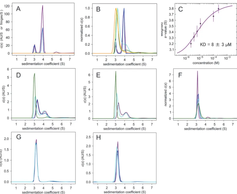
ΔIBB-h-importin-α1.The AUC-SV forΔIBB-h-importin-α1 shows that it is in a
h-importin-α1.However, h-importin-α1 remains monomeric in a variety of protein
concen-tration (Fig. 1F). Neither of the SV40 nor NP NLS peptides changed the monomeric state (Fig. 1G, H). These results show that the self-bound IBB domain autoinhibits not only NLS binding but also homodimerization.
Fig 1. AUC-SV measurements.(A) c(s) distributions ofΔIBB-h-importin-α1, 247μM (purple), 100μM (blue), 14μM (cyan), 11μM (green), 7.3μM (yellow) and 4.2μM (orange). (B) Normalized c(s) distributions ofΔIBB-h-importin-α1. (C) KD value estimation by the fitting curve of the sw vs protein concentration. (D) 25μMΔIBB-h-importin-α1 + SV40 NLS of 0μM (purple), 1μM (blue), 10μM (cyan) and 100μM (green). (E) 25μMΔIBB-h-importin-α1 + nucleoplasmin NLS of 0μM (purple), 10μM (cyan), and 100μM (green). (F) Normalized c(s) distributions of h-importin-α1, 61μM (purple), 34μM (blue), 8.6μM (cyan), and 4.3μM (green). (G) 10μM h-importin-α1 + SV40 NLS of 0μM (purple), 10μM (blue), 100μM (cyan). (H) 10μM h-importin-α1 + nucleoplasmin NLS of 0μM (purple), 10μM (blue), 100μM (cyan).
X-ray crystallography of
Δ
IBB-h-importin-
α
1
Only the diffractive crystals ofΔIBB-h-importin-α1 were obtained, not those of
h-importin-α1. The crystals diffracted X-rays to 2.6 Å resolution in SPring-8. The molecular replacement
technique was used to solve the structure (Table 1). The crystal structure ofΔIBB-importin-α1 is shown inFig. 2A. The pseudo 2-fold axis lies in close proximity to the N239 of chains A and
Table 1. Crystallographic data collection and refinement statistics ofΔIBB-h-importin-α1.
Space group TetragonalP43212
Cell constants (Å) a = b= 139.30,c= 141.29
Wavelength (Å) 1.00
Resolution (Å) 50.0–2.60 (2.69–2.60)
Oscillation angle (deg.) 1.0
σcut-off 0.0
Rmergea 0.065 (0.391)
No. of measurements 352,972 (3,701)
No. of independent reflections 40,652
Completeness (%) 96.96 (90.25)
Multiplicity 8.7 (4.8)
Mean<I/σ(I)> 31.83 (4.86)
Wilson B-factor (Å) 48.59
Refinement statistics
Resolution (Å) 46.7–2.63 (2.79–2.63)
No. of reflections used 40,616 (5,982)
Completeness (%) 96.96 (90.25)
Rworkb 0.1930 (0.2843)
Rfreec 0.2207 (0.3062)
No. of atoms
Protein (chain A, chain B) 6,496, 6,507
PEG 11
Water 192
Average B factor
Protein (chin A, chain B) 45.879, 38.836
PEG 71.622
Water 39.057
R.m.s. deviations
Bond lengths (Å) 0.003
Bond angles (°) 0.671
Ramachandran plot (%)
Favored region 95.6
Allowed region 4.04
Outer region 0.36
Clashscored 12.08
aRmerge=ΣΣ
i||I(h)—I(h)i| /ΣΣiI(h), whereI(h) is the mean intensity after rejections.
bRwork=Σ| F
p—Fpc/Σ|Fp|.
cRfree, the same asRworkbut calculated on 4.93% of data excluded from re finement.
dClashscore, calculated by MolProbity.
Values in parentheses are for highest resolution shells.
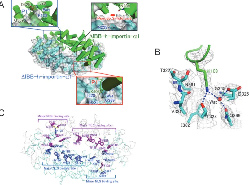
B, which are in the disallowed region in the Ramachandran plot. Structural stress is probably put on them owing to the dimerization. The K108 residue is extensively involved in the dimer formation and is represented in the close-up views. The side chain of K108 enters the P10site
of the minor NLS binding site [31], making hydrogen bonds with D325, T328, and Q369 of an-other protomer and one water molecule (Fig. 2B). The P10site comprises V321, T322, D325,
T328, N361, I362, G365, and Q369, which are conserved in the minor NLS binding site of importin-αproteins (Fig. 3). The major (W142, N146, W184, N188, W231, N235, W273,
Fig 2. Crystal structure of homodimericΔIBB-h-importin-α1.(A) The closed homodimer structure ofΔIBB-h-importin-α1. One of the protomers is shown as a surface drawing. Both of the P10-binding pockets are depicted in close-up views. The K108 inserts in the P10-binding pocket, making hydrogen bonds
with D325, T328, and Q369 of another protomer, and Wat (a water molecule). The pseudo 2-fold axis is drawn in the close-up view for the center of the dimer. (B) The P10binding pocket with 2fo-fc electron density maps (>1.0σ). Residues involved in the P10-binding pocket are V321, T322, D325, T328, N361, I362,
and Q369. One water molecule is depicted as Wat. The K108 of another protomer is inserted into the P10-binding pocket, making hydrogen bonds depicted
with dotted blue lines. (C) A ribbon drawing of the homodimericΔIBB-importin-α1 in cyan and gray. Residues of each protomer are in blue and orange. Major and minor NLS binding sites are indicated. Residues involved in the major NLS binding sites are W142, N146, W184, N188, W231, N235, W273, and Y277 in bold characters. The residues in the minor NLS biding sites are D325, T328, W357, N361, Q369, W399, and N403. Each of the K108 makes hydrogen bonds with D325, T328, Q369, and one water molecule in the minor NLS binding site of another protomer. Because the major and minor NLS binding sites are extensively buried in the dimerization interface as shown in the drawing, NLS signals are inaccessible to the sites. All molecular pictures were prepared with PyMol [40].
doi:10.1371/journal.pone.0115995.g002
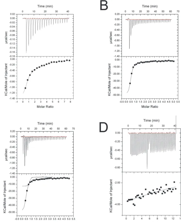
Fig 3. ITC fitting curves.(A) h-importin-α1 + SV40 NLS peptide. (B)ΔIBB-h-importin-α1 + SV40 NLS peptide. (C)ΔIBB-h-importin-α1 + nucleoplasmin NLS. (D) h-importin-α1 + nucleoplasmin.
Y277) and minor (W357, N361, W399, N403) NLS binding sites are buried in the dimer inter-face (Fig. 2C), thus NLS peptides cannot access the NLS binding sites. Because the major NLS binding site involves typically P1-P7 binding pockets [32], it has higher NLS binding affinity than the minor NLS binding site with P10-P20.
The P10-binding pocket in the minor NLS binding site plays a common role in a variety of
importin-αs; the P10accepts the K/R residues of the typical NLS signal motif KRXX [31–33],
(Fig. 4). In the present crystal structure ofΔIBB-h-importin-α1, each of the K108 residues
en-ters into the P10-binding pocket of another protomer so that the minor NLS binding sites are
in a state of autoinhibition (Fig. 2B), and, together with the major NLS binding sites, are buried in the dimer interface (Fig. 2C). As a result, the closed homodimer is stabilized owing to the limited access of NLS peptides to the major and minor NLS binding sites.
Isothermal titration calorimetry (ITC) assay for binding of NLS peptides
to h-importin-
α
1 and
Δ
IBB-h-importin-
α
1
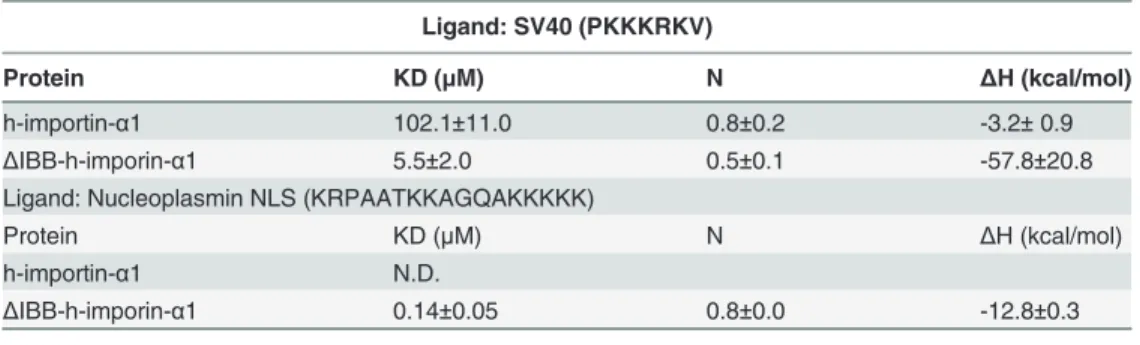
h-importin-α1.We carried out a binding assay of h-importin-α1 andΔIBB-h-importin-α1 to
study autoinhibition resulting from homodimerization. SV40 NLS bound to h-importin-α1
with a KD value of ~100μM (Table 2). While the fitting to the ITC data, one-site binding model was used because two-site model did not converge to adequate values. The AUC-SV ex-periments reveal that h-importin-α1 is monomeric during SV40 NLS binding (Fig. 1G). This result suggests that both the IBB domain and the NLS peptides bind to the internal NLS bind-ing sites and prevent homodimerization.
On the other hand, the bipartite nucleoplasmin NLS peptide did not bind to h-importin-α1
(Fig. 3D). This result suggests that NLS binding is restricted by the IBB domain.
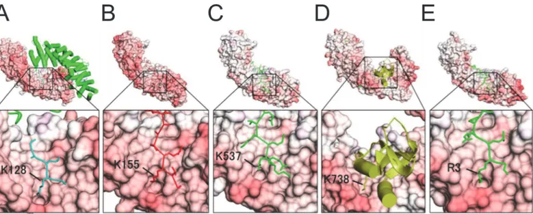
Fig 4. Crystal structures of complexes of importin-αs and NLS ligands with close-up views showing the P10-binding pocket in the minor NLS
binding sites.(A) K128 of SV40 NLS peptide (Yeast importin-α: PDB ID: 1BK6), (B) K155 ofXenopusnucleoplasmin NLS (Yeast importin-α: 1EE5), (C) K537 ofXenopusnucleoplasmin NLS (importin-α2: 1PJN), (D) K738 of influenza virus polymerase subunit Pb2 (importin-α5: 2JDQ), and (E) R3 of the IBB domain of the CBP20 (ΔIBB-importin-α1: 3FEY).
doi:10.1371/journal.pone.0115995.g004
ΔIBB-h-importin-α1.ΔIBB-h-importin-α1 bound to SV40 NLS with KD = ~6μM and to
nucleoplasmin NLS peptides with KD = ~0.14μM (Table 2). During the NLS binding, the monomer–dimer equilibrium shifted to the monomer side (Fig. 1D, E), which suggests that the NLS binding sites are involved in the dimer interface even in solution, as in the
crystal structure.
Discussion
Homodimerization found in importin-
α
s
It has been shown or suggested that the importin-αs from yeast andXenopuseggs form dimers. Yeast importin-α(PDB IB: 1BK6) provided the first crystal structure of an importin-αthat was shown to dimerize not only in the crystal but also in solution, as revealed by dynamic light scat-tering (DLS) [13]. The SV40 NLS peptides are, however, able to bind to the NLS binding sites because the homodimer is an“open”structure where the NLS binding sites are exposed to the solution (Fig. 4A). The open homodimerization probably attributes to H119 at the top of the loop; this loop corresponds to the K108 loop ofΔIBB-importin-α1. H119 cannot enter the P10
-binding pocket in the minor NLS -binding site, and this inability causes the protomers to slide along the long axis of the molecules. The NLS binding parameters of yeast importin-αwere
an-alyzed by fluorescence depolarization assay and it was found that yeast importin-α(1–542) binds to neither SV40 NLS-GFP nor Myc NLS-GFP [34]. On the other hand, yeastΔ
IBB-importin-α(89–530) binds to both SV40 and Myc NLS, with KDs of 9 ± 4 nM and 6 ± 3 nM, respectively [35]. In addition, an enzyme-linked immunosorbent assay (ELISA)-based assay re-vealed that yeast importin-α(1–542) does not bind to SV40 NLS, whereas yeastΔ IBB-impor-tin-α(88–530) binds very tightly [35]. These results suggest that the IBB domain is mainly
responsible for the autoinhibition activity of yeast importin-α, while the“open homodimeriza-tion”does not restrict the NLS binding. Thus, the NLS binding affinity of yeast importin-α
changes dramatically from 0% (off), when the IBB autoinhibits itself, to 100% (on), when it does not, that is keenly contrast to the closed-homodimer of h-importin-α1.
Moreover, it has been reported that wild importin-αfromXenopuseggs is homodimerized during purification [14], and a variety of studies have revealed that purifiedXenopus
importin-αis in dimer–monomer equilibration in the concentration range of 0.6~1.2 mM, with a
Table 2. Binding parameters of h-importin-α1 andΔIBB-h-importin-α1 by ITC.
Ligand: SV40 (PKKKRKV)
Protein KD (μM) N ΔH (kcal/mol)
h-importin-α1 102.1±11.0 0.8±0.2 -3.2±0.9
ΔIBB-h-imporin-α1 5.5±2.0 0.5±0.1 -57.8±20.8
Ligand: Nucleoplasmin NLS (KRPAATKKAGQAKKKKK)
Protein KD (μM) N ΔH (kcal/mol)
h-importin-α1 N.D.
ΔIBB-h-imporin-α1 0.14±0.05 0.8±0.0 -12.8±0.3
KD: dissociation constant, N: number of binding sites, ΔH: binding enthalpy, N.D. not detectable.
dissociation constant KD of ~20μM [36]. We found that theXenopusimportin-αhas an argi-nine residue (R105) that is aligned to the K108 of importin-α1 (S2 Fig.). Thus, the R105 possibly interacts with the P10-binding pocket of another protomer, promoting the
closed-homodimerization.
The first structural knowledge about the autoinhibition mechanism was obtained from the crystal structure of mouse-importin-α2 (m-importin-α2) [37], [38]. In that crystal structure, the IBB domain binds to the major, but not the minor, NLS binding site; thus the NLS binding affinity is partly autoinhibited. It has been unclear whether m-importin-α2 forms a homodimer or not [37], although the m-importin-α2 has a K102 and the P10-binding pocket consists of the
same residues as in h-importin-α1 (S2 Fig.). Further studies should be addressed about the multimerization property of the m-importin-α2.
It is reported thatΔIBB-h-importin-α1 (70–529) complexes with the cap-binding complex (CBC) [39]. In theΔIBB-h-importin-α1 (70–529)-CBC complex, the NH1 and NH2 of R3
(P10) in the canonical type of NLS (RRXX; P10P20XX) at the N-terminus of CBC (CBP80 +
CBP20) form hydrogen bonds with OD2 of D325 and OG1 of T328 in the ridge of the P10
-binding pocket (Fig. 4E), which may shift the monomer-dimer equilibration to the monomeric side.
A possible autoinhibition state of h-importin-
α
1
Together with the results obtained from the AUC-SV, ITC, and crystallography, we can add a potential autoinhibition mode of the h-importin-α1 to the conventional scenario, as shown in
Fig. 5. In the canonical scheme, the self-bound IBB domain autoinhibits NLS binding to the NLS binding sites in h-importin-α1. Our current study reveals that the self-bound IBB domain
also autoinhibits homodimerization. The IBB domain is dissociated from the NLS binding sites,e.g., by the competitive binding of NLS peptides to the NLS binding sites and/or the bind-ing of the IBB domain to importin-β(Fig. 5A). Upon the IBB domain dissociation, the h-importin-α1 can homodimerize as an autoinhibition state (Fig. 5B), which possibly lowers the
KD (~6μM) for NLS comparing to that of yeast (~10 nM) [34]. Through the association/disso-ciation of the IBB domain and molecular h-importin-α1, the NLS binding to the h-importin-α1 is controlled by both of the self-binding of IBB domain and homodimerization.
Fig 5. Autoinhibition states of h-importin-α1 by IBB binding and homodimerization.(A) A conventional scheme of autoinhibition by self-binding of IBB domain. The IBB domain bound in the NLS binding sites autoinhibits NLS binding. In addition, the current work reveals the IBB domain also prevents it from homodimerization. In turn, the bound IBB domain dissociates from the NLS binding sites by binding of NLS peptides and/or importin-β. (B) A potential homodimer autoinhibition mode. The current study reveals that a novel autoinhibition state by homodimerization, which possibly corresponds to NLS binding regulation.
doi:10.1371/journal.pone.0115995.g005
Conclusions
In h-importin-α1, NLS binding affinity is autoinhibited by the self-binding of IBB domain. In addition, the closed homodimerization possibly works as an autoinhibition state when the IBB domain dissociates from the NLS binding sites. There still remains unresolved subjects whether the current closed-homodimer is physiologically relevantin vivo. Further studies are required to address the functional aspects of the closed-homodimer, by mutagenesis and cellular biological experiments.
Supporting Information
S1 Fig. Anion exchange chromatography with the Resource Q column and fraction charac-terization by 12.5% sodium dodecyl sulfate polyacrylamide gel electrophoresis (SDS-PAGE).(A) h-importin-α1. Each number assigned to the resulting elution position coincides
with the lane of 12.5% SDS-PAGE. The blue line depicts the affinity column chromatography elution profile and the green line depicts the gradient of the NaCl concentration of the elution buffer. Fractions 39–44 were merged and concentrated. (B)ΔIBB-h-importin-α1. Fractions
40–45 were merged and concentrated. (EPS)
S2 Fig. Sequence alignment of importin-αs, with residue numbers and secondary structures
of h-importin-α1.Sequences of human importin-α1 (UniProtKB: P52292), mouse importin-α2 (P52293), yeast importin-α(Q02821), andXenopusimportin-α1 (P52171). The position of
K108 of importin-α1 is represented by a blue triangle. The residues (V321, T322, D325, N361,
I362, G365) involved in the P10-binding pocket around the minor NLS binding site are
con-served among the importin-αs. Thus, the P10-binding pocket should be conserved.
(EPS)
Acknowledgments
We thank Drs. Yuuki Nakamura, Go Ueno, Takaaki Hikima, and Masaki Yamamoto for their help in collecting the X-ray diffraction data using the mail-in system at SPring-8. We thank Drs. Takehiro Suzuki and Katsuhiko Kamada for assistance with the analytical ultracentrifuge experiment. We thank Dr. Yoichi Takeda for instructing us in the ITC experiment. We are grateful to the Support Unit for Bio-material Analysis, and the RIKEN BSI Research Resources Center for help with sequence analysis and peptide synthesis.
Author Contributions
Conceived and designed the experiments: HM YA. Performed the experiments: HM AS GM YT SU. Analyzed the data: HM AS GM YT YM ND YA SU. Contributed reagents/materials/ analysis tools: HM AS GM. Wrote the paper: HM YA.
References
1. Paine PL, Moore LC, Horowitz SB (1975). Nuclear envelope permeability Nature 254: 109–114. doi:
10.1038/254109a0PMID:1117994
2. Stoffler D, Fahrenkrogt B, AebiI U (1999). The nuclear pore complex: from molecular architecture to functional dynamics. Curr. Opin. Cell Biol. 11: 391–401. PMID:10395558
3. Stoffler D, Goldie KN, Feja B, Aebi U (1999). Calcium-mediated structural changes of native nuclear pore complexes monitored by time-lapse atomic force microscopy. J. Mol. Biol. 287: 741–752. PMID:
10191142
5. Tran EJ, Bolger TA, Wente SR (2007). Snapshot: nuclear transport. Cell, 131: 420. PMID:17956740 6. Görlich D, Mattaji IW (1996). Nucleocytoplasmic transport. Science 271: 1513–1518. PMID:8599106
7. Eigg EA (1997). Nucleocytoplasmic transport: signals, mechanisms and regulation. Nature 386: 779–
787. PMID:9126736
8. Weis K (1998). Importins and exportins: how to get in and out of the nucleus. Trends in Biochem. Sci. 23: 185–189.
9. Cuomo CA, Kirch SA, GyuRis J, Brent R, Oettinger MA (1994). Rch1, a protein that specifically interacts with the RAG-1 recombination-activating protein. Proc. Natl. Acad. Sci. USA 91: 6156–6160. PMID:
8016130
10. Goldfarb DS, Corbett AH., Mason DA, Harreman MT, Adam SA (2004). Importin alpha: a multipurpose nuclear-transport receptor. Trends Cell Biol. 14: 505–514. PMID:15350979
11. Panté N, Kann M (2002). Nuclear pore complex is able to transport macromolecules with diameters of ~39 nm. Mol. Cell Biol. 13: 425–434.
12. Kamata M, Nitahara-Kasahara Y, Miyamoto Y, Yoneda Y, Aida Y (2005). Importin-αpromotes passage through the nuclear pore complex of human immunodeficiency virus type 1 Vpr. J. Virol. 79: 3557–
5364. PMID:15731250
13. Conti E, Uy M, Leighton L, Blobel G, Kuriyan J (1998). Crystallographic analysis of the recognition of a nuclear localization signal by the nuclear import factor karyopherin alpha. Cell 94: 193–204. PMID:
9695948
14. Görlich D, Prehn S, Laskey RA, Hartmann E (1994). Isolation of a protein that is essential for the first step of nuclear protein import. Cell 79, 767–778. PMID:8001116
15. Nitahara-Kasahara Y, Kamata M, Yamamoto T, Zhang X, Miyamoto Y, et al. (2007). Novel nuclear im-port of Vpr promoted by imim-portinαis crucial for human immunodeficiency virus type 1 replication in macrophages. J. Viol. 81: 5284–5293.
16. Schuck P (2000). Size-distribution analysis of macromolecules by sedimentation velocity ultracentrifu-gation and lamm equation modelling. Biophys. J. 78: 1606–1619. PMID:10692345
17. Schuck P (2003) On the analysis of protein self-association by sedimentation velocity analytical ultra-centrifugation. Anal. Biochem. 320: 104–124. PMID:12895474
18. Ueno G, Kanda H, Hirose R, Ida K, Kumasaka T, et al. (2006). RIKEN structural genomics beamlines at the SPring-8; high throughput protein crystallography with automated beamline operation. J Struct Funct Genomics 7: 15–22. PMID:16645781
19. Okazaki N, Hasegawa K, Ueno G, Murakami H, Kumasaka T, et al. (2008). Mail-in data collection at SPring-8 protein crystallography beamlines. J. Synchrotron Radiat. 15: 288–291. doi:10.1107/
S0909049507064679PMID:18421161
20. Otwinowski Z, Minor W (1997). Processing of X-ray diffraction data collected in oscillation mode. Meth-ods in Enzymology Macromolecular Crystallography part A 276: 307–326.
21. Keegan RM, Winn MD (2007). Automated search-model discovery and preparation for structure solu-tion by molecular replacement. Acta Cryst. D 63: 447–457.
22. Terwilliger TC (2003). Automated main-chain model-building by template-matching and iterative frag-ment extension. Acta Cryst. D 59: 38–44.
23. Yao M, Zhou Y, Tanaka I (2006). Lafire: software for automating the refinement process of protein-structure analysis. Acta. Cryst. D 62: 189–196.
24. Langer G, Cohen SX, Lamzin VS, Perrakis A (2008). Automated macromolecular model building for X-ray crystallography using ARP/wARP version 7. Nat. Protoc. 3: 1171–1179. doi:10.1038/nprot.2008.
91PMID:18600222
25. McRee DE (1999). XtalView/Xfit—A versatile program for manipulating atomic coordinates and
elec-tron density. J. Struct. Biol. 125: 156–165. PMID:10222271
26. Emsley P, Cowtan K(2004). Coot: model-building tools for molecular graphics. Acta Cryst. D 60: 2126–
2132.
27. Brünger AT, Adams PD, Clore GM, DeLano WL, Gros P, et al. (1998). Crystallography & NMR system: a new software suite for macromolecular structure determination. Acta Cryst. D 54: 905–921.
28. Adams PD, Afonine PV, Bunkóczi G, Chen VB, Davis IW (2010). PHENIX: a comprehensive Python-based system for macromolecular structure solution. Acta Cryst. D 66: 213–221.
29. Laskowski RA (1993). PROCHECK: a program to check the stereochemical quality of protein struc-tures. J. Appl. Cryst. 26: 283–291.
30. Davis IW, Murry LW, Richardson JS, Richardson DS (2004). Molprobity: structure validation and all-atom contact analysis for nucleic acids and their complexes. Nucleic Acids Res. 32:W615–619. PMID:
15215462
31. Fontes MR, Teh T, Kobe B (2000). Structural basis of recognition of monopartite and bipartite nuclear localization sequences by mammalian importin-α. J. Mol. Biol. 297: 1183–1194. PMID:10764582
32. Conti E, Kuriyan J (2000). Crystallographic analysis of the specific yet versatile recognition of distinct nuclear localization signals by karyopherinα. Structure 8: 229–338.
33. Nair R, Carter P, Rost B (2003). Nlsdb: database of nuclear localization signals. Nucleic Acids Res. 31: 397–399. PMID:12520032
34. Hodel AE, Harreman MT, Pulliam KF, Harben ME, Holmes JS, et al. (2006). Nuclear localization signal receptor affinity correlates within vivolocalization inSaccharomyces cerevisiae. J. Biol. Chem. 281: 23545–23556. PMID:16785238
35. Fanara P, Hodel MR, Corbett AH, Hodel AE (2000). Quantitative analysis of nuclear localization signal (NLS)-importinαinteraction through fluorescence depolarization evidence for auto-inhibitory regulation of NLS binding. J. Biol. Chem. 275: 21218–21223. PMID:10806202
36. Percipalle P, Jonathan P, Butler G, Finch JT, Jans DA, et al. (1999). Nuclear localization signal recogni-tion causes release of importin-αfrom aggregates in the cytosol. J. Mol. Biol. 292: 263–273. PMID:
10493874
37. Kobe B. (1999). Autoinhibition by an internal nuclear localization signal revealed by the crystal structure of mammalian importin a. Nat. Struct. Biol. 6: 388–397. PMID:10201409
38. Kobe B. (1999). Nuclear targeting signal recognition: a key control point in nuclear transport? Nat. Struct. Biol. 6: 388–397. PMID:10201409
39. Dias SMG, Wilson K, Rojas KF, Ambrosio ALB, Cerione RA. (2009). The molecular basis for the regula-tion of the cap-binding complex by the importins. Nat. Struct. Mol. Biol. 16: 930–937. doi:10.1038/
nsmb.1649PMID:19668212