TRIM30
Is a Negative-Feedback Regulator
of the Intracellular DNA and DNA
Virus-Triggered Response by Targeting STING
Yanming Wang1,2☯, Qiaoshi Lian1☯, Bo Yang2, Shanshan Yan2, Haiyan Zhou1, Lan He1,
Guomei Lin1, Zhexiong Lian2, Zhengfan Jiang3, Bing Sun1,4*
1Institute of Biochemistry and Cell Biology, Shanghai Institutes for Biological Sciences, Chinese Academy of Sciences, Shanghai, China,2School of Life Sciences, University of Science and Technology of China, Hefei, China,3State Key Laboratory of Protein and Plant Gene Research, Key Laboratory of Cell Proliferation and Differentiation of the Ministry of Education, School of Life Sciences, Peking University, Beijing, China; Peking University-Tsinghua University Joint Center for Life Sciences, Beijing, China,4Key Laboratory of Molecular Virology & Immunology, Institute Pasteur of Shanghai, Chinese Academy of Sciences, Shanghai, China
☯These authors contributed equally to this work. *[email protected]
Abstract
Uncontrolled immune responses to intracellular DNA have been shown to induce autoim-mune diseases. Homeostasis regulation of imautoim-mune responses to cytosolic DNA is critical for limiting the risk of autoimmunity and survival of the host. Here, we report that the E3 ubi-quitin ligase tripartite motif protein 30α(TRIM30α) was induced by herpes simplex virus type 1 (HSV-1) infection in dendritic cells (DCs). Knockdown or genetic ablation of TRIM30α
augmented the type I IFNs and interleukin-6 response to intracellular DNA and DNA viruses.Trim30α-deficient mice were more resistant to infection by DNA viruses.
Biochemi-cal analyses showed that TRIM30αinteracted with the stimulator of interferon genes (STING), which is a critical regulator of the DNA-sensing response. Overexpression of TRIM30αpromoted the degradation of STING via K48-linked ubiquitination at Lys275 through a proteasome-dependent pathway. These findings indicate that E3 ligase TRIM30α
is an important negative-feedback regulator of innate immune responses to DNA viruses by targeting STING.
Author Summary
Negative-feedback regulation is a broad and pivotal biological event to maintain the homeostasis of the host. Viral DNA species derived from DNA viruses or retroviruses can activate STING signaling to produce pro-inflammatory cytokines and type I interferon, which further recruit immune cells or induce interferon stimulated genes (ISGs) to clear viral infection respectively. However, excessive STING-signaling activation has been shown to induce autoimmune disorders. Thus, it is important to finely turn off STING
sig-naling. Here we demonstrate that TRIM30αis rapidly induced followed by STING
a11111
OPEN ACCESS
Citation:Wang Y, Lian Q, Yang B, Yan S, Zhou H, He L, et al. (2015) TRIM30αIs a Negative-Feedback
Regulator of the Intracellular DNA and DNA Virus-Triggered Response by Targeting STING. PLoS Pathog 11(6): e1005012. doi:10.1371/journal. ppat.1005012
Editor:Laurent Coscoy, University of California Berkeley, UNITED STATES
Received:January 18, 2015
Accepted:June 8, 2015
Published:June 26, 2015
Copyright:© 2015 Wang et al. This is an open access article distributed under the terms of the
Creative Commons Attribution License, which permits unrestricted use, distribution, and reproduction in any medium, provided the original author and source are credited.
Data Availability Statement:All relevant data are within the paper and its Supporting Information files.
Funding:This work was supported by grants from the national key project of 973 (2013CB530504) and the National Natural Science Foundation of China (31030029, 31230024, 81361120409). The funders had no role in study design, data collection and analysis, decision to publish, or preparation of the manuscript.
activation.Trim30α-deficient mice show more resistance to infection by DNA viruses.
Meanwhile, knockdown or genetic ablation of TRIM30αaugments the type I IFNs and
IL-6 responses to intracellular DNA and DNA viruses. Biochemical analyses show that
TRIM30αinteracts with STING and promotes the degradation of STING via K48-linked
ubiquitination at Lys275. These findings demonstrate that induced TRIM30αis a
nega-tive-feedback regulator of STING pathway activation triggered by DNA and DNA viruses, which helps the host to avoid excessive response and maintain homeostasis.
Introduction
The innate immune system is the first barrier of defense against pathogens, and a variety of germline-encoded pattern recognition receptors (PRRs), including Toll-like receptors (TLRs), C-type lectin receptors (CLRs), RIG-I-like receptors (RLRs) and NOD-like receptors (NLRs), have evolved in this system [1]. PRRs recognize the pathogen-associated molecular patterns (PAMPs) of microbes, which activate the innate immune response. The major PAMPs include nucleic acids derived from diverse bacterial, viral and eukaryotic pathogens. Thus far, many RNA receptors have been verified, among which TLR3 and TLR7/8, which are located in endo-somes, sense double-stranded (ds)RNA and single-stranded (ss)RNA [2,3]. Retinoic-acid-inducible gene I (RIG-I) and melanoma differentiation-associated gene 5 (MDA5), which are located in the cytoplasm, detect cytosolic RNA [4,5].
In the past 5 years, the molecular basis of DNA sensing by the innate immune system has begun to be revealed, and a variety of DNA receptors have been proposed [6]. For example, TLR9 senses the CpG motifs-contained DNA [7]. In addition, AIM2 (absent in melanoma 2) binds to DNA and activates the inflammasome complex via the adaptor protein ASC
(apopto-sis-associated speck-like protein containing a CARD) [8–10]. Furthermore, RNA polymerase
III detects cytosolic DNA and induces type I interferons through the RIG-I pathway [11,12]. Moreover, many other DNA receptors, including DAI, IFI16, DDX41 and cGAS, trigger type
I interferon response via adaptor STING [13–16]. Although aspects of the molecular basis of
STING-mediated DNA sensing by the innate immune system have been determined, the pre-cise mechanism by which this pathway is fine-tuned remains unclear. Normal activation of the DNA signaling pathway contributes to appropriate host defense. However, uncontrolled stimulation by DNA results in excessive production of inflammatory cytokines and type I IFNs, which may cause severe autoimmune diseases, such as systemic lupus erythematosus (SLE) [17]. Several negative regulators of the STING-meditated DNA signaling pathway have been described. For example, the E3 ubiquitin ligase TRIM21 induces the Lys48 (K48)-linked ubiquitination and degradation of DDX41 and negatively regulates the innate immune response to intracellular dsDNA [18]. Another E3 ubiquitin ligase, RNF5, which is constitu-tive expressed, inhibits virus-triggered signaling by targeting STING for ubiquitination and degradation [19]. Hiroyasu Konno et al demonstrated that cyclic dinucleotides (CDN) trigger the phosphorylation of STING by UNC-51-like kinase (ULK1), which inhibits STING activity and thus prevents the persistent transcription of innate immune genes [20]. A recent study has identified NLRC3 as another negative regulator of STING-induced innate immune response [21].
Previous work in our laboratory has demonstrated that TRIM30αnegatively regulates TLR4
signaling by targeting TAB2 and TAB3 for degradation [22]. TRIM30αis also found to
nega-tively regulate NLRP3 inflammasome activation by modulating reactive oxygen species
pro-duction [23]. In this study, we report that TRIM30αis a negative-feedback regulator of innate
immune responses to intracellular DNA and DNA viruses by promoting degradation of STING via K48-linked ubiquitination at Lys275.
Results
TRIM30α
knockdown increases the host response to cytoplasmic DNA
and DNA virus infection in D2SC cells
TRIM30αhas been reported to be induced by TLR ligands in an NF-κB-dependent way and
then negatively regulates TLR signaling by a‘feedback’mechanism. We found TRIM30αcan
also be induced upon intracellular poly(dA:dT) stimulation in bone marrow-derived dendritic
cells (BMDCs) (S1A Fig). To address whether TRIM30αregulates the immune response to
DNA-mediated signaling, we used TRIM30αsiRNA (T3) to silence endogenous TRIM30α
expression in D2SC cells, a mouse mDC cell line. The results showed that the T3 siRNA
effi-ciently reduced the TRIM30αprotein expression compared with control siRNA (SC) (Fig 1A).
Various DNA ligands were used to stimulate D2SC cells, including poly(dA:dT), interferon stimulatory DNA (ISD), the genomic DNA of C57BL/6 mice and cyclic di-GMP (c-di-GMP).
Knockdown of TRIM30αenhanced the production of type I IFN and IL-6 compared with
con-trols (Fig 1B). In contrast, the induction of type I IFN by the synthetic analog of viral dsRNA
poly (I:C) (the signaling of which is largely governed by MDA5) was reduced when TRIM30α
was knocked down. To further confirm the function of TRIM30αin the anti-DNA viral
response, we infected D2SC cells with the DNA viruses HSV-1 or vaccinia virus (VACV). The
data showed that IFN-βand IL-6 production was dramatically increased in D2SC cells
trans-fected with TRIM30α-specific siRNA (Fig 1C). TRIM30αknockdown also promoted the type I
IFN and IL-6 expression at the mRNA level after stimulation with poly(dA:dT), ISD or HSV-1 (S1B and S1C Fig). We next examined the IFN-stimulated genes, such as the chemokine IP-10.
Notably, TRIM30αknockdown enhanced the production of IP-10 mRNA upon ISD
stimula-tion, but had no effect on the poly(I:C)-mediated signaling (Fig 1D).
Intracellular nucleic acids and most viruses activate NF-κB, which is indispensable for
TRIM30αexpression. We found that poly(I:C), poly(dA:dT), ISD, c-di-GMP, genomic DNA
and DNA viruses, including HSV-1 and VACV, facilitated the expression of TRIM30α(S1D
Fig). Moreover, TRIM30αexpression was considerably decreased by treatment with two
NF-κB inhibitors, the IκB kinase-2 (IKK-2) inhibitor TPCA-1 (2-[(aminocarbonyl)
amino]-5-(4-fluorophenyl)-3-thio-phenecarboxamide) and PDTC (pyrrolidine dithiocarbamate), in BMDCs stimulated for 6 h with poly(I:C), ISD or HSV-1 (S1E Fig).
Type I IFN and IL-6 expression is dependent upon the transcription factors IRF3 and
NF-κB. Therefore, the amount of phosphorylated-IRF3 and-p65 was determined in D2SC cells
after exposure to poly(dA:dT) at different time points. We found that TRIM30αknockdown
resulted in high level of phosphorylation of IRF3 and p65 after poly(dA:dT) treatment (Fig 1E). Collectively, these results indicate that specifically knocking down TRIM30αconsiderably enhances type I IFN and IL-6 activation and the expression of IP-10 in response to cytoplasmic DNA and DNA viruses.
TRIM30α
deficiency increases the host response to cytoplasmic DNA
and DNA virus infection in dendritic cells
To further demonstrate the function of TRIM30αin immune response to DNA-mediated
sig-naling, we generatedTrim30α-deficient mice, in which the second exon was knocked out by
homologous recombination (S2 Fig). Immunoblot analysis confirmed that TRIM30α
deficiency resulted in much higher expression of type I IFN and IL-6 in response to multiple DNA ligands compared with wild-type DCs, similar to the results observed in D2SC cells. In
contrast, TRIM30αdeficiency did not affect poly(I:C) signaling (Fig 2B). Consistently with
pre-vious results, TRIM30αdeficiency dramatically increased IFN-βand IL-6 expression induced
by HSV-1 and VACV infection at protein level (Fig 2C). Beside the role in DNA sensing
path-way, we also determine the function of TRIM30αagainst RNA viruses. As shown inS3 Fig,
TRIM30αknockdown or deficiency dramatically potentiated IFN-βand IL-6 production by
infection with VSV in D2SC cells and BMDCs (S3 Fig). The results suggest that TRIM30α
neg-atively regulate immune response both to DNA and RNA viruses.
Finally, we assessed IRF3 and p65 phosphorylation in BMDCs after poly(dA:dT)
stimula-tion and found that TRIM30αdeficiency promoted much higher expression of the
phosphory-lation of endogenous IRF3 and p65 (Fig 2D). These results suggest that TRIM30αplays a
Fig 1. TRIM30αknockdown promotes immune signaling by cytoplasmic DNA and DNA virus infection in D2SC cells.(A) Immunoblot analysis of the knockdown of exogenous TRIM30αin D2SC cells treated with control siRNA (SC) or TRIM30αsiRNA (T3) for 24 h and then stimulated for 16 h with poly(dA: dT) (1μg/ml).β-actin served as a loading control throughout the experiment. (B, C) ELISA of type I IFN and IL-6 in D2SC cells treated with siRNA SC or T3 and then stimulated for 16 h with poly(dA:dT) (1μg/ml), ISD (1μg/ml), genomic DNA (2μg/ml), c-di-GMP (8μg/ml) or poly(I:C) (5μg/ml) (C) or with HSV-1 (MOI 10) or vaccinia virus (VACV) (MOI 10). (D) Real-time PCR of IP-10 mRNA in D2SC cells treated with siRNA SC or T3 and then stimulated for 8 h with ISD (1μg/ml) or poly(I:C) (5μg/ml). (E) Immunoblot analysis of p-IRF3, p-p65 and TRIM30αin lysates of D2SC cells treated with siRNA SC or T3 and then stimulated for 1–5 h with poly(dA:dT) (1μg/ml). Densitometry analysis to quantify ratio of p-IRF3 or p-p65 toβ-actin is shown on the right. The data are representative of three independent experiments and are presented as mean±SEM.*p<0.05,**p<0.01 and***p<0.001.
doi:10.1371/journal.ppat.1005012.g001
negative role in regulating type I IFN and IL-6 production in response to cytoplasmic DNA and DNA viruses.
TRIM30α
deficiency inhibits HSV-1 infection
To evaluate the function of TRIM30αin host antiviral responses in vitro, we knocked down the
expression of TRIM30αin mouse fibroblast L929 cells and then infected these cells with the
DNA virus HSV-1. As expected, TRIM30αknockdown suppressed HSV-1 infection (Fig 3A).
In contrast, overexpression of TRIM30αmarkedly facilitated HSV-1 replication (Fig 3B). These
data suggest that TRIM30αis an important negative regulator in anti-viral defense. To further
identify the role of TRIM30αin vivo host defense, wild type andTrim30α-deficient mice were
challenged by intraperitoneal (i.p.) injection with HSV-1, and virus titers were examined 20 h later. Significantly more HSV-1 replication was detected in wild type mice (Fig 3C).
Moreover, genomic DNA copies of HSV-1 were dramatically reduced in the brain, lung and
liver ofTrim30α-deficient mice in comparison to wild type mice upon HSV-1 infection for 2
days (Fig 3D). Interestingly, the liver showed much more difference between WT and KO mice
Fig 2. TRIM30αdeficiency promotes immune signaling by cytoplasmic DNA and DNA virus infection in BMDCs.(A) Immunoblot analysis of TRIM30α expression in the splenocytes of wild type (WT) andTrim30α-/-(KO) mice. (B, C) ELISA of type I IFN and IL-6 in wild-type andTrim30α-/-BMDCs mock
treated or stimulated for 16 h by transfection with poly(dA:dT) (1μg/ml), ISD (1μg/ml), c-di-GMP (8μg/ml), genomic DNA (2μg/ml) or poly(I:C) (5μg/ml) (B); or with HSV-1 (MOI 10) or VACV (MOI 10) (C). (D) Immunoblot analysis of phosphorylated IRF3 (p-IRF3), p-p65 and TRIM30αin lysates of wild type and Trim30α-/-BMDCs stimulated for 1
–5 h with poly(dA:dT) (1μg/ml). Densitometry analysis to quantify ratio of p-IRF3 or p-p65 toβ-actin is shown on the right. The data are representative of three independent experiments and are presented as mean±SEM.*p<0.05,**p<0.01 and***p<0.001.
compared to brain and lung, which was worthy to be explored. Together these data suggest
that TRIM30αknockdown or deficiency inhibited HSV-1 replication both in vitro and in vivo.
TRIM30α
deficiency protects mice from infection with HSV-1
To investigate the role of TRIM30αin host antiviral innate response, we isolated CD11c+
sple-nocytes from wild-type andTrim30α-deficient mice and treated these cells for 16 h with ISD,
HSV-1 or poly(I:C). TheTrim30α-deficient CD11c+splenocytes produced high levels of IFN-β
and IL-6 upon stimulation with ISD and HSV-1. However, TRIM30αdeficiency had little effect
on poly(I:C)-triggered response (Fig 4A and 4B). Besides DCs, macrophages are also involved in immune response triggered by intracellular DNA or DNA virus. We then obtained
perito-neal macrophages (PM) from wild type andTrim30α-/-mice and then the above PM were
stim-ulated with ISD or infected with HSV-1. The real-time PCR analysis demonstrated that
Trim30α-/-PM produced more type I IFN and ISGs, suggesting that the negative role of
TRIM30αin regulating DNA-sensing signaling pathway was not restricted in DCs (S4A and
S4B Fig).
To further elucidate the function of TRIM30αin vivo, wild-type andTrim30α-deficient
mice were infected intravenously (i.v.) with HSV-1. Then we isolated various organs to evaluate
immune response induced by DNA viruses. The levels of IFN-βand TNF-αmRNAs in liver
and lung were markedly potentiated inTrim30α-deficient mice (Fig 4C and 4D). In addition,
we observed higher IFN-βand IL-6 levels in the serum ofTrim30α-deficient mice than in
wild-type mice followed by HSV-1 infection (Fig 4E). Survival experiments demonstrated that the
survival rate was significantly prolonged in theTrim30α-deficient mice infected with HSV-1
Fig 3. TRIM30αdeficiency inhibits DNA virus infection.(A, B) Viral titers in L929 cells transfected with control siRNA SC and T3 (A), empty vector (Vec) or TRIM30αplasmid (B) for 24 h and then infected with HSV-1 (MOI 10) for 20 h. The titers of HSV-1 were determined by standard plaque assay. (C) Viral titers in wild type andTrim30α-/-mice intraperitoneally injected with HSV-1 (1×10⁷plaque-forming units (PFU)).
HSV-1 titers were measured 20 h later by plaque assay of peritoneal wash fluid. (D) Real-time PCR of HSV-HSV-1 genomic DNA in the brain, lung and liver from wild type andTrim30α-/-mice infected with HSV-1 (2×10⁷PFU)
(i.v.) for 2 days. The data are representative of three independent experiments and are presented as mean±SEM.*p<0.05,***p<0.001.
doi:10.1371/journal.ppat.1005012.g003
(Fig 4F). Thus, TRIM30αnegatively controls DNA virus-triggered signaling, and TRIM30α
deficiency protects mice from DNA virus infection.
TRIM30α
interacts with STING
To identify the molecular mechanisms by which TRIM30αinhibits the DNA-triggered
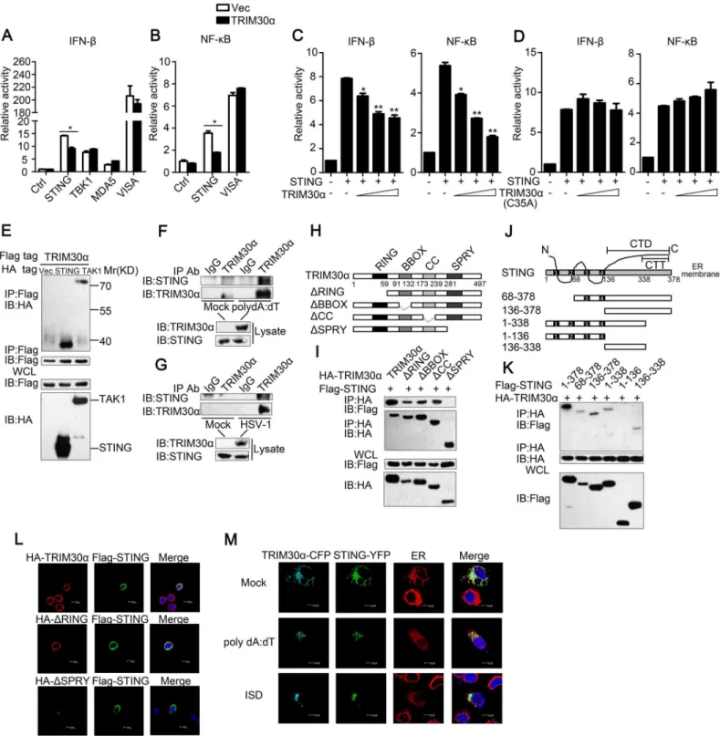
response, luciferase assays were performed to detect the target of TRIM30α. We found that
TRIM30αinhibited the IFN-βreporter activation mediated by STING, a key adaptor protein
for most DNA-sensing pathways [24,25]. However, TRIM30αdid not affect the downstream
kinase of STING, TBK1, and had no influence on MDA5- and virus-induced signaling adaptor
(VISA)-induced signaling (Fig 5A). In addition, TRIM30αoverexpression significantly
inhib-ited STING-induced NF-κB reporter activation but did not affect VISA (Fig 5B). It is
hypothe-sized that TRIM30αtargets STING to inhibit DNA-mediated response. As shown inFig 5C,
exogenous expression of TRIM30αinhibited STING-induced IFN-βand NF-κB reporter
acti-vation in a dose dependent manner (Fig 5C). In contrast, TRIM30α(C35A), which contains an
enzymatically inactive mutant of the RING domain had no effect on STING-induced IFN-β
Fig 4. TRIM30αdeficiency protects the mice from DNA virus infection.(A, B) ELISA of IFN-βand IL-6 in wild-type andTrim30α-/-CD11c+splenocytes
stimulated for 24 h with ISD (1μg/ml), HSV-1 (MOI 10) or poly(I:C) (5μg/ml). (C, D) Real-time PCR of IFN-βand TNF-αin the lung and liver from wild type and Trim30α-/-mice infected with HSV-1 (2×10⁷PFU) (i.v.) for the indicated times. (E) ELISA of IFN-βand IL-6 in serum of wild type andTrim30α-/-mice 6 h after
intravenous infection with HSV-1 (1.2×10⁷PFU) (n = 5). (F) Survival of age- and sex-matched wild-type andTrim30α
-/-mice (n = 5 per group) infected with HSV-1 (2×10⁷PFU) (i.v.) and monitored daily for 15 d. The data are representative of three independent experiments and are presented as mean±SEM. *p<0.05,**p<0.01 and***p<0.001.
Fig 5. TRIM30αinteracts with STING.(A) Luciferase activity of MEF cells transfected with the IFN-βluciferase reporter, together with STING, TBK1, MDA5 or VISA, and the empty vector (Vec) or the TRIM30αplasmid. (B) Luciferase activity of MEF cells transfected with the NF-κB luciferase reporter, together with STING or VISA, and the empty vector (Vec) or the TRIM30αplasmid. (C and D) Luciferase activity of MEF cells cotransfected with the IFN-βpromoter or the NF-κB promoter and empty vector or increasing amounts of TRIM30α(C) or C35A (D) (0.2, 0.4 and 0.6μg) together with STING. (E) Immunoblot analysis of lysates from HEK293T cells transfected with Flag-TRIM30αtogether with HA-tagged vector, STING or TAK1 plasmids, followed by immunoprecipitation (IP) with anti-Flag Ab and immunoblot analysis with anti-HA Ab. (F, G) Immunoblot analysis in lysates of D2SC cells mock treated or stimulated with poly(dA:dT) (1μg/ml) (F); or with HSV-1 (MOI 10) (G) for 8 h, followed by immunoprecipitation with anti-TRIM30αAb and immunoblot analysis with anti-STING Ab. (H) A schematic presentation of full-length TRIM30αand its mutants. RING, ring-finger domain; BBOX, B-box domain; CC, coiled-coil domain; SPRY, SPRY domain. (I) Immunoblot analysis of lysates from HEK293T cells transfected with the indicated plasmids, followed by immunoprecipitation (IP) with anti-HA Ab
and NF-κB reporter activation (Fig 5D). All these suggest that TRIM30αsuppresses STING signaling dependent on its RING domain. Furthermore, co-immunoprecipitation assays were
performed and the results showed that TRIM30αinteracted with STING when co-transfected
into HEK293T cells (Fig 5E). TGF beta-activated kinase (TAK1) interacts with TRIM30α,
which has been previously reported, was used as a positive control [22]. We next performed
endogenous co-immunoprecipitation experiments and verified that endogenous TRIM30α
could interact with STING in D2SC cells after poly(dA:dT) or HSV-1 stimulation (Fig 5F and 5G). To further demonstrate the interaction, TRIM30αand STING were quickly translated in
vitro, and immunoprecipitation analysis indicated that TRIM30αcould interact with STING
directly (S5 Fig).
To explore the domains that govern the association between TRIM30αand STING,
HA-tagged full-length TRIM30αand various TRIM30αtruncations were expressed in HEK293T
cells. Co-immunoprecipitation assays showed that only the SPRY domain of TRIM30αbound
to STING (Fig 5H and 5I). Previous studies have reported that STING is a multiple transmem-brane domain-containing protein and that the C-terminal amino acids of STING include a
globular carboxy-terminal domain (CTD, amino acids 136–378) and carboxy-terminal tail
(CTT, amino acids 338–378) [26,27]. We found that only the C-terminal amino acids 136–338
of STING were required for interaction with TRIM30α(Fig 5J and 5K). Flag-tagged STING,
HA-tagged TRIM30αand TRIM30αtruncations were expressed in L929 cells. Confocal
microscopy experiments showed that STING was co-localized with full-length TRIM30αand
ΔR, but notΔSPRY, confirming that the SPRY domain of TRIM30αis required for interaction
with STING (Fig 5L). We then expressed CFP-TRIM30αand YFP-STING in L929 cells and
detected the co-localization of these two proteins (Fig 5M). Interestingly, it was also found that STING translocated from the endoplasmic reticulum (ER) to perinuclear sites in response to poly(dA:dT) and ISD, in agreement with previous studies [25]. These results suggest that
TRIM30αinteracts with STING to inhibit DNA-mediated immune response.
TRIM30α
enhances the degradation of STING
It is very important to determine the mechanism by which interaction of TRIM30αwith
STING suppress the intracellular DNA-sensing pathway. As shown inFig 6A,Trim30α
-defi-cient BMDCs maintained a higher expression of STING than wild type BMDCs after poly(dA: dT) stimulations of different durations (Fig 6A). In contrast, the expression of TBK1 and IRF3,
two adaptors downstream of STING, maintained unchanged inTrim30α-deficient BMDCs,
suggesting that TRIM30αmay suppress the stability of STING. We next treated wild type
BMDCs with poly(dA:dT) in the presence or absence of MG132, the inhibitor of proteasome. We observed a high level expression of STING in BMDCs upon MG132 treatment than con-trol, indicating that STING undergoes degradation after stimulation with intracellular DNA in a proteasome way (Fig 6B). Moreover, MG132 treatment significantly promoted much higher
expression of phosphorylation of TBK1 and IRF3 than control cells, which mimics TRIM30α
deficiency upon intracellular DNA stimulation. Confocal microscopy further demonstrated
and immunoblot analysis with anti-Flag Ab. (J) A schematic presentation of full-length STING and its mutants. (Transmembrane) TM1, 21-41aa; TM2, 47-67aa; TM3, 87-106aa; TM4, 115-135aa; CTD, carboxy-terminal domain; CTT, carboxy-terminal tail. (K) Immunoblot analysis of lysates from HEK293T cells transfected with the indicated plasmids and then performed as in I. (L) Confocal microscopy of L929 cells transfected for 24 h with Flag-tagged STING and HA-tagged full-length TRIM30α,ΔRING orΔSPRY mutant of TRIM30α. Immunofluorescence was performed using anti-HA (red) and anti-Flag (green). (M) Confocal microscopy of L929 cells transfected for 24 h with cyan fluorescent protein-labeled TRIM30a (CFP-TRIM30α) and yellow fluorescent protein-labeled STING (YFP-STING) and then mock treated or stimulated for 4 h with poly(dA:dT) (1μg/ml) or ISD (1μg/ml). Nuclei were stained with the DNA-intercalating dye DAPI. Staining of calnexin served as a marker of the endoplasmic reticulum (ER). The data are representative of three independent experiments and are presented as mean±SEM.*p<0.05 and**p<0.01.
that STING was co-localized with proteasome during ISD stimulation, suggesting that STING may undergo degradation via proteasome in response to DNA stimulus (Fig 6C).
Because TRIM30αpromotes the degradation of TAB2 and TAB3 [22], we hypothesized that
TRIM30αalso promotes the degradation of STING. To address this, TRIM30αwas
overex-pressed in L929 cells. The expression of endogenous STING in L929 cells was diminished in a dose-dependent manner and rescued by MG132 (Fig 6D). Furthermore, we determined
whether STING degradation is dependent upon the RING domain of TRIM30α, which confers
it E3 ubiquitin ligase activity. TRIM30α(C35A) and TRIM30α(ΔR) attenuated STING
degra-dation when expressed in L929 cells compared with full-length TRIM30α(Fig 6E). Collectively,
these results indicated that the interaction of TRIM30αwith STING enhances STING
degrada-tion in a proteasome pathway, which is dependent on its RING domain.
TRIM30α
targets STING for K48-linked ubiquitination at Lys275
Most TRIM proteins contain RING domains, which allow them to mediate ubiquitylationevents [28]. Therefore, we hypothesized that TRIM30αis an E3 ubiquitin ligase for STING. To
address this, TRIM30αand STING were co-transfected in HEK293T cells.
Immunoprecipita-tion and immunoblot analysis showed that TRIM30αcould ubiquitinate STING in a
dose-dependent manner (Fig 7A). Moreover, we observed that TRIM30αoverexpression enhanced
Fig 6. TRIM30αenhances the degradation of STING.(A) Immunoblot analysis of STING, TBK1, IRF3 and TRIM30αin lysates from wild type and Trim30α-/-BMDCs stimulated for 2
–24 h with poly(dA:dT) (1μg/ml). (B) Immunoblot analysis of STING in lysates from wild type BMDCs stimulated for 2–24 h with poly(dA:dT) (1μg/ml) in the presence or absence of 20 mM MG132. (C) Confocal microscopy of L929 cells transfected for 24 h with Flag-tagged STING and then mock treated or stimulated for 12 h with ISD (1μg/ml). Immunofluorescence was performed using anti-Flag (red) and anti-20S proteasomeβ1 (green). Nuclei were stained with the DNA-intercalating dye DAPI. (D) Immunoblot analysis of STING in lysates of L929 cells transfected with increasing doses of Myc-tagged TRIM30α(0, 0.8 and 1.2μg) and then treated for 6 h with DMSO (negative control) or 20 mM MG132. Densitometry analysis to quantify ratio of STING toβ-actin is shown on the below. (E) Immunoblot analysis of STING in lysates of L929 cells transfected with full-length TRIM30α, C35A orΔR (1μg) and then treated as in D.
doi:10.1371/journal.ppat.1005012.g006
Fig 7. TRIM30αis an E3 ubiquitin ligase and targets STING for K48-mediated ubiquitination at Lys275.(A) Immunoblot analysis of lysates from HEK293T cells transfected with plasmids for Flag-STING, HA-ubiquitin and increasing concentrations of Myc-TRIM30α(0, 1, 1.5 and 2μg), followed by immunoprecipitation with anti-Flag, and analyzed via immunoblot with anti-HA Ab. (B) Immunoblot analysis of lysates from HEK293T cells transfected with various combinations of plasmids for Myc-TRIM30α, Flag-STING, HA-K48-ubi and HA-K63-ubi and then performed as in A. (C) Immunoblot analysis of lysates from HEK293T cells transfected with the indicated plasmids and Myc-tagged TRIM30α(C35A),ΔR and then performed as in B. (D) Immunoblot analysis of lysates in wild-type andTrim30α-/-BMDCs stimulated for 8 h with ISD (1μg/ml), followed by immunoprecipitation with anti-STING Ab and
analyzed with anti-Ub, anti-K48- and anti-K63-linked ubiquitin. (E) Immunoblot analysis of STING ubiquitination in vitro. TRIM30αand STING were quickly translated in vitro, and then, the biotin-ubiquitin E1 and indicated E2s were added for the ubiquitination assays. Ubiquitination was assessed by anti-ubi. (F) Immunoblot analysis of lysates from HEK293T cells transfected with Myc-tagged TRIM30α, Flag-tagged wild-type and mutant STING together with HA-tagged ubiquitin, then performed as in A. (G) Luciferase assays of MEF cells cotransfected with the IFN-βpromoter and empty vector or TRIM30αtogether with wild-type and mutant STING plasmids. (H) Real-time PCR of IFN-βand IP-10 mRNA in L929 cells transfected with Flag-tagged vector, wild type STING and K275R mutant, 24 h after transfection, then infected the above cells with HSV-1 (MOI 10) for 10 h. The data are representative of three independent experiments and are presented as mean±SEM.*p<0.05,**p<0.01 and***p<0.001.
K48-linked ubiquitination of STING but not K63-linked ubiquitination (Fig 7B). K48-linked ubiquitination is normally linked to proteasomes-mediated degradation of proteins, which is
in line with our early data (Fig 6D and 6E). As shown inFig 7C, TRIM30α(C35A) andΔR
dra-matically attenuated the ubiquitination of STING compared with full-length TRIM30α,
sug-gesting that TRIM30αubiquitinates STING dependent upon its RING domain activity (Fig
7C). Next, we examined the ubiquitination of STING in primary cells and observed that wild type and K48-linked ubiquitination of STING in wild type BMDCs was increased in
compari-son toTrim30α-deficient BMDCs stimulated with ISD for 8 h (Fig 7D). Collectively, these
find-ings indicate that TRIM30αis an E3 ubiquitin ligase for STING and mediates STING
degradation via the proteasome pathway. To further investigate whether TRIM30αdirectly
ubiquitinates STING, TRIM30αand STING were quickly translated in vitro. In vitro
ubiquiti-nation showed that the TRIM30αprotein directly mediated ubiquitination of STING (Fig 7E).
In order to map the ubiquitination sites on STING that are targeted by TRIM30α, we
replaced each of the seven STING lysine (K) residues that not in transmembrane region with
arginine (R). We then expressed TRIM30α, wild-type and mutant STING in HEK293T cells.
As shown inFig 7F, the ubiquitination of mutation of K275 to arginine was obviously
attenu-ated (Fig 7F). In addition, luciferase assays showed that TRIM30αsignificantly suppressed
wild-type and STING mutants-induced IFN-βreporter activity except for STING K275R (Fig
7G). Moreover, STING K275R maintained high level in HSV-1 triggered IFN-βand IP-10 pro-duction in comparison to wild type STING (Fig 7H). Taken together, the results suggest that
Lys275 was the ubiquitination site of STING targeted by TRIM30α.
Discussion
STING has been identified as an essential adaptor protein for controlling TLR-independent cytosolic DNA signaling [29]. Cytosolic DNA derived from DNA viruses, bacteria or parasites is sensed by DNA receptors that subsequently activate STING. Activated STING recruits and
activates the cytosolic kinases IKK and TBK1, which in turn activate NF-κB and IRF3,
respec-tively [30]. In addition, cyclic dinucleotides (CDNs) can directly bind and activate STING. However, the regulation of STING signaling remains to be fully elucidated. In this context, we
discovered the novel function of TRIM30αin negatively regulating the STING pathway.
TRIM30αdeficiency or knockdown enhanced the production of type I IFNs and the
inflamma-tory cytokine IL-6 upon stimulation with intracellular DNA or infection with the DNA viruses
HSV-1 or VACV in BMDCs and D2SC cells. Moreover,Trim30α-deficient mice were resistant
to HSV-1 compared with wild type mice. In vitro and in vivo studies demonstrated the negative
role of TRIM30αin cytosolic DNA-mediated responses. Co-immunoprecipitation and
immu-noblot analyses indicated that TRIM30αinteracted with STING through binding of the SPRY
domain of TRIM30αwith C-terminal amino acids 136–338 of STING. We further investigated
that TRIM30αmediated K48-linked ubiquitination of STING at Lys275, which promoted
STING degradation via the proteasome-dependent pathway. In our experiments, we found
that TRIM30αexpression was induced by DNA virus-triggered NF-κB activation, which then
induced STING degradation, and that these events negatively regulated the STING-mediated signaling pathway.
Our laboratory has previously shown that TRIM30αexpression is dependent on NF-κB
activation and then targeting of TAB2 and TAB3 for degradation, thus attenuating the TLR
sig-naling pathway [22]. Based on our previous work, we speculate that TRIM30αmay act as a
brake to prevent excessive immune response activation. In TLR and STING-mediated
signal-ing, TRIM30αserves as an important feedback regulator and appropriately controls excessive
inflammatory responses or type I IFNs production. Our results suggest that TRIM30αmay be
a negative regulator involved in other immune pathways in general. This hypothesis remains to be explored.
In the present study, we determined that TRIM30αis an E3 ubiquitin ligase for STING, the
activity of which is dependent upon its RING domain. However, TRIM30αinduces TAB2
deg-radation via lysosomes but not the ubiquitin-proteasome pathway. This observation suggests
that TRIM30αis a multifunctional protein, degrading different substrates via distinct
path-ways. However, the characteristics of the target elements and the manner by which TRIM30α
chooses the degradation pattern remain unclear.
Once activated, STING leads to increased expression of inflammatory cytokines and/or type I IFNs. Therefore, prevention of STING activity is of utmost importance for avoiding severe autoimmune disorders. Glen N. Barber demonstrated that when STING undergoes autophagy-dependent delivery, it is phosphorylated by serine/threonine UNC-51-like kinase [20]. Phos-phorylation of Ser366 in STING was found to inhibit STING-dependent IRF3 activity but not
NF-κB activity. However, TRIM30αcan efficiently attenuate both the IRF3 and NF-κB
path-ways, dampening persistent immune activation. TRIM30αis a fine-tuned regulator in the
down-regulation of STING-mediated signaling.
Materials and Methods
Ethics statement
C57BL/6 mice were purchased from the Shanghai Laboratory Animal Center (SLAC). All mice were bred and kept in specific pathogen-free (SPF) conditions in the Shanghai Institute of Bio-chemistry and Cell Biology. All animal care and use protocols were performed in accordance with the Regulations for the Administration of Affairs Concerning Experimental Animals approved by the State Council of People's Republic of China. The animal experiments were approved by the Institutional Animal Care and Use Committee of the Shanghai Institute of Bio-chemistry and Cell Biology, Chinese Academy of Sciences (Approval Number: IBCBSPF0028).
Mice
TRIM30α-deficient mice were generated on a 129 background and backcrossed to C57BL/6 for
at least 7 generations by the Shanghai Research Center for Model Organisms. Mice 6–8 weeks
of age that were matched by body weight and sex were used in the experiments.
Reagents and cDNA constructs
The poly(I:C), ISD and c-di-GMP were from Invitrogen. Poly(dA:dT) and lipopolysaccharide (LPS) were from Sigma. Genomic DNA from C57BL/6 mice was made in-house. The following antibodies were used for immunoblot analysis or immunoprecipitation: anti-HA
(CO-MMS-101R; Covance), anti-Flag (F3165; Sigma), anti-STING (3337; Cell Signaling), anti-TRIM30α
(previously described) [22], K63-specific anti-ubiquitin (05–1313; Millipore), K48-specific
anti-ubiquitin (05–1307; Millipore), anti-IRF3 (sc-9082; Santa Cruz), anti-phosphorylated
IRF3 (4947s; Cell Signaling), anti-p65 (4764s; Cell Signaling) and anti-phosphorylated p65 (3033s; Cell Signaling). The following antibodies were used for confocal microscopy: anti-20S
proteasomeβ1 (SC-67345; Santa Cruz); anti-calnexin (C4731; Sigma) and Alexa Fluor 647
immunoprecipitation reagent was purchased from Santa Cruz. Anti-HA beads were purchased
from Sigma. The ELISA kits were acquired from the following sources: murine IFN-β(PBL),
murine IFN-α(PBL) and murine IL-6 (R&D). The mouse STING sequence was amplified by
PCR using cDNA from BMDCs and subsequently cloned into a pcDNA3 vector (Invitrogen).
All TRIM30αand STING deletion mutants/point mutants were constructed by PCR and
sub-cloned into a pcDNA3 vector. The other plasmids were either generated or obtained as described previously [22,31].
Cell culture, transfection and stimulation
D2SC cells were provided by Yong-Jun Liu (UT MD Anderson Cancer Center, Texas, USA)
and maintained in Iscove’s modified Dulbecco’s medium containing 10% (vol/vol)
heat-inacti-vated FCS and 1% (vol/vol) penicillin-streptomycin (Invitrogen-Gibco). L929, Vero, MEF, HEK293T and HEK293 cells were cultured in DMEM supplemented with 10% (vol/vol) FBS,
penicillin (100 U/ml) and streptomycin (100 U/ml). Single-cell suspensions of CD11c+
spleno-cytes were isolated from spleens with CD11c MicroBeads (130-052-001; Miltenyi Biotec)
fol-lowing the manufacturer’s instructions. The procedure for generating BMDCs has been
described previously [32]. X-tremeGENE was used for transient transfection of plasmid DNA into L929 and MEF. Transfection of 293T was performed with Turbofect. For stimulation, poly
(dA:dT) (1μg/ml), ISD (1μg/ml), c-di-GMP (8μg/ml), genomic DNA (2μg/ml) or poly(I:C)
(5μg/ml) were delivered into cells using Lipofectamine 2000.
Viruses and infection
HSV-1 and VACV were kindly provided by Xuetao Cao (Second Military Medical University, Shanghai, China) and Zhengfan Jiang (Peking University, Shanghai, China), respectively. Cells were infected with HSV-1 (MOI 10) or VACV (MOI 10) for 1.5 h and subsequently washed with PBS and cultured in fresh media. Cytokine production was analyzed 16 h or 24 h later. For the in vivo cytokine production study, age- and sex-matched groups of mice were
intrave-nously infected with HSV-1 (1.2×10⁷PFU per mouse). HSV-1 viral titer was determined by the
plaque-forming assay on Vero cells.
RNA-mediated interference
The TRIM30αsiRNA T3 and negative control siRNA have been described previously [22].
D2SC cells were transfected with siRNA delivered by Lipofectamine 2000. At 24 h after trans-fection, the cells were used for further experiments.
Real-time PCR
Total RNA was extracted from cells or tissues using TRIzol reagent (Invitrogen). The RNA was then used in a RT reaction using the Prime Script RT Master Mix kit (TaKaRa). All gene tran-scripts were analyzed by quantitative PCR with SYBR Green
Master Mix (ABI) using an ABI PRISM 7900HT Sequence Detection System (PE Applied Biosystems).
Primers for PCR are listed as follows: IP-10:
sp 5’-GGGCCAGTGAGAATGAGGG-3’,
as 5’-GCTCGCAGGGATGATTTCAA-3’;
HSV-1 genomic DNA:
sp 5’-TGGGACACATGCCTTCTTGG-3’,
as 5’-ACCCTTAGTCAGACTCTGTTACTTACCC-3’;
IFN-α1
sp 5’-CCTGAACATCTTCACATCAAAGGA-3’,
as 5’-AGCTGCTGGTGGAGGTCATT-3’;
IFN-β:
sp 5’-CCTGGAGCAGCTGAATGGAA-3’,
as 5’-TTGAAGTCCGCCCTGTAGGT-3’;
TNF-α:
sp 5’-AAGCCTGTAGCCCACGTCGTA-3’,
as 5’-GGCACCACTAGTTGGTTGTCTTTG-3’;
ISG12
sp 5’-TTGCCAATGGAGGTGGAGTT-3’,
as 5’-AGGACCCCTGCTGATTGGA-3’;
ISG20
sp 5’-CGCTGCAGCATTGTGAACA-3’,
as 5’-CGGGTCGGATGTACTTGTCA-3’;
ISG56
sp 5’-CTCAGAGCAGGTCCAGTTCCTT-3’,
as 5’-GGCCAGGAGGTTGTGCAT-3’;
HPRT:
sp 5’-TGCTCGAGATGTCATGAAGGAG-3’,
as 5’-CAGAGGGCCACAATGTGATG-3’;
Acta2:
sp 5’-ATGACCCAGATTATGTTTGAGACC-3’;
as 5’-CCAGAGTCCAGCACAATACC-3’.
In vitro ubiquitination assay
TRIM30αand STING proteins were expressed with a TNT Quick-coupled Transcription/
Translation Systems kit (Promega). In vitro ubiquitination assay was performed with a
ubiqui-tination kit (Enzo Life Science) following the manufacturer’s instructions.
Immunoprecipitation and immunoblot analysis
These experiments were performed as described previously [22]. In brief, HEK293T cells were transfected with various combinations of plasmids. At 24 h after transfection, lysates of the cells were prepared in lysis buffer and incubated with the Protein A/G Plus-Agarose immuno-precipitation reagent together with the indicated Ab overnight at 4°C. Complexes were washed three times with lysis buffer and analyzed by immunoblot. For endogenous co-immunoprecipi-tation experiments, lysates of D2SC cells stimulated with poly(dA:dT) for 12 h were incubated
with anti-TRIM30αor rabbit IgG Ab and analyzed by immunoblot. For ubiquitination,
BMDCs were stimulated with 1μg/ml ISD for 8 h delivered by Lipofectamine 2000 and then
collected for immunoblot analysis with anti-STING.
Confocal microscopy
L929 cells were transfected with expressing plasmids for cyan fluorescent protein-labeled
TRIM30αand yellow fluorescent protein-labeled STING. After 24 h, cells were stimulated for
4 h with 1μg/ml poly(dA:dT), 1μg/ml ISD or left unstimulated. After stimulation, cells were
Nuclei were stained with 4, 6-diamidino-2-phenylindole, and fluorescent images were captured with a Leica TCS SP2 laser confocal microscope.
Luciferase reporter gene assay
MEF cells were transfected with an IFN-βluciferase reporter plasmid and aRenillaluciferase
plasmid as an internal control plus the indicated expression plasmids. Empty control vector
was added so that a total of 1μg of DNA was transfected into each well of cells. Then, 24 h after
transfection, cells were lysed, and reporter activity was analyzed with the Dual-Luciferase Reporter Assay system (Promega).
Statistics
The data are presented as the mean ± SEM from at least three independent experiments.
Stu-dent’s t-test was used to compare two independent groups. For all tests, values ofp<0.05 were
considered statistically significant.
Proteins accession numbers
The accession numbers in the UniProtKB/SwissProt database for the proteins in the
manu-script are followed: TRIM30α, P15533; IFN-α, P01572; IFN-β, P01575; IL-6, P08505; IRF3,
P70671; p65, Q04207; STING, Q3TBT3; TBK1, Q9WUN2; MDA5,Q9BYX4; VISA, Q7Z434.
Supporting Information
S1 Fig. TRIM30αprotein is induced by cytoplasmic nucleic acid and TRIM30αknockdown promotes type I IFN and IL-6 production.(A) Immunoblot analysis of TRIM30αin lysates
from BMDCs stimulated for 2–12 h with poly(dA:dT) (1μg/ml). (B and C) Real-time PCR of
IFN-α1, IFN-βand IL-6 mRNA in D2SC cells treated with siRNA SC or T3 and then
stimu-lated for 8 h with poly(dA:dT) (1μg/ml), ISD (1μg/ml) or poly(I:C) (5μg/ml) (B) or infected
for 4 h with HSV-1 (MOI 10). (D) Immunoblot analysis of TRIM30αin lysates of BMDCs
stimulated for 16 h with poly(I:C) (5μg/ml), poly(dA:dT) (1μg/ml), ISD (1μg/ml), c-di-GMP
(8μg/ml) and genomic DNA (2μg/ml) or infected for 16 h with HSV-1 (MOI 10) or VACV
(MOI 10). (E) Immunoblot analysis of TRIM30αin lysates from BMDCs stimulated for 6 h
with poly(I:C) (5μg/ml), ISD (1μg/ml) and HSV-1 (MOI 10), pretreated for 1 h with the
indi-cated signaling inhibitors, 10μM TPCA-1 and 100μM PDTC. The data are representative of
three independent experiments and are presented as mean ± SEM.p<0.05,p<0.01 and
p<0.001.
(TIF)
S2 Fig. Generation ofTrim30α-deficient mice.(A) The design of theTrim30αknockout mouse is shown. Exon 2 was knocked out by homologous recombination. (B) PCR genotyping ofTrim30αknockout mice. (C) Photos of 6-week-old male WT andTrim30α-/-littermates. (D)
Genotypes of the offsprings from the breeding ofTrim30αheterozygous mice.
(TIF)
S3 Fig. TRIM30αknockdown or deficiency enhances IFN-βand IL-6 production against RNA virus.(A and B) ELISA of IFN-βand IL-6 in D2SC cells treated with siRNA SC or T3 for
24 h, or in wild type (WT) andTrim30α-/-(KO) BMDCs mock treated or infected with VSV
(MOI 1) for 16 h. The data are representative of three independent experiments and are
pre-sented as mean ± SEM.p<0.05,p<0.01 andp<0.001.
(TIF)
S4 Fig. TRIM30αdeficiency promotes production type I IFN and ISGs production in peri-toneal macrophages (PM).(A and B) Real-time PCR of IFN-α1, IFN-β(A), ISG12, ISG20,
ISG56 and IP-10 mRNA in peritoneal macrophage from WT andTrim30α-/-mice treated with
ISD (1μg/ml) or HSV-1 (MOI 10) for 8 h. The data are representative of three independent
experiments and are presented as mean ± SEM.p<0.05,p<0.01 andp<0.001.
(TIF)
S5 Fig. TRIM30αinteracts with STING in vitro.HA-tagged STING and Myc-tagged vector
or TRIM30αwere quickly translated in vitro, and STING and TRIM30αproteins were mixed
together, followed by immunoprecipitation with anti-HA, and analyzed via immunoblot with anti-Myc.
(TIF)
Acknowledgments
We thank Zhengfan Jiang and Yong-Jun Liu for helpful comments about this paper, Zhengfan Jiang and Xuetao Cao for providing reagents.
Author Contributions
Conceived and designed the experiments: BS YW QL BY ZL ZJ. Performed the experiments: YW QL BY SY HZ LH GL. Analyzed the data: YW QL BY. Contributed reagents/materials/ analysis tools: ZJ. Wrote the paper: YW QL.
References
1. Takeuchi O, Akira S (2010) Pattern Recognition Receptors and Inflammation. Cell 140: 805–820. doi: 10.1016/j.cell.2010.01.022PMID:20303872
2. Alexopoulou L, Holt AC, Medzhitov R, Flavell RA (2001) Recognition of double-stranded RNA and acti-vation of NF-kappa B by Toll-like receptor 3. Nature 413: 732–738. PMID:11607032
3. Heil F, Hemmi H, Hochrein H, Ampenberger F, Kirschning C, et al. (2004) Species-specific recognition of single-stranded RNA via toll-like receptor 7 and 8. Science 303: 1526–1529. PMID:14976262 4. Yoneyama M, Kikuchi M, Natsukawa T, Shinobu N, Imaizumi T, et al. (2004) The RNA helicase RIG-I
has an essential function in double-stranded RNA-induced innate antiviral responses. Nature Immunol-ogy 5: 730–737. PMID:15208624
5. Yoneyama M, Kikuchi M, Matsumoto K, Imaizumi T, Miyagishi M, et al. (2005) Shared and unique func-tions of the DExD/H-box helicases RIG-I, MDA5, and LGP2 in antiviral innate immunity. Journal of Immunology 175: 2851–2858.
6. Paludan Søren R, Bowie Andrew G (2013) Immune Sensing of DNA. Immunity 38: 870–880. doi:10. 1016/j.immuni.2013.05.004PMID:23706668
7. Hemmi H, Takeuchi O, Kawai T, Kaisho T, Sato S, et al. (2000) A Toll-like receptor recognizes bacterial DNA. Nature 408: 740–745. PMID:11130078
8. Burckstummer T, Baumann C, Bluml S, Dixit E, Durnberger G, et al. (2009) An orthogonal proteomic-genomic screen identifies AIM2 as a cytoplasmic DNA sensor for the inflammasome. Nature Immunol-ogy 10: 266–272. doi:10.1038/ni.1702PMID:19158679
9. Hornung V, Ablasser A, Charrel-Dennis M, Bauernfeind F, Horvath G, et al. (2009) AIM2 recognizes cytosolic dsDNA and forms a caspase-1-activating inflammasome with ASC. Nature 458: 514–U516. doi:10.1038/nature07725PMID:19158675
10. Rathinam VAK, Jiang ZZ, Waggoner SN, Sharma S, Cole LE, et al. (2010) The AIM2 inflammasome is essential for host defense against cytosolic bacteria and DNA viruses. Nature Immunology 11: 395– 403. doi:10.1038/ni.1864PMID:20351692
12. Chiu Y-H, MacMillan JB, Chen ZJ (2009) RNA Polymerase III Detects Cytosolic DNA and Induces Type I Interferons through the RIG-I Pathway. Cell 138: 576–591. doi:10.1016/j.cell.2009.06.015 PMID:19631370
13. Takaoka A, Wang Z, Choi MK, Yanai H, Negishi H, et al. (2007) DAI (DLM-1/ZBP1) is a cytosolic DNA sensor and an activator of innate immune response. Nature 448: 501–U514. PMID:17618271 14. Zhang Z, Yuan B, Bao M, Lu N, Kim T, et al. (2011) The helicase DDX41 senses intracellular DNA
mediated by the adaptor STING in dendritic cells. Nat Immunol 12: 959–965. doi:10.1038/ni.2091 PMID:21892174
15. Sun L, Wu J, Du F, Chen X, Chen ZJ (2013) Cyclic GMP-AMP synthase is a cytosolic DNA sensor that activates the type I interferon pathway. Science 339: 786–791. doi:10.1126/science.1232458PMID: 23258413
16. Unterholzner L, Keating SE, Baran M, Horan KA, Jensen SB, et al. (2010) IFI16 is an innate immune sensor for intracellular DNA. Nature Immunology 11: 997–1004. doi:10.1038/ni.1932PMID:20890285 17. Stetson DB, Ko JS, Heidmann T, Medzhitov R (2008) Trex1 Prevents Cell-Intrinsic Initiation of
Autoim-munity. Cell 134: 587–598. doi:10.1016/j.cell.2008.06.032PMID:18724932
18. Zhang Z, Bao M, Lu N, Weng L, Yuan B, et al. (2013) The E3 ubiquitin ligase TRIM21 negatively regu-lates the innate immune response to intracellular double-stranded DNA. Nat Immunol 14: 172–178. doi:10.1038/ni.2492PMID:23222971
19. Zhong B, Zhang L, Lei C, Li Y, Mao A-P, et al. (2009) The Ubiquitin Ligase RNF5 Regulates Antiviral Responses by Mediating Degradation of the Adaptor Protein MITA. Immunity 30: 397–407. doi:10. 1016/j.immuni.2009.01.008PMID:19285439
20. Konno H, Konno K, Barber GN (2013) Cyclic dinucleotides trigger ULK1 (ATG1) phosphorylation of STING to prevent sustained innate immune signaling. Cell 155: 688–698. doi:10.1016/j.cell.2013.09. 049PMID:24119841
21. Zhang L, Mo J, Swanson Karen V, Wen H, Petrucelli A, et al. (2014) NLRC3, a Member of the NLR Family of Proteins, Is a Negative Regulator of Innate Immune Signaling Induced by the DNA Sensor STING. Immunity.
22. Shi M, Deng W, Bi E, Mao K, Ji Y, et al. (2008) TRIM30 alpha negatively regulates TLR-mediated NF-kappa B activation by targeting TAB2 and TAB3 for degradation. Nat Immunol 9: 369–377. doi:10. 1038/ni1577PMID:18345001
23. Hu Y, Mao K, Zeng Y, Chen S, Tao Z, et al. (2010) Tripartite-motif protein 30 negatively regulates NLRP3 inflammasome activation by modulating reactive oxygen species production. J Immunol 185: 7699–7705. doi:10.4049/jimmunol.1001099PMID:21048113
24. Ishikawa H, Barber GN (2008) STING is an endoplasmic reticulum adaptor that facilitates innate immune signalling. Nature 455: 674–678. doi:10.1038/nature07317PMID:18724357
25. Ishikawa H, Ma Z, Barber GN (2009) STING regulates intracellular DNA-mediated, type I interferon-dependent innate immunity. Nature 461: 788–792. doi:10.1038/nature08476PMID:19776740 26. Sun W, Li Y, Chen L, Chen H, You F, et al. (2009) ERIS, an endoplasmic reticulum IFN stimulator,
acti-vates innate immune signaling through dimerization. Proc Natl Acad Sci U S A 106: 8653–8658. doi: 10.1073/pnas.0900850106PMID:19433799
27. Burdette DL, Vance RE (2012) STING and the innate immune response to nucleic acids in the cytosol. Nature Immunology 14: 19–26.
28. Ozato K, Shin D-M, Chang T-H, Morse HC (2008) TRIM family proteins and their emerging roles in innate immunity. Nature Reviews Immunology 8: 849–860. doi:10.1038/nri2413PMID:18836477 29. Barber GN (2014) STING-dependent cytosolic DNA sensing pathways. Trends Immunol 35: 88–93.
doi:10.1016/j.it.2013.10.010PMID:24309426
30. Wu J, Sun L, Chen X, Du F, Shi H, et al. (2013) Cyclic GMP-AMP is an endogenous second messenger in innate immune signaling by cytosolic DNA. Science 339: 826–830. doi:10.1126/science.1229963 PMID:23258412
31. Yang B, Wang J, Wang Y, Zhou H, Wu X, et al. (2013) Novel Function of Trim44 Promotes an Antiviral Response by Stabilizing VISA. The Journal of Immunology 190: 3613–3619. doi:10.4049/jimmunol. 1202507PMID:23460740
32. Hou W, Wu Y, Sun S, Shi M, Sun Y, et al. (2003) Pertussis toxin enhances Th1 responses by stimula-tion of dendritic cells. J Immunol 170: 1728–1736. PMID:12574336