Tumor Suppression in
Drosophila
Emiliana Tognon1, Nadine Wollscheid1, Katia Cortese2, Carlo Tacchetti2,3, Thomas Vaccari1*
1IFOM, Istituto FIRC di Oncologia Molecolare @ IFOM-IEO Campus, Milano, Italy,2DIMES, Dipartimento di Medicina Sperimentale, Anatomia Umana, Universita` di Genova, Genova, Italy,3Experimental Imaging Center, Scientific Institute San Raffaele, Milano, Italy
Abstract
Multivesicular endosome (MVE) sorting depends on proteins of the Endosomal Sorting Complex Required for Transport (ESCRT) family. These are organized in four complexes (ESCRT-0, -I, -II, -III) that act in a sequential fashion to deliver ubiquitylated cargoes into the internal luminal vesicles (ILVs) of the MVE. Drosophila genes encoding ESCRT-I, -II, -III components function in sorting signaling receptors, including Notch and the JAK/STAT signaling receptor Domeless. Loss of ESCRT-I, -II, -III inDrosophila epithelia causes altered signaling and cell polarity, suggesting that ESCRTs genes are tumor suppressors. However, the nature of the tumor suppressive function of ESCRTs, and whether tumor suppression is linked to receptor sorting is unclear. Unexpectedly, a null mutant inHrs,encoding one of the components of the ESCRT-0 complex, which acts upstream of ESCRT-I, -II, -III in MVE sorting is dispensable for tumor suppression. Here, we report that two Drosophilaepithelia lacking activity of Stam, the other known components of the ESCRT-0 complex, or of bothHrsand Stam,accumulate the signaling receptors Notch and Dome in endosomes. However, mutant tissue surprisingly maintains normal apico-basal polarity and proliferation control and does not display ectopic Notch signaling activation, unlike cells that lack ESCRT-I, -II, -III activity. Overall, ourin vivodata confirm previous evidence indicating that the ESCRT-0 complex plays no crucial role in regulation of tumor suppression, and suggest re-evaluation of the relationship of signaling modulation in endosomes and tumorigenesis.
Citation:Tognon E, Wollscheid N, Cortese K, Tacchetti C, Vaccari T (2014) ESCRT-0 Is Not Required for Ectopic Notch Activation and Tumor Suppression in Drosophila. PLoS ONE 9(4): e93987. doi:10.1371/journal.pone.0093987
Editor:Francois Schweisguth, Institut Pasteur, France
ReceivedFebruary 12, 2014;AcceptedMarch 10, 2014;PublishedApril 9, 2014
Copyright:ß2014 Tognon et al. This is an open-access article distributed under the terms of the Creative Commons Attribution License, which permits unrestricted use, distribution, and reproduction in any medium, provided the original author and source are credited.
Funding:This work was supported by AIRC (Associazione Italiana Ricerca sul Cancro- http://www.airc.it) startup grant#6118 (TV), by Telethon Italia (http://www. telethon.it) grant#GGP13225 (TV), by Compagnia San Paolo (Torino- http://www.compagnia.torino.it) grant#2011.1172 (CT), and by AIRC IG grant#12035 (CT). The funders had no role in study design, data collection and analysis, decision to publish, or preparation of the manuscript.
Competing Interests:The authors have declared that no competing interests exist. * E-mail: [email protected]
Introduction
Epithelial tissue development and homeostasis relies on proper coordination of cell polarity and cell growth. Cell-cell communi-cation enables such coordination via a number of conserved signaling pathways. Consistent with this, deregulation of signal transduction frequently alters cell polarity and growth and is commonly observed in pathology.
A major modulator of signaling outputs is endocytic trafficking [1]. Underscoring the importance of endocytosis in modulation of a number of signaling pathways, endocytic proteins are increas-ingly found mutated in cancer [2]. In most pathways, initiation of the signaling cascade occurs at the plasma membrane, when ligands meet their cognate receptors. Subsequent internalization of ligand-receptor cargo complexes usually leads to transport to early endosomes. Following endosomal entry, receptors can be recycled back to the plasma membrane for further rounds of signaling, or destined degradation in the lysosome. Both fates can potentiate or attenuate signaling depending on the specific mechanisms of signaling activation of each receptor and on the handling of other signaling components by the endocytic machinery. For example, while some receptors continue to signal in endosomes, as is the case of some Receptor Tyrosine Kineses (RTKs), others require recycling back to the plasma membrane, such as the Transferrin receptor [1].
Endosomal sorting is the entry point into the degradative fate and it involves sorting of ubiquitylated cargoes on the limiting membrane of endosomes and the formation of Multi Vesicular Endosomes (MVEs). Endosomal sorting and MVE biogenesis are controlled by Endosomal Sorting Required for Transport (ESCRT) proteins. Four multi-subunit ESCRT complexes (ESCRT-0, -I, -II, -III) act in a sequential fashion to deliver cargoes into the internal luminal vesicles (ILVs) of the nascent MVE [3–5]. The process is thought to start when the ESCRT-0 components Hrs and Stam, acting as an heterodimer, clusters ubiquitylated cargoes in flat clathrin-coated domains of the endosomal membrane. ESCRT-0 then is thought to recruit the ESCRT-I complex and subsequent action of ESCRT-II and -III complexes leads to de-ubiquitylation of cargoes and their sequestration in forming ILVs [6–9]. The full extent of cargoes subjected to endosomal sorting, and how sorting affects signaling modulation precisely is largely unknown.
Mutants for ESCRT components in metazoan animals, such as
genes display altered apico-basal polarity and unrestrained proliferation leading to formation of tumor-like masses, indicating that endosomal sorting, possibly by regulating signal transduction play a major role in tumor suppression [17,18]. Loss of tumor suppression inDrosophilaESCRT mutants requires ectopic activity of Notch, JAK/STAT, and dpp and JNK signaling, as down-modulation of these pathways in ESCRT mutant rescues the overproliferation or the loss of polarity, or both. For instance, ESCRT mutant cells display (and rely on) ectopic, ligand-independent activation of Notch signaling for cell-autonomous proliferation and on ectopic JAK/STAT signaling activation for cell-autonomous non cell-autonomous proliferation [11]. Such dramatic increase of proliferative signaling alters cell cycle regulation and is counteracted by JNK- and Hippo-mediated mediated activation of apoptosis [14,16]. Thus, while the proliferative defects of ESCRT mutants are well documented, how apico-basal polarity is compromised is still obscure. Despite this, consistent with conservation in the involvement of ESCRTs in tumor suppression, a number of ESCRT-I, -II, -III components have been found mis-expressed in various cancers (see for review [19]).
Unexpectedly, while all theDrosophilaESCRT-I, -II, -III genes analyzed so far behave as tumor suppressors and prevent ectopic ligand independent Notch activation, Drosophila Hrs, which encodes for one of the two obligate ESCRT-0 components, is required for endosomal sorting, and signaling attenuation by RTKs, but it appears dispensable for tumor suppression. In addition, in a Hrs mutant, Notch fails to be degraded but it is otherwise normally activated [20–22]. It has been recently reported that mutants inStam,which encodes for the Hrs partner in ESCRT-0, and Hrs Stam double mutants affect endosomal sorting, MVE biogenesis and alter RTK signaling [23,24]. However, it is not clear whetherStamorHrs Stamdouble mutants display loss of tumor suppression or altered Notch trafficking and signaling [23]. Thus, we decided to analyze epithelial tissues that lack function of Stam or both Hrs and Stam during Drosophila
development.
Here we show that differently from ESCRT-I, -II, -III mutants,
Stam orHrs, Stam double mutants do not present loss of tumor suppression or ectopically active Notch signaling. However, similarly to singleHrs mutants and other ESCRT mutants,Stam
orHrs, Stam double mutants display endosomal accumulation of ubiquitinated cargoes, including Notch and the JAK/STAT receptor Domeless. Unexpectedly, our data indicate that ESCRT-0 is dispensable for tumor suppression and ectopic Notch signaling activation, and shed light on the mechanism of ESCRT-mediated tumor suppression and of endosomal Notch activation.
Materials and Methods
Fly Strains and Genetics
Drosophilalines referred to in the text areHrsD28
[20],Stam2L2896
[24], and the double mutant HrsD28 Stam2L2896 (Bloomington Drosophila Stock Center (BDSC)#3914, #41804 and#41806, respectively). Predominantly mutant eye and wing discs (referred to in the text as mutant discs) were generated with the eyeFLP cell lethal system as described [25]. Mutant eye disc clones were
generated with the eyeFLP mosaic system as described previously [26]. Mutant FE cell clones were generated by using the heat shock-mosaic system [27] and the GR1 system [28]. For most of the mosaic experiments, female flies were heat-shocked at 37uC for 1 h two times a day for 2 days and then incubated at 25uC for 4 days before dissection. Detailed genotypes are available upon request.
The Hrs, Stam recombinants devoid of l(2)gl lesions were generated via standard genetic procedures. After we made sure that both theHrsD28andStam2L2896single mutants did not contain
l(2)gllesions by complemention assay with the null allele l(2)gl4,
HrsD28 females were crossed with Stam2L2896 males to generate recombinogenic F1 females. These were then crossed to a balancer stock and the F2 male progeny was stocked and crossed back to
Hrs and Stam mutants and relative deficiencies (Hrs deficiency: BDSC#9543; Stamdeficiency BDSC#7821). Males that failed complementation with both loci but complemented l(2)gl4 or a
l(2)gl deficiency (BDSC #3634) were kept as independent recombinant fly lines.
Immunostainings and Confocal Microscopy
Ovaries and discs were dissected in PBS, fixed in 4% PFA for 20 minutes at room temperature and then rinsed three times in phosphate buffered saline with 0,1% Triton X-100. To increase permeabilization of the antibody in the tissue, ovaries have been treated for 10 min with 1% triton X-100. Before incubation with primary antibody ovaries and discs have been incubated with a blocking solution composed of 5% BSA in PBS-Triton 0,1%. Primary antibodies were used for immunostaining against the following antigens: Hnt, Cut, Notch ECD, Notch ICD, (all from Developmental Studies Hybridoma Bank- DSHB); Dome (A gift from Stephane Noselli). Avl (Lu and Bilder, 2005); Ubiquitin FK2 (Biomol); activated Caspase-3 (Signal Transduction Technologies). Secondary antibodies conjugated to Alexa-488, Alex-568 were used (Molecular Probes). Phallodin-TRITC from sigma was used to mark F-actin while DAPI (496-diamidino-2-phenylindole) to stain the nuclei. The images were obtained using a Zeiss LSM 510-Meta confocal microscope or aa TCS microscope (Leica). Images were edited with Adobe Photoshop CS and were assembled with Adobe Illustrator.
Trasmission Electron Microscopy
microscope (FEI company, Eindhoven, The Netherlands) and processed with Adobe Illustrator CS5. Quantifications were performed with Image J on an set of approximately 20 micrographs per sample.
Notch-trafficking Assay
Wild-type or eyFLP/+; FRT40A Stam2L2896/FRT40A P(mini-w, cl), eyFLP/+;FRT40A HrsD28Stam2L2896l(2)glFRT40A P(mini-w, cl) eye discs were dissected in Schneider’s Drosophila medium and after dissection the medium was replaced. Imaginal discs were cultured for 20 and 60 min, respectively, in presence of anti-Notch ECD antibody that recognizes the extracellular portion of Notch. Following medium changes the organs were fixed immediately for the 0 min time point or after 60 min or 300 min for the different time points. Localization of the anti-Notch EDC antibodies was revealed using secondary antibody, and co-staining with anti-Avl was performed in a subset of samples.
RT- PCR
Total RNA from wing imaginal discs (40 discs per sample) was extracted using TRIZOL Reagent (Invitrogen) and RNeasy Mini Kit (Qiagen) according to the manufacturer’s protocol. Concen-tration and purity was determined by measuring optical density at 260 and 280 nm using a Nanodrop spectrophotometer. 1mg of total RNA was reverse transcribed using a SuperScript VILO cDNA Synthesis kit (Invitrogen) according to the manufacturer’s protocol. 5 ng of cDNA was amplified (in triplicate) in a reaction volume of 15ml containing the following reagents: 7.5ml of
TaqMan PCR Mastermix 26 No UNG (Applied Biosystems, Foster City, CA), 0.75ml of TaqMan Gene expression assay 206
(Applied Biosystems, Foster City, CA). For each sampls 300 nM of primers and 100 nM of Roche probes were used. RT-PCR was carried out on the ABI/Prism 7900 HT Sequence Detector System (Applied Biosystems), using a pre-PCR step of 10 min at 95uC, followed by 40 cycles of 15 s at 95uC and 60 s at 60uC. The following primers were used:
Hrs: fwd tcaaccagaaagatgtcactcc; rev ccaggagggaatagcagga; Stam: fwd ggaatctttgggcagtcgt; rev ccagttgtcgttggtattagtttc; Vps25: fwd ccttcccacccttctttaca; rev tgcctgaggtatttgagaaagag; RpL32-RA: fwd cggatcgatatgctaagctgt; rev cgacgcactctgttgtcg;
Results
The Reported ESCRT-0 Double Mutant Allele Contains a
l(2)gl Mutation
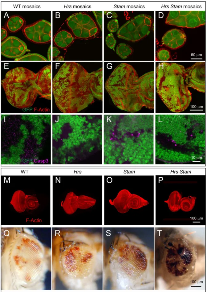
To compare the phenotype of the Stam or of the Hrs, Stam
double mutant to that ofHrsor of ESCRT-I, -II, -III mutants, we generated clones of cells mutant forStam2L2896(Mutant cells are GFP-negative; see Material and Methods) in the follicular epithelium (FE) of the Drosophila ovary. As it is the case of FE cells mutant for HrsD28, Stam mutant FE cells display normal epithelial morphology (Fig. 1A–C). Similarly, we observed no detectable phenotype when we generated mosaic eye imaginal discs (Fig. 1E–G) or eye imaginal discs consisting predominantly of
mutant cells (Fig. 1M–O) for bothHrsandStam. In contrast, cells homozygous for a recently reported HrsD28, Stam2L2896
double mutant allele [23] formed large clones of mesenchymal-like cells (Fig. S1A). Additionally, mosaic eye imaginal discs or eye imaginal discs consisting predominantly of cells mutant for both Hrs and Stam showed a similar loss of epithelial architecture phenotype (Fig. S1B,D).
BothHrsandStamare on chromosome 2L, which harbors in a sub-telomeric position the tumor suppressor l(2)gl, a gene frequently lost by spontaneous deletion [29]. Thus, we wondered whether theHrs, Stamdouble mutant chromosome present in the Bloomington stock center carried a mutation inl(2)gl. Failure to complement the null allele l(2)gl4 indicated a possible lesion in
l(2)glon the chromosome carrying bothHrsandStammutations. To test if it was indeed the case, we recombined away the distal part of chromosome 2L containing l(2)gl from the Hrs Stam
chromosome and retested for complementation. We isolated several independent recombinants that fail to complementHrsand
Stamdeficiencies but complementl(2)gl4,a further indication of the presence ofl(2)glmutation in the originalHrs Stamchromosome (Hrs Stam l(2)gl triple mutant henceforth; see Material and Methods).
ESCRT-0 Components are not Required for Tumor Suppression in Drosophila
To test whether the HrsD28, Stam2L2896 mutant chromosome devoid of the l(2)gl mutation still possessed tumor-promoting ability, we analyzed mosaic FE, mosaic eye discs, or eye discs consisting predominantly of cells mutant for the recombined allele. Interestingly, these do not display loss of tissue architecture (Fig. 1D, H, P), as is the case of singleHrsorStammutant alleles, suggesting that thel(2)gllesion in the original double mutant allele was responsible for the loss of tumor suppression phenotypes. These data indicate that simultaneous loss of both ESCRT-0 components do not lead to loss of tissue architecture, a striking difference to ESCRT-I, -II, -III mutations, which are tumorigenic [11,17,18]. Consistent with this surprising difference, we found that eye discs consisting predominantly of cells mutant forHrs, or
Stamor bothHrsandStamprogress to form adult eyes. These are smaller than wild-type and have a rough appearance but contain some mutant photoreceptors (Fig. 1Q–T) The scarcity of mutant adult photoreceptors might be due to cell death, as we occasionally see apoptotic cells in clones ofHrs, orStamor bothHrsandStam
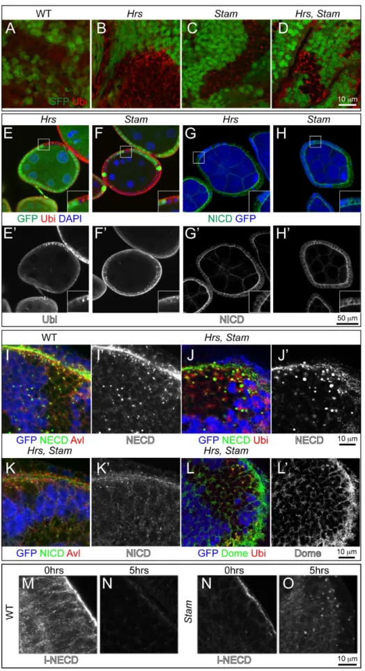
double mutants (Fig. 1I–L). In sheer contrast to these, a number of ESCRT-I, -II, -III mutations, such as those mapping toTsg101, vps28, Vps25, vps20,when made homozygous in eye discs, display a Mutant Eye No Eclosion (MENE) phenotype that have been associated loss of tumor suppression in Drosophila [30]. Overall, these data suggest that the activity ofHrsand Stamis not tumor suppressive in two different Drosophila epithelial tissues. negative), as revealed by an antibody against mono- and poly- ubiquitin chains (Ubi). High magnification of the boxed areas is shown in insets. (G–H) Mutant FE cells (GFP-negative) show accumulation of the Notch receptor. Notch receptor has been revealed using anti-NICD specific to the intracellular domain of Notch. Apical as well as intracellular accumulations of Notch ICD epitope is seen inHrsandStamFE mutant cells. High magnification of the boxed areas is shown in insets. (I–K) Co-localization with anti Notch ECD (NECD) or Notch ICD (NICD) and Avl, marking early endosomes, in mosaic eye imaginal discs. Notch ECD is mainly accumulated in early endosomes in GFP-negative mutant tissue. (L–L’) Mosaic eye imaginal discs were stained with Ubi and anti-Domeless (Dome). Hrs, Stammutant cells (GFP-negative) accumulate ubiquitylated cargoes and moderate levels of Dome, compared to WT. (M–O) Endocytic trafficking assay with anti-Notch ECD to label Notch at the surface of living imaginal discs. In WT tissue, after labeling (0 hrs), Notch is present mostly at the apical surface of the cell. After a 5-hour chase (5 hrs) Notch is completely degraded in WT but still present in endosomes inStammutant discs, indicating that Notch is internalized but it is not degraded.
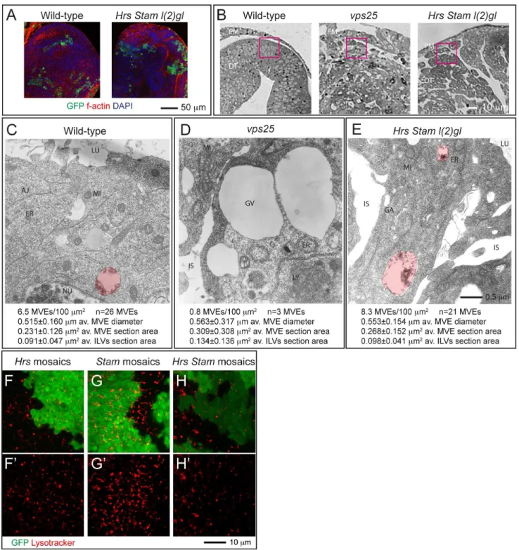
Figure 3. ESCRT-0 mutants do not affect endosomal maturation.(A) Example of eye tissue almost completely homozygous for a WT (left) or mutant chromosome (right). WT cells are marked with GFP and represent 10% of the disc tissue. (B) Phase contrast images of cross-sections used for EM show the indicative regions used for ultrastructural analysis (pink boxes). Note the absence of monolayer architecture in sections of mutant tissues. (C–E) Electron micrograph of sections of eye disc tissue of the indicated genotype. A portion of the apical part of 2–3 epithelial cells above the level of the basal nuclei is shown. While MVEs (highlighted in red) are absent inVps25mutant cells, they are present in ESCRT-0 mutant cells. Quantification of MVE density, diameter, section area and ILV content is presented below each panel. (F–H) Incorporation of Lysotracker in mosaic discs. A single subapical confocal cross-section is shown in each panel, showing no difference in acidification in WT (GFP-positive) versus mutant cells. Labels are as follows: PM: peripodial membrane, DT: disc tissue, LU: Apical lumen, AJ: Adherens Junctions, ER: Endoplasmic Reticulum, GA: Golgi apparatus, MI: Mitocondrium, NU: Nucleus, GV: giant vacuoles, IS: interstitial space between unpolarized cells.
doi:10.1371/journal.pone.0093987.g003
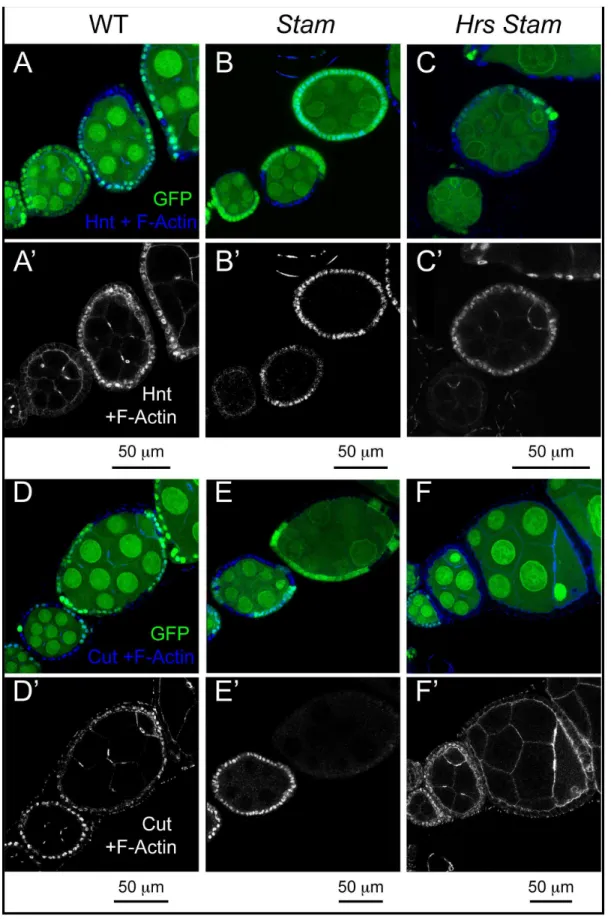
Figure 4. ESCRT-0 mutations do not alter Notch signaling in FE cells.(A–F) Mosaic egg chambers at stages 5–7 of oogenesis stained to detect the Notch targets Hnt (A–C) and Cut (D–F) and f-Actin.StamorHrs Stammutant cells are marked by the absence of GFP. In bothStamandHrs Stam mutant FE cells, Hnt is normally expressed and Cut normally downregulated after stage 6, indicating no impairment of Notch signaling activation. (A’–F’) show single channels.
Impaired ESCRT-0 Activity Leads to Accumulation of Ubiquitin, Notch and Dome
To test whether ESCRT-0 mutants are able to sort ubiquity-lated cargoes, we immunostained mosaic eye disc and FE cells containing clones of cells mutant forHrs,orStamor bothHrsand
Stamwith an antibody specific to mono- and poly-ubiquitin chains. In contrast to WT cells, but similarly to previous reports ofHrsand of ESCRT-I, -II, -III mutants [20–22], Hrs, Stam and Hrs Stam
mutant cells, as well as Hrs Stam l(2)gl triple mutant cells accumulated ubiquitin (Fig. 2A–F; Fig. S1E–F).
The Notch receptor is a cargo prominently subjected to endosomal sorting in Drosophila discs and FE cells [11–13]. To assess whether Notch is sorted and degraded in endosomes of ESCRT-0 mutant cells, we immunolocalized Notch inHrs,Stam
single or doubleHrs, Stammutant cells and inHrs Stam l(2)gltriple mutant cells. Compared to WT cells, mutant eye disc cells displayed accumulation of Notch, as assessed with an antibody that recognizes the extracellular domain of Notch (NECD). Accumulation is less evident using an antibody to the intracellular portion (NICD), and is not present inl(2)glmutant discs (Fig. 2G– K; Fig. S1G, I–K). Similarly, we found accumulation of Domeless (Dome), the single-pass non-tyrosine-kinase receptor for JAK/ STAT signaling (Fig. 2L; Fig. S1H).
To follow sorting and degradation of transmembrane proteins over time, we performed a Notch endocytic trafficking assay in living imaginal discs [22]. Briefly, we cultured freshly dissected discs in insect media in presence of a Notch antibody that recognizes an extracellular epitope. We then washed and chased internalization of the bound antibody overtime. In contrast to WT discs, but likeHrs andVps25mutant discs [22], Stammutant, or
Hrs Stam l(2)gltriple mutant disc cells displayed various degrees of intracellular signal after a 5 hrs chase, indicating that they fail to degrade endosomal Notch (Fig. 2K–N; Figure S1J–K). Co-staining of Notch with the early endosomal marker Avalanche (Avl) reveals that undegraded Notch and ubiquitin accumulate for the most part in early endosomes (Fig. 2I; Fig. S1I). Overall, these data are consistent with a general defect in endosomal sorting and degradation of ubiquitylated cargoes, including signaling recep-tors, in ESCRT-0 mutants.
ESCRT-0 is not Required for Endosome Maturation Given the accumulation of ubiquitin and of Notch and Dome in ESCRT-0 mutant cells, we next assayed whether mutant cells possess mature endosomes. One aspect of endosome maturation involves formation of ILVs during MVE biogenesis. To test whether ILV formation occurs in ESCRT-0 mutants, we analyzed the morphology of mutant cells at the ultrastructural level. To this end, we generated eye discs mutant forStam, orHrs, Stam l(2)glor
Vps25, encoding the eponymous ESCRT-II component. Discs containing a minimal amount of non homozygous cells (Fig. 3A; GFP-positive), were sectioned and the tissue facing the peripodial membrane covering the disc was analyzed (Fig. 3B). In sections from control discs containing WT cells, we could observe several MVEs with an average diameter of roughly 500 nm and a little less than half of their section represented by ILVs (Fig. 3C). In contrast, inVps25mutant cells, we detected very few MVEs with irregular size and ILV content (Fig. 3D), as previously reported for
Drosophilamutants in ESCRT-II components [18]. In these cells, we often observe the presence of very large (diameter.1500 nm) clear vacuoles. Due to the loss of apico-basal polarity of Vps25
mutant cells, we also find large interstitial spaces (Fig. 3D). In tissue mutant for Hrs, Stam, l(2)gl we find MVEs that are indistinguishable in abundance and features to those of WT cells (Fig. 3E), despite the presence of tissue disorganization similar to
that ofVps25cells, due to thel(2)glmutation which,per se, does not affect trafficking (Fig. S1J). These data are consistent with previous results inDrosophilaGarland cells [23] and indicate that, different to ESCRT-II, ESCRT-0 components are dispensable for MVE biogenesis in epithelial tissue.
Another aspect of endosomal maturation is the progressive acidification of the lumen of endosomes. To test whetherHrs, or
StamorHrs and Stammutant cells possess acidic endo-lysosomal organelles, we cultured mosaic discs in presence of Lysotraker, a vital dye that concentrates in acidic compartments. Compared with WT cells, clones ofHrsmutant cells incorporate equal levels of Lysotracker, consistent with previous evidence [31](Fig. 3E). Similarly,StamorHrs Stam mutant cells are indistinguishable to surrounding WT cells, indicating no impairment of the ability to acidify endocytic organelles (Fig. 3F–G). Taken together, these data suggest that loss of Hrs, Stam or both do not affect endosomal maturation.
ESCRT-0 is not Required for Notch Signaling Activation or Downregulation
Accumulation of Notch in endosomes of ESCRT-I, -II, -III mutants correlates with ectopic and ligand-independent Notch signaling [18]. In contrast, mutations that disrupt earlier steps of endocytic vesicle trafficking such as those affectingRab5andavl, inhibit activation of Notch [22]. Therefore, it is unclear what to expect in ESCRT-0 mutants. To assay Notch activation in mutant FE cells, we monitored expression of the transcription factors Hindsight (Hnt) and Cut, whose expression is modulated by Notch activation at mid-oogenesis. In fact, upon expression of the ligand Delta in germline cells at stage 6 of oogenesis, Notch signaling is activated in neighboring FE cells. As a result, FE cells downreg-ulate Cut expression, unpregdownreg-ulate Hnt expression, arrest mitotic cell cycles and begin to endoreplicate [32,33]. Surprisingly, the pattern of Hnt and Cut expression detected by immunofluores-cence in small clones ofHrs,orStam,orHrs, Stammutant FE cells at stage 5–7 was unchanged, when compared to WT cells, indicating that Notch activation is not altered in ESCRT-0 mutants (Fig. 4A–F). In clear contrast, precocious expression of Hnt before stage 6 is observed in ESCRT-I mutant FE cells [22]. InHrs Stam l(2)gltriple mutant cells, ectopic Hnt expression and failure to downregulate Cut expression is visible only in multi-layering cells that do not contact the germline and are likely to not be reached by the ligand (Fig. S1L–M). Overall, our data confirm and extend the notion that ESCRT-0 activity is not required for Notch signaling and that its loss do not promote ectopic, ligand-independent activation, as observed in other ESCRT-I, -II, -III mutants [18,20,21].
Discussion
The ESCRT-0 Complex is Dispensable for Tumor Suppression inDrosophila
In this study, we study the effects of impairment of ESCRT-0 function on Drosophila epithelial tissue development in vivo. We found that the recently reported [23,24]HrsD28Stam2L2896double mutant allele carries a third mutation inl(2)gl,which we show is responsible for the loss of tumor suppressor (TS) phenotype of triple mutant tissue (Fig. S1). We analyzed independent HrsD28 Stam2L2896
reported in the literature are due to impairment ofl(2)glactivity [23], we advise future use of our recombinant chromosome devoid of thel(2)gllesion.
The fact that ESCRT-0 is dispensable for tumor suppression (TS) marks a striking difference to mutations in most genes encoding components of the downstream ESCRT-I, -II, -III complexes. The discrepancy between the tissue architecture phenotypes of ESCRT-0 and other ESCRTs could be explained by different scenarios that we discuss below.
1) ESCRT TS function is not linked to endosomal sorting: ESCRT-III is very ancient, it is present in archaea and unicellular organisms [34], in which its membrane bending capacity is mostly used in the last step of cytokinesis [35,36]. In contrast, ESCRT-0 has evolved recently, is dispensable for completion of cytokinesis, and might represent a specialization to sort a subset of cargoes in endosomes [37]. However, we do not favor the idea that the tumor suppression activity of ESCRT complexes correlates with their involvement in cytokinesis. In fact, while cell division and cytokinesis defect have been extensively linked to tumorigenesis, ESCRT-II, another ESCRT complex that behaves as TS, is dispensable for cytokinesis [35,36].
2) ESCRT TS function is linked to endosomal sorting and residual Hrs or Stam function might be present in mutants: We think this is unlikely because bothHrsD28andStam2L2896
are null alleles to the best of our knowledge. In fact, HrsD28
expresses only the amino terminal first quarter of the protein, and is devoid of most functional domains [20], while
Stam2L2896line harbors a non sense mutation leading to an early stop codon at amino acid 6 [24]. Both genes have no paralogs inDrosophila. In addition, quantitative RT-PCR also shows that in bothHrsandHrs Stam l(2)glmutants only 50% of theHrstranscript is present. In bothStamandHrs Stam l(2)gl
mutant tissues, only 20–30% of theStamtranscript is present (Fig. S2), indicating that in either backgrounds both mutant
HrsandStamtranscripts are possibly subjected to non sense-mediated decay, and further decreasing the likelihood of residual function.
3) ESCRT TS function is linked to endosomal sorting, but the relevant cargoes do not require Hrs or Stam for their sorting: Several studies suggested that alternative ESCRT-0 proteins may work in parallel, or even instead of Hrs and Stam. Good candidates are two families of proteins, GGAs and Tom1 (target of Myb1), both present inDrosophila,that have similar characteristics to those of ESCRT-0 components. These in fact contain VHSs, Ubiquitin binding, and Clathrin binding domains typical of ESCRT-0 components, they recruit ubiquitylated proteins to endosomal membranes, and they interact with I and Clathrin [38–40]. Thus, ESCRT-0 complex could be dispensable for sorting of TS-relevant proteins. Interestingly, endocytosis of junctional adhesion proteins, such as E-Cadherin, directly regulates polarity in
Drosophila epithelia [41]. Consistent with a minor role of ESCRT-0 in controlling polarity, a study showed that mutation inDrosophila Hrsdoes not affect the localization of DE-Cadherin [21]. In contrast, junctional adhesion proteins appear sensitive to function of more downstream ESCRTs. Indeed, ESCRT-I and -III have been shown to be required for degradation of adhesive molecules, such as Claudin-1, and for maintenance of polarity in vertebrate epithelial cells [42]. Thus, we predict that proteins that play a role in ensuring correct epithelial architecture and polarity, such as those
involved in cell-cell adhesion, might not require ESCRT-0 for their sorting and degradation.
ESCRT-0 is Dispensable for Ectopic Notch Activation in Endosomes
InStamand double mutants we observed accumulation of Notch receptor in endosomes, especially when immunolocalizing with an anti-Notch ECD, which recognizes the extracellular portion of Notch. It is not clear why the accumulation is less evident by immunolocalization of the intracellular portion of Notch with anti-Notch ICD. It is possible that either the latter accumulated less that the former, perhaps due to the fact that Notch is normally activated in mutant cells, or the two antibodies possess different efficiency in recognizing their epitopes. However the case, we were surprised to find no ectopic activation of Notch signaling. A trivial possibility to explain the difference with ESCRT-I, -II, -III mutants is that Notch trafficking and degradation might be quantitatively less affected than in ESCRT-0 mutant. Although our assays are not quantitative, the genetic nature of the ESCRT-0 mutants renders this possibility rather unlikely.
Another possibility is that recycling from endosomes to the plasma membrane might be important to prevent ectopic Notch receptor activation and recycling might still functioning in ESCRT-0 mutants, however it might not in ESCRT-I, -II, -III mutants. At present, whether the Notch receptor is recycled to the plasma membrane, and the status of recycling on different ESCRT mutant in Drosophila epithelia are unknown, preventing us to conclude on the likelihood of such an hypothesis. However, we observe an accumulation of NECD in endosomes of ESCRT-0 mutants that is comparable to that of ESCRT-I, -II, -III, an evidence that appears to contrast with the possibility of substantial recycling of Notch in ESCRT-0 mutants.
Finally, Notch accumulation in endosomes in ESCRT-0 mutants might not yield ectopic activation because such forced and ligand-independent Notch activation might require the cargo clustering by ESCRT-0 on the limiting membrane of endosomes. Alternatively, endosomes of ESCRT-0 mutant cells might not be mature enough to permit ligand-independent activation. These two not necessarily mutually exclusive possibilities are supported by the fact that Hrs and Stam act with Clathrin to trap and concentrate cargoes to be degraded on the endosomal membrane [9], and by evidence in Drosophila suggesting that ligand-independent Notch activation occurs in late endosome/lysosomes and depends on endosome acidification and maturation [31,43– 46]. Our data clearly indicate that two aspects of endosomal maturation, MVE biogenesis and endolysosomal acidification occur normally in ESCRT-0 mutants, suggesting that these could support later events required for ectopic Notch activation. The fact that ectopic Notch activation is not observed in the mutants thus points to cargo clustering as a potential prerequisite for ectopic activation of Notch. Whether cargo clustering is required for efficient Notch cleavage requires further studies.
Supporting Information
Figure S1 l(2)gl is responsible for the loss of tumor suppression phenotype in triple mutants.(A–B) Epithelial disorganization inHrs, Stam l(2)gltriple mutant tissues revealed by staining to detect sub-cortical f-Actin. FE cells homozygous for
Hrs, Stam l(2)gl(marked by the absence of GFP) in a stage 5–6 egg chamber are misshapen and multilayered. Eye imaginal cells homozygous for the mutations also show mesenchymal-like cells and autonomous disruption of epithelial organization. (C) Mosaic eye imaginal discs stained with antibody anti-activated Caspase 3 to mark apoptotic cells. In Hrs Stam l(2)gl mutant tissue (GFP negative) apoptosis is activated. (D) Eye imaginal disc formed by mutant cells homozygous forHrs, Stam l(2)glstained to detect sub-cortical f-Actin show a tumor-like phenotype. (E–H)Hrs Stam l(2)gl
mosaic eye and FE cells (marked by the absence of GFP) stained to detect ubiquitin, Notch and the Domeless receptors. Separate channels are shown in E’F’G’H’. Cells homozygous forHrs Stam l(2)gl show accumulation of ubiquitin, Notch and Domeless intracellularly. (I–K) Co-localization with anti Notch ECD (NECD) or Notch ICD (NICD) and Avl, marking early endosomes, in mosaic eye imaginal discs. Notch ECD is mainly accumulated in early endosomes in GFP-negativeHrs, Stam l(2)gl
triple mutant tissue, but not in (2)gl mutant tissue. (L–M) Endocytic trafficking assay with anti-Notch ECD to label Notch at the surface of living Hrs Stam l(2)gl mutant eye disc. Notch receptor fails to be degraded in mutant cells and it remains accumulated intracellularly after 5 hrs after the endo of labeling
(0 hrs). (N–O) Mosaic egg chambers at stages 5–7 of oogenesis stained for Hnt and Cut. Hrs Stam l(2)gl homozygous cells are marked by the absence of GFP. InHrs, Stam l(2)gltriple mutant cells Hnt expression and failure to downregulate Cut expression is visible only in multilayering cells that are likely not reached by the ligand, indicating that Notch activation is not affected in mutant cells that are exposed to the ligand and Notch is not ectopically activated in those that are not. WT controls for all panels are presented in Fig. 1–2, 4.
(JPG)
Figure S2 Mutant transcripts for Hrs and Stam are subjected to non sense-mediated decay.Quantitative RT-PCR experiment on mRNA extracted from eye imaginal discs from single Hrs or Stam or double Hrs, Stam or triple Hrs, Stam, l(2)gl mutant tissue compared to control indicates reduction ofHrs
orStammRNA expression in corresponding mutant extracts. (TIF)
Acknowledgments
We thank Stephane Noselli, the BDSC and the DHSB for reagents, and Simona Polo for critically reading the manuscript.
Author Contributions
Conceived and designed the experiments: TV. Performed the experiments: ET NW KC. Analyzed the data: TV ET NW KC CT. Wrote the paper: TV ET.
References
1. Sigismund S, Confalonieri S, Ciliberto A, Polo S, Scita G, et al. (2012) Endocytosis and signaling: cell logistics shape the eukaryotic cell plan. Physiological reviews 92: 273–366.
2. Mellman I, Yarden Y (2013) Endocytosis and cancer. Cold Spring Harbor perspectives in biology 5: a016949.
3. Babst M, Katzmann D, Estepa-Sabal E, Meerloo T, Emr SD (2002) Escrt-III: an endosome-associated heterooligomeric protein complex required for mvb sorting. Developmental cell 3: 271–282.
4. Babst M, Katzmann D, Snyder W, Wendland B, Emr SD (2002) Endosome-associated complex, ESCRT-II, recruits transport machinery for protein sorting at the multivesicular body. Developmental cell 3: 283–289.
5. Katzmann D, Babst M, Emr SD (2001) Ubiquitin-dependent sorting into the multivesicular body pathway requires the function of a conserved endosomal protein sorting complex, ESCRT-I. Cell 106: 145–155.
6. Katzmann D, Stefan C, Babst M, Emr SD (2003) Vps27 recruits ESCRT machinery to endosomes during MVB sorting. The Journal of cell biology 162: 413–423.
7. Bilodeau PS, Urbanowski JL, Winistorfer SC, Piper RC (2002) The Vps27p– Hse1p complex binds ubiquitin and mediates endosomal protein sorting. Nature Cell Biology: 8.
8. Piper R, Cooper A, Yang H, Stevens T (1995) VPS27 controls vacuolar and endocytic traffic through a prevacuolar compartment in Saccharomyces cerevisiae. The Journal of cell biology 131: 603–617.
9. Sachse M, Urbe´ S, Oorschot V, Strous GJ, Klumperman J (2002) Bilayered clathrin coats on endosomal vacuoles are involved in protein sorting toward lysosomes. Molecular biology of the cell 13: 1313–1328.
10. Rusten TE, Vaccari T, Stenmark H (2012) Shaping development with ESCRTs. Nature Cell Biology 14: 38–45.
11. Vaccari T, Bilder D (2005) The Drosophila tumor suppressor vps25 prevents nonautonomous overproliferation by regulating notch trafficking. Developmen-tal cell 9: 687–698.
12. Thompson B, Mathieu J, Sung H, Loeser E, Rorth P, et al. (2005) Tumor suppressor properties of the ESCRT-II complex component Vps25 in Drosophila. Dev Cell 9: 711–720.
13. Moberg K, Schelble S, Burdick S, Hariharan I (2005) Mutations in erupted, the Drosophila ortholog of mammalian tumor susceptibility gene 101, elicit non-cell-autonomous overgrowth. Developmental cell 9: 699–710.
14. Robinson BS, Moberg KH (2011) Drosophila endocytic neoplastic tumor suppressor genes regulate Sav/Wts/Hpo signaling and the c-Jun N-terminal kinase pathway. Cell cycle (Georgetown, Tex) 10: 4110–4118.
15. Herz H, Chen Z, Scherr H, Lackey M, Bolduc C, et al. (2006) vps25 mosaics display non-autonomous cell survival and overgrowth, and autonomous apoptosis. Development (Cambridge, England) 133: 1871–1880.
16. Woodfield SE, Graves HK, Hernandez JA, Bergmann A (2013) De-regulation of JNK and JAK/STAT signaling in ESCRT-II mutant tissues cooperatively contributes to neoplastic tumorigenesis. PLoS ONE 8: e56021.
17. Herz H-M, Woodfield SE, Chen Z, Bolduc C, Bergmann A, et al. (2009) Common and Distinct Genetic Properties of ESCRT-II Components in Drosophila. PLoS ONE 4: e4165.
18. Vaccari T, Rusten TE, Menut L, Nezis IP, Brech A, et al. (2009) Comparative analysis of ESCRT-I, ESCRT-II and ESCRT-III function in Drosophila by efficient isolation of ESCRT mutants. Journal of cell science 122: 2413–2423. 19. Stuffers S, Brech A and Stenmark H (2008) ESCRT proteins in physiology and
disease. Experimental cell research.
20. Lloyd T, Atkinson R, Wu M, Zhou Y, Pennetta G, et al. (2002) Hrs regulates endosome membrane invagination and tyrosine kinase receptor signaling in Drosophila. Cell 108: 261–269.
21. Jekely G, Rorth P (2003) Hrs mediates downregulation of multiple signalling receptors in Drosophila. EMBO reports 4: 1163–1168.
22. Vaccari T, Lu H, Kanwar R, Fortini ME, Bilder D (2008) Endosomal entry regulates Notch receptor activation in Drosophila melanogaster. The Journal of cell biology 180: 755–762.
23. Chanut-Delalande H, Jung AC, Baer MM, Lin L, Payre F, et al. (2010) The Hrs/Stam complex acts as a positive and negative regulator of RTK signaling during Drosophila development. PLoS ONE 5: e10245.
24. Chanut-Delalande H, Jung AC, Lin L, Baer MM, Bilstein A, et al. (2007) A genetic mosaic analysis with a repressible cell marker screen to identify genes involved in tracheal cell migration during Drosophila air sac morphogenesis. Genetics 176: 2177–2187.
25. Newsome T, Asling B, Dickson BJ (2000) Analysis of Drosophila photoreceptor axon guidance in eye-specific mosaics. Development 127: 851–860. 26. Tapon N, Ito N, Dickson BJ, Treisman J, Hariharan I (2001) The Drosophila
tuberous sclerosis complex gene homologs restrict cell growth and cell proliferation. Cell 105: 345–355.
27. Lee T, Luo L (2001) Mosaic analysis with a repressible cell marker (MARCM) for Drosophila neural development. Trends Neurosci 24: 251–254.
28. Goentoro LA, Yakoby N, Goodhouse J, Schu¨pbach T, Shvartsman SY (2006) Quantitative analysis of the GAL4/UAS system inDrosophila oogenesis. Genesis 44: 66–74.
29. Roegiers F, Kavaler J, Tolwinski N, Chou Y-T, Duan H, et al. (2009) Frequent Unanticipated Alleles of lethal giant larvae in Drosophila Second Chromosome Stocks. Genetics 182: 407–410.
30. Menut L, Vaccari T, Dionne H, Hill J, Wu G, et al. (2007) A mosaic genetic screen for Drosophila neoplastic tumor suppressor genes based on defective pupation. Genetics 177: 1667–1677.
31. Vaccari T, Duchi S, Cortese K, Tacchetti C, Bilder D (2010) The vacuolar ATPase is required for physiological as well as pathological activation of the Notch receptor. Development 137: 1825–1832.
32. Deng W, Althauser C, Ruohola-Baker H (2001) Notch-Delta signaling induces a transition from mitotic cell cycle to endocycle in Drosophila follicle cells. Development (Cambridge, England) 128: 4737–4746.
33. Lopez-Schier H, St Johnston D (2001) Delta signaling from the germ line controls the proliferation and differentiation of the somatic follicle cells during Drosophila oogenesis. Genes & development 15: 1393–1405.
34. Samson RY, Obita T, Freund SM, Williams RL, Bell SD (2008) A role for the ESCRT system in cell division in archaea. Science (New York, NY) 322: 1710– 1713.
35. Carlton JG, Martin-Serrano J (2007) Parallels between cytokinesis and retroviral budding: a role for the ESCRT machinery. Science (New York, NY) 316: 1908– 1912.
36. Morita E, Sandrin V, Chung H-Y, Morham SG, Gygi SP, et al. (2007) Human ESCRT and ALIX proteins interact with proteins of the midbody and function in cytokinesis. The EMBO journal 26: 4215–4227.
37. Leung K, Dacks J, Field M (2008) Evolution of the multivesicular body ESCRT machinery; retention across the eukaryotic lineage. Traffic (Copenhagen, Denmark).
38. Blanc C, Charette SJ, Mattei S, Aubry L, Smith EW, et al. (2009) Dictyostelium Tom1 participates to an ancestral ESCRT-0 complex. Traffic (Copenhagen, Denmark) 10: 161–171.
39. Katoh Y, Shiba Y, Mitsuhashi H, Yanagida Y, Takatsu H, et al. (2004) Tollip and Tom1 form a complex and recruit ubiquitin-conjugated proteins onto early endosomes. The Journal of biological chemistry 279: 24435–24443. 40. Gullapalli A, Wolfe BL, Griffin CT, Magnuson T, Trejo JA (2006) An essential
role for SNX1 in lysosomal sorting of protease-activated receptor-1: Evidence for retromer-, Hrs-, and Tsg101-independent functions of sorting nexins. Molecular biology of the cell 17: 1228–1238.
41. Leibfried A, Fricke R, Morgan MJ, Bogdan S, Bellaiche Y (2008) Drosophila Cip4 and WASp define a branch of the Cdc42-Par6-aPKC pathway regulating E-cadherin endocytosis. Current Biology 18: 1639–1648.
42. Dukes JD, Fish L, Richardson JD, Blaikley E, Burns S, et al. (2011) Functional ESCRT machinery is required for constitutive recycling of claudin-1 and maintenance of polarity in vertebrate epithelial cells. Molecular biology of the cell 22: 3192–3205.
43. Kobia F, Duchi S, Deflorian G, Vaccari T (2013) Pharmacologic Inhibition Of Vacuolar H+ATPase Reduces Physiologic And Oncogenic Notch Signaling. Molecular Oncology 8: 207–220.
44. Hori K, Sen A, Kirchhausen T, Artavanis-Tsakonas S (2011) Synergy between the ESCRT-III complex and Deltex defines a ligand-independent Notch signal. The Journal of cell biology 195: 1005–1015.
45. Schneider M, Troost T, Grawe F, Martinez-Arias A, Klein T (2013) Activation of Notch in lgd mutant cells requires the fusion of late endosomes with the lysosome. Journal of cell science 126: 645–656.