Notch Extracellular Domain as Revealed by Electron
Microscopy
Deborah F. Kelly1, Robert J. Lake1¤a, Teije C. Middelkoop1¤b, Hua-Ying Fan2, Spyros Artavanis-Tsakonas1*, Thomas Walz1,3*
1Department of Cell Biology, Harvard Medical School, Boston, Massachusetts, United States of America,2Epigenetics and Progenitor Cells Program, Fox Chase Cancer Center, Philadelphia, Pennsylvania, United States of America,3Howard Hughes Medical Institute, Harvard Medical School, Boston, Massachusetts, United States of America
Abstract
Background:The Notch receptor links cell fate decisions of one cell to that of the immediate cellular neighbor. In humans, malfunction of Notch signaling results in diseases and congenital disorders. Structural information is essential for gaining insight into the mechanism of the receptor as well as for potentially interfering with its function for therapeutic purposes.
Methodology/Principal Findings:We used the Affinity Grid approach to prepare specimens of the Notch extracellular domain (NECD) of the Drosophila Notch and human Notch1 receptors suitable for analysis by electron microscopy and three-dimensional (3D) image reconstruction. The resulting 3D density maps reveal that the NECD structure is conserved across species. We show that the NECD forms a dimer and adopts different yet defined conformations, and we identify the membrane-proximal region of the receptor and its ligand-binding site.
Conclusions/Significance:Our results provide direct and unambiguous evidence that the NECD forms a dimer. Our studies further show that the NECD adopts at least three distinct conformations that are likely related to different functional states of the receptor. These findings open the way to now correlate mutations in the NECD with its oligomeric state and conformation.
Citation:Kelly DF, Lake RJ, Middelkoop TC, Fan H-Y, Artavanis-Tsakonas S, et al. (2010) Molecular Structure and Dimeric Organization of the Notch Extracellular Domain as Revealed by Electron Microscopy. PLoS ONE 5(5): e10532. doi:10.1371/journal.pone.0010532
Editor:Hilal Lashuel, Swiss Federal Institute of Technology Lausanne, Switzerland ReceivedFebruary 26, 2010;AcceptedApril 16, 2010;PublishedMay 7, 2010
Copyright:ß2010 Kelly et al. This is an open-access article distributed under the terms of the Creative Commons Attribution License, which permits unrestricted use, distribution, and reproduction in any medium, provided the original author and source are credited.
Funding:This work was supported by National Institutes of Health (NIH) grants GM062580 (to Stephen C. Harrison) and NS26084 and CA098402 (to Spyros Artavanis-Tsakonas). Thomas Walz is an investigator of the Howard Hughes Medical Institute. The funders had no role in study design, data collection and analysis, decision to publish, or preparation of the manuscript.
Competing Interests:The authors have declared that no competing interests exist.
* E-mail: [email protected] (SA-T); [email protected] (TW)
¤a Current address: Epigenetics and Progenitor Cells Program, Fox Chase Cancer Center, Philadelphia, Pennsylvania, United States of America
¤b Current address: Hubrecht Institute, Royal Netherlands Academy of Arts and Sciences and University Medical Center Utrecht, Utrecht, The Netherlands
Introduction
The signaling framework governing metazoan development is defined by a few fundamental cell interaction mechanisms, which, synergistically, control cellular fates. The Notch signaling pathway is unique among them in that it links the fate of a cell to that of the immediate neighbor through interactions of the Notch receptor expressed on one cell with membrane-bound ligands expressed on the adjacent cellular neighbor. The developmental action of Notch is highly pleiotropic, affecting a broad spectrum of tissues, cellular fates, and developmental events. Its fundamental importance in metazoan biology is underscored by evolutionary conservation and by the spectrum of mutant phenotypes associated with malfunction of Notch signaling [1,2,3]. In humans, Notch malfunction has been linked to neurovascular abnormalities, cancer and pleiotropic congenital syndromes, rendering Notch signaling a therapeutic target (e.g. [4]).
The paradigmatic Drosophila Notch receptor [5] features a large extracellular domain (NECD) (,186 kDa), a single
mem-brane-spanning a-helix, and a smaller intracellular domain
(,100 kDa). Upon ligand interactions the intracellular domain is
into the mechanism of the receptor as well as for potentially interfering with its function for therapeutic purposes, we sought to use EM combined with 3D image reconstruction to obtain first structural information of the NECD.
Results
Protein expression and EM imaging
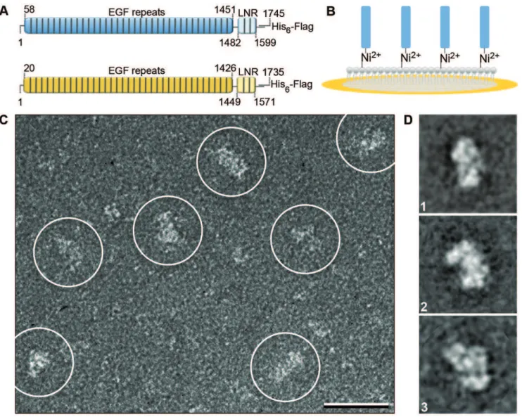
We used Sf9 insect cells to express a secreted form of the Drosophila NECD (residues 1–1745) containing a C-terminal tandem Flag-His6 tag (Figure 1A). The expression level was,
however, very low (,0.05 mg/l cell culture) and we were unable to purify the recombinant NECD using conventional chromato-graphic purification procedures. We therefore decided to test whether we could prepare specimens suitable for EM with the recently developed Affinity Grid approach [9], which is based on the idea of monolayer purification [10]. Affinity Grids are carbon-coated EM grids with a pre-deposited lipid monolayer that contains lipids whose head groups are functionalized with a
Nickel-nitrilotriacetic acid (Ni-NTA) group (Figure 1B). Because of the high affinity of Ni-NTA groups for His tags, pure preparations of a His-tagged protein can be obtained from impure protein solutions or even cell extracts, simply by incubating Affinity Grids with the solution containing the His-tagged protein and washing off the unbound proteins. When we incubated Affinity Grids with the Sf9 cell medium containing the Flag-His6-tagged NECD and
prepared the grids by negative staining, images revealed a heterogeneous particle population. Many particles appeared, however, to be sufficiently large to represent the NECD (Figure 1C). After image classification, several projection averages showed particles with defined, but variable shapes with a length of
,200 A˚ and a width of,100 A˚ (Figure 1D and Figure S1). Density maps of the extracellular domain of Drosophila Notch
To calculate 3D reconstructions, we prepared the NECD using a simplified cryo-negative staining procedure and collected 50u/0u
image tilt pairs (Figure S2A). Classification of the particles from
Figure 1. EM of the NECD.(A) Constructs used of the Drosophila (blue, residues 1–1745) and human (yellow, residues 1–1735) NECDs contained 36-EGF repeats (residues 58–1451, Drosophila and 20–1426, human) followed by 3-LNR motifs (residues 1482–1599, Drosophila and 1426–1571, human) and a C-terminal tandem Flag-His6tag. (B) Schematic drawing of an Affinity Grid, which features a pre-deposited lipid monolayer. The lipid monolayer contains Ni-NTA lipids that can recruit His-tagged NECD. (C) Raw image of negatively stained Drosophila NECD (marked by white circles; scale bar is 25 nm) and (D) representative class averages (side length of the individual panels is 43 nm).
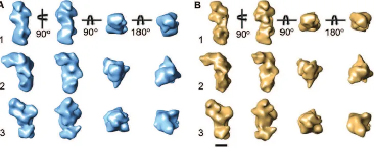
the images of the untilted specimens produced averages similar to those obtained with the negatively stained samples (Figure S2B). We combined classes that showed similar features (indicated by numbers in Figure S2B, Table S1) and calculated three individual 3D reconstructions using the random conical tilt approach [11]. The density maps at 25-A˚ resolution (Figure S4A) enclose similar volumes, but show remarkably different features (Figure 2A). Differences in the overall structure of the three density maps suggest that the NECD can undergo bending motions, but the differences in the fine structures suggest that conversion from one conformation to the others does not involve simple rigid body movements of large domains but substantial rearrangements of the individual EGF and LNR domains.
Density maps of the extracellular domain of human Notch1
While the significant differences in the fine structures between the three density maps of the Drosophila NECD are potentially interesting, as they may reflect distinct functional states, they also raised concerns regarding the accuracy of the 3D reconstructions. We reasoned that if the structural features revealed by the EM analysis of the Drosophila NECD are significant, they would be conserved across species. We therefore expressed the extracellular domain of human Notch1 (residues 1–1735) (Figure 1A), which shares 47% sequence identity with that of the Drosophila receptor.
We prepared cryo-negatively stained samples on Affinity Grids and collected 50u/0uimage tilt pairs (Figure S3A). Several of the averages obtained from classification of the particles from the images of the untilted specimens showed similar features as those of the Drosophila NECD (marked in Figure S3B) and these classes were chosen for 3D reconstruction. The three resulting density maps of the human Notch1 extracellular domain, also at a resolution of 25 A˚ (Figure S4B), are remarkably similar to the density maps of the Drosophila NECD (compare Figure 2A with Figure 2B). This result shows that the NECD structure is conserved in the Drosophila Notch and human Notch1 receptors, and that the features seen in the density maps are reproducible and thus presumably of functional importance.
Orienting the NECD structure
We performed antibody labeling to localize the membrane-proximal region of the receptor (using a monoclonal antibody against the C-terminal Flag tag) and the ligand-binding region using a monoclonal antibody raised against EGF repeats 12–20 of the Drosophila Notch receptor that encompasses at least part of the ligand-binding region defined by EGF repeats 11–13 [8,12] (Figure 3A, Figure S5). The anti-Flag tag antibody identifies one of the ends of the extended NECD as the position where the receptor inserts into the membrane (Figure 3B). By contrast, the antibody against EGF repeats 12–20 shows that the ligand-binding region is not exposed on the most membrane-distal surface of the NECD but rather on the side of the receptor (Figure 3B). Binding of a ligand to Notch could thus result in a trans, side-by-side interaction, overlapping the two membrane-bound molecules and thus bringing two neighboring cellular membranes into close apposition.
The NECD forms a dimer
The EM density maps of the extracellular domains of the Drosophila Notch and human Notch1 receptors encompass a molecular weight of about 380 kDa (Table S1). This value suggests that the NECD forms a homodimer (expected molecular weight of 372 kDa). We used scanning transmission electron microscopy (STEM) to verify that the Drosophila NECD is indeed dimeric. Continuous carbon STEM grids were coated with a Ni-NTA lipid-containing monolayer, analogous to how Affinity Grids are prepared [9], and tagged Drosophila NECD was directly adsorbed from the conditioned Sf9 insect cell medium onto these ‘‘STEM Affinity Grids’’. Analysis of 273 particles selected from 10 images of freeze-dried, unstained NECD specimens gave an average molecular mass of 381623 kDa, and fitting of the particle mass histogram with a Gaussian curve gave a peak value of 412 kDa (Figure 2C). Both values are consistent with the NECD forming a dimer.
Discussion
The extracellular domains of many cell surface receptors are composed of individual domains connected by flexible linkers. The
Figure 2. 3D reconstructions of the NECD. (A) Density maps of the Drosophila NECD corresponding to the averages shown in (B). (B) Corresponding density maps of the human NECD. Scale bar is 5 nm.
extracellular domain of the Down syndrome cell adhesion molecule (Dscam), for example, consists of ten Ig-like domains. EM images of negatively stained samples of a Dscam construct containing eight of the Ig-like domains showed it to be very flexible and to adopt many different conformations [13]. With 36 EGF and three LNR motifs, the organization of the NECD is even more complex than the extracellular domain of Dscam. However, despite its highly modular organization, our EM studies show that the NECD adopts defined and at least three distinct conformations that are conserved from fly to human. The biological significance of the three different conformations observed for the NECD remains to be determined, but it is reasonable to suggest as a working hypothesis that they may reflect distinct functional states of the receptor.
Unambiguous evidence for a multimeric structure of the Notch receptor has been lacking. The earliest evidence suggesting homotypic interactions came from the genetic behavior of the Abruptex mutations in Drosophila. These ligand-dependent
gain-of-function, point mutations affecting the NECD display negative complementation, a behavior most plausibly explained by postu-lating the existence of homotypic interactions [14]. Co-immuno-precipitation experiments supported the existence of homotypic interactions [15]. In addition, studies based on whole cell cross-linking and denaturing gel electrophoresis also seem to be consistent with this notion [16,17]. However, immunoprecipitation from cell extracts suffers from the possibility that inferred interactions may not be direct, but mediated by other cellular components. Moreover, size estimations by denaturing gel electrophoresis of the Notch receptor, a protein whose extracellular domain harbors about 250 cysteine residues, has been very unreliable in our hands. Thus, we consider that conclusions of the oligomeric state of Notch on the basis of these studies to be suggestive but not conclusive. By contrast, our EM and STEM results now conclusively demonstrate that soluble NECD exists as a dimer.
Our EM analysis revealing that the NECD forms a dimer and adopts distinct conformations now opens the way to correlate the
Figure 3. Characterization of the Drosophila NECD.(A) Projection averages (top row) and schematic representations (bottom row) of the NECD labeled with antibodies specific for the ligand-binding site (LBS Ab) and for the Flag tag (Flag Ab) in comparison with the corresponding average of unlabeled NECD (Reference). The side length of the individual panels is 43 nm. (B) EM density map of Drosophila NECD showing the positions of the ligand-binding site (LBS) and the membrane-insertion site (C terminus) deduced from the antibody labeling. (C) Histogram indicating the mass distribution of particles imaged by STEM.
functional state of mutations as revealed by genetic analyses to the receptor structure. It will be particularly interesting to analyze whether mutations such as the Abruptex mutations or mutations underlying the catastrophic neurodegenerative disease CADASIL in humans, which also correlate with extracellular point mutations [18], affect the oligomeric state and/or the conformation of the NECD. It would be equally interesting to examine whether the different NECD conformations represent different functional states, and in particular whether cis and trans interactions with the ligands Delta and Serrate induce the NECD to favor a particular conformation, which would make it possible to identify which NECD conformations represent the active and inhibited states of the receptor.
Methods
Expression of NECDs
The extracellular domains of Drosophila Notch (amino acids 1– 1745) and human Notch1 (1–1735) were cloned into pFastBac1 with C-terminal tandem His6 and Flag tags. Proteins were
expressed in Sf9 cells using a baculovirus expression system. The secreted NECDs were directly purified from the culture media using Affinity Grids [9].
Affinity Grid purification of NECDs
Affinity Grids were prepared as described [9]. 50 mM imidazole (final concentration) was added to the insect cell media containing the Flag-His6-tagged NECD of either Drosophila Notch or human
Notch1. 3-ml aliquots of the cell media were incubated for 2
minutes on continuous carbon (CC) Affinity Grids covered with a lipid monolayer composed of 2% or 20% Ni-NTA (1,2-dioleoyl-sn
-glycero-3-[N(5-amino-1-carboxypentyl)iminodi acetic acid] succi-nyl-nickel salt) lipid and DLPC (1,2-dilauryl-sn
-glycero-3-phos-phatidylcholine) as filler lipid. Lipids were purchased from Avanti Polar Lipids (Alabaster, AL).
Specimen preparation
Negative staining. 3-ml aliquots of medium containing
Flag-His6-tagged NECD were incubated for 2 minutes on 2% CC
Affinity Grids. The grids were blotted from the side, washed with one drop of 0.75% uranyl formate and stained for 20 seconds with another drop of 0.75% uranyl formate. The grids were blotted and allowed to air-dry.
Cryo-negative staining. 3-ml aliquots of medium containing
Flag-His6-tagged NECD were incubated for 2 minutes on 20%
CC Affinity Grids. The solution was gently removed with a Hamilton syringe and 3-ml aliquots of a solution containing 1%
trehalose (w/v) and 1% (w/v) uranyl formate were added to the grids. The grids were blotted for 3 seconds and plunged into liquid ethane using a Vitrobot (FEI Company, Hillsboro, Oregon) operated at 22uC and 65% relative humidity.
Electron microscopy
Specimens were imaged in an FEI Tecnai 12 electron microscope (FEI, Hillsboro, OR) equipped with an LaB6filament
and operated at an acceleration voltage of 120 kV. Images of untilted, negatively stained samples were collected at room temperature, and image pairs of cryo-negatively stained samples at tilt angles of 50u and 0u were collected at liquid nitrogen temperature using a Gatan 626 cryo-specimen holder (Gatan, Pleasanton, CA). Images were recorded using low-dose procedures on imaging plates at a nominal magnification of 67,0006and
defocus values of about21.8mm (tilted specimens) and21.5mm
(untilted specimens). Imaging plates were scanned with a Ditabis
scanner (Pforzheim, Germany) using a step size of 15mm, a gain
setting of 20,000 and a laser power setting of 30% [19]. The images were binned over 262 pixels for a final sampling of 4.5 A˚ /
pixel at the specimen level.
Image processing
For negatively stained specimens of the Drosophila NECD, 6917 particles were interactively selected from 56 images using WEB, the display program associated with the SPIDER software package [20], which was used for further image processing. The particles were windowed into individual 96696 pixels images,
rotationally and translationally aligned, and subjected to 10 cycles of multi-reference alignment. Each round of multi-reference alignment was followed by K-means classification into 20 classes. The references used for the first multi-reference alignment were randomly chosen from the raw images.
For cryo-negatively stained specimens, 13,802 particles pairs were interactively selected from 112 50u/0u image pairs of Drosophila NECD, and 12,393 particle pairs from 112 50u/0u
image pairs of human NECD. The particles were windowed into individual 96696 pixels images, and the particles from the images of the untilted specimens classified into 20 classes as described above.
Particles from classes that showed the same projection average (denoted as 1, 2 and 3 in Figures S2B and Figure S3B) were combined (summarized in Table S1). The particle images from the images of the tilted specimens were then used to calculate six individual 3D reconstructions (3 for the Drosophila and 3 for the human NECD) using the back-projection algorithm implemented in SPIDER. The orientation parameters of the particles (all particles from the images of the tilted specimens and 10% of the particles from the images of the untilted specimens) were improved over 10 refinement cycles using FREALIGN v 7.05 [21], which was also used to correct for the contrast transfer function (CTF) of the microscope. The correct defocus value for each particle image was deduced from the position of each particle in the image and the tilt angles and defocus values of the images, which were determined with CTFTILT [22]. FREALIGN was first run for one cycle using mode 3 (systematic parameter search) with an angular step of 7uto determine initial orientation parameters for each particle relative to the starting model. The resulting parameters were iteratively refined over 9 additional cycles run in mode 1 (local parameter refinement) including data in the 200 -10 A˚ resolution range. The resolution of the final density maps were estimated to be 25 A˚ using Fourier shell correlation (FSC) and the FSC = 0.5 cut-off criterion [23] (Figure S4), and the density maps were low-pass filtered to that resolution.
Preparation of STEM Affinity Grids and NECD samples
All STEM experiments were conducted at the Brookhaven National Laboratory facilities headed by Dr. Joseph Wall. Lipid monolayers were prepared according to [10] and contained 20% Ni-NTA lipid. A thin layer of continuous carbon was deposited on STEM holey carbon grids. The grids were then placed on the 20% Ni-NTA lipid-monolayer for 1 minute, gently lifted off with forceps, blotted from the side, and allowed to air-dry. 3-ml aliquots
of insect cell medium containing Flag-His6-tagged Drosophila
Antibody labeling of the Drosophila NECD
Monoclonal antibody C458.2H for the ligand-binding site was raised against Drosophila EGF repeats 12–20 [26], and Flag tag-specific M2 antibody was purchased from Sigma-Aldrich (St. Louis, MO). Antibodies were diluted to 0.1 mg/ml in buffer containing 20 mM Hepes, pH 7.4, 150 mM NaCl, 5 mM MgCl2
and 10 mM CaCl2. 10ml of the antibody solutions were mixed
with 10ml insect cell medium containing Flag-His6-tagged
Drosophila NECD and 50 mM imidazole. 3-ml aliquots of these mixtures were applied to 2% CC Affinity Grids and incubated for 2 minutes prior to blotting and negative staining. The samples were imaged in the electron microscope as described above. 8890 particles were selected from 168 images of Drosophila NECD labeled with the antibody against the ligand-binding site, and 10,214 particles were selected from 168 images of Drosophila NECD labeled with the anti-Flag antibody. The particles in each data set were classified into 100 classes as described above.
Supporting Information
Table S1 3D reconstructions.
Found at: doi:10.1371/journal.pone.0010532.s001 (0.04 MB DOC)
Figure S1 Averages obtained by classifying 6917 particle images of negatively stained Drosophila NECD into 20 classes. The side length of the individual panels is 43 nm.
Found at: doi:10.1371/journal.pone.0010532.s002 (4.28 MB TIF)
Figure S2 (A) Images of untilted (left panel) and 50utilted (right panel) specimens of Drosophila NECD in cryo-negative stain. Circles indicate individual particles. Black line indicates tilt axis. (B) Averages obtained by classifying the 13,802 particles selected from the images of untilted specimens of cryo-negatively stained Drosophila NECD into 20 classes. The side length of the individual panels is 43 nm.
Found at: doi:10.1371/journal.pone.0010532.s003 (7.26 MB TIF)
Figure S3 (A) Images of untilted (left panel) and 50utilted (right panel) specimens of human NECD in cryo-negative stain. Circles indicate individual particles. Black line indicates tilt axis. (B) Averages obtained by classifying the 12,393 particles selected from the images of untilted specimens of cryo-negatively stained human NECD into 20 classes. The side length of the individual panels is 43 nm.
Found at: doi:10.1371/journal.pone.0010532.s004 (7.46 MB TIF)
Figure S4 Fourier shell correlation (FSC) curves (green, black and red) indicate a resolution of 25 A˚ for all Drosophila (A) and human (B) NECD density maps.
Found at: doi:10.1371/journal.pone.0010532.s005 (0.81 MB TIF)
Figure S5 Antibody labeling of Drosophila NECD. Class average (left) and a gallery of particle images (right) for Drosophila NECD labeled with the antibody against the ligand-binding site (LBS Ab - top row) and for Drosophila NECD labeled with the anti-Flag antibody (Flag Ab - bottom row). The side length of the individual panels is 43 nm.
Found at: doi:10.1371/journal.pone.0010532.s006 (2.65 MB TIF)
Acknowledgments
We thank Beth Lin and Dr. Joseph S. Wall at the Brookhaven National Laboratory STEM Facility for their assistance in sample preparation and STEM data collection.
Author Contributions
Conceived and designed the experiments: DFK SAT TW. Performed the experiments: DFK RJL TCM. Analyzed the data: DFK. Contributed reagents/materials/analysis tools: HYF SAT. Wrote the paper: DFK SAT TW.
References
1. Artavanis-Tsakonas S, Rand MD, Lake RJ (1999) Notch signaling: cell fate control and signal integration in development. Science 284: 770–776. 2. Fortini ME (2009) Notch signaling: the core pathway and its posttranslational
regulation. Dev Cell 16: 633–647.
3. Hurlbut GD, Kankel MW, Artavanis-Tsakonas S (2009) Nodal points and complexity of Notch-Ras signal integration. Proc Natl Acad Sci U S A 106: 2218–2223.
4. Hansson EM, Lendahl U, Chapman G (2004) Notch signaling in development and disease. Semin Cancer Biol 14: 320–328.
5. Wharton KA, Johansen KM, Xu T, Artavanis-Tsakonas S (1985) Nucleotide sequence from the neurogenic locus notch implies a gene product that shares homology with proteins containing EGF-like repeats. Cell 43: 567–581. 6. Fortini ME, Artavanis-Tsakonas S (1994) The suppressor of hairless protein
participates in notch receptor signaling. Cell 79: 273–282.
7. Kelly DF, Lake RJ, Walz T, Artavanis-Tsakonas S (2007) Conformational variability of the intracellular domain of Drosophila Notch and its interaction with Suppressor of Hairless. Proc Natl Acad Sci U S A 104: 9591–9596. 8. Gordon WR, Arnett KL, Blacklow SC (2008) The molecular logic of Notch
signaling - a structural and biochemical perspective. J Cell Sci 121: 3109–3119. 9. Kelly DF, Abeyrathne PD, Dukovski D, Walz T (2008) The Affinity Grid: a
pre-fabricated EM grid for monolayer purification. J Mol Biol 382: 423–433. 10. Kelly DF, Dukovski D, Walz T (2008) Monolayer purification: a rapid method
for isolating protein complexes for single-particle electron microscopy. Proc Natl Acad Sci U S A 105: 4703–4708.
11. Radermacher M, Wagenknecht T, Verschoor A, Frank J (1987) Three-dimensional reconstruction from a single-exposure, random conical tilt series applied to the 50S ribosomal subunit of Escherichia coli. J Microsc 146: 113–136.
12. Rebay I, Fleming RJ, Fehon RG, Cherbas L, Cherbas P, et al. (1991) Specific EGF repeats of Notch mediate interactions with Delta and Serrate: implications for Notch as a multifunctional receptor. Cell 67: 687–699.
13. Meijers R, Puettmann-Holgado R, Skiniotis G, Liu JH, Walz T, et al. (2007) Structural basis of Dscam isoform specificity. Nature 449: 487–491.
14. Foster GG (1975) Negative complementation at the notch locus of Drosophila melanogaster. Genetics 81: 99–120.
15. Vooijs M, Schroeter EH, Pan Y, Blandford M, Kopan R (2004) Ectodomain shedding and intramembrane cleavage of mammalian Notch proteins is not regulated through oligomerization. J Biol Chem 279: 50864–50873. 16. Sakamoto K, Chao WS, Katsube K, Yamaguchi A (2005) Distinct roles of EGF
repeats for the Notch signaling system. Exp Cell Res 302: 281–291. 17. Kidd S, Baylies MK, Gasic GP, Young MW (1989) Structure and distribution of
the Notch protein in developing Drosophila. Genes Dev 3: 1113–1129. 18. Joutel A, Corpechot C, Ducros A, Vahedi K, Chabriat H, et al. (1996) Notch3
mutations in CADASIL, a hereditary adult-onset condition causing stroke and dementia. Nature 383: 707–710.
19. Li Z, Hite RK, Cheng Y, Walz T (2009) Evaluation of imaging plates as recording medium for images of negatively stained single particles and electron diffraction patterns of two-dimensional crystals. J Electron Microsc 59: 53–63. 20. Frank J, Radermacher M, Penczek P, Zhu J, Li Y, et al. (1996) SPIDER and
WEB: processing and visualization of images in 3D electron microscopy and related fields. J Struct Biol 116: 190–199.
21. Grigorieff N (2007) FREALIGN: high-resolution refinement of single particle structures. J Struct Biol 157: 117–125.
22. Mindell JA, Grigorieff N (2003) Accurate determination of local defocus and specimen tilt in electron microscopy. J Struct Biol 142: 334–347.
23. Bottcher B, Wynne SA, Crowther RA (1997) Determination of the fold of the core protein of hepatitis B virus by electron cryomicroscopy. Nature 386: 88–91. 24. Wall JS, Hainfeld JF, Simon MN (1998) Scanning transmission electron
microscopy of nuclear structures. Methods Cell Biol 53: 139–164.
25. Wall JS, Simon MN (2001) Scanning transmission electron microscopy of DNA-protein complexes. Methods Mol Biol 148: 589–601.