Research Article
DEFEROXAMINE ACCELERATED HEALING IN OPEN
EXCISION WOUND MODEL IN RATS
Mahendra Ram, Vishakha Singh
*, Dhirendra Kumar, Sanjay Kumawat, Madhu C.
Lingaraju, Rajesh Kumar
1, Raju Prasad
2, B. K .Roy
2, Dinesh Kumar
Received 10 March 2016, revised 16 May 2016
Division of Pharmacology and Toxicology, Indian Veterinary Research Institute, Izatnagar-243 122, Uttar Pradesh, India.
1 Department of Livestock Product Technology, Ranchi College of Veterinary Science and Animal
Husbandry, Kanke, Ranchi-6, Jharkhand, India.
2 Department of Pharmacology and Toxicology, Ranchi College of Veterinary Science and Animal
Husbandry, Kanke, Ranchi-6, Jharkhand, India.
*Corresponding author.e - mail: [email protected].
ABSTRACT: Forty apparently healthy male Wister rats were used in this study and full thickness cutaneous wounds were created under pentobarbitone anesthesia. All the rats were divided into two groups, of which one (control) was treated with ointment base and other with DFO ointment (0.1%). Wound size measurement and tissue collection were done on days 3, 7, 11 and 14 post-wounding. Histopathological changes were assessed by H&E staining. The percent wound healing was significantly higher on days 7, 11 and 14 in DFO-treated rats as compared to control. DFO markedly facilitated cutaneous wound healing in rats by recruitment of inflammatory cells, deposition of fibroblasts, formation of new blood vessels and epithelialization to the wound site. Therefore, topical application of DFO ointment might be of great use in cutaneous wound healing in rats.
Key words: Deferoxamine, Cutaneous wound healing, Acceleration.
INTRODUCTION
Wound healing is an integral part of recovery of damaged skin (Norris et al., 1990), which serves as a vindicatory barrier against any harmful environmental insult that may result in loss of the integrity of skin and ultimately lead to morbidity or even death (Singer and Clark, 1999). Skin-wound healing starts immediately after injury and consists of three overlapping phases: haemostasis, inflammation, proliferation and maturation.
Optimum healing of a cutaneous wound requires a well orchestrated integration of complex cellular and molecular events of cell migration, proliferation, extracellular matrix deposition and remodelling (Wu et al., 2007). Local hypoxia in wounds could accelerate wound closure mediated by the hypoxia-inducible factor-1 (HIF-1). Importantly, HIF-1 has been shown to play a pivotal role during the process of wound healing (Xing et al., 2011).
enhance wound healing by supporting the body mechanisms. But this passive wound healing process proves inadequate for some obstinate/ recalcitrant wounds or when immunity or other body functions are compromised. Although several substances/drugs/herbal products have been tried to fasten/enhance the wound healing process, search for satisfactory alternative is still going on.
Deferoxamine is a chelating agent used to remove excess iron from the body (Miller, 1989). Iron is co-factor required for the prolyl hydroxylation of HIF-1∝, a reaction that leads to its ultimate degradation. DFO inhibits prolyl hydroxylation by inhibiting prolyl hydroxylase enzyme by removing iron from the environment. This localized iron chelation leads to the constitutive and sustained presence of HIF-1∝ that subsequently causes the increased transcription of VEGF and other downstream angiogenic molecules, resulting in a variety of effects on the growth of new blood vessels (Hou
et al., 2013). The increased HIF-1∝ mediates various processes including cell metabolism, proliferation, survival and angiogenesis and regulates a number of target genes such as
VEGF, erythropoietin and stromal cell-derived factor-1∝ (SDF-1∝) (Rey and Semenza, 2010). In view of the above facts we hypothesized that DFO could increase neo-vascularization through the accumulation and migration of inflammatory cells, fibroblast proliferation, extracellular matrix deposition and remodelling, which can accelerate cutaneous wound healing in rats.
MATERIALS AND METHODS
Animals used
Forty animals were used in this study, which were divided into two groups i.e., group I (control) and group II (treatment) consisted of twenty animals in each group [Animal experimentation was done following IAEC permission no IAEC/6/2013].
Creation of wound in rats
All animals were anaesthetized by intraperitoneal (i.p.) injection of pentobarbitone sodium (@ 50 mg/kg). Approximately 2 x 2 cm2 (400 mm2) open excision-type wound was created on the back (dorsal thoracic region) of the rats to the depth of loose subcutaneous
tissue. After recovery from anesthesia, animals were housed individually in polypropylene cage in the departmental animal shed of IVRI, Izatnagar, Bareilly, UP, India.
Application of ointment
All rats were divided into two groups consisting of 20 animals and each group was further subdivided into 4 subgroups (i.e., days 3, 7, 11 and 14) with 5 animals in each group.
1. Group I (control): Wound was topically treated with ointment base (5% hard paraffin, 90% soft paraffin and 5% lanolin) twice daily. 2. Group II (DFO-treated): Wound was topically treated with 0.1% DFO ointment, twice daily.
Wound contraction measurement
Wound area was measured on days 0, 3, 7, 11 and 14 post-wounding by tracing its contour using a transparent paper. The area (mm2) within the boundaries of each tracing was determined planimetrically and expressed as per cent wound contraction. The values were expressed as percent values of the 0 day measurement and were calculated by the Wilson’s formula (Agren
et al., 1997):
Collection of tissue for histopathological study
Five rats from group I and five rats of all the subgroups of group II were sacrificed on days 3, 7, 11 and 14 to collect granulation tissue and those were preserved in 10% neutral buffered formalin for histopathological study. The fixed
granulation tissues were embedded in paraffin. 5 µm thick tissue sections were stained with H&E as per standard procedure. The stained sections were visualized under light microscope (OLYMPUS, BX 41, USA) at 40x magnification. Ten random fields (40X) from different sections in each group were evaluated and scoring (1-15) was done according to standard method (Greenhalgh et al., 1990).
STATISTICAL ANALYSIS
Results are expressed as mean ± S.E. with five animals in each group. The statistical significance between the control and DFO-treated groups was analyzed by applying two-way ANOVA using Graph Pad Prism v 4.03 software program (San Diego, CA, USA) and the differences between the experimental groups were considered statistically significant at P<0.05.
RESULTS
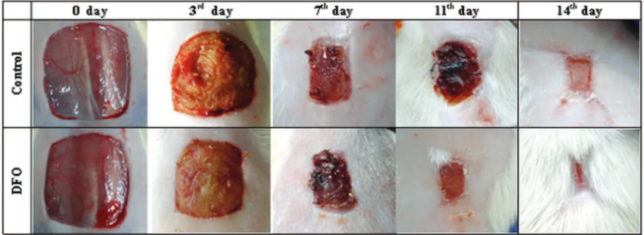
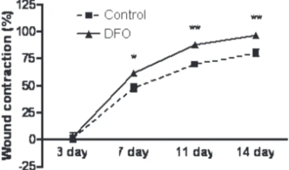
Fig. 1a shows the gross appearance of wound in DFO-treated and control rats on different Fig.1b: Per cent wound contraction in DFO-treated rats in comparison to control on days 3, 7, 11 and 14 post-wounding. Data are expressed
as mean ± SEM of 5 rats, *P<0.05, **P<0.005
3 rd day
7
th day
11
th day
14
th day
Control
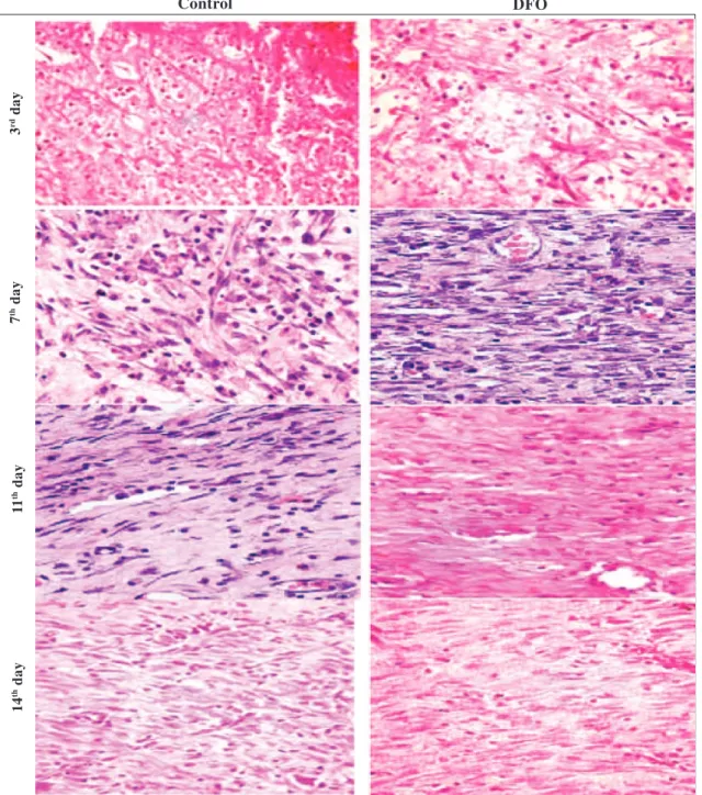
Fig. 2 (a). Representative images of H&E stained histopathological sections of granulation tissues of
control and DFO-treated rats (magnification 40x and scale bar 50 µµµµµm) on days 3, 7, 11 and 14
post-wounding.
days (0, 3, 7, 11, and 14) and it also depicts progressively greater wound closer in DFO-treated rats on day 7 onwards, as compared to control. The per cent wound closer was significantly greater on days 7, 11 and 14 in DFO-treated rats, as compared to respective control rats (Fig. 1b).
Histopathological findings
The representative pictures of H&E stained sections of granulation tissue of DFO-treated and control rats on different days given in Fig. 2. On day 3, the migration of inflammatory cells was more in DFO-treated granulation tissues, as compared to control. The proliferation of fibroblasts, collagen deposition and formation of blood vessels were more evident in DFO-treated ted, as compared to control group. On day 7, DFO-treated rats, showed more fibroblast proliferation, more blood vessels in perpendicular direction and few
inflammatory cells. In converse, the control group showed loose and thin granulation tissue, less fibroblasts and more inflammatory cells. On day 14, the DFO-treated wounds showed thick, intact and fully formed epithelial layer (Fig. 2a), dense collagen fibers with parallel arrangement resembling normal skin and H & E score is significant higher from day 3 onwards (Fig. 2b) which represents better granulation tissue formation. On the other hand, there was thin, loose and immature epithelial layer, few inflammatory cells and less and loose collagen deposition was evident in control group.
DISCUSSION
Wound healing is a complex process of synchronized interplay of a variety of inflammatory cells, cytokines and growth factors and the alteration of which leads to impairment of the wound healing. Lots of cellular and molecular-biological studies demonstrated that many cytokines, growth factors, and proteases are closely involved in the wound-healing process to complete normal tissue repair after damage (Donald and Zachary, 2004). Deferoxamine, an iron chelator has been used for the induction of HIF-1∝ accumulation even under normoxia (Duscher et al., 2015). The HIF-1∝ inducing property of deferoxamine and its indirect effect on several vital cellular processes, there is no available report on the effect of deferoxamine on wound healing after topical application. In this study Fig. 1b shows a significant increase in percent wound closure in deferoxamine-treated groups from day 7 onwards to day14, as compared to control. The results indicate marked acceleration of wound healing process after deferoxamine application and almost complete healing on day 14.
In this study, we hypothesized that DFO enhance wound healing by the recruitment of Fig. 2 (b). Histological scoring of H&E stained
granulation tissue sections of control and DFO-treated rats. Data are expressed as mean ± SEM
of 5 sections, **P<0.01, ***P<0.001 versus control
several cells which are crucial for granulation tissue formation and its proper deposition during wound healing process. In this study, Fig. 1b shows a significant increase in percent wound contraction in DFO-treated groups from day 7 to 14, as compared to control. The results indicate marked acceleration of wound healing process after topical application DFO and almost complete healing on day 14.
The wound gap is filled by the granulation tissue which in early stage are of immature type consisting of inflammatory cells, angioblasts, fibroblasts, collagen fibres, etc which in later stage becomes more matured and permanent and takes place in the gap formed by the formation of wound. The further maturity of such matured granulation tissue on the surface is responsible for wound closer, if it is not being completely covered by epidermal epithelial cells. The formation of granulation tissue is followed by two early events one is haemostasis and second is inflammation. More influx of inflammatory cells is prerequisite for early scavenging of irritant and tissue debris as well as release of growth factors for proliferation of fibroblast cells, angioblast cells and synthesis of matrix. This parameter has been used for comparison of effect of DFO histopathologically in this study. Likewise, number of cells involved in formation of granulation tissue has also been taken into consideration for comparison. Moreover, early maturation of granulation tissue has been used as an important indicator for assessing the effect of DFO in promoting wound healing histopathologically, on the basis of the fact that, immature fibroblast cells are round, plump, while mature one is elongated and smaller in size (Werner et al., 2005). Similarly, more maturity of granulation tissue is also assessed by thin and loosely dispersed collagen fibres
while in case of mature granulation tissue, they are found in wavy bundles and running parallel to each other (Ram et al., 2015). In this study, the effect of DFO was found to cause early maturation of granulation tissue in DFO-treated rats compared to control rats.
Angiogenesis is a process of proliferative phase of wound healing where new vessels appear as early as day 3 after wounding (Ram
et al., 2016). Angiogenesis is closely related to the formation of granulation tissue and depends on the complex control system of proangiogenic and antiangiogenic factors produced by a variety of cells. Once granulation is complete, angiogenesis ceases, as endothelial cells undergo apoptosis which is evident in histopathological findings (Fig. 2).
Superior granulation tissue is characterized by the presence of fibroblasts with extracellular matrix (ECM) formation and well formed blood vessels in the perpendicular direction which is stimulated by growth factors. In addition to the formation of ECM in healing tissue, its progressive degradation and remodeling in a regulated manner is essential to form mature healed wound tissue. DFO-treated rats in this study showed formation of more fibroblast and collagen bundles in a time-dependent manner as evidenced in histopathological observations and its score (Fig. 2), which indicates that DFO facilitated a well synchronized process of wound repair.
ACKNOWLEDGEMENT
The authors are thankful to the Director and the Joint Director (Academic), Indian Veterinary Research Institute for providing fund and necessary facilities to conduct the study.
REFERENCES
Agren MS, Mertz PM, Franzen I (1997) A comparative study of three occlusive dressing in the treatment of full thickness wounds in pigs. J Am Acad Dermatol 36: 53-58.
Donald M, Zachary JF (2004) Pathological Basis
of Veterinary Disease, 4th edn., Elsevier Publication.
Duscher D, Neofytou E, Wong VW, Maan ZN, Rennert RC, Inayathullah M, Januszyk M, Rodrigues M, Malkovskiy AV, Whitmore AJ, Walmsley GG, Galvez MG, Whittam AJ, Brownlee M, Rajadas J, Gurtner GC (2015) Transdermal deferoxamine prevents pressure-induced diabetic ulcers. Proc Natl Acad Sci USA 112(1): 94-99.
Greenhalgh DG, Sprugel KH, Murray MJ, Ross R (1990) PDGF and FGF stimulahealing in the genetically diabetic mouse. Am J Pathol 136: 1235-1246.
Hou Z, Nie C, Si Z, Mac Y (2013) Deferoxamine enhances neovascularization and accelerates wound healing in diabetic rats via the accumulation of
hypoxia-inducible factor-1∝. Diabetes Res Clin Pract
101: 62-71.
Miller MJ (1989) Syntheses and therapeutic potential of hydroxamic acid based siderophores and analogs. Chem Reviews 89 (7): 1563-1579.
Norris SO, Provo B, Stotts NA (1990) Physiology of wound healing and risk factors that impede the healing process. AACN Clin Issues Crit Care Nurs 1: 545-552.
Ram M, Singh V, Kumawat S, Kumar D, Kant V, Tandan SK, Kumar D (2016) Bilirubin modulated cytokines, growth factors and angiogenesis to improve cutaneous wound healing process in diabetic rats. Int Immunopharmacol 30: 137-149.
Ram M, Singh V, Kumawat S, Kumar D, Lingaraju MC, Singh TU, Rahal A, Tandan SK, Kumar D (2015) Deferoxamine modulates cytokines and growth factors to accelerate cutaneous wound healing in diabetic rats. Eur J Pharmacol 764: 9-21.
Rey S, Semenza GL (2010) Hypoxia-inducible factor-1-dependent mechanisms of vascularization and vascular remodelling. Cardiovasc Res 86: 236-242.
Singer AJ, Clark RAF (1999) Cutaneous wound healing. New Engl J Med 341(10): 738-746.
Werner N, Kosiol S, Schiegl T, Ahlers P, Walenta K, Link A, Bohm M, Nickenig G (2005) Circulating endothelial progenitor cells and cardiovascular outcomes. N Engl J Med 353(10): 999-1007.
Wu Y, Wang J, Scott PG, Tredget EE (2007) Bone marrow-derived stem cells in wound healing: a review. Wound Repair Regen 15: 18-26.
Xing D, Liu L, Marti GP, Zhang X, Reinblatt M, Milner SM, Harmon JW (2011) Hypoxia and hypoxiainducible factor in the burn wound. Wound
Repair Regen 19: 205-213.