The Combination of Three Natural
Compounds Effectively Prevented Lung
Carcinogenesis by Optimal Wound Healing
Linxin Liu1☯, Hong Li1☯, Zhenzhen Guo1, Xiaofang Ma1, Ning Cao1, Yaqiu Zheng1, Shengnan Geng1, Yongjian Duan2, Guang Han1*, Gangjun Du1*
1Institute of Pharmacy, Pharmacy College of Henan University, Jinming District, Kaifeng, Henan Province 475004, China,2Department of Oncology, The first hospital Affiliated to Henan University, Kaifeng, Henan Province 475001, China
☯These authors contributed equally to this work. *[email protected](GH);[email protected](GD)
Abstract
The tumor stroma has been described as“normal wound healing gone awry”. We explored whether the restoration of a wound healing-like microenvironment may facilitate tumor heal-ing. Firstly, we screened three natural compounds (shikonin, notoginsenoside R1 and aco-nitine) from wound healing agents and evaluated the efficacies of wound healing
microenvironment for limiting single agent-elicited carcinogenesis and two-stage carcino-genesis. The results showed that three compounds used alone could promote wound heal-ing but had unfavorable efficacy to exert wound healheal-ing, and that the combination of three compounds made up treatment disadvantage of a single compound in wound healing and led to optimal wound healing. Although individual treatment with these agents may prevent cancer, they were not effective for the treatment of established tumors. However, combina-tion treatment with these three compounds almost completely prevented urethane-induced lung carcinogenesis and reduced tumor burden. Different from previous studies, we found that urethane-induced lung carcinogenesis was associated with lung injury independent of pulmonary inflammation. LPS-induced pulmonary inflammation did not increase lung carci-nogenesis, whereas decreased pulmonary inflammation by macrophage depletion pro-moted lung carcinogenesis. In addition, urethane damaged wound healing in skin excision wound model, reversed lung carcinogenic efficacy by the combination of three compounds was consistent with skin wound healing. Further, the combination of these three agents reduced the number of lung cancer stem cells (CSCs) by inducing cell differentiation, resto-ration of gap junction intercellular communication (GJIC) and blockade of the epithelial-to-mesenchymal transition (EMT). Our results suggest that restoration of a wound healing microenvironment represents an effective strategy for cancer prevention.
OPEN ACCESS
Citation:Liu L, Li H, Guo Z, Ma X, Cao N, Zheng Y, et al. (2015) The Combination of Three Natural Compounds Effectively Prevented Lung Carcinogenesis by Optimal Wound Healing. PLoS ONE 10(11): e0143438. doi:10.1371/journal. pone.0143438
Editor:Hua Zhou, Macau University of Science and Technology, MACAO
Received:August 7, 2015
Accepted:November 4, 2015
Published:November 23, 2015
Copyright:© 2015 Liu et al. This is an open access article distributed under the terms of theCreative Commons Attribution License, which permits unrestricted use, distribution, and reproduction in any medium, provided the original author and source are credited.
Data Availability Statement:All relevant data are within the paper and its Supporting Information files.
Funding:This work was supported by National Natural Science Foundation of China (No. 81173094, 81373974, 81472745).
Introduction
Although conventional anticancer therapies, which consist of surgical resection, radiotherapy and chemotherapy, are effective in the management of many patients, they are ineffective for approximately half of cancer patients [1]. Resistance to conventional anticancer therapies in patients with advanced solid tumors has prompted the need for alternative cancer therapies [2]. Accumulating evidence suggests that cells and factors of the tumor microenvironment decisively contribute to not only the survival of primary neoplastic cells but also to the subse-quent key events of neoplastic disease progression, including tumor growth, invasion, and metastasis [3]. Epidemiologically, chronic wound states are well-known risk factors for cancer development [4]. Furthermore, aberrant wound healing with chronic inflammation can report-edly promote malignant transformation [5]. These phenomena have further solidified the view of cancer as“a wound that does not heal”[6]. Studies of the role of aberrant wound healing in
cancer pathologies will be important for the discovery of novel therapeutics that can promote wound healing or abrogate carcinogenesis in the tumor microenvironment.
Clearing a wound bed of nonviable tissue is increasingly acknowledged as an important step that may facilitate the healing process for a variety of wound types, particularly chronic wounds [7]. Necrotic tissue in the wound bed will significantly delay and in some cases prevent healing, as it may serve as a reservoir for bacterial growth, contain elevated levels of inflammatory mediators that promote chronic inflammation at the wound site, and impair the cellular migra-tion that is necessary for wound repair [8]. However, the factors that dictate the delicate bal-ance between normal wound repair and aberrant wound healing are yet to be fully elucidated. We hypothesized that appropriate regeneration, sufficient oxygen supply and waste elimina-tion may regulate aberrant wound healing and facilitate tumor eliminaelimina-tion. To stringently test this wound healing hypothesis, we screened three natural compounds (shikonin, notoginseno-side R1 and aconitine) from wound healing agents and created an optimal wound healing microenvironment. Specifically, we observed the effects of three compounds on carcinogenesis in mice with lung cancer induced by urethane (ethylcarbamate). We found that wound heal-ing–inducing therapy caused normal cells to lose their malignant transformation potential and
improve aberrant wound healing, which may in turn allow for the control of tumorigenesis.
Materials and Methods
Reagents
Shikonin, Aconitine and Notoginsenoside R1, 98% or higher purity (HPLC), were purchased from Sigma-Aldrich, Inc. (St. Louis, MO,USA) (S1 Fig). Urethane (ethylcarbamate), lipopoly-saccharide (LPS), basic fibroblast growth factor (bFGF), heparin, 12-O-tetradecanoylphorbol-13-acetate (TPA), Clodronate and Evans blue were purchased from Sigma Chemical Co. Bleo-mycin (BLM) from The antibodies used include: E-cadherin, N-cadherin, Vimentin, Nanog, Oct4, Snail1, CD133, Ki-67, cleaved-caspase 3, connexin 43 and fibronectin were obtained from BD Pharmingen. Horseradish peroxidase (HRP)-conjugated goat anti-mouse IgG poly-clonal antibody, Peroxidase substrate DAB (30, 30-diaminobenzidine) and AEC
(3-amino-9-ethylcarbazole) were from Nichirei Bioscience (Tokyo, Japan). The mouse quantitative ELISA kits (TNF-α, MPO, ROS and 8-OHdG) were obtained from R&D Systems.
Ethics statement
Guide for the Care and Use of Laboratory Animals and the regulation of animal protection committee to minimize the suffering and injury.
Animals
The female BALB/c mice, 5–7 weeks of age, were obtained from Beijing Weitong Lihua Animal
Co. All mice were housed in individual ventilated cages under a 12 h light-dark cycles (lights on 7:00 AM to 7:00 PM). The animals were fed standard rodent chow and water and were maintained under pathogen-free conditions within the institutional animal facility. The ani-mals were monitored daily and euthanized humanely by overdose carbon dioxide at the end of the experiment or the first sign of shortness of breath, reduced locomotion and reduced body weight (greater than 20% total body weight). All surgery was performed under general anesthe-sia by intraperitoneal injection of 45mg/kg pentobarbital sodium, and all efforts were made to minimize suffering.
Excision wound model and pharmacological properties of screened
compounds
BALB/c mice were intraperitoneally anesthetized with pentobarbital sodium (45 mg⁄kg), the
back hair of the animals was shaved, and a full-thickness excisional wound (approximately 10 mm in diameter) was created on the animal’s dorsum using sharp sterilized scissors. The
wounds were kept open, and the mice were housed individually. The gross area of the skin wound was photographed by a digital camera D7000 (Nikon, Japan) every week. The wounds were not sutured or covered and healed by secondary intention. Wound healing was analyzed as a percentage of the original wound area. The wound was considered to be completely closed when the wound area was grossly equal to zero.
To screen wound healing agents, mice were treated with selected natural compounds, including alkaloids, saponins, and naphthoquinones, by oral gavage once daily five days before excisional wound for two weeks. Control mice received vehicle in the same manner (0.5% carboxymethylcellulose in PBS).
Pharmacological properties of screened compounds were examined by TPA- induced ear edema, macrophage phagocytosis and blood perfusion. Briefly, mice were treated with screened compounds (2 mg/kg shikonin, 0.2 mg/kg aconitine and 20 mg/kg Notoginsenoside R1 indi-vidually) by oral gavage once daily for ten days. Following the last intragastric administration of compounds, the back blood perfusion of mice was recorded using a Laser Speckle Flowmeter (PeriCam PSI, Sweden). Subsequently, ear edema was induced on the right ear with 20μL TPA (1.0 mg dissolved in 20μL acetone) delivered to both the inner and outer surfaces of the ear, the left ear was selected as negative control which received 20μL acetone. After 3 h, mice received an intraperitoneal injection of 1 mg zymosan in 5 mL normal saline. Next 1 h, mice were euthanzed by overdose carbon dioxide, periotoneal exudates was collected to determine the differential counts based on nuclear morphology using Cytofuge and Wright Giemsa stain-ing. Moreover, 7-mm-diameter ear punch biopsies were individually weighed on an electronic balance, and the edema size was calculated based on the weight difference between both ears.
Forty mice were used in excision wound model and another forty mice were used in mea-sure of pharmacological properties, each group included ten mice. At the end of the experi-ments, none of mice died or showed signs of distress or discomfort.
Urethane-induced lung adenocarcinoma model
injection of urethane (600 mg/kg body weight) dissolved in sterile 0.9% NaCl once weekly for 10 weeks. In the first experiment, the mice were treated with the selected compounds (2 mg/kg shikonin, 0.2 mg/kg aconitine and 20 mg/kg Notoginsenoside R1 individually or in combina-tion) suspended in 0.5% carboxymethylcellulose by oral gavage once daily for 22 weeks follow-ing the first injection of urethane. In the second experiment, the mice were treated with the selected compounds by oral gavage once daily for 15 weeks following the last injection of ure-thane. Control mice received vehicle (0.5% carboxymethylcellulose in PBS) by oral gavage. During the studies, the health of the mice was monitored daily, and body weights were mea-sured weekly. At the end of the experiment, under anesthesia with pentobarbital sodium (45mg/kg), the lungs were weighed, filled with fixative overnight, transferred to 70% ethanol for 24 h, and examined using a dissecting microscope to count surface polyps in a blinded fash-ion. The tumor diameter on the surface of the lung was measured with a digital caliper. Tumor volume was expressed in mm3 and calculated as [l×w2] × 0.5, where l and w denote length and width, respectively. After tumors were counted, lung tissues were processed for paraffin embedding and histological or immunohistochemical analysis.
The lung tumor incidence, multiplicity, and tumor load (sum of tumor per lung in average) were determined according to previously established criteria [10].
To observe whether urethane-induced lung carcinogenesis is associated with pulmonary inflammation or lung injury, mice were intratracheally injected 3 times at 3-week intervals with either bleomycin (BLM 200μg in 50μL PBS) to induce lung injury or lipopolysaccharide (LPS 20μg in 50μL PBS) to induce pulmonary inflammation following the first injection of urethane. To deplete macrophage, liposome-encapsulated clodronate was prepared as described previously [11]. Specifically, 100μL (1 mg) of clodronate liposome suspension in PBS per mouse was injected intraperitoneally 3 times at 3-week intervals following the first injection of urethane. BALB/c mice were also treated with the compounds according to above protocol following the first injection of urethane.
Next day following the last injection of urethane, five mice each group were euthanzed by overdose carbon dioxide and 10% lung homogenates was performed for assays of ROS and 8-OHdG (lung injury) as well as myeloperoxidase (MPO) and TNFα(pulmonary
inflamma-tion) by quantitative sandwich enzyme immunoassay technique according to the manufactur-er’s instructions.
To examine interaction between carcinogenesis and wound healing microenvironment, excisional wound (1 cm2) was created on the animal’s dorsum of three weeks before the end of
experiment.
In this study, each group included twenty-two mice, at the end of the experiment, there were no less than twenty mice in each group. In all experimental procedure, one to two mice each group with clear evidence of myocardial infarction were excluded from the study and humanely euthanzed. The endpoint of the experiment was the first sign of shortness of breath, reduced locomotion and reduced body weight (greater than 20% total body weight) because of lung tumors.
Histological study
Histological sections (5μm) were stained with hematoxylin and eosin and analyzed. Prolifer-ative lesions in the lungs were classified as hyperplasia, adenoma or adenocarcinoma based on recommendations published by the Mouse Models of Human Cancers Consortium [12]. Lung pathology scored the lung injury according to previously published criteria [13]: grade 0, nor-mal tissue; grade 1, (<20% of the slide), 2 (20%-50% of the slide) and 3(>50% of the slide).
Immunohistochemical study
Lung histological sections (5μm) from the paraffin blocks were obtained in silanized slides. The sections were deparaffinized, rehydrated and washed in PBS. Antigen retrieval was per-formed in 10 mM sodium citrate at pH 6.0 for 20 min using a microwave oven. After blocking with 3% hydrogen peroxide for 10 min, the sections were incubated in 10% fetal bovine serum (FBS) in PBS for 2 h. A blocking solution of 0.1% Triton-X and 0.1% bovine serum albumin supplemented with 2% horse serum was used for the nuclear marker stains. The slides were incubated with primary antibodies in an appropriate blocking buffer overnight at 4°C. Cell pro-liferation was assessed with a primary monoclonal antibody against Ki-67; cell apoptotic changes were assessed with a primary monoclonal antibody against cleaved-caspase 3; and ade-nocarcinoma was distinguished using a primary monoclonal antibody against lactate dehydro-genase and TIF-1α. Subsequently, the sections were rinsed with PBS and incubated for 1 h at
room temperature with horseradish peroxidase (HRP)-conjugated goat anti-mouse IgG poly-clonal antibody. The signal was developed using the peroxidase substrate DAB which generated a brown reaction product. The sections were counterstained with hematoxylin and photo-graphed under a microscope. Each case was scored according to the intensity and area of stain-ing. A combined staining score (intensity + extension) of between 0 and 3 was considered to be low expression, whereas a score between 4 and 6 was considered to be high expression.
Lung barrier permeability assay
The lung barrier function of the mice was assessed with the Evans blue (EB) dye extra-barrier technique to determine the alveolar epithelium integrity [14]. Thirty minutes prior to sacrifice, 50 mg/kg EB was injected via the tail vein. The mice were subsequently sacrificed, and the lungs were removed to prepare the homogenate. Formamide was added to the homogenate, which was then incubated at 37°C for 24 h and centrifuged at 3,000 rpm for 30 min to obtain the supernatant. Subsequently, the optical density of EB in the supernatant was spectrophoto-metrically determined at 620 nm and calculated according to the EB standard curve.
Cell culture
Human alveolar basal epithelial cancer cells A549, human normal bronchial epithelium cells BEAS-2B, Human umbilical vein endothelial cells ECV304 and Mouse fibroblasts L929 were purchased from the Chinese Academy of Sciences. A549 cells were grown in RPMI1640 medium supplemented with 10% (v/v) FBS. BEAS-2B cells and ECV304 cells were cultured in DMEM/F12 medium supplemented with 10% (v/v) FBS. L929 cells were cultured in 4.5 g/l glu-cose DMEM supplemented with 10% FBS. All cells were grown in a humidified atmosphere containing 5% CO2and 95% air at 37°C. For in vitro experiments, Shikonin, Aconitine and Notoginsenoside R1 were dissolved at a concentration of 50 mM in DMSO as a stock solution (stored at-20°C) and then further diluted in cell culture medium to create working concentra-tions. The maximum final concentration of DMSO was less than 0.1% for each treatment, and was also used as a control.
Cell transformation assay
0.33% and transferred to the top of the bottom agar. Colonies were examined under a light microscope after 2 weeks of culture at 37°C and 5% CO2.
A tumor sphere assay was performed in ultralow adherent 6-well plates under serum-free, stem cell-selective conditions as previously described [15]. Briefly, A549 cells (5 × 10³ cells/well) were resuspended in 0.8% methylcellulose (MC)-based serum-free RPMI1640 medium supplemented with 20 ng/mL EGF, 20 ng/mL bFGF, 4 mg/mL heparin and speci-fied concentrations of compounds. The cells were further allowed to grow for 14 d, and the numbers of spheres were counted with a microscope. All experiments were repeated in triplicate.
Cell proliferation
A549 or L929 cells or ECV304 cells (1×10⁴cells/well) were incubated in a 96-well plate over-night at 37°C. Cells were treated with specified concentrations of compounds, each condition was tested in six replicates. During the final 4 h of the 48-h incubation, the supernatants were discarded and 100μL MTT (0.5 mg/ml) was added to each well. After 4 h, the MTT was dis-carded and 100μL of DMSO was added to each well. The absorbance of the samples at 570 nm was determined using a plate reader (Elx-800; Bio-Tek, USA). The results of the cell prolifera-tion assay are presented as a percentage of the control cells. All experiments were repeated in triplicate.
Cell differentiation assay
A549 cells (1 × 10⁴cells/well) were incubated in a 6-well plate overnight at 37°C. Cells were treated with specified concentrations of compounds, and the medium was exchanged every 2 d for 8 d. The cells were detached by trypsinization, and cell differentiation was assessed by flow cytometry based on the expression levels of CD133, Nanog, Oct4, E-cadherin, N-cadherin, vimentin and Snail. The data were analyzed to calculate the percentage of the cell population in each group using FlowJo 7.6 software. All experiments were repeated in triplicate.
Epithelial-to-mesenchymal transition (EMT) and gap junction
intercellular communication (GJIC) assays
For the EMT assay, BEAS-2B cells were grown to 70% confluency in a 6-well plate. The cells were then treated with control DMEM/F12, urethane, specified concentrations of compounds for 48 h. The cells were collected, resuspended and analyzed by flow cytometry for the expres-sion levels of E-cadherin, N-cadherin, vimentin and fibronectin using sandwiched antibodies. The geometric mean fluorescence of FITC-conjugated anti-IgG was used to measure protein expression.
Western blot analysis
Lung samples from tumor-bearing animals were homogenized and sonicated in RIPA buffer on ice. The lysates were then centrifuged at 12,000 rpm for 15 min at 4°C, and the protein con-centration was determined using the Bradford protein assay.
Forty micrograms of protein from tissues was separated on a 12% SDS-PAGE gel and elec-troblotted onto a polyvinylidene difluoride membrane. The immunoblot was incubated with blocking solution (5% skim milk) at room temperature for 1 h, followed by incubation over-night with a primary antibody against Nanog, Oct4, E-cadherin, N-cadherin, vimentin or con-nexin 43 at 4°C. The blots were washed and incubated with a 1:1,000 dilution of HRP-conjugated secondary antibody for 1 h at room temperature. After washing, the blots were developed using ECL detection reagent (GE Healthcare) and quantitated by densitometry using Image Quant image analysis system (Storm Optical Scanner). All the experiments were performed independently three times at least.
Statistical analysis
All data are expressed as the mean ± standard deviation and were analyzed with SPSS 17.0 soft-ware. The differences between groups were evaluated using one-way analysis of variance (ANOVA) followed by the Tukey test or Dunnett's test.P0.05 was considered statistically significant.
Results
The screened three compounds promoted wound healing
Cancer is often known as a wound that does not heal. Here, we aimed to determine whether a wound healing microenvironment could prevent the development of cancer. To establish a wound healing microenvironment, we first screened wound healing agents in a skin excision wound model. Among the screened compounds, shikonin, notoginsenoside R1 and aconitine resulted in significantly smaller wound areas during the experimental period than the other compounds (Fig 1AandS1 Table). After 14 d, all wounds treated with shikonin, aconitine and notoginsenoside R1 showed complete closure (10/10), whereas only 50% (5/10) of control wounds showed complete closure. Of note, many hairs were observed in the regenerated skin upon gross evaluation, especially in the aconitine group compared with the other groups.
In laser speckle imaging, significantly greater blood flow was detected in the wound areas in the notoginsenoside R1 and aconitine groups but not in shikonin group relative to the control group (Fig 1B). Exposing the ears of the mice to TPA to induce edema resulted in marked increases in skin thickness, but this change (the ratio of 2×normal ear thickness and edema ear thickness) was significantly less pronounced in the shikonin group (95.64 ± 5.17%) relative to the TPA control group (82.25 ± 8.92%). Treatment with shikonin reduced the water content in TPA-treated ears by approximately 55% (Fig 1C). The results of the macrophage phagocytosis assay showed that notoginsenoside R1 and aconitine considerably enhanced the macrophage phagocytosis of zymosan in vivo by approximately 25% and 50%, respectively (Fig 1D).
Histopathology of mouse lung carcinogenesis
than twenty percent of mice. Most tumors arose centrally within the bronchi at different levels, including the main, lobar, segmental and subsegmental bronchi.
Urethane-induced lung histological alterations were significantly improved by treatment of screened wound healing agents (S2 Fig).
The combination of three wound healing agents decreased incidence of
mouse lung carcinogenesis
To observe the effect of the wound healing microenvironment on lung carcinogenesis, we used three of the screened compounds (shikonin, aconitine and notoginsenoside R1) wich promote wound healing to treat mice following the first or last carcinogen application.
All of the three compounds appeared to prevent carcinogenesis when administered alone following the first carcinogen treatment based on the lung tumor incidence and load, whereas
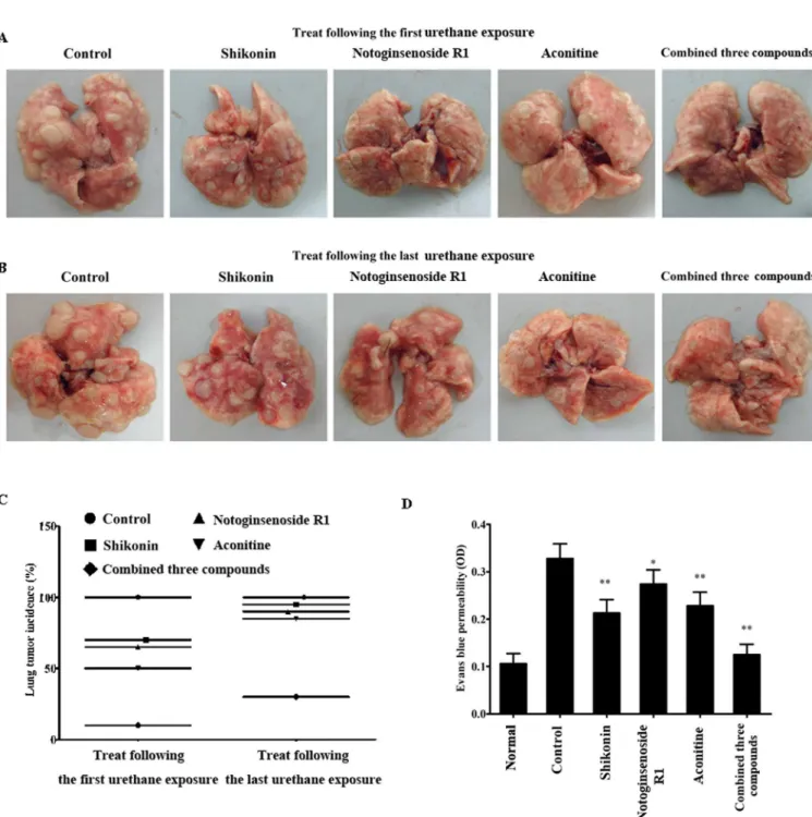
Fig 1. The pharmacological properties of screened three wound healing agents.(A) Screened three compounds promoted wound closure in a skin excision wound model. (B) Notoginsenoside R1 and aconitine increased blood flow perfusion on laser speckle imaging. (C) Shikonin exerted anti-inflammatory efficacy in the TPA-induced ear edema model. (D) Notoginsenoside R1 and aconitine promoted macrophage phagocytosis of zymosan. The results are presented as mean±SE (n = 10/group).**p<0.01, vs. control group.
the combination of the three drugs almost completely abrogated urethane-induced lung carci-nogenesis based on either lung tumor incidence or load (Fig 2A). However, individual treat-ment with any of these compounds after the last carcinogen treattreat-ment did have no impact on tumor incidence, although the tumor load was decreased (Fig 2BandFig 3A). Nevertheless, the combination of these three agents remained effective even after the induction of carcinogenesis.
Fig 2. The screened three wound healing agents suppressed lung carcinogenesis and their combination led to optimal preventive efficacy.(A) Three wound healing agents were administered to animals following the first carcinogen treatment and were found to prevent lung carcinogenesis. (B) They were administered following the last carcinogen treatment but did not prevent lung carcinogenesis, whereas the combination of these three agents remained effective. (C) Summary data of lung tumor incidence (n = 20). (D) Three wound healing agents restored the lung barrier in urethane-induced lung
carcinogenesis. The results are presented as mean±SE (n = 10/group).*p<0.05,**p<0.01, vs control group.
As shown inFig 2C, when the mice were treated following the first carcinogen treatment, the incidences of lung tumorigenesis in the shikonin, aconitine and notoginsenoside R1 groups were 70% (14/20), 50% (10/20) and 65% (13/20), respectively, and these values were signifi-cantly lower than that in the control group 100% (20/20). In the combined treatment group, only 10% (2/20) of mice had developed tumors at terminal sacrifice. When the mice were treated following the last carcinogen treatment, the incidence of lung tumorigenesis in the combined group was 30% (6/20), which was also considerably lower than that in the control group 100% (20/20). The incidence of lung tumorigenesis in the shikonin, aconitine and noto-ginsenoside R1 groups was 95% (19/20), 85% (17/20) and 90% (18/20), respectively, and did not differ significantly in mean tumor size from that in the control group (p>0.05), indicating
that the used dose of three compounds did not decrease tumor proliferation.
Fig 3. The screened three wound healing agents desereased lung tumor overall volume and did not negatively impact health or significantly affect the body weights of mice.(A) Three wound healing agents desereased lung tumor overall volume. (B-D) Three wound healing agents did not negatively impact health or significantly affect the body weights of mice. The results are presented as mean±SE (n = 10/group).*p<0.05,**p<0.01, vs control group.
In addition, we evaluated lung vascular barrier by EB dye which should not cross through vascular barrier under normal conditions. Urethane-induced lung carcinogenesis increased lung concentration of EB which indicated the increased permeability of the lung vascular bar-rier. Consistent with decreased carcinogenic incidence, the combination treatment restored the lung vascular barrier to normal levels, whereas single compound did not completely restore the lung vascular barrier (Fig 2D), suggesting that the combination of three compounds led to opti-mal wound healing.
In this study, the three screened compounds did not appear to be toxic, negatively impact health or significantly affect the body weights of mice at the experimental dose alone or in com-bination (Fig 3B–3D).
Urethane-induced lung carcinogenesis was associated with lung injury
independent of pulmonary inflammation
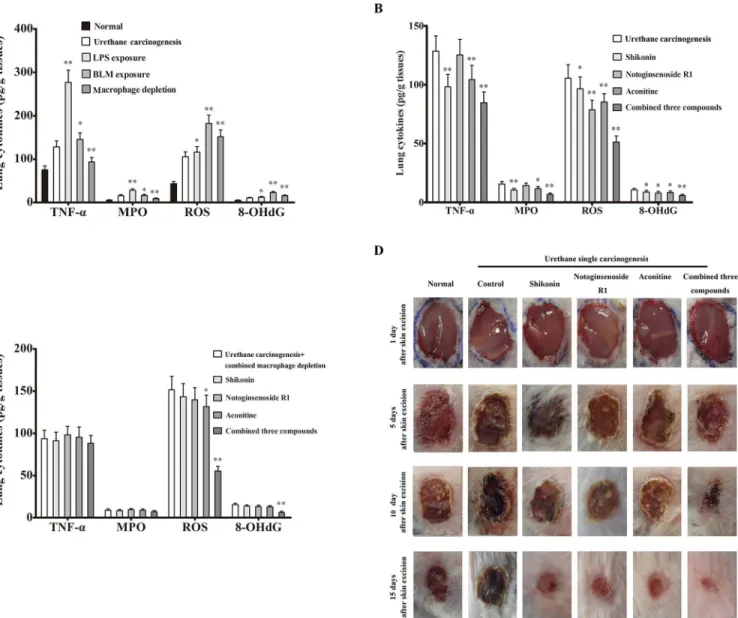
To observe whether urethane-induced lung carcinogenesis is associated with pulmonary inflammation or lung injury, mice were intratracheally injected with either BLM or LPS. As shown inFig 4A–4C, the increased levels of MPO and TNF-αwere more in LPS-exposed lungs
than those in BLM-exposed lungs compared with normal lung control, in contrast, the increased levels of ROS and 8-OHdG were more in BLM-exposed lungs than those in LPS-exposed lungs, indicating that LPS mainly induce pulmonary inflammation and that BLM mainly induce lung injury although both can not be clearly distinguished. In addition, urethane damaged wound healing in skin excision wound model, and the combination of three com-pounds led to optimal wound healing in this model (Fig 4D).
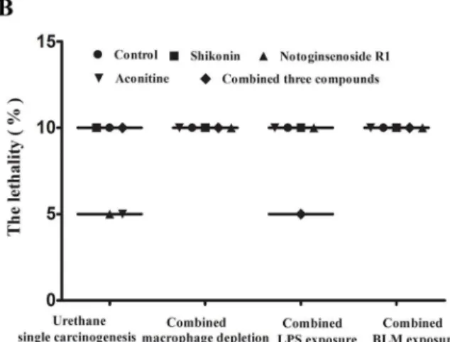
In this study, the data from a separate experiment showed that LPS-induced pulmonary inflammation did not result in increased lung carcinogenesis, whereas macrophage depletion decreased pulmonary inflammation but promoted lung carcinogenesis. In contrast, bleomycin-induced lung injury increased lung carcinogenesis. Importantly, all of three conditions
decreased carcinogenic preventive efficacy of the single compound but had little effect on com-bined efficacy (Fig 5A–5DandFig 6A). The reversed lung carcinogenic efficacy by combina-tion of three compounds was consistent with skin wound healing, and the injured lungs of non-tumor-bearing mice that were treated with the three drugs alone or in combination had returned to normal and showed complete wound healing at terminal sacrifice (S4 Fig), indicat-ing that the carcinogenic preventive efficacy of three compounds could be derived from resto-ration of a wound healing-like microenvironment.
In this study, the combined models or the three screened compounds did not appear to be toxic, negatively impact health or significantly affect the body weights of mice at the end of the experiments (Fig 6B–6E).
The wound healing-like microenvironment induced by the combination of
three compounds decreased lung cancer stem cells
the levels of stem cell markers and increased the levels of E-cadherin and connexin 43 to sup-press tumorigenesis (Fig 7AandS3 Fig).
The ability to proliferate and escape apoptosis is a fundamental property of CSCs. To confirm the presence of CSCs during lung tumorigenesis, immunohistochemical assays using Ki-67 as a proliferative marker and cleaved caspase-3 as an apoptotic marker were conducted with paraffin-embedded lung tissue slides. Ki-67 staining was increased more than 5-fold in carcinogenetic lung tissues compared with normal lungs. Simultaneously, the area that stained positive for cleaved cas-pase-3 was lower than 20% of the area observed in normal lungs. Furthermore, treatment with the screened compounds alone or combination decreased Ki-67 staining and increased cleaved cas-pase-3 staining after carcinogen-induced tumorigenesis was suppressed (Fig 7B).
Fig 4. The combination of three wound healing agents improved urethane-damaged wound healing environment.(A) LPS exposure mainly decreased MPO and TNFα, whereas BLM exposure and macrophage depletion mainly promoted ROS and 8-OHdG. (B) Three wound healing agents improved urethane-damaged wound healing environment (n = 5). (C) Macrophage depletion reversed improved wound healing environment by three compounds alone but had a slight effect on combination efficacy (n = 5). (D) Three wound healing agents promoted wound healing in urethane-induced carcinogenic model (n = 10). The results are presented as mean±SE.*p<0.05,**p<0.01, vs urethane carcinogenic group.
The wound healing microenvironment induced by the combination of
three compounds promoted lung cancer cell differentiation
Dedifferentiation is a fundamental characteristic of tumor cells, especially CSCs [18]. To fur-ther study the effect of the wound healing microenvironment on tumor-initiating cells, we treated A549 cells in vitro with the combined compounds as described above. The flow cytome-try results showed that the percentage of the population expressing stem cell markers including CD133, Oct4 and Nanog, and EMT markers including N-cadherin, vimentin and snail, greatly decreased in response to treatment with aconitine, especially for the combination of three com-pounds. However, the effect was not observed for treatment with shikonin or notoginsenoside R1. Simultaneously, exposure to aconitine or the combination of three compounds increased the population that expressed E-cadherin, a normal epithelial marker (Fig 8A).
Differentiated cells are characterized by a decreased capacity for self-renewal. To confirm that a wound healing microenvironment can induce differentiation, we carried out tumor sphere formation and soft agar colony formation assays. Consistent with the above results,
Fig 5. Urethane-induced lung carcinogenesis was associated with lung injury independent of pulmonary inflammation.(A) Urethane-induced lung carcinogenesis was promoted by BLM- or macrophage depletion-induced lung injury but was not affected by LPS-induced pulmonary inflammation. (B-D) LPS exposure, BLM exposure and macrophage depletion reversed carcinogenic preventive efficacy of single compound but had slight effect on combined efficacy shown as lung tumor incidence, lung tumor number and lung weight, respectively. The results are presented as mean±SE (n = 20/group).*p<0.05, **p<0.01, vs control group.
A549 cells exposed to chronic aconitine or combination of three compounds formed fewer tumor spheres and soft agar colonies than those exposed to control medium. Treatment with other compounds did not affect the number of tumor spheres and colonies compared with con-trol cells (Fig 8B).
The wound healing microenvironment induced by the combination of
three compounds prevented lung cell malignant transformation
The promotion of EMT and loss of GJIC are important characteristics of cell malignant trans-formation. To support the chemopreventive effect of the wound healing microenvironment on carcinogenesis, we treated BEAS-2B cells in vitro with three compounds alone or in combina-tion to observe urethane-induced cell malignant transformacombina-tion. Flow cytometric analysis showed a decrease in the population of BEAS-2B cells expressing N-cadherin, vimentin and fibronectin in response to aconitine or combination of three compounds, but the effect was not observed for cells treated with shikonin or notoginsenoside R1. Simultaneously, approximately 10% to 45% of shikonin-treated BEAS-2B cells or BEAS-2B cells treated with the combination of three compounds were E-cadherin-positive cells, whereas less than 10% of urethane-treated cells were E-cadherin positive (Fig 8C).
Fig 6. The combined models increased lung tumor overall volume but did not negatively impact health or significantly affect the body weights of mice.(A) The combined models increased lung tumor overall volume and decreased carcinogenic preventive efficacy of a single compound. (B-E) The combined models did not negatively impact health or significantly affect the body weights of mice. The results are presented as mean±SE (n = 20/group). **p<0.01, vs control group.
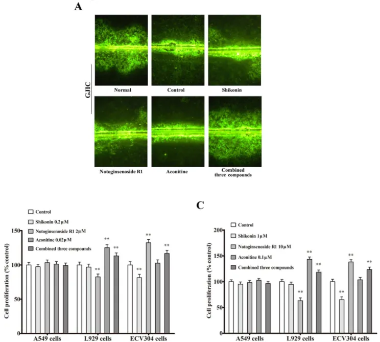
In contrast, urethane-treated control cells lost GJIC, whereas GJIC was markedly restored in BEAS-2B cells treated with the combination of three compounds; this effect was not observed in shikonin- or aconitine -treated cells (Fig 9A).
To confirm that cancer preventive efficiency was from restoration of a wound healing-like microenvironment but not direct anti-tumor proterties, we treated A549, L929 and ECV304 cells with three compounds alone or in combination. A549 cells were not affected by the used dose of three compounds alone or in combination (more than in vivo plasma concentration). L929 cells were decreased by notoginsenoside R1, not affected by shikonin but were promoted by aconotine and combination of three compounds. ECV304 cells were decreased by shikonin, not affected by aconotine but were promoted by notoginsenoside R1 and the combination of three compounds (Fig 9B and 9C).
Discussion
The naphthoquinone shikonin is is the major active principle of Lithospermi Radix which is an effective traditional Chinese herb in various types of wound-healing [19]. The combination of shikonin's anti-inflammatory activity together with its wound-healing properties makes it a great potential therapeutic agent for the injury treatment [20], pharmaceutical formulations
Fig 7. The wound healing microenvironment decreased lung CSCs and induced cell apoptosis.(A) The combination of three compounds reduced several putative stem cell markers and restored normal E-cadherin and connexin 43 expression examined by western blot analysis in urethane-induced lung carcinogenesis.(B) The combination of three compounds decreased expression of the proliferative marker Ki-67 and increased expression of the apoptotic marker cleaved caspase-3 examined by immunohistochemical assays in urethane-induced lung carcinogenesis. The results are presented as mean±SE (n = 5/group)**p<0.01vscontrol group. The immunohistochemical images were taken at ×40 magnification.
with wound-healing properties based on shikonin have been in the market for many years although its mechanism of action remains unknown [21].The saponin notoginsenoside R1 (NR1) is the major bioactive component in panax notoginseng which is widely used in Asian countries in the treatment of microcirculatory diseases [22]. The pro-angiogenic action of NR1 in vivo and in vitro provided scientific evidence to be used for the treatment of cardiovascular diseases, traumatic injuries and wound-healing [23]. Aconitine is a major bioactive diterpenoid alkaloid with high content derived from herbal aconitum plants which is widely used to treat various diseases, such as shock caused by acute myocardial infarction, coronary heart disease and angina pectoris in China for thousands of years [24]. However, little information is avail-able on the role of aconitine in wound healing. It was reported that Baikal aconite alkaloids showed manifest wound healing effects on the excision skin wound model [25]. It may be a mechanism of reparative activity to direct stimulation of fibroblasts by aconitine. In our study, although three compounds used alone could promote wound healing, all of them also had unfavorable efficiency to exert wound healing, such as shikonin exerted anti-inflammatory effi-ciency but decreased angiogenesis and macrophage phagocytosis which was disadvantage in oxygen supply and waste elimination, notoginsenoside R1 stimulated angiogenesis but
Fig 8. The wound healing microenvironment prevented lung cell malignant transformation.(A) The combination of three compounds reduced the population expressing stem cell markers or EMT markers examined by flow cytometry and in A549 cells. (B) The combination of three compounds decreased A549 cell self-renew shown as the number of tumor spheres and soft agar colonies. (C) The combination of three compounds decreased the population expressing EMT markers examined by flow cytometry in urethane-treated BEAS-2B cells. The results are presented as mean±SE (n = 5/group)**p<0.01vs
control group.
decreased fibroblasts which was disadvantage in wound healing, and aconitine promoted oxy-gen supply but intensely stimulated fibroblasts which was unfavorable for correct repair pro-cess. The combination of three compounds made up treatment disadvantage of single
compound in wound healing and obtained optimal wound healing. Although three compounds have demonstrated their ability to suppress cancer cell proliferation [26–28], the used dose of
three compounds alone or in combination had no effect on tumor growth indicated by tumor size in this study. We did not also observe decreased cancer cell proliferation by three com-pounds alone or in combination in vitro.
Fig 9. The wound healing microenvironment restored GJIC but did not directly decrease tumor cells. (A) The combination of three compounds reversed GJIC loss in urethane-treated BEAS-2B cells. Images were taken at ×10 magnification. (B and C) The combination of three compounds led to angiogenesis and optimal fibroblast proliferation but did not directly decrease A549 cancer cells. The results are presented as mean±SE (n = 5/group)
**p<0.01vscontrol group.
One of the core questions in cancer biology relates to the identity and nature of the cancer cell of origin leading to tumor initiation [29]. Evidence from mouse skin models of carcinogen-esis suggests that initiated cells at different stages within a stem cell hierarchy require varying degrees of reprogramming depending on their degree of differentiation [30]. For tumorigene-sis, various interrelated determinants govern this complex tumor-host interaction, including GJIC, which plays a central role in coordinating intercellular signal-transduction pathways to control tissue homeostasis [31]. In addition, EMT in response to carcinogen exposure supports the involvement of GJIC in the carcinogenesis process [32]. In this study, the wound healing microenvironment decreased the number of lung CSCs, as evidenced by decreased stem cell markers and cell proliferation as well as increased expression of epithelial markers and cell apo-ptosis. This environment also induced lung cancer cell differentiation, as evidenced by
decreased stem cell markers and a capacity of self-renewal, prevented lung cell malignant trans-formation, as evidenced by decreased EMT and increased GJIC, and delayed tumor progres-sion, as evidenced by the halted progression of hyperplasia to carcinoma in situ or invasive tumors. Our results provide clear evidence that tumor initiation is necessary but not sufficient for tumor formation, and a wound healing microenvironment may induce terminal differentia-tion, which may in turn cause tumor initiating cells to lose their self-renewal ability and allow their malignant potential to be controlled.
The previous models of chronic wound-associated cancer were usually based on chronic inflammatory microenvironment where regenerative proliferation places the dividing cells at greater risks for acquiring mutations and subsequently selects for those cells that incurred mutations that impart proliferative and/or survival advantages to the cells [33]. Although chronic inflammatory conditions increase cancer risk [34], the hypothesis that the anti-inflam-matory activities of celecoxib and aspirin would reduce mouse lung tumorigenesis was not sup-ported in urethane-induced lung carcinogenic model [35]. In this study, we used the mouse-urethane model which exhibits similar histological appearance and molecular changes to human lung adenocarcinoma to evaluate the efficacies of wound healing microenvironment for limitted single agent-elicited carcinogenesis and two-stage carcinogenesis. We found that urethane caused lung injury independent on pulmonary inflammation, and that LPS-induced pulmonary inflammation did not result in increased lung carcinogenesis. In contrast, macro-phage depletion decreased pulmonary inflammation but promoted lung carcinogenesis. As expected, bleomycin-induced lung injury increased lung carcinogenesis and the injury increased lung carcinogenesis could be reversed by combination of three compounds but not single compound. In addition, urethane damaged wound healing in skin excision wound model, reversed lung carcinogenic efficiency by the combination of three compounds was con-sistent with skin wound healing, indicating that the carcinogenic preventive efficacy of three compounds could be derived from restoration of a wound healing-like microenvironment.
In summary, our results suggested that restoration of a wound healing microenvironment represents a simple and effective strategy for cancer prevention. Furthermore, a deficiency in wound healing may be a sensitive index for cancer prognosis.
Supporting Information
S1 Fig. Compound structural formula.(TIF)
S2 Fig. Urethane-induced lung histological alteration (× 20).
S3 Fig. Weastern blot.
(TIF)
S4 Fig. Wound healing agents restore lung histological structure in urethane-treated mice without lung tumor.
(TIF)
S1 Table. The screened wound healing agents in mouse skin excision wound model.
(TIF)
Acknowledgments
The authors thank Prof. Ming Bai and Prof. Ming-San Miao for generous assistance. We thank AJE (American Journal Experts) for editing the manuscript. This work was supported by National Natural Science Foundation of China (No. 81173094, 81373974, 81472745).
Author Contributions
Conceived and designed the experiments: GJD. Performed the experiments: LXL HL ZZG XFM NC YQZ SNG. Analyzed the data: GJD LXL HL. Contributed reagents/materials/analysis tools: YJD GH. Wrote the paper: GJD LXL HL. Provided facility for histopathology: YJD GH.
References
1. Siegel R, Naishadham D, Jemal A. Cancer statistics. 2012, CA Cancer J Clin. 2012; 62 (1): 10–29. doi:10.3322/caac.20138PMID:22237781
2. Huang L, Li F, Sheng J, Xia X, Ma J, Zhan M, et al. DrugComboRanker: drug combination discovery based on target network analysis. Bioinformatics. 2014; 30 (12): i228–36. doi:10.1093/bioinformatics/ btu278PMID:24931988
3. Brennen WN, Rosen DM, Wang H, Isaacs JT, Denmeade SR. Targeting carcinoma-associated fibro-blasts within the tumor stroma with a fibroblast activation protein-activated prodrug. J Natl Cancer Inst. 2012; 104 (17): 1320–1334. doi:10.1093/jnci/djs336PMID:22911669
4. Wu S, Rhee KJ, Albesiano E, Rabizadeh S, Wu X, Yen HR, et al. A human colonic commensal pro-motes colon tumorigenesis via activation of T helper type 17 T cell responses. Nat Med. 2009; 15 (9): 1016–1022. doi:10.1038/nm.2015PMID:19701202
5. Huber S, Gagliani N, Zenewicz LA, Huber FJ, Bosurgi L, Hu B, et al. IL-22BP is regulated by the inflam-masome and modulates tumorigenesis in the intestine. Nature. 2012; 491 (7423): 259–263. doi:10. 1038/nature11535PMID:23075849
6. Dvorak HF. Tumors: wounds that do not heal-redux. Cancer Immunol Res. 2015; 3 (1): 1–11. doi:10. 1158/2326-6066.CIR-14-0209PMID:25568067
7. Madhok BM, Vowden K, Vowden P. New techniques for wound debridement. Int Wound J. 2013; 10 (3): 247–251. doi:10.1111/iwj.12045PMID:23418808
8. Maeda TM, Kimura CK, Takahashi KT, Ichimura KI. Increase in skin perfusion pressure after maggot debridement therapy for critical limb ischaemia. Clin Exp Dermatol. 2014; 39 (8): 911–914. doi:10. 1111/ced.12454PMID:25283968
9. Du G, Sun T, Zhang Y, Lin H, Li J, Liu W, et al. The mitochondrial dysfunction plays an important role in urethane-induced lung carcinogenesis. Eur J Pharmacol. 2013; 715 (1–3): 395–404. doi:10.1016/j. ejphar.2013.04.031PMID:23707353
10. Stearman RS, Dwyer-Nield L, Zerbe L, Blaine SA, Chan Z, Bunn PA Jr, et al. Analysis of orthologous gene expression between human pulmonary adenocarcinoma and a carcinogen induced murine model. Am J Pathol. 2005; 167 (6): 1763–1775. PMID:16314486
11. Zeisberger SM, Odermatt B, Marty C, Zehnder-Fjallman AHM, Ballmer-Hofer K, et al. Clodronate-lipo-some-mediated depletion of tumour-associated macrophages: a new and highly effective antiangio-genic therapy approach. Br J Cancer. 2006; 95 (3): 272–281. PMID:16832418
13. Yunhe F, Bo L, Xiaosheng F, Fengyang L, Dejie L, Zhicheng L, et al. The effect of magnolol on the Toll-like receptor 4/nuclear factor kappaB signaling pathway in lipopolysaccharide-induced acute lung injury in mice. Eur J Pharmacol. 2012; 689 (1–3): 255–261. doi:10.1016/j.ejphar.2012.05.038PMID: 22683864
14. Tang M, Tian Y, Li D, Lv J, Li Q, Kuang C, et al. TNF-αmediated increase of HIF-1αinhibits VASP expression, which reduces alveolar-capillary barrier function during acute lung injury (ALI). PLoS One. 2014; 9(7): e102967. doi:10.1371/journal.pone.0102967PMID:25051011
15. Paranjape AN, Balaji SA, Mandal T, Krushik EV, Nagaraj P, Mukherjee G, et al. Bmi1 regulates self-renewal and epithelial to mesenchymal transition in breast cancer cells through Nanog. BMC Cancer. 2014; 14:785. doi:10.1186/1471-2407-14-785PMID:25348805
16. Ke Q, Li L, Cai B, Liu C, Yang Y, Gao Y, et al. Connexin 43 is involved in the generation of human-induced pluripotent stem cells. Hum Mol Genet. 2013; 22 (11): 2221–2233. doi:10.1093/hmg/ddt074 PMID:23420013
17. Wang B, Jacob ST. Role of cancer stem cells in hepatocarcinogenesis. Genome Med. 2011; 3 (2): 11. doi:10.1186/gm225PMID:21345246
18. Grimwade D, Mistry AR, Solomon E, Guidez F. Acute promyelocytic leukemia: a paradigm for differenti-ation therapy. Cancer Treat Res. 2010; 145: 219–235. doi:10.1007/978-0-387-69259-3_13PMID: 20306254
19. Andújar I, Ríos JL, Giner RM, Recio MC. Pharmacological properties of shikonin—a review of literature since 2002. Planta Med. 2013; 79 (18): 1685–1697. doi:10.1055/s-0033-1350934PMID:24155261 20. Andújar I, Ríos JL, Giner RM, Recio MC. Shikonin promotes intestinal wound healing in vitro via
induc-tion of TGF-βrelease in IEC-18 cells. Eur J Pharm Sci. 2013; 49 (4): 637–641. doi:10.1016/j.ejps. 2013.05.018PMID:23727294
21. Papageorgiou VP, Assimopoulou AN, Ballis AC. Alkannins and shikonins: a new class of wound heal-ing agents. Curr Med Chem. 2008; 15 (30): 3248–3267. PMID:19075667
22. Yoshikawa M, Murakami T, Ueno T, Yashiro K, Hirokawa N, Murakami N, et al. Bioactive saponins and glycosides. VIII. Notoginseng (1): new dammarane-type triterpene oligoglycosides, notoginsenosides-A, -B, -C, and -D, from the dried root of Panax notoginseng (Burk.). Chem Pharm Bull (Tokyo). 1997; 45 (6):1039–1045.
23. Hong SJ, Wan JB, Zhang Y, Hu G, Lin HC, Seto SW, et al. Angiogenic effect of saponin extract from Panax notoginseng on HUVECs in vitro and zebrafish in vivo. Phytother Res. 2009; 23(5):677–686. doi:10.1002/ptr.2705PMID:19107746
24. Fu M, Wu M, Wang JF, Qiao YJ, Wang Z. Disruption of the intracellular Ca2+ homeostasis in the car-diac excitation-contraction coupling is a crucial mechanism of arrhythmic toxicity in aconitine-induced cardiomyocytes. Biochem Biophys Res Commun. 2007; 354 (4): 929–936. PMID:17276394 25. Zyuz'kov GN, Krapivin AV, Nesterova YV, Povetieva TN, Zhdanov VV, Suslov NI, et al. Mechanisms of
regeneratory effects of baikal aconite diterpene alkaloids. Bull Exp Biol Med. 2012; 153 (6): 846–850. PMID:23113300
26. Wu H, Xie J, Pan Q, Wang B, Hu D, Hu X. Anticancer agent shikonin is an incompetent inducer of can-cer drug resistance. PLoS One. 2013; 8(1): e52706. doi:10.1371/journal.pone.0052706PMID: 23300986
27. Du J, Lu X, Long Z, Zhang Z, Zhu X, Yang Y, et al. In vitro and in vivo anticancer activity of aconitine on melanoma cell line B16. Molecules. 2013; 18 (1): 757–767. doi:10.3390/molecules18010757PMID: 23299553
28. Wang CZ, Xie JT, Fishbein A, Aung HH, He H, Mehendale SR, et al. Antiproliferative effects of different plant parts of Panax notoginseng on SW480 human colorectal cancer cells. Phytother Res. 2009; 23 (1): 6–13. doi:10.1002/ptr.2383PMID:19048608
29. Ho PL, Kurtova A, Chan KS. Normal and neoplastic urothelial stem cells: getting to the root of the prob-lem. Nat Rev Urol 2012; 9 (10): 583–594. doi:10.1038/nrurol.2012.142PMID:22890301
30. Song IY, Balmain A. Cellular reprogramming in skin cancer. Semin Cancer Biol. 2015; 32: 32–39. doi: 10.1016/j.semcancer.2014.03.006PMID:24721247
31. Piccoli C, D'Aprile A, Scrima R, Ambrosi L, Zefferino R, Capitanio N. Subcytotoxic mercury chloride inhibits gap junction intercellular communication by a redox- and phosphorylation-mediated mecha-nism. Free Radic Biol Med. 2012; 52 (5): 916–927. doi:10.1016/j.freeradbiomed.2011.12.018PMID: 22240155
33. Sun D, Ren H, Oertel M, Sellers RS, Zhu L. Loss of p27Kip1 enhances tumor progression in chronic hepatocyte injury-induced liver tumorigenesis with widely ranging effects on Cdk2 or Cdc2 activation. Carcinogenesis. 2007; 28 (9): 1859–1866. PMID:17434927
34. Grivennikov SI. Inflammation and colorectal cancer: colitis-associated neoplasia. Semin Immuno-pathol. 2013; 35 (2): 229–244. doi:10.1007/s00281-012-0352-6PMID:23161445