Research Article
Deregulation of Annexin-A1 and Galectin-1 Expression
in Precancerous Gastric Lesions: Intestinal Metaplasia and
Gastric Ulcer
Ana Flávia Teixeira Rossi,
1Márcia Cristina Duarte,
1Ayla Blanco Poltronieri,
1Marina Curado Valsechi,
1Yvana Cristina Jorge,
1Dalísio de-Santi Neto,
2Paula Rahal,
1Sonia Maria Oliani,
1and Ana Elizabete Silva
11Department of Biology, S˜ao Paulo State University (UNESP), Cˆampus S˜ao Jos´e do Rio Preto,
Rua Crist´ov˜ao Colombo 2265, 15054-000 S˜ao Jos´e do Rio Preto, SP, Brazil
2Legal Medicine Department and Pathology Service, Hospital de Base, Avenida Brigadeiro Faria Lima 5544,
15090-000 S˜ao Jos´e do Rio Preto, SP, Brazil
Correspondence should be addressed to Ana Elizabete Silva; [email protected]
Received 14 October 2013; Revised 15 January 2014; Accepted 15 January 2014; Published 25 February 2014
Academic Editor: Fulvio D’Acquisto
Copyright © 2014 Ana Fl´avia Teixeira Rossi et al. his is an open access article distributed under the Creative Commons Attribution License, which permits unrestricted use, distribution, and reproduction in any medium, provided the original work is properly cited.
Objective. Annexin-A1 (ANXA1/AnxA1) and galectin-1 (LGALS1/Gal-1) are mediators that play an important role in the
inlammatory response and are also associated with carcinogenesis. We investigated mRNA and protein expression in precancerous gastric lesions that participate in the progression cascade to gastric cancer, such as intestinal metaplasia (IM) and gastric ulcer (GU).Methods. Quantitative real-time PCR (qPCR) and immunohistochemical techniques were used to analyze the relative quantiication levels (RQ) ofANXA1andLGALS1mRNA and protein expression, respectively.Results.Increased relative expression levels ofANXA1were found in 100% of cases, both in IM (mean RQ = 6.22 ± 0.06) and in GU (mean RQ = 6.69 ± 0.10). However, theLGALS1presented basal expression in both groups (IM: mean RQ = 0.35 ± 0.07; GU: mean RQ = 0.69 ± 0.09). Immunohistochemistry revealed signiicant positive staining for both the AnxA1 and Gal-1 proteins in the epithelial nucleus and cytoplasm as well as in the stroma of the IM and GU groups (� < 0.05) but absence or low immunorectivity in normal mucosa.
Conclusion. Our results bring an important contribution by evidencing that both the AnxA1 and Gal-1 anti-inlammatory proteins
are deregulated in precancerous gastric lesions, suggesting their involvement in the early stages of gastric carcinogenesis, possibly due to an inlammatory process in the gastric mucosa.
1. Introduction
Precancerous lesions are related to the development of tumors in several organs, such as intestinal-type gastric cancer that develops through the progression of various
sequential lesions that frequently start with a Helicobacter
pylori infection. his infection causes supericial gastritis that can progress to chronic atrophic gastritis, intestinal
metaplasia, dysplasia, and, inally, carcinoma [1]. Intestinal
metaplasia, characterized by the diferentiation of gastric
stem cells into intestinal-phenotype cells [2], is associated
with more than 80% of intestinal-type adenocarcinoma [3].
Besides this metaplasia-dysplasia-carcinoma pathway, gastric
carcinogenesis can originate from gastric ulcer [4], a lesion in
the mucosa that develops in low acid concentration sites and
severe inlammation [5,6]. Eighty-ive percent of gastric ulcer
cases occur in the presence ofH. pyloriinfection [5,7].
he progression of the lesions cascade depends on many genetic factors in both the host and the bacterium, besides
environmental factors [8]. H. pylori may persist for many
years in the host, causing a chronic inlammation. his results in a great amount of inlammatory mediators and the reactive oxygen and nitrogen species that induce genetic and epigenetic changes in protooncogene and tumor suppressor
genes, inluencing the emergence of cancer [9]. Such bacteria populations are heterogeneous and contain virulence factors
such as cagA (cytotoxin-associated gene A antigen), which
produces a protein that acts in many cellular events such as cytoskeleton rearrangement, cellular polarity breaking, and
mitogenic and proapoptotic responses [10]. his virulence
genotype, however, is not found in all the strains; its occur-rence has a relation with major gastric mucosa inlammation
[11] and high risk of gastric cancer development [12].
Outstanding among the inlammatory mediators
that activate the immune response cascade are the anti-inlammatory proteins annexin-A1 (AnxA1) and galectin-1
(Gal-1), involved in various inlammatory processes [13,14].
AnxA1 belongs to a protein superfamily characterized by binding to cellular membranes in a calcium-dependent manner, acting on several cellular and molecular processes
[13]. hroughout the inlammatory process, this protein
externalizes in the plasmatic membrane, blocking the interaction between leukocytes and endothelium in order to stop the inlammatory cells transmigration to the damaged
site [15]. Its anti-inlammatory action is also related to the
induction of neutrophil apoptosis [16] and inhibition of
phospholipase A2 (PLA2) [17]. Furthermore, it also has a
connection to cellular diferentiation mechanisms, growth,
signal transduction, and cytoskeleton formation [18]. hus,
changes in their expression and subcellular location can
contribute to inlammatory diseases and cancer [19–22].
Galectin-1 belongs to a family of�-galactoside-binding
protein [23]. It has many cellular functions, such as apoptosis
[24], cellular signaling, adhesion, migration [25], and
prolif-eration [26], and its action depends on its cellular
concentra-tion and locaconcentra-tion [14]. Its anti-inlammatory function relates
to the induction of apoptosis of T-activated cells [24, 27]
and the decreased production of proinlammatory cytokines
[28,29]. Furthermore, galectin-1 promotes diferentiation of
Treg cells controlling their immunosuppressive mechanisms
[30,31] and suppresses dendritic cells maturation favoring a
tolerogenic microenvironment [32]. In the tumor
microenvi-ronment, the presence of Gal-1 contributes to
immunosup-pression [31], angiogenesis promotion [33], metastasis [34],
and cell transformation [35]. Several types of cancers show
changed expression of this anti-inlammatory mediator [36,
37], associated with the increase of cellular proliferation [36]
and metastasis [38].
Studies on annexin-A1 and galectin-1 in gastric cancer and precancerous lesions are few and reveal contradictory results. Some of them found AnxA1 and Gal-1
downregula-tion [39–41], while others reported increase of these proteins
in the lesions and tumor tissue [42–46].
Considering that gastric cancer has a high incidence
and poor prognosis when diagnosed at a late stage [47],
the study of precancerous lesions can provide important information about the initial phases of gastric carcinogenesis, thereby contributing to prevention strategies which may lead to a decrease in its incidence through the identiication of potential biomarkers. Recently, we showed a similarly increased expression of annexin-A1 and galectin-1 in chronic gastritis and in gastric cancer, evidencing deregulation in the expression of these proteins during the initial step of
gastric carcinogenesis [46]. Based on this inding, we decided
to investigate the expression and location pattern of both proteins in other precancerous gastric lesions that participate in the progression cascade to gastric cancer.
In this study, we evaluated the quantitative mRNA
expres-sion of genesANXA1andLGALS1and the expression of both
proteins in intestinal metaplasia and gastric ulcer lesions. In addition, we also investigated the occurrence of association
between the expression levels and infection by H. pylori
and itscagAvirulence genotype, besides other risk factors
associated with gastric carcinogenesis.
2. Materials and Methods
2.1. Samples. his research was approved by the local Research Ethics Committee (CEP-IBILCE/UNESP, number 059/11), and written informed consent was obtained from all participants, who also illed out a questionnaire from which we obtained family and occupational data, besides information on smoking and drinking habits.
DNA and cDNA samples of gastric biopsies (antrum and corpus) of the lesion area and adjacent normal mucosa stored
in our laboratory from a previous study [48] were used.
All subjects were recruited from the Service of Endoscopy, Hospital de Base, S˜ao Jos´e do Rio Preto, SP, Brazil. A total of 75 samples were evaluated, 36 of which of intestinal metaplasia of individuals without gastric cancer (IM—19 women, 17
men; mean age: 60.53 ± 12.21 years), 29 of gastric ulcer
(GU—8 women, 21 men; mean age: 54.62 ± 12.42 years),
and 10 from individuals with histologically normal gastric mucosa without dyspepsia, used as controls (C—3 women, 7
men; mean age:34.20 ± 9.73years). Subjects with prepyloric
or nonsteroid anti-inlammatory drug-induced ulcers were
excluded from this study [48].
2.2. Molecular Diagnosis for H. pylori and cagA Genotype.
DNA samples of normal mucosa adjacent to gastric lesions were subjected to multiplex PCR containing primers for
bacterial genesUreAandtsaAand for the human constitutive
gene CYP1A1, to verify DNA integrity and eiciency of
the PCR reaction (Table 1). We used 1X bufer, 0.15�M
of each deoxyribonucleotide, 2 mM MgCl2, 0.6�M of each
primer, 100 ng genomic DNA, and 1.8 U PlatinumTaqDNA
Polymerase (Invitrogen) in a inal volume of 25�L. he
reaction was processed in an automatic thermocycler, with
denaturation performed at 94∘C for 3 minutes, followed by 35
cycles at 94∘C for 45 seconds, 60∘C for 30 seconds, and 72∘C
for 1 minute, and a inal extension of 10 minutes at 72∘C. he
reaction products were subjected to electrophoresis on 3.0% agarose gel stained with ethidium bromide.
he H. pylori-positive samples were subjected to a
sec-ond PCR assay, to investigate thecagAvirulence genotype.
he reaction solution contained 1X bufer, 0.1�M of each
deoxyribonucleotide, 2 mM MgCl2, 0.6�M of each primer
(Table1), 300 ng genomic DNA, and 1 UTaqDNA Polymerase
(Invitrogen). he reaction conditions were 94∘C for 5 minutes
for denaturation, 40 cycles of 1 minute at 94∘C, 1 minute
Table 1: Sequence of primers and size of the fragments generated to determineH. pyloriinfection andcagAgenotype.
Gene Position GenBank∗access Sequence
5�-3� Fragment size (bp)
CYP1A1 45844322
45844547 NW 004078084.1
F: TTTGGAAGTGCTCACAGCAG
R: CTCACCCCTGATGGTGCTAT 226
UreA 754333
754648 CP006610.1
F: TTCCTGATGGGACCAAACTC
R: TTACCGCCAATGTCAATCAA 316
tsaA 37
449 AY762757.1
F: CCTGCCGTTTTAGGAAACAA
R: TCCGCATTCCTACCTAATGG 413
cagA 1
244 JF798698.1
F: TGACTAACGAAACTATTGATC
R: CAGGATTTTTGATCGCTTTATT 244
∗http://www.ncbi.nlm.nih.gov/genbank/.
minutes at 72∘C. he reaction products were subjected to
electrophoresis on 1.0% agarose gel. We used positive and negative controls in all experiments.
2.3. Relative mRNA Quantiication by Quantitative PCR (qPCR). Relative quantiication of mRNA was performed in an ABI Prism 7300 Sequence Detection System (Applied Biosystems, California, USA), using the SybrGreen PCR Core Reagent (Applied Biosystems, California, USA) protocol and
speciic primers for the genesANXA1, LGALS1,andACTB,
as described in previous studies [46]. GeneACTBwas used
as endogenous control (reference gene) because it had shown
the lowest variation ampliication in gastric tissue [48].
Beyond the IM and GU groups, 10 samples of normal gastric mucosa were mixed and submitted to quantitative PCR and used as a calibrator for analysis (standard sample).
Relative quantiication (RQ) of genesANXA1andLGALS1
was performed according to the model proposed by Pfal
[49], compared with pool of samples from normal gastric
mucosa and normalized to the ACTB reference,
accord-ing to the formula � = (� target)ΔCt target(control−sample)/
(�endogenous)ΔCt endogenous(control−sample). RQ was expressed
as Log2 mean±SD, and cases with a value of RQ>2 were
considered upregulated.
2.4. Immunohistochemistry. Immunohistochemical analysis was performed on 25 samples of IM, 10 of GU, and 6 of normal mucosa. he GU group samples included lesion, peripheral, healing, and regeneration areas. Ater deparaf-inization, histological sections were submitted to antigen retrieval in citrate bufer and blocking endogenous peroxide
activity. he sections were incubated overnight at 4∘C with
the primary rabbit polyclonal anti-ANXA1 and anti-Gal-1 antibodies (Zymed Laboratories, Cambridge, UK) diluted to 1 : 1000 and 1 : 500, respectively, in 1% bovine serum albumin (BSA in phosphate-bufered saline (PBS)). Ater incubation with a secondary biotinylated antibody (Dako, Cambridge, UK), staining was detected using a peroxidase-conjugated
streptavidin complex, revealed with 3,3�-diaminobenzidine
substrate (Dako, Cambridge, UK), and counterstained with hematoxylin. All experiments were carried out with a nega-tive control consisting of a tissue section incubated with PBS
and 1% BSA instead of the primary antibody, with all other steps kept equal, conirming that the staining is speciic.
Immunostaining was evaluated in stroma, epithelial nuclei, and cytoplasm by densitometric analysis using an arbitrary scale from 0 to 255, performed with AxioVision sotware under a Zeiss-Axioskop II light microscope. Sixty points, equally distributed in each one of the regions, were
scored, and the resulting values were expressed as mean±
SE.
2.5. Statistical Analysis. Fisher’s exact test was used to deter-mine if there were any signiicant diferences among the groups regarding the presence of bacteria and the virulence
of genotypecagA.To determine whether an association exists
between groups and relative gene expression, we used the
unpaired�-test with Welch’s correction. he same test was
used to investigate the existence of an association of risk
factors (age, gender, smoking, and drinking) andH. pylori
infection with the values of relative gene expression. he nonparametric Mann-Whitney test was used to determine whether there was a correlation between the virulence of
genotypecagAand the relative gene expression. For protein
expression, the mean values obtained by densitometry for each region were compared in groups C, IM, and GU, using the ANOVA followed by the Bonferroni test. To determine
the association between H. pyloriinfection and the values
of protein expression we used the nonparametric
Mann-Whitney test. A value was considered signiicant if� < 0.05.
3. Results
3.1. Molecular Diagnosis for H. pylori and cagA Genotype.
Forty-six percent (29/63) of all gastric lesion samples were
found to be positive for H. pylori; 38.9% (14/36) of them
belonged to the IM group and 55.5% (15/27) to the GU group.
Among them, 75.9% (22/29) were also positive for thecagA
genotype, 85.7% (12/14) belonging to the IM group and 66.7% (10/15) to the GU group. No signiicant diference regarding
the bacterium infection (� = 0.21) or the presence of the
cagAgenotype (� = 0.39) was found between the IM and GU
groups. All 10 samples of normal mucosa were conirmed by
Intestinal metaplasia 0 2 4 6 8 10
1 3 5 7 9 11 13 15 17 19 21 23 25 27 29 31 33 35
−2 −4 Rela ti ve exp re ssio n (L og 2 ) (a) Gastric ulcer ANXA1 LGALS1 0 2 4 6 8 10
1 3 5 7 9 11 13 15 17 19 21 23 25 27 29
−2 −4 Rela ti ve exp re ssio n (L og 2 ) (b) 0 1 2 3 4 5 6 7 8 9 Intestinal
metaplasia Gastric ulcer
M ea n s o f r ela ti ve exp re ssio n (L og 2 ) −1 (c)
Figure 1: Distribution of relative gene expression (Log2RQ) ofANXA1andLGALS1(a and b) and mean values of relative gene expression in intestinal metaplasia and gastric ulcer groups (c).
3.2. Relative Expression of mRNA Determined by q-PCR. he
relative gene expression ofANXA1was upregulated in 100%
of both the IM (mean RQ =6.22 ± 1.43) and the GU cases
(mean RQ =6.69 ± 1.56). However, forLGALS1,both groups
showed basal gene expression, with overexpression in only
36.1% (13/36) of the IM cases (mean RQ =0.35 ± 1.37) and
44.8% (13/29) of the GU cases (mean RQ = 0.69 ± 1.65)
(Figure1and Table 2). here was no signiicant diference
between the IM and GU groups regarding the mean gene
expression values for either theANXA1or theLGALS1gene
(� = 0.21and� = 0.38, resp.).
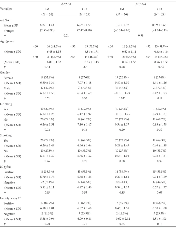
We also evaluated a possible association among the
relative expression levels ofANXA1andLGALS1mRNA and
the risk factors age, gender, smoking, drinking, H. pylori
infection, andcagAgenotype (Table2). We found a signiicant
increase only in the mean level of LGALS1 mRNA of the
women (0.80 ± 1.30) compared to the men (−0.15 ± 1.29) in
the IM group (� = 0.03).
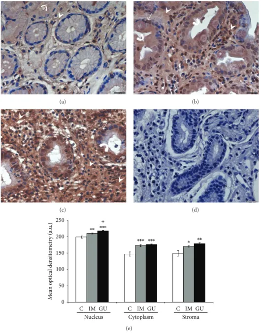
3.3. AnxA1 and Gal-1 Expressions in Precancerous Gastric Lesions. In normal mucosa, the AnxA1 and Gal-1 proteins showed low expression in the stroma, while in the epithelium there was no immunostaining for Gal-1, and AnxA1 presented
low immunoreactivity (Figures 2(a) and 3(a)). In the IM
and GU groups, both proteins exhibited high immunos-taining in the epithelial nuclei and cytoplasm as well as in the stroma. For Gal-1, these groups showed high staining throughout the extension of the epithelial cytoplasm (Figures
3(b)and3(c)). However, regarding the AnxA1 protein, the
GU samples presented higher immunostaining in the basal
portion of the epithelial cytoplasm (Figure2(c)), while the
IM group showed immunoreactivity in the whole cytoplasm
(Figure2(b)). he speciicity of the reactions was conirmed
by negative controls (Figures2(d)and3(d)).
he mean optical densitometry values for AnxA1 ranged from 147.5 to 218.2 in the three regions analyzed (epithelial nucleus, cytoplasm, and stroma) and, for Gal-1, from 131.6 to 215.8. here was a signiicant diference in the expression of both proteins in the epithelial cytoplasm and nuclei and in the stroma of the IM and GU groups compared to the
control group (� < 0.05). Moreover, we found a signiicant
diference (� < 0.05) between the IM and GU groups
regarding the nuclear expression of the AnxA1 protein in the
gastric epithelium (Figures2(e)and3(e)).
4. Discussion
his is the irst study that revealed increased annexin-A1 expression in human intestinal metaplasia and gastric ulcer and increased galectin-1 expression in gastric ulcer, two precancerous gastric lesions, indicating that these anti-inlammatory mediators can exert efects on the initial steps of stomach carcinogenesis. Our results also demonstrated the location of these proteins in the afected tissue and showed
that gene expression alterations occur regardless ofH. pylori
infection andcagAvirulence genotype.
he relative gene expression of ANXA1 was 6.2- and
6.7-fold increased, respectively, in intestinal metaplasia and gastric ulcer and these results were conirmed by AXNA1
protein expression analysis. RegardingLGALS1,the relative
Table 2: Relative gene expression ofANXA1andLGALS1mRNA in the intestinal metaplasia (IM) and gastric ulcer (GU) groups and comparison according to risk factors.
Variables
ANXA1 LGALS1
IM GU IM GU
(� = 36) (� = 29) (� = 36) (� = 29)
mRNA
Mean±SD 6.22 ± 1.43 6.69 ± 1.56 0.35 ± 1.37 0.69 ± 1.65
(range) (2.55–8.90) (2.42–8.80) (−3.54–2.86) (−4.04–3.13)
� 0.21 0.38
Age (years)
<60 16 (44.5%) <55 15 (51.7%) <60 16 (44.5%) <55 15 (51.7%) (Mean±SD) 6.48 ± 1.55 6.81 ± 1.71 0.62 ± 1.11 0.63 ± 1.84
≥60 20 (55.5%) ≥55 14 (48.3%) ≥60 20 (55.5%) ≥55 14 (48.3%) (Mean±SD) 6.00 ± 1.32 6.55 ± 1.43 0.14 ± 1.53 0.76 ± 1.50
� 0.34 0.66 0.28 0.83
Gender
Female 19 (52.8%) 8 (27.6%) 19 (52.8%) 8 (27.6%)
(Mean±SD) 6.30 ± 1.34 7.07 ± 1.18 0.80 ± 1.30 1.41 ± 1.26
Male 17 (47.2%) 21 (72.4%) 17 (47.2%) 21 (72.4%)
(Mean±SD) 6.12 ± 1.55 6.54 ± 1.69 −0.15 ± 1.29 0.42 ± 1.73
� 0.71 0.35 0.03∗ 0.11
Drinking
Yes 10 (27.8%) 11 (39.3%) 10 (27.8%) 11 (39.3%)
(Mean±SD) 6.12 ± 1.26 6.17 ± 1.97 −0.13 ± 1.75 0.29 ± 1.81
No 26 (72.2%) 17 (60.7%) 26 (72.2%) 17 (60.7%)
(Mean±SD) 6.26 ± 1.51 7.10 ± 1.17 0.54 ± 1.17 0.88 ± 1.58
� 0.78 0.18 0.29 0.39
Smoking
Yes 26 (72.2%) 18 (64.3%) 26 (72.2%) 18 (64.3%)
(Mean±SD) 6.26 ± 1.49 6.66 ± 1.64 0.29 ± 1.49 0.46 ± 1.88
No 10 (27.8%) 10 (35.7%) 10 (27.8%) 10 (35.7%)
(Mean±SD) 6.11 ± 1.32 6.86 ± 1.52 0.53 ± 1.01 0.98 ± 1.21
� 0.76 0.75 0.58 0.39
H. pylori
Positive 14 (38.9%) 15 (55.5%) 14 (38.9%) 15 (55.5%)
(Mean±SD) 6.70 ± 1.75 6.88 ± 1.35 0.29 ± 1.61 0.94 ± 1.59
Negative 22 (61.1%) 12 (44.5%) 22 (61.1%) 12 (44.5%)
(Mean±SD) 5.91 ± 1.11 6.47 ± 1.86 0.39 ± 1.23 0.67 ± 1.77
� 0.15 0.53 0.85 0.69
GenotypecagAa
Positive 12 (85.7%) 10 (66.7%) 12 (85.7%) 10 (66.7%)
(Mean±SD) 6.88 ± 1.81 6.82 ± 1.60 0.45 ± 1.58 0.50 ± 1.68
Negative 2 (14.3%) 5 (33.3%) 2 (14.3%) 5 (33.3%)
(Mean±SD) 5.58 ± 0.96 6.99 ± 0.81 −0.62 ± 2.12 1.81 ± 1.03
� 0.20 0.77 0.55 0.16
(a)
∗
(b)
(c) (d)
M
ea
n
o
p
ti
ca
l den
si
to
met
ry (a.u
.)
∗ ∗∗
∗∗ ∗∗∗
∗∗∗ ∗∗∗
100
C IM GU C IM GU C IM GU
Nucleus Cytoplasm Stroma
250
200
150
50
0
+
(e)
Figure 2: Expression of annexin-A1 protein in gastric mucosa (a–c). (a) Expression of AnxA1 in normal mucosa (C), predominantly in the cytoplasm of epithelial cells (curved arrow) and stroma (arrowhead). (b) Increased expression ater intestinal metaplasia (IM) in the nucleus (arrow), cytoplasm (curved arrow), and stromal region (arrowhead). (c) Higher AnxA1 expression can be observed in peripheral area of gastric ulcer (GU) especially in stroma (arrowhead) and epithelial cells (curved arrow and arrow). (d) Absence of immunoreactivity in the reaction control. Asterisk: goblet cell. Hematoxylin of Harris counterstain. Bar: 20�m. (e) Densitometry analyses (mean±SE).∗�in relation to C (∗� < 0.05;∗∗� < 0.01; ∗∗∗� < 0.001);+� < 0.05in relation to IM. a.u.: arbitrary unit.
immunohistochemistry techniques performed are reliable due to the absence of quantiication and immunostaining, respectively, in the negative controls. Several studies have shown minimal or limited correlation between mRNA and protein levels, particularly when using average expression
values [50,51]. his is justiied by the existence of
posttran-scriptional processes such as translation, posttranslational mechanisms, and degradation, which inluence the protein
abundance in a speciic tissue [50–52]. herefore, mRNA
expression levels do not always predict corresponding protein
levels [52].
he increase of annexin-A1 expression in the lesions analyzed indicates a possible involvement in the progression of gastric carcinogenesis from early lesions, as observed in
our previous study in chronic gastritis [46] and now in
intestinal metaplasia and gastric ulcer, to gastric cancer. he role of this protein in carcinogenesis is still uncertain, and
its efect may seem tissue speciic [22] and dependent on its
subcellular location [53]. Particularly in gastric cancer, the
few studies show conlicting results, some of them revealing
overexpression of AnxA1 [44, 46,53] and others
(a)
∗
(b)
(c) (d)
M
ea
n
o
p
ti
ca
l den
si
to
met
ry (a.u
.) ∗∗∗
∗∗∗
∗∗∗ ∗∗∗ ∗∗∗ ∗∗∗
100
C IM GU C IM GU C IM GU
Nucleus Cytoplasm Stroma
250
200
150
50
0
(e)
Figure 3: Expression of galectin-1 protein in the gastric mucosa (a–c). (a) Expression of Gal-1 in normal mucosa (C) in the stroma (arrowhead). (b) Intense immunostaining was observed in intestinal metaplasia (IM) in the nucleus (arrow), cytoplasm (curved arrow), and stromal region (arrowhead). (c) Peripheral area of gastric ulcer (GU) with high expression in stroma (arrowhead) and epithelial cells (curved arrow and arrow). (d) Absence of immunoreactivity in the reaction control. Asterisk: goblet cell. Hematoxylin of Harris counterstain. Bar: 20�m. (e) Densitometry analyses (mean±SE).∗�in relation to C (∗∗∗� < 0.001). a.u.: arbitrary unit.
[45] observed increased expression of this protein in healing
areas of gastric ulcer induced in rats, but we did not ind any study in intestinal metaplasia. Interestingly, other studies in cancers that originate from multistep processes also show alterations in the AnxA1 expression, both in precursor lesions and carcinoma, indicating a greater proximity between
pre-cancerous and pre-cancerous stages [19,21]. For example, in oral
squamous cell carcinoma, this protein presented decreased expression in the plasmatic membrane of tumor cells and
premalignant lesions compared to normal oral mucosa [21].
Similarly, Alves et al. [19] also observed reduced expression of
AnxA1 in premalignant lesions diagnosed as oral leukoplakia and in laryngeal squamous cell carcinoma.
he molecular pathway of geneANXA1in the modulation
of carcinogenesis is related to its action as a substrate to the epidermal growth factor receptor (EGFR) and protein kinase C (PKC), which implies its involvement in
sig-nal transduction pathways to cancer [54, 55]. he AnxA1
expression inluences the mitogen-activated protein kinases (MAPKs) pathway that is associated with the regulation of biological functions such as cell proliferation, diferentiation,
pathway members remains uncertain. Increased ANXA1
expression was found to be associated with constitutive expression of extracellular signal-regulated kinases- (ERK-)1
and 2 in macrophages [54] and vascular smooth muscle cells,
contributing to the reduction in the cell proliferation rate
through downregulation of cyclin D1 [57]. Yet, in prostate
cancer, the involvement of AnxA1 in this pathway was not by an antiproliferative but rather a proapoptotic action through
p38 and JNK (c-Jun N-terminal kinase) activation [56]. hus,
the action of AnxA1 on proliferation seems to depend on the tissue type. An antiproliferative activity was found in
lung adenocarcinoma [58], macrophages, and smooth muscle
tissue [57]. On the other hand, proliferation stimulation
was observed in hepatocytes, in which AnxA1 was related
to EGF [59], and in breast cancer; in the latter, it was
associated with formyl peptide receptor (FPR2) binding
and increased levels of cyclin D1 [60]. In gastric cancer
patients, the high expression of this protein was related to the promotion of invasiveness and shorter survival, and this rela-tionship occurred through the FPR/ERK/ITGB1BP1 pathway
[53].
he Gal-1 protein shows increased expression in various types of cancer and is associated in most of the cases with
aggressiveness and metastatic potential [36, 37, 61–63]. In
gastric cancer, however, Bektas et al. [40] did not ind
expres-sion of this protein in most of the cases studied, whereas
Jorge et al. [46] observed increased expression of Gal-1 in the
stroma and epithelium of cancerous gastric mucosa. Chen
et al. [43] observed increased expression of the LGALS1
mRNA in the stomach cell line TMC-1, suggesting that this
protein is important in the metastasis process. Anotherin
vitrostudy showed that the tumor suppressor geneRASSF1A
positively regulates theLGALS1mRNA level, leading to the
suppression of the NF-kB signaling pathway, which indicates
that Gal-1 expression may be related to cell cycle arrest [64].
Regarding gastric lesions, we previously observed elevated
protein expression in chronic gastritis [46], but Bektas et
al. [40] did not detect any increased expression in
tumor-associated metaplasia and dysplasia, and we found no reports about this inding in gastric ulcer. However, in the present study, Gal-1 presented increased expression in both intestinal metaplasia not associated with cancer and gastric ulcer. In contrast, in colorectal adenoma, a precancerous lesion of the colon presented downregulation of Gal-1 compared to normal mucosa, showing a change in gene expression in early
stages of colorectal carcinogenesis [65].
Galectin-1 has many functions involving carcinogenesis. High expression of this protein in the tumor microenvi-ronment contributes to the development and progression of the tumor by promoting environmental
immunosuppres-sion, angiogenesis, and metastasis [30]. Gal-1 induces
Fas-mediated apoptosis in immature thymocytes and activated
T lymphocytes [66], resulting in the activation of caspase
8 and increased mitochondrial membrane potential [27];
in lymphoblastoid Jurkat cells, the apoptosis triggered by Gal-1 occurs via JNK/c-Jun/AP-1 (activating protein-1
tran-scription factor) [24]. hus, Gal-1 contributes to conferring
immune privilege to tumors through apoptosis of T cells. Gal-1 released in the tumor microenvironment also acts on
activated endothelial cells, promotingH-Rassignaling via the
Raf/MAPK/MEK/ERK pathway, which results in the prolif-eration and migration of endothelial cells and consequent
generation of new vessels [67]. Taken together, these data,
combined with the results of the present study, indicate that galectin-1 can also inluence the gastric carcinogenesis process, contributing to the progression of the lesions cascade from the initial stages.
Besides the change in expression levels, the location of these anti-inlammatory proteins may play an important role
in the development of diferent pathological conditions [19].
We analyzed the location of both proteins in the premalignant lesions, which presented increased expression in relation to normal mucosa in the stroma and in the epithelial cytoplasm and nucleus. AnxA1 is normally detected in the cytoplasm
of many tissues [20]. Exposure to hydrogen peroxide, heat,
arsenic, and EGF promotes its translocation to the nucleus
[68]. In gastric cancer, nuclear expression of this protein
was found to be associated with advanced disease and poor
prognosis [41], and in oral carcinoma it is a predictor of lower
survival [20]. However, we cannot airm how signiicant the
nuclear expression of this protein was in the precancerous lesions studied here. Diferently, Gal-1 has both cytoplasmic
expressions as nuclear [63], while stromal expression is
associated with diferentiation stage and metastasis in gastric
cancer [40]. Our results showed signiicantly higher
expres-sion of this protein in the stroma of intestinal metaplasia and gastric ulcer samples compared to normal mucosa, which
may contribute, as shown by Hittelet et al. [69] in colon
cancer, to the progression from a precancerous to a cancerous stage.
When we analyzed the association ofANXA1andLGALS1
mRNA expression levels with the risk factors in the two lesions studied, we only found an association between
the relative expression of LGALS1 and gender in the IM
group, with women showing higher expression than men
(RQ = 0.80 versus −0.15). We did not ind any
associa-tion between the expression levels of both genes and H.
pyloriinfection, nor with the cagAgenotype. Nevertheless, considering the important role of bacterial infection in the development and progression of gastric cancer, investiga-tions in larger populainvestiga-tions are needed to conirm these results.
Regarding the role of the higher expression of
galectin-1 in women, Von Wolf et al. [70] reported elevated
expres-sion of this protein in human endometrium during the menstrual cycles and in maternal deciduas, suggesting a role of this protein in maintaining pregnancy. Other
stud-ies suggest that sex steroids may regulate LGALS1
expres-sion in female reproductive tissues of humans and rats
[70, 71]. Choe et al. [71] had observed increased
expres-sion of this gene in the uterus of ovariectomized rats
six hours ater treatment with 17�-estradiol and 12 hours
ater inoculation of progesterone, the efects being blocked by antagonists of these hormones. It has therefore been suggested that estrogen and progesterone receptors are
involved in galectin-1 expression [72], so the higherLGALS1
5. Conclusions
In conclusion, our results evidence that both the AnxA1 and Gal-1 anti-inlammatory proteins are overexpressed in precancerous gastric lesions such as intestinal metaplasia and gastric ulcer. hese results, together with the data of our
previous study in chronic gastritis and gastric cancer [46],
allow suggesting their involvement in gastric carcinogen-esis from the early stages through the tumor progression cascade, possibly due to the inlammatory process of the gastric mucosa. However, the deregulated expression occurs
regardless of the H. pylori infection and cagA virulence
genotype. Further investigations are necessary to clarify the mechanisms of action of these proteins in the progression from precancerous lesions to cancer.
Conflict of Interests
he authors declare that there is no conlict of interests regarding the publication of this paper.
Acknowledgments
he authors are grateful to Dr. Sebasti˜ao Roberto Taboga and Luiz Roberto Faleiros for their help with histological sections and Caroline de Freitas Zanon for contributing to the immunohistochemical standardization of galectin-1. his study was supported by the Brazilian agencies FAPESP (2011/11550-3 and 2012/15036-8) and CNPq (304870/2012-9).
References
[1] P. Correa, “A human model of gastric carcinogenesis,”Cancer
Research, vol. 48, no. 13, pp. 3554–3560, 1988.
[2] H. Watanabe, “Intestinal metaplasia—the efect of acid on the gastric mucosa and gastric carcinogenesis,”Journal of
Toxico-logic Pathology, vol. 23, no. 3, pp. 115–123, 2010.
[3] R. A. Busuttil and A. Boussioutas, “Intestinal metaplasia: a premalignant lesion involved in gastric carcinogenesis,”Journal
of Gastroenterology and Hepatology, vol. 24, no. 2, pp. 193–201,
2009.
[4] J. A. Todd, C. J. Richards, A. Dixon, and R. J. Robinson, “Gastric ulcer and malignancy—is there a need for follow-up endoscopy?”Alimentary Pharmacology & herapeutics, vol. 19, no. 9, pp. 989–991, 2004.
[5] J. G. Kusters, A. H. M. van Vliet, and E. J. Kuipers, “Pathogenesis
ofHelicobacter pyloriinfection,”Clinical Microbiology Reviews,
vol. 19, no. 3, pp. 449–490, 2006.
[6] B. Bauer and T. F. Meyer, “he human gastric pathogen
Helicobacter pyloriand its association with gastric cancer and
ulcer disease,”Ulcers, vol. 2011, Article ID 340157, 23 pages, 2011. [7] A. S. T. de Carvalho, “Peptic ulcer,”Jornal de Pediatria, vol. 76,
2, pp. S127–S134, 2000.
[8] C. Resende, A. hiel, J. C. Machado, and A. Ristim¨aki, “Gastric cˆancer: basic aspects,”Helicobacter, vol. 16, supplement 1, pp. 38–44, 2011.
[9] T. Chiba, H. Marusawa, and T. Ushijima, “Inlammation-associated cancer development in digestive organs: mechanisms and roles for genetic and epigenetic modulation,” Gastroenterol-ogy, vol. 143, no. 3, pp. 550–563, 2012.
[10] D. B. Polk and R. M. Peek Jr., “Helicobacter pylori:gastric cancer and beyond,”Nature Reviews, vol. 10, no. 6, pp. 403–414, 2010. [11] M. Hatakeyama, “Helicobacter pyloriand gastric
carcinogen-esis,”Journal of Gastroenterology, vol. 44, no. 4, pp. 239–248, 2009.
[12] B. X. Truong, V. T. C. Mai, H. Tanaka et al., “Diverse charac-teristics of thecagAgene ofHelicobacter pyloristrains collected from patients from Southern Vietnam with gastric cancer and peptic ulcer,”Journal of Clinical Microbiology, vol. 47, no. 12, pp. 4021–4028, 2009.
[13] V. Bizzarro, A. Petrella, and L. Parente, “Annexin A1: novel roles in skeletal muscle biology,”Journal of Cellular Physiology, vol. 227, no. 8, pp. 3007–3015, 2012.
[14] F. Cedeno-Laurent and C. J. Dimitrof, “Galectin-1 research in T cell immunity: past, present and future,”Clinical Immunology, vol. 142, no. 2, pp. 107–116, 2012.
[15] L. H. K. Lim and S. Pervaiz, “Annexin 1: the new face of an old molecule,”he FASEB Journal, vol. 21, no. 4, pp. 968–975, 2007. [16] J. P. Vago, C. R. C. Nogueira, L. P. Tavares et al., “Annexin A1 modulates natural and glucocorticoid-induced resolution of inlammation by enhancing neutrophil apoptosis,”Journal of
Leukocyte Biology, vol. 92, no. 2, pp. 249–258, 2012.
[17] R. J. Flower and G. J. Blackwell, “Anti-inlammatory steroids induce biosynthesis of a phospholipase A2 inhibitor which prevents prostaglandin generation,”Nature, vol. 278, no. 5703, pp. 456–459, 1979.
[18] S. M. Oliani and C. D. Gil, “Prote´ına antiinlamat´oria anexina 1: mecanismos celulares e relevˆancia cl´ınica,”Revista Arquivos de
Ciˆencias da Sa´ude, vol. 13, pp. 186–191, 2006.
[19] V. A. F. Alves, S. Nonogaki, P. M. Cury et al., “Annexin A1 subcellular expression in laryngeal squamous cell carcinoma,”
Histopathology, vol. 53, no. 6, pp. 715–727, 2008.
[20] C.-Y. Lin, Y.-M. Jeng, H.-Y. Chou et al., “Nuclear localization of annexin A1 is a prognostic factor in oral squamous cell carcinoma,”Journal of Surgical Oncology, vol. 97, no. 6, pp. 544– 550, 2008.
[21] H. Nomura, K. Uzawa, Y. Yamano et al., “Down-regulation of plasma membranous annexin A1 protein expression in prema-lignant and maprema-lignant lesions of the oral cavity: correlation with epithelial diferentiation,”Journal of Cancer Research and
Clinical Oncology, vol. 135, no. 7, pp. 943–949, 2009.
[22] W.-Y. Kang, W.-T. Chen, Y.-C. Huang, Y.-C. Su, and C.-Y. Chai, “Overexpression of annexin 1 in the development and diferentiation of urothelial carcinoma,”Kaohsiung Journal of
Medical Sciences, vol. 28, no. 3, pp. 145–150, 2012.
[23] F.-T. Liu and G. A. Rabinovich, “Galectins: regulators of acute and chronic inlammation,”Annals of the New York Academy of
Sciences, vol. 1183, pp. 158–182, 2010.
[24] B. Brandt, E. F. Abou-Eladab, M. Tiedge, and H. Walzel, “Role of the JNK/c-Jun/AP-1 signaling pathway in galectin-1-induced T-cell death,”Cell Death & Disease, vol. 1, article e23, 2010. [25] R. C. Hughes, “Galectins as modulators of cell adhesion,”
Biochimie, vol. 83, no. 7, pp. 667–676, 2001.
[26] K. Scott and C. Weinberg, “Galectin-1: a bifunctional regulator of cellular proliferation,”Glycoconjugate Journal, vol. 19, no. 7–9, pp. 467–477, 2002.
[28] G. A. Rabinovich, G. Daly, H. Dreja et al., “Recombinant galectin-1 and its genetic delivery suppress collagen-induced arthritis via T cell apoptosis,” he Journal of Experimental
Medicine, vol. 190, no. 3, pp. 385–398, 1999.
[29] L. Santucci, S. Fiorucci, N. Rubinstein et al., “Galectin-1 sup-presses experimental colitis in mice,”Gastroenterology, vol. 124, no. 5, pp. 1381–1394, 2003.
[30] K. Ito, K. Stannard, E. Gabutero et al., “Galectin-1 as a potent target for cancer therapy: role in the tumor microenvironment,”
Cancer and Metastasis Reviews, vol. 31, no. 3-4, pp. 763–778,
2012.
[31] T. Dalotto-Moreno, D. O. Croci, J. P. Cerliani et al., “Targeting galectin-1 overcomes breast cancer-associated immunosuppres-sion and prevents metastatic disease,”Cancer Research, vol. 73, no. 3, pp. 1107–1117, 2013.
[32] R. Soldati, E. Berger, A. C. Zenclussen et al., “Neuroblas-toma triggers an immunoevasive program involving galectin-1-dependent modulation of T cell and dendritic cell compart-ments,”International Journal of Cancer, vol. 131, no. 5, pp. 1131– 1141, 2012.
[33] D. O. Croci, M. Salatino, N. Rubinstein et al., “Disrupting galectin-1 interactions with N-glycans suppresses hypoxia-driven angiogenesis and tumorigenesis in Kaposi’s sarcoma,”
he Journal of Experimental Medicine, vol. 209, no. 11, pp. 1985–
2000, 2012.
[34] Y. L. Hsu, C. Y. Wu, J. Y. Hung, Y. S. Lin, M. S. Huang, and P. L. Kuo, “Galectin-1 promotes lung cancer tumor metastasis by potentiating integrin �6�4 and Notch1/Jagged2 signaling pathway,”Carcinogenesis, vol. 34, no. 6, pp. 1370–1381, 2013. [35] A. Paz, R. Haklai, G. Elad-Sfadia, E. Ballan, and Y. Kloog,
“Galectin-1 binds oncogenic H-Ras to mediate Ras membrane anchorage and cell transformation,”Oncogene, vol. 20, no. 51, pp. 7486–7493, 2001.
[36] H.-J. Kim, H.-K. Jeon, Y. J. Cho et al., “High galectin-1 expression correlates with poor prognosis and is involved in epithelial ovarian cancer proliferation and invasion,”European
Journal of Cancer, vol. 48, no. 12, pp. 1914–1921, 2012.
[37] H. Wu, P. Chen, R. Liao et al., “Overexpression of galectin-1 is associated with poor prognosis in human hepatocellular carcinoma following resection,”Journal of Gastroenterology and
Hepatology, vol. 27, no. 8, pp. 1312–1319, 2012.
[38] H. Barrow, J. M. Rhodes, and L.-G. Yu, “he role of galectins in colorectal cancer progression,”International Journal of Cancer, vol. 129, no. 1, pp. 1–8, 2011.
[39] G. Yu, J. Wang, Y. Chen et al., “Tissue microarray analysis reveals strong clinical evidence for a close association between loss of annexin A1 expression and nodal metastasis in gastric cancer,”Clinical & Experimental Metastasis, vol. 25, no. 7, pp. 695–702, 2008.
[40] S. Bektas, B. Bahadir, B. H. Ucan, and S. O. Ozdamar, “CD24 and galectin-1 expressions in gastric adenocarcinoma and clinicopathologic signiicance,”Pathology & Oncology Research, vol. 16, no. 4, pp. 569–577, 2010.
[41] F. Zhu, C. Xu, Z. Jiang et al., “Nuclear localization of annexin A1 correlates with advanced disease and peritoneal dissemination in patients with gastric carcinoma,”Anatomical Record, vol. 293, no. 8, pp. 1310–1314, 2010.
[42] J. W. Lim, H. Kim, and K. H. Kim, “Cell adhesion-related gene expression byHelicobacter pyloriin gastric epithelial AGS cells,”
International Journal of Biochemistry & Cell Biology, vol. 35, no.
8, pp. 1284–1296, 2003.
[43] Y.-R. Chen, H.-F. Juan, H.-C. Huang et al., “Quantitative proteomic and genomic proiling reveals metastasis-related protein expression patterns in gastric cancer cells,”Journal of
Proteome Research, vol. 5, no. 10, pp. 2727–2742, 2006.
[44] C.-M. Wu, Y.-S. Lee, T.-H. Wang et al., “Identiication of dif-ferential gene expression between intestinal and difuse gastric cancer using cDNA microarray,”Oncology Reports, vol. 15, no. 1, pp. 57–64, 2006.
[45] G. R. Martin, M. Perretti, R. J. Flower, and J. L. Wallace, “Annexin-1 modulates repair of gastric mucosal injury,”
Ameri-can Journal of Physiology, vol. 294, no. 3, pp. G764–G769, 2008.
[46] Y. C. Jorge, M. M. Mataruco, L. P. Ara´ujo et al., “Expression of annexin-A1 and galectin-1 anti-inlammatory proteins and mRNA in chronic gastritis and gastric cancer,” Mediators of
Inlammation, vol. 2013, Article ID 152860, 11 pages, 2013.
[47] B.-G. Jang and W. H. Kim, “Molecular pathology of gastric carcinoma,”Pathobiology, vol. 78, no. 6, pp. 302–310, 2011. [48] M. C. Duarte, E. Babeto, K. R. M. Leite et al., “Expression
ofTERT in precancerous gastric lesions compared to gastric cancer,”Brazilian Journal of Medical and Biological Research, vol. 44, no. 2, pp. 100–104, 2011.
[49] M. W. Pfal, “A new mathematical model for relative quantii-cation in real-time RT-PCR,”Nucleic Acids Research, vol. 29, no. 9, article e45, 2001.
[50] G. Chen, T. G. Gharib, C.-C. Huang et al., “Discordant protein and mRNA expression in lung adenocarcinomas,”Molecular &
Cellular Proteomics, vol. 1, no. 4, pp. 304–313, 2002.
[51] D. Greenbaum, C. Colangelo, K. Williams, and M. Gerstein, “Comparing protein abundance and mRNA expression levels on a genomic scale,”Genome Biology, vol. 4, no. 9, article 117, 2003.
[52] Y. Guo, P. Xiao, S. Lei et al., “How is mRNA expression predictive for protein expression? A correlation study on human circulating monocytes,”Acta Biochimica et Biophysica Sinica, vol. 40, no. 5, pp. 426–436, 2008.
[53] T. Y. Cheng, M. S. Wu, J. T. Lin et al., “Annexin A1 is associated with gastric cancer survival and promotes gastric cancer cell invasiveness through the formyl peptide receptor/extracellular signal-regulated kinase/integrin beta-1-binding protein 1 path-way,”Cancer, vol. 118, no. 23, pp. 5757–5767, 2012.
[54] L. C. Alldridge, H. J. Harris, R. Plevin, R. Hannon, and C. E. Bryant, “he annexin protein lipocortin 1 regulates the MAPK/ERK pathway,”he Journal of Biological Chemistry, vol. 274, no. 53, pp. 37620–37628, 1999.
[55] V. Gerke and S. E. Moss, “Annexins: from structure to function,”
Physiological Reviews, vol. 82, no. 2, pp. 331–371, 2002.
[56] C.-H. Hsiang, T. Tunoda, Y. E. Whang, D. R. Tyson, and D. K. Ornstein, “he impact of altered annexin I protein levels on apoptosis and signal transduction pathways in prostate cancer cells,”Prostate, vol. 66, no. 13, pp. 1413–1424, 2006.
[57] L. C. Alldridge and C. E. Bryant, “Annexin 1 regulates cell proliferation by disruption of cell morphology and inhibition of cyclin D1 expression through sustained activation of the ERK1/2 MAPK signal,”Experimental Cell Research, vol. 290, no. 1, pp. 93–107, 2003.
[58] J. D. Croxtall and R. J. Flower, “Lipocortin 1 mediates dexamethasone-induced growth arrest of the A549 lung ade-nocarcinoma cell line,”Proceedings of the National Academy of
Sciences of the United States of America, vol. 89, no. 8, pp. 3571–
[59] C. de Coupade, R. Gillet, M. Bennoun, P. Briand, F. Russo-Marie, and E. Solito, “Annexin 1 expression and phosphoryla-tion are upregulated during liver regeneraphosphoryla-tion and transforma-tion in antithrombin III SV40 T large antigen transgenic mice,”
Hepatology, vol. 31, no. 2, pp. 371–380, 2000.
[60] T. Khau, S. Y. Langenbach, M. Schuliga et al., “Annexin-1 signals mitogen-stimulated breast tumor cell proliferation by activation of the formyl peptide receptors (FPRs) 1 and 2,”he FASEB
Journal, vol. 25, no. 2, pp. 483–496, 2011.
[61] G. A. Rabinovich, “Galectin-1 as a potential cancer target,”
British Journal of Cancer, vol. 92, no. 7, pp. 1188–1192, 2005.
[62] S. J. Rodig, J. Ouyang, P. Juszczynski et al., “AP1-dependent galectin-1 expression delineates classical hodgkin and anaplas-tic large cell lymphomas from other lymphoid malignancies with shared molecular features,”Clinical Cancer Research, vol. 14, no. 11, pp. 3338–3344, 2008.
[63] Y.-M. Ding, J.-H. Dong, L.-L. Chen, and H.-D. Zhang, “Increased expression of galectin-1 is associated with human oral squamous cell carcinoma development,”Oncology Reports, vol. 21, no. 4, pp. 983–987, 2009.
[64] D. Zheng-Hao, W. Ji-Fang, X. de-Sheng, and Z. Jian-Hua, “Galectin-1 is up-regulated byRASSF1Agene in human gastric carcinoma cell line SGC7901,”Acta Pathologica, Microbiologica,
et Immunologica Scandinavica, vol. 120, no. 7, pp. 582–590, 2012.
[65] F. F. Lam, L. Jankova, O. F. Dent et al., “Identiication of distinctive protein expression patterns in colorectal adenoma,”
Proteomics, vol. 4, no. 1, pp. 60–70, 2010.
[66] N. L. Perillo, K. E. Pace, J. J. Seilhamer, and L. G. Baum, “Apoptosis of T cells mediated by galectin-1,”Nature, vol. 378, no. 6558, pp. 736–739, 1995.
[67] V. L. hijssen, B. Barkan, H. Shoji et al., “Tumor cells secrete galectin-1 to enhance endothelial cell activity,”Cancer Research, vol. 70, no. 15, pp. 6216–6224, 2010.
[68] H. J. Rhee, G.-Y. Kim, J. W. Huh, S.-W. Kim, and D. S. Na, “Annexin I is a stress protein induced by heat, oxidative stress and a sulhydryl-reactive agent,” European Journal of
Biochemistry, vol. 267, no. 11, pp. 3220–3225, 2000.
[69] A. Hittelet, H. Legendre, N. Nagy et al., “Upregulation of galectins-1 and -3 in human colon cancer and their role in regulating cell migration,”International Journal of Cancer, vol. 103, no. 3, pp. 370–379, 2003.
[70] M. von Wolf, X. Wang, H.-J. Gabius, and T. Strowitzki, “Galectin ingerprinting in human endometrium and decidua during the menstrual cycle and in early gestation,”Molecular
Human Reproduction, vol. 11, no. 3, pp. 189–194, 2005.
[71] Y. S. Choe, C. Shim, D. Choi, C. S. Lee, K. K. Lee, and K. Kim, “Expression of galectin-1 mRNA in the mouse uterus in under the control of ovarian steroids during blastocyst implantation,”
Molecular Reproduction and Development, vol. 48, no. 2, pp.
261–266, 1997.
[72] N. G. han, R. Romero, O. Erez et al., “Emergence of hormonal and redox regulation of galectin-1 in placental mammals: implication in maternal-fetal immune tolerance,”Proceedings of
the National Academy of Sciences of the United States of America,
Submit your manuscripts at
http://www.hindawi.com
Stem Cells
International
Hindawi Publishing Corporation
http://www.hindawi.com Volume 2014
Hindawi Publishing Corporation
http://www.hindawi.com Volume 2014 INFLAMMATION
Hindawi Publishing Corporation
http://www.hindawi.com Volume 2014
Behavioural
Neurology
Endocrinology
International Journal ofHindawi Publishing Corporation
http://www.hindawi.com Volume 2014
Hindawi Publishing Corporation
http://www.hindawi.com Volume 2014
Disease Markers
Hindawi Publishing Corporation
http://www.hindawi.com Volume 2014
BioMed
Research International
Oncology
Journal ofHindawi Publishing Corporation
http://www.hindawi.com Volume 2014
Hindawi Publishing Corporation
http://www.hindawi.com Volume 2014
Oxidative Medicine and Cellular Longevity
Hindawi Publishing Corporation
http://www.hindawi.com Volume 2014
PPAR Research
The Scientiic
World Journal
Hindawi Publishing Corporationhttp://www.hindawi.com Volume 2014
Immunology Research
Hindawi Publishing Corporation
http://www.hindawi.com Volume 2014
Journal of
Obesity
Journal ofHindawi Publishing Corporation
http://www.hindawi.com Volume 2014
Hindawi Publishing Corporation
http://www.hindawi.com Volume 2014 Computational and Mathematical Methods in Medicine
Ophthalmology
Journal ofHindawi Publishing Corporation
http://www.hindawi.com Volume 2014
Diabetes Research
Journal ofHindawi Publishing Corporation
http://www.hindawi.com Volume 2014
Hindawi Publishing Corporation
http://www.hindawi.com Volume 2014
Research and Treatment
AIDS
Hindawi Publishing Corporation
http://www.hindawi.com Volume 2014
Gastroenterology Research and Practice
Hindawi Publishing Corporation
http://www.hindawi.com Volume 2014
Parkinson’s
Disease
Evidence-Based Complementary and Alternative Medicine
Volume 2014