Volume 2013, Article ID 152860,11pages http://dx.doi.org/10.1155/2013/152860
Research Article
Expression of Annexin-A1 and Galectin-1 Anti-Inflammatory
Proteins and mRNA in Chronic Gastritis and Gastric Cancer
Yvana Cristina Jorge,
1Mayra Mioto Mataruco,
1Leandro Pires Araújo,
1Ana Flávia Teixeira Rossi,
1Juliana Garcia de Oliveira,
1Marina Curado Valsechi,
1Alaor Caetano,
2Kenji Miyazaki,
3Célia Sebastiana de Jesus Fazzio,
4Jorge Alberto Thomé,
5Paula Rahal,
1Sonia Maria Oliani,
1and Ana Elizabete Silva
11Department of Biology, S˜ao Paulo State University (UNESP), Campus de S˜ao Jos´e do Rio Preto, Rua Crist´ov˜ao Colombo 2265,
15054-000 S˜ao Jos´e do Rio Preto, SP, Brazil
2Rio Preto Endoscopy Center, S˜ao Jos´e do Rio Preto, SP, Brazil 3Endoscopy Service, Hospital de Base, S˜ao Jos´e do Rio Preto, SP, Brazil
4Legal Medicine Department and Pathology Service, Hospital de Base, S˜ao Jos´e do Rio Preto, SP, Brazil 5Institute of Anatomical Pathology and Cytopathology, S˜ao Jos´e do Rio Preto, SP, Brazil
Correspondence should be addressed to Sonia Maria Oliani; smoliani@ibilce.unesp.br and Ana Elizabete Silva; anabete@ibilce.unesp.br
Received 7 May 2012; Accepted 28 December 2012
Academic Editor: Eeva Moilanen
Copyright © 2013 Yvana Cristina Jorge et al. his is an open access article distributed under the Creative Commons Attribution License, which permits unrestricted use, distribution, and reproduction in any medium, provided the original work is properly cited.
Objective. he anti-inlammatory proteins annexin-A1 and galectin-1 have been associated with tumor progression. his scenario prompted us to investigate the relationship between the gene and protein expression of annexin-A1 (ANXA1/AnxA1) and galectin-1 (LGALS1/Gal-1) in an inlammatory gastric lesion as chronic gastritis (CG) and gastric adenocarcinoma (GA) and its association withH. pyloriinfection.Methods. We analyzed 40 samples of CG, 20 of GA, and 10 of normal mucosa (C) by the quantitative real-time PCR (qPCR) technique and the immunohistochemistry assay.Results. HighANXA1mRNA expression levels were observed in 90% (36/40) of CG cases (mean relative quantiication RQ =4.26 ± 2.03) and in 80% (16/20) of GA cases (mean RQ =4.38 ± 4.77). However,LGALS1mRNA levels were high (mean RQ =2.44 ± 3.26) in 60% (12/20) of the GA cases, while low expression was found in CG (mean RQ =0.43 ± 3.13;� < 0.01). Normal mucosa showed modest immunoreactivity in stroma but not in epithelium, while stroma and epithelium displayed an intense immunostaining in CG and GA for both proteins.Conclusion. hese results have provided evidence that galectin-1 and mainly annexin-A1 are overexpressed in both gastritis and gastric cancer, suggesting a strong association of these proteins with chronic gastric inlammation and carcinogenesis.
1. Introduction
Chronic inlammation has been recognized as a process that can trigger cancer, due to the host immune response with local expression of cytokines, chemokines, adhesion mole-cules, and pro- and anti-inlammatory proteins that stimu-late processes such as proliferation, survival, cell migration, and neovascularization. he strongest association between chronic inlammation and malignancy is observed in gastric
cancer induced byHelicobacter pyloriinfection [1].
he inlammatory process resulting fromH. pylori
infec-tion triggers a cascade of events initialized by chronic gastritis that evolves to gastric atrophy, intestinal metaplasia,
dysplasia, and inally carcinoma [2]. his bacterium is present
in 90% of all chronic gastritis patients [3] and in 77% of
noncardia gastric cancers [4].
Both the intrinsic factors of the host and the bacterial virulence are associated with the development of gastric
can-cer induced byH. pylori. Among the bacterial genes, there is
cancer [5]. he CagA protein is internalized by the host epi-thelial cells, disrupting the cell cycle and inducing cell
inva-sion through the activation of matrix metalloproteases [6].
Bacterial lipopolysaccharides activate several cell
pro-cesses, including expression of annexin-A1 (AnxA1) [7] and
galectin-1 (Gal-1) [8], which are both anti-inlammatory
pro-teins. During the inlammatory response, AnxA1 is translo-cated from the cell cytoplasm to the membrane, resulting in a decrease in the transmigration of inlammatory cells
to the site of injury [9]. Furthermore, AnxA1 plays its
anti-inlammatory role by inhibiting the activities of
phospholi-pase A2 and inducible nitric oxide synthase [10]. It is also
associated with modiications of the cytoskeleton, transport of molecules, ion lux, diferentiation and migration, cell
growth, and apoptosis [11].
Galectin-1 (Gal-1), a member of a family of carbohydrate-binding proteins, may act in the same manner as AnxA1 in the transmigration of inlammatory cells, being important also in T-cell apoptosis by binding to T-cell receptors, thus
triggering the Faz/caspase cascade [12]. It also contributes
to diferent events associated with carcinogenesis, including tumor transformation, cell cycle regulation, apoptosis, cell
adhesion, migration, and inlammation [13,14].
Nevertheless, there are only a few studies in the liter-ature that evaluated the expression pattern of those anti-inlammatory mediators in gastric mucosa, but their speciic functions are unclear. While in gastric cell lines the Gal-1
protein expression increased [15,16], in gastric
adenocarci-noma was observed low expression in tumor cells [17]. In
turn, AnxA1 was studied in gastric tissue with discrepant indings, such as increased expression during gastric mucosal
damage healing [18], loss of expression in metastatic gastric
cancers [19], higher expression in difuse-type gastric cancer
compared to the intestinal type [20], and decreased
expres-sion in gastric adenocarcinoma, but with positive staining in
advanced stage and peritoneal dissemination [21].
hus, further research in this ield might improve our understanding of the possible role of those anti-inlammatory proteins in carcinogenesis, which is at present fairly limited and controversial. Moreover, such studies may lead to the possible identiication of AnxA1 and Gal-1 as potential biomarkers in gastric cancer progression.
herefore, the aim of this study was to investigate the
relative expression levels ofANXA1andLGALS1mRNA by
quantitative real-time PCR (qPCR) and the expression of both proteins by immunohistochemical assay in inlamma-tory gastric lesions such as chronic gastritis compared to gastric cancer. We also investigated a possible relationship
between mRNA expression levels andH. pyloriinfection and
itscagAvirulence genotype in both lesions evaluated.
2. Materials and Methods
2.1. Subjects and Samples. Gastric biopsies were obtained from seventy (70) individuals submitted to endoscopy at the Endoscopy Service of the Hospital de Base and at Rio Preto Endoscopy Center, both in S˜ao Jos´e do Rio Preto, SP, Brazil. he histopathological data were supplied, respectively, by the
Legal Medicine Department and by the Pathology Service and the Institute of Anatomical Pathology and Cytopathology (IAPC), in the same city. For each individual, three (03) biopsy samples from the antrum region were collected for molecular studies and one for immunohistochemical analy-sis.
he gastric adenocarcinoma (GA) group comprised 20
individuals (14 male and 6 female; mean age63.4 ± 14years)
with a histopathologically conirmed diagnosis of gastric
ade-nocarcinoma [22]. Among the studied samples, 12 cases were
diagnosed as intestinal-type adenocarcinoma (IGC) and 8 as difuse-type adenocarcinoma (DGC). he chronic gastritis (CG) group was composed of 40 individuals (18 male and 22
female; mean age52.5 ± 15years) with a histopathologically
conirmed diagnosis of chronic gastritis [23].
he biopsies for the control (C) group were obtained from 10 healthy individuals (7 male and 3 female; mean
age35 ± 10.8 years) with no dyspeptic gastric complaints
and diagnosed as histopathologically normal gastric mucosa. Epidemiological data on the study population were collected using a standard interviewer-administered questionnaire, with questions about current and past occupation, smoking habits, alcohol intake, and family history of cancer. None of the 70 subjects were under antibiotic or anti-inlammatory treatment neither radiotherapy nor chemotherapy. About 60% of all patients in the GA group were smokers and 40% were drinkers, while all patients in the CG and C groups were nonsmokers and nondrinkers. Smokers were deined as individuals who consumed at least 100 cigarettes during their lifetime, and alcohol consumers were those who drank more
than four times a week [24].
he Research Ethics Committee of the participating insti-tution approved this research (CEP IBILCE/UNESP number 058/09), and written informed consent was obtained from all individuals studied.
2.2. Isolation of Total Nucleic Acids and Reverse-Transcription PCR (RT-PCR). Soon ater collection, the biopsies were
stored in RNA later solution (Applied Biosystems) at−20∘C,
to preserve their integrity until RNA extraction. he nucleic acid extraction was performed according to the protocol accompanying the reagent TRIzol (Invitrogen) that allows the simultaneous extraction of RNA and DNA. RNA and DNA concentrations were determined in a NanoDrop ND1000 spectrophotometer (Uniscience) by measuring absorbance at
260 and 280 nm. DNA samples were stored at−20∘C and used
for the molecular diagnosis ofH. pylori.
Aterwards, reverse-transcription (RT) PCR was
per-formed in an automated thermocycler, using 2.5�g of total
RNA in the presence of 1.25�L of oligo-d(T)16 (0.5�g/�L),
2.0�L of RNAse Inhibitor (80 U/�L), and a High Capacity
cDNA Archive Kit (Applied Biosystems), in a total volume
of 50�L, according to the manufacturer’s instructions. he
reactions were carried out for 10 minutes at 25∘C, followed by
120 minutes at 37∘C. he integrity of all cDNA preparations
was tested by a PCR assay of a 613 bpACTB(�-actin) gene
fragment, used as control for abundant transcripts, whose
primer sequences were F: 5�
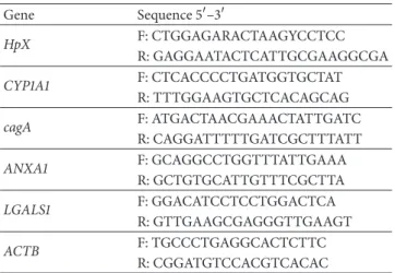
Table 1: Primers sequences used in multiplex PCR to determineH. pyloriinfection andcagAgenotype and in q-PCR assays.
Gene Sequence 5�–3�
HpX F: CTGGAGARACTAAGYCCTCC
R: GAGGAATACTCATTGCGAAGGCGA
CYP1A1 F: CTCACCCCTGATGGTGCTAT
R: TTTGGAAGTGCTCACAGCAG
cagA F: ATGACTAACGAAACTATTGATC
R: CAGGATTTTTGATCGCTTTATT
ANXA1 F: GCAGGCCTGGTTTATTGAAA
R: GCTGTGCATTGTTTCGCTTA
LGALS1 F: GGACATCCTCCTGGACTCA
R: GTTGAAGCGAGGGTTGAAGT
ACTB F: TGCCCTGAGGCACTCTTC
R: CGGATGTCCACGTCACAC
2.3. Molecular Diagnoses for H. pylori-cagA. To determine
the presence of H. pylori infection, DNA samples were
subjected to a multiplex PCR reaction containing primers
for the bacterial gene HpX [25] and for CYP1A1 (human
housekeeping gene, which attests the integrity of the DNA).
In summary, we used 5.0�L of 10X bufer, 5.0�L of dNTPs
(1.23 mmol/L, Invitrogen), 2.0�L MgCl2(25 mmol/L), 2.0�L
of primers (10 nmol/�L, Invitrogen), 24.5�L of dH2O, 5.0�L
of genomic DNA, and 0.5�L of Taq DNA Polymerase
(5 U/�L, Invitrogen). he material was processed in an
auto-mated thermocycler and was initially subjected to a
temper-ature of 94∘C for 5 minutes for denaturation. Subsequently,
it was subjected to 40 ampliication cycles at 94∘C for 45
seconds, at 60∘C for 30 seconds, and at 72∘C for 90 seconds,
followed by a inal extension cycle of 7 minutes at 72∘C.
he ampliication products were visualized on 2.0% agarose gel stained with ethidium bromide. Fragments of 150 bp and
226 bp corresponding to genesCYP1A1andHpX, respectively,
were observed.
heH. pylori-positive samples were then subjected to a
second PCR run, to investigate the virulence genotypecagA
of the bacterium [26]. he parameters used were the same as
for the previous reaction, except the annealing temperature,
which in this case was 52∘C. he product was visualized on
1.0% agarose gel stained with ethidium bromide, and a 232 bp fragment was observed. Positive and negative controls were used in all experiments. he primer sequences are listed in
Table 1.
2.4. Quantitative Analysis of the Relative Amount of ANXA1 and LGALS1 mRNA by Quantitative Real-Time PCR (q-PCR). he relative quantiication q-PCR assay forANXA1 and LGALS1 mRNA expression was performed in an ABI Prism 7300 Sequence Detector System (Applied Biosystems, Foster City, CA, USA), according to the instructions for the SYBR Green PCR Core Reagent (Applied Biosystems), using
primers speciic for genesANXA1 and LGALS1[27]. Gene
ACTBwas used as endogenous control (reference gene) of the
reaction, because it had shown the lowest variation compared
to�-tubulinand�2-microglobulingenes in a previous study
[28]. he primer sequences are presented inTable 1.
he expression levels of theANXA1,LGALS1, andACTB
mRNA were tested in triplicate (cDNA from the same RT reaction, but in separated wells). Controls with no template cDNA were used for each assay (negative control). Samples of normal gastric mucosa were mixed to form a pool that was used as a calibrator (standard sample). he q-PCR assays
were performed in a total volume of 50�L, containing 10�L
of SYBR Green Master Mix (Applied Biosystems), 25 ng of
cDNA, and 0.4�M ofANXA1and 0.5�MLGALS1primers.
Ater initial incubation at 50∘C for 2 min to allow
uracil-N-glycosylase (UNG) digestion and at 95∘C for 10 min to
acti-vate the AmpliTaq Gold DNA polymerase (both provided by the Universal PCR Master Mix), the samples were ampliied
by subjecting them to 40 biphasic cycles of 95∘C for 15 sec and
60∘C for 1 min.
he luorescence signal was measured in the extension
phase of the PCR reaction, and a threshold value (C�) of
luorescence in the exponential part of the ampliication curve was selected. he larger the quantities of the material at start, the lower the CT values. Relative quantiication (RQ)
of genesANXA1andLGALS1was obtained as described by
Pfal [29] and normalized with the�-actincontrol reference
gene and normal gastric mucosa. he transcript levels were
considered to be upregulated if RQ>2.0.
2.5. Immunohistochemistry. Deparainized sections (4�m)
were incubated in citrate bufer, pH 6.0, at 96∘C for 30
minutes, washed with distilled water, incubated with 3% hydrogen peroxide in methanol (30 minutes), and washed in phosphate-bufered saline (PBS, pH 7.4). he primary antibodies rabbit polyclonal anti-Gal-1 and rabbit polyclonal anti-ANXA1 (Zymed Laboratories, Cambridge, UK) were diluted to 1 : 500 or 1 : 2000, respectively, in 1% bovine serum
albumin (BSA) and applied overnight at 4∘C. As negative
controls, some sections were incubated with 1% BSA without any primary antibody. Fragments were then washed in PBS, incubated with the universal LSAB kit/HRP secondary anti-body (Dako, USA) according to the manufacturer’s protocol,
washed in PBS, and developed with 3,3�-diaminobenzidine in
chromogen solution (Dako, USA). he sections were washed thoroughly in distilled water, counterstained with hema-toxylin and mounted on glass slides. Densitometric analysis for AnxA1 and Gal-1 immunostaining was performed using an arbitrary scale from 0 to 255 with the AxioVision sotware on a Zeiss-Axioskop II light microscope, and the data were
expressed as mean±SE.
2.6. Statistical Analysis. Fisher’s exact test was used to deter-mine if there were signiicant diferences between groups regarding the presence of bacteria and the cagA genotype, the histological type of tumor, and the gender. he data obtained
from mRNA quantiication were expressed as mean±SD.
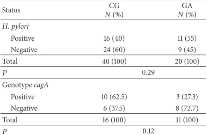
Table 2: Distribution of infection byH. pyloriandcagAstrains into chronic gastritis (CG) and gastric adenocarcinoma (GA) groups.
Status CG
N(%)
GA N(%) H. pylori
Positive 16 (40) 11 (55)
Negative 24 (60) 9 (45)
Total 40 (100) 20 (100)
� 0.29
GenotypecagA
Positive 10 (62.5) 3 (27.3)
Negative 6 (37.5) 8 (72.7)
Total 16 (100) 11 (100)
� 0.12
�: number of individuals.
genotype, histological type of tumor, gender, smoking, and
drinking, we used the nonparametric �-test with Welch’s
correction. he value of protein expression was expressed as
mean±SE. he mean densitometry analysis results obtained
for proteins AnxA1 and Gal-1 were compared by ANOVA, followed—if signiicant—by the Bonferroni test. hese analy-ses were performed using the GraphPad InStat and GraphPad
Prism 4 sotware. he value was considered signiicant if� <
0.05.
3. Results
3.1. Molecular Diagnoses for H. pylori-cagA. he frequencies
of cases with positive molecular diagnosis forH. pyloriand
genotype cagA in groups CG and GA are presented in
Table 2. All 10 samples of normal mucosa were conirmed by
molecular testing asH. pylorinegative.
Out of a total of 60 samples from the case groups, 45% were H. pyloripositive: 40% (16/40) in the CG group and 55% (11/20) in the GA group, with no signiicant diference
between the groups (� = 0.29). Regarding the genotypecagA,
48% ofH. pylori-positive samples werecagApositive: 62.5%
(10/16) in the CG and 27.3% (3/11) in the GA group. Again,
there was no signiicant diference between the groups (� =
0.12).
3.2. Relative Gene Expression Analysis. he relative
expres-sion levels ofANXA1 mRNA, ater normalization with the
ACTB reference gene and comparison with the normal
mucosa, were increased in 90% (36/40) of the CG cases (mean
RQ =4.26 ± 2.03) and in 80% (16/20) of the GA cases (mean
RQ = 4.38 ± 4.77), so there was no statistically signiicant
diference between the groups (� = 0.33). ForLGALS1, the
mRNA relative expression values found were lower; only the GA group showed overexpression in 60% (12/20) of the cases
(mean RQ =2.44 ± 3.26). he CG group showed constitutive
expression (mean RQ =0.43 ± 3.13), with only 7.5% (3/40)
of the cases presenting increased expression. hus, the mean
level ofLGALS1mRNA expression was signiicantly higher
in the GA than in the CG group (� < 0.01) (Table 3 and
Figure 1).
In another analysis, we investigated the GA group for
a possible association betweenANXA1andLGALS1mRNA
expression and risk factors such drinking, smoking, and
his-tological type of gastric cancer (Table 4), but no association
was observed. In addition, comparing the variables, gender, H. pyloriinfection, andcagA+genotype, in the CG and the
GA groups (Table 5), a signiicant diference was found in
the GA group for the gender and the mean level ofANXA1
expression (� = 0.04), due to a 2 times greater expression
of this gene in the females than in the males of this group.
Likewise, the cagA+ genotype also showed an association
with theANXA1expression level in the GA group, due to a
higher mRNA expression in thecagA-positive compared to
thecagA-negative cases (mean RQ 6.40 versus 2.77,� < 0.01). None of the other investigated factors appears to be associated
with the levels ofANXA1andLGALS1mRNA.
3.3. Protein Expression Measured by Immunohistochemistry Assay. Protein expression was evaluated in normal mucosa, CG, intestinal (IGC), and difuse- (DGC-) type gastric cancer. Modest expression of AnxA1 and Gal-1 was observed in the stroma of normal mucosa, while the epithelium did not
show any expression of these proteins (Figures2(a)and3(a),
resp.). However, in the inlammatory process of CG mucosa, intense immunostaining of AnxA1 and Gal-1 was seen in
the basal portion of the epithelium (Figures2(b)and 3(b),
resp.). Positive immunostaining for AnxA1 and Gal-1 was
also observed in DGC (Figures2(c)and3(c), resp.) and IGC
(Figures2(d)and3(d), resp.) tumor cells. In some areas of
gastric cancer samples, it was possible to identify epithelial cells showing AnxA1 and Gal-1 expression.
he mean optical densitometry values of AnxA1 and Gal-1
expression are presented in Figures2(e)and3(e), respectively.
he AnxA1 mean density was 90.79 for normal mucosa (C group), while for CG and GA, it was, respectively, 168.57 and 190.20. hus, a signiicant diference was found comparing
the normal mucosa with the CG and GA groups (� < 0.01
for both). he mean density of the Gal-1 protein was lower in all three groups (86.52, 136.97, and 146.30 in C, CG, and GA, resp.), showing a statistically signiicant diference between
the normal mucosa and the CG and GA groups (� < 0.01for
both).
In another densitometry analysis to determine the cyto-plasmic and nuclear immunoreactivity of AnxA1 separately in all groups, although observed nuclear immunostaining mainly in gastric cancer, the data obtained in our results did not show a statistically signiicant diference between normal
mucosa and CG(� = 0.19)and normal mucosa and GA
(� = 0.18)(data not shown).
4. Discussion
Table 3: Comparison ofANXA1andLGALS1mRNA relative expression levels between the chronic gastritis (CG) and the gastric adenocar-cinoma (GA) groups.
Variable ����1 �����1
CG GA CG GA
Relative expression (mean±SD) 4.26 ± 2.03 4.38 ± 4.77 0.43 ± 3.13 2.44 ± 3.26
Minimum −2.97 −7.88 −10.78 −6.95
Maximum 8.90 9.29 10.91 9.28
� 0.33 <0.01∗
∗Signiicant diference.
Table 4: Relative expression ofANXA1andLGALS1mRNA in the gastric adenocarcinoma group related to drinking, smoking, and histological type of cancer.
Variable ����1 �����1
Drinking
Yes 8 (40%) 8 (40%)
(mean±SD) 2.29 ± 5.76 1.74 ± 1.49
No 12 (60%) 12 (60%)
(mean±SD) 5.98 ± 2.61 2.82 ± 3.91
� 0.15 0.39
Smoking
Yes 12 (60%) 12 (60%)
(mean±SD) 4.01 ± 5.54 2.44 ± 2.18
No 8 (40%) 8 (40%)
(mean±SD) 4.20 ± 4.17 2.45 ± 4.21
� 0.93 0.99
Histology
Intestinal 12 (60%) 12 (60%)
(mean±SD) 5.82 ± 2.89 3.60 ± 2.79
Difuse 8 (40%) 8 (40%)
(mean±SD) 1.55 ± 6.02 0.71 ± 3.32
� 0.09 0.06
factors, we investigated the presence of H. pylori in both
lesions and its virulence genotypecagA, since this bacterium
oten triggers the progression of the gastric carcinogenesis cascade. To the best of our knowledge, this is the irst study that has evaluated the expression of AnxA1 and Gal-1 in chronic gastritis.
We have found a high relative expression of ANXA1
mRNA already in the CG inlammatory process, in which 90% of cases showed an increased expression
level(mean RQ = 4.26), which became 3 to 8 times higher
ater normalization with the ACTB reference gene and
comparison with normal mucosa. Similarly, in the GA group, 80% of cases presented upregulated expression levels, which
were 3 to 9 times higher(mean RQ = 4.38)than that in
normal mucosa. ForLGALS1, our study showed a slightly
increased relative expression only in 60% of the GA cases
(mean RQ = 2.44), with an increase of 2 to 7 times, but in
chronic gastritis the mean value was low(mean RQ = 0.43).
In general, the immunohistochemical analysis conirmed the results of mRNA expression by qPCR, although in CG
the relative expression levels of LGALS1 mRNA was not
equivalently increased as protein expression.
Studies on the expression ofANXA1mRNA in neoplastic
processes are still limited and the results are conlicting. For example, loss of expression was found in squamous cell
carcinoma of the esophagus [30], prostatic adenocarcinoma
[31], sinonasal adenocarcinoma [32], larynx [33], and breast
cancer [34]. On the other hand, overexpression has been
reported in colorectal adenocarcinoma [35], urothelial
car-cinoma [36], lung adenocarcinoma [37], and oral cancers
[38], thus suggesting that changes in the expression levels of
ANXA1may be related to the tissue or tumor type.
Martin et al. [18] reported that normal gastric mucosa
shows weak expression of the protein AnxA1, while in the ulcer healing process the expression was increased, pro-moting the reduction of the ulcer. In contrast, Yu et al.
[19] observed overexpression of both gene and protein in
normal mucosa but loss of expression in 64% of primary gastric tumors, mainly correlated with advanced stage and
metastasis. More recently, Zhu et al. [21] observed that AnxA1
protein is expressed in both gastric adenocarcinoma (45%) and normal tissues (69%), but with diferent subcellular
distribution. Similar results were reported by Cheng et al. [39]
that observed high AnxA expression, both mRNA and pro-tein, associated with metastasis, invasion, and poor survival in gastric cancer patients. he authors also proposed a new mechanism of how AnxA1 regulates the gastric cancer cell invasion through activation FPR/ERK/ITGB1BP1 pathway.
In the present study, the immunohistochemical analysis for AnxA1 showed modest immunostaining in stromal cells of normal mucosa and intense expression in stroma and epithelium of both CG and GA, thus conirming the results of the mRNA expression analysis. he assay used in this study did not allow a clear diferentiation of the cellular localization of the protein, although positive immunostaining was observed in the cytoplasm of epithelial cells and the nucleus of cancer cells, with lower intensity and frequency in chronic gastritis. However, when the densitometry analysis to determine cytoplasmic and nuclear immunoreactivity of AnxA1 separately was performed, the data obtained did not show a statistically signiicant diference among nor-mal mucosa, chronic gastritis, and gastric adenocarcinoma. Although, there are some reports that show translocation
of AnxA1 during carcinogenesis, as Alves et al. [40] that
showed 87.5% positivity for AnxA1 in larynx tumors and increased immunoreactivity in the membrane compared to the cytoplasm and the nucleus. However, compared to the normal tissue, the nuclear and cytoplasmic expression was
Table 5: Relative expression ofANXA1andLGALS1mRNA in chronic gastritis (CG) and gastric adenocarcinoma (GA) groups according to gender, infection byH. pylori, and presence ofcagA+ genotype.
Variable ����1 �����1
CG GA CG GA
Gender
Female 22 (55%) 6 (30%) 22 (55%) 6 (30%)
(mean±SD) 4.40 ± 1.80 6.67 ± 2.00 0.29 ± 3.55 4.81 ± 3.26
Male 28 (45%) 14 (70%) 28 (45%) 14 (70%)
(mean±SD) 4.08 ± 2.32 3.25 ± 5.16 0.60 ± 2.61 1.66 ± 2.96
� 0.63 0.04∗ 0.75 0.10
H. pylori
Positive 16 (40%) 11 (55%) 16 (40%) 11 (55%)
(mean±SD) 4.04 ± 2.59 3.91 ± 5.48 −0.05 ± 3.97 3.33 ± 3.16
Negative 24 (60%) 9 (45%) 24 (60%) 9 (45%)
(mean±SD) 4.40 ± 1.60 4.35 ± 4.05 0.75 ± 2.46 1.36 ± 3.23
� 0.47 0.61 0.24 0.73
GenotypecagA
Positive 10 (62.5%) 3 (27.3%) 10 (62.5%) 3 (27.3%)
(mean±SD) 2.91 ± 2.74 6.40 ± 3.00 −1.57 ± 3.62 2.58 ± 1.26
Negative 6 (37.5%) 8 (72.7%) 6 (37.5) 8 (72.7%)
(mean±SD) 5.49 ± 1.56 2.77 ± 6.52 1.89 ± 3.74 2.80 ± 3.10
� 0.14 <0.01∗ 0.09 0.51
∗Signiicant diference.
translocation from the cellular to the nuclear membrane. While in gastric adenocarcinoma AnxA1 showed positive nuclear staining correlated with advanced disease stage and peritoneal dissemination but in normal tissues was
predom-inantly localized in the cytoplasm [21]. In addition, weak
nuclear staining also occasionally occurred in sporadic cells of gastric tumors (14/118 cases) evaluated by Cheng et al.
[39]. But, in general, the AnxA1 immunostaining was mainly
cytoplasmatic in the epithelial cells.
Changes in the expression pattern of AnxA1 must be related with factors that inluence its translocation and export, such as the cellular concentration of calcium, consid-ering that, when the intracellular calcium level is increased,
AnxA1 is translocated to membranes [42]. Furthermore, it is
reported that protein phosphorylation is also required for this
process [20]. In tumor cells, calcium and other factors may be
altered, resulting in an abnormal location of AnxA1. However, this relationship needs to be better understood and may be the connection between inlammation, signal transduction,
diferentiation, and cellular transport in cancer [40].
To date, Gal-1 is involved in various important aspects of carcinogenesis, so mRNA and protein expressions have been examined in several types of cancer. Overexpression has been
reported in colorectal cancer [43], Hodgkin’s lymphoma [44],
squamous cell carcinomas of the larynx, and carcinomas of
the hypopharynx [45]. Conversely, there is also a report of low
expression, as in chronic inlammation of nasal polyposis
[27].
Two research groups evaluated the expression of Gal-1 in gastric cell lines and both agreed that this protein is
overexpressed in tumor cells. Chen et al. [16] showed higher
expression of the protein in TMC-1 compared to SC-M1 cells,
proposing Gal-1 as a biomarker for metastasis. Lim et al. [15]
investigated AGS cells infected byH. pyloriand observed high
expression levels of Gal-1 in this cell type. Recently, Estofolete
et al. [46] observed by immunostaining that both Gal-1
and -3 were highly expressed in an experimental model of
N-methyl-N�-nitro-N-nitrosoguanidine- (MNNG-) induced
gastric carcinogenesis.
he immunohistochemistry assay performed showed modest staining for Gal-1 in the stroma of normal mucosa, while in CG and GA the staining was stronger in stroma
and epithelium. In contrary, theLGALS1mRNA levels were
lower in the CG group in comparison with the GA group. Several studies have shown lack of correlation between RNA expression and protein expression proiles using diferent methodologies. Some authors state that in fact the use of mRNA expression patterns is insuicient for understanding the expression of protein products, as additional posttran-scriptional mechanisms, including protein translation, post-translational modiication, and degradation, may inluence
the level of a protein present in a given cell or tissue [47–50].
he comparison between the CG and GA groups regard-ingANXA1and LGALS1mRNA levels and risk factors did
not show any association with the LGALS1 mRNA levels.
However, a positive association was observed between
over-expression ofANXA1 and female gender in the GA group,
since in women the mRNA expression was twice as high as in
men (mean RQ = 6.67 versus 3.25), and theH. pylori-cagA+
infection showed an mRNA mean level approximately twice
higher in patients infected bycagA+strains (mean RQ = 6.40
Chronic gastritis (CG)
V
al
ues o
f r
ela
ti
ve
exp
ressio
n (log2) 5
10 15
0
−5
−10
ANXA1
(a)
Gastric adenocarcinoma (GA)
Va
lu
es
o
f r
el
at
iv
e
exp
ressio
n (log2) 5
10 15
0
−5
−10
ANXA1
(b)
Chronic gastritis (CG) LGALS1
V
al
ues o
f r
ela
ti
ve
exp
ressio
n
(log2) 5
10 15
0
−5
−15 −10
(c)
Gastric adenocarcinoma (GA) LGALS1
Va
lu
es
o
f r
el
at
iv
e
exp
ressio
n (log2)
12 10 8 6 4 2 0 −2 −4 −6 −8
(d)
M
eans
of
v
alu
es
of
rel
at
ive
exp
ressio
n (log2)
10
7.5
5
2.5
0
CG GA
ANXA1
LGALS1
(e)
Figure 1: Values of relative gene expression (log2) ofANXA1(a, b) andLGALS1(c, d) and means of values of relative gene expression (e) in the chronic gastritis (CG) and gastric adenocarcinoma (GA) groups.
It is well known that there is sexual dimorphism in the immune and inlammatory responses in humans. Women produce more vigorous cellular and humoral reactions, are more resistant to certain infections, and sufer a higher
incidence of autoimmune diseases than males [51]. It is
possible that hormonal diferences may explain part of this
dimorphism. It has also been suggested that the ANXA1
expression may difer in several types of cancer due to
hormonal inluence [52].
Ang et al. [52] conducted a very elegant study on the
inlu-ence of AnxA1 in MCF-7 breast cancer cells, in which they
showed that 17 �-estradiol (active metabolite of estrogen)
regulates ANXA1 expression. Moreover, 17�-estradiol was
shown to activate cyclic-AMP- (cAMP-) responsive element (CRE) binding (CREB) proteins to induce a transcriptional
activity. he promoter region ofANXA1was examined and
found to contain a similar, near identical, 8-nucleotide sequence (TGATGTCA) to the CRE consensus sequence (TGACGTCA). So, estrogen could also promote the
tran-scription of ANXA1. Yet, those authors proposed another
theory to explain the connection between estrogen and
AnxA1 levels. Elevated 17�-estradiol activates the ERK1/2
pathway, increasing cell proliferation, and this serves as a
sign to stimulate ANXA1 transcription in order to reduce
(a) (b)
(c) (d)
M
ea
n
o
p
tical den
si
to
m
etr
y (a.u
.)
200 250
150
100
50
0
CG
C GA
AnxA1
∗
∗∗
(e)
Figure 2: Endogenous annexin-A1 (ANXA1) expression in the gastric mucosa. Immunoreactivity of ANXA1 in sections of gastric mucosa tissue by rabbit polyclonal antibody ANXA1. (a) Histological analysis showing modest immunoreactivity in stroma and negative for epithelial cells in normal mucosa. Note the immunopositivity in basal portion of epithelial in (b) chronic gastritis and intense stromal-epithelial immunostaining in (c) difuse-type adenocarcinoma and (d) intestinal-type adenocarcinoma. Hematoxylin counterstain. Bar: 20�m. (e) Densitometry analyses on the gastric mucosa tissues immunostained for AnxA1 in normal mucosa (C), chronic gastritis (CG), and gastric adenocarcinoma (GA) groups. Comparison between the C group and the CG (∗) and GA (∗∗) groups was made by the ANOVA test, with
� < 0.01.
caused by the activation of ERK1/2 by estrogen. However, in our study, in the GA group the mean age of women
was elevated (67.4±18.4 years), which characterizes the
postmenopausal phase that have decreased estrogen levels, thus not justifying the relation between increased expression ofANXA1and estrogen. herefore, further studies are needed in order to clarify this issue.
he relationship between the presence of virulence factor
CagAand gastric carcinogenesis is well documented. Western
populations infected with CagA-positive strains generally
have an accentuated inlammatory response, with increased
risk of developing peptic ulcer and stomach cancer [53].
he phosphorylation of the CagA protein activates SHP-2 (protein tyrosine phosphatase), which then inhibits the FAK (focal adhesion kinase), an enzyme that modulates
adhesion, migration, and cell survival [4]. Consequently,
(a) (b)
(c) (d)
M
ea
n
o
p
tical den
si
to
m
etr
y (a.u
.)
200 Gal-1
100
0
CG
C GA
∗ ∗∗
(e)
Figure 3: Endogenous galectin 1 (Gal-1) expression in the gastric mucosa. Immunoreactivity of Gal-1 in sections of gastric mucosa tissue by rabbit polyclonal anti-Gal-1. (a) Negative immunostaining in epithelium and modest positivity in stroma in normal mucosa. Immunopositivity in basal portion of epithelial in chronic gastritis (b) and, intense stromal-epithelial immunostaining in difuse-type adenocarcinoma (c) and intestinal-type adenocarcinoma (d). Hematoxylin counterstain. Bar: 20�m. (e) Densitometry analyses on the gastric mucosa tissues immunostained for Gal-1 in normal mucosa (C), chronic gastritis (CG), and gastric adenocarcinoma (GA) groups. Comparison between the C group and the CG (∗) and GA (∗∗) groups was made by the ANOVA test, with� < 0.01.
characterized by elongation and spreading of host cells [54].
Furthermore, CagA participates in cell signaling by activating thePIK3CAandKRASpathways [1], ERK (MAPK) [55,56], and MEK/ERK and JAK1 signaling pathway in gastric cancer
cells [57]. To our knowledge, there are no reports about the
relationship to the CagA virulence factor and the expression of ANXA1 and Gal-1 anti-inlammatory proteins. However,
Lin et al. [58] observed overexpression of AnxA4 in tumor
cells of patients infected withH. pyloriand in gastric cancer
SCM-1 cells aterH. pyloriinfection. Recently, Lin et al. [59]
observed that infection by H. pylori induced a change in
AnxA1 and AnxA4 localization, causing a translocation from the cytoplasm to the plasma membrane, probably for
epithe-lial cell membrane repair in the consequence ofH.
pylori-generated membrane disruptions. So, due to the action of CagA bacterial protein in diferent cell signaling pathways, it is possible that it may also contribute to activation of ANXA1 expression mainly, considering that this protein plays a key
role as intracellular Ca2+lux, modiications of the
5. Conclusions
In conclusion, this study showed overexpression of both ANXA1mRNA and protein already in a precursor lesion such as CG, similar to GA, in which higher expression levels were
observed inH. pylori-cagA+cases, suggesting upregulation
of this gene in early stages of gastric carcinogenesis. In
turn,LGALS1(mRNA or protein) was slightly overexpressed
in both lesions, indicating also its participation in gastric carcinogenesis. However, as in several types of cancers the role of these proteins is not yet fully understood, further investigations are needed to help clarify the molecular mech-anisms by which they act in this kind of lesion.
Acknowledgment
his study was supported by the Brazilian agencies FAPESP, CNPq, and CAPES.
References
[1] T. Chiba, H. Marusawa, and T. Ushijima, “Inlammation-associ-ated cancer development in digestive organs: mechanisms and roles for genetic and epigenetic modulation,”Gastroenterology, vol. 143, pp. 550–563, 2012.
[2] P. Correa, “A human model of gastric carcinogenesis,”Cancer Research, vol. 48, no. 13, pp. 3554–3560, 1988.
[3] T. Robbins and R. S. Cotran, “Stomach,” inPathologic Bases of Diseases, pp. 787–801, Saunders, Philadelphia, Pa, USA, 6th edi-tion, 2005.
[4] L. T. Nguyen, T. Uchida, K. Murakami, T. Fujioka, and M. Moriyama, “Helicobacter pylorivirulence and the diversity of gastric cancer in Asia,”Journal of Medical Microbiology, vol. 57, no. 12, pp. 1445–1453, 2008.
[5] J. Q. Huang, G. F. Zheng, K. Sumanac, E. J. Irvine, and R. H. Hunt, “Meta-analysis of the relationship between cagA seropos-itivity and gastric cancer,”Gastroenterology, vol. 125, no. 6, pp. 1636–1644, 2003.
[6] H. Isomoto, Y. Nishi, K. Ohnita et al., “he relationship between plasma and gastric ghrelin levels and strain diversity in Heli-cobacter pylori virulence,”American Journal of Gastroenterol-ogy, vol. 100, no. 6, pp. 1425–1427, 2005.
[7] C. D. John, F. N. Gavins, N. A. Buss, P. O. Cover, and J. C. Buckingham, “Annexin A1 and the formyl peptide receptor fam-ily: neuroendocrine and metabolic aspects,”Current Opinion in Pharmacology, vol. 8, no. 6, pp. 765–776, 2008.
[8] J. Almkvist and A. Karlsson, “Galectins as inlammatory medi-ators,”Glycoconjugate Journal, vol. 19, no. 7-9, pp. 575–581, 2002. [9] T. S. Gastardelo, A. S. Damazo, J. Dalli, R. J. Flower, M. Perretti, and S. M. Oliani, “Functional and ultrastructural analysis of annexin A1 and its receptor in extravasating neutrophils during acute inlammation,”American Journal of Pathology, vol. 174, no. 1, pp. 177–183, 2009.
[10] L. Parente and E. Solito, “Annexin 1: more than an anti-phos-pholipase protein,”Inlammation Research, vol. 53, no. 4, pp. 125–132, 2004.
[11] V. Gerke, C. E. Creutz, and S. E. Moss, “Annexins: linking Ca2+ signalling to membrane dynamics,”Nature Reviews Molecular Cell Biology, vol. 6, no. 6, pp. 449–461, 2005.
[12] P. Matarrese, A. Tinari, E. Mormone et al., “Galectin-1 sensitizes resting human T lymphocytes to Fas (CD95)-mediated cell
death via mitochondrial hyperpolarization, budding, and is-sion,”Journal of Biological Chemistry, vol. 280, no. 8, pp. 6969– 6985, 2005.
[13] G. A. Rabinovich, “Galectin-1 as a potential cancer target,” British Journal of Cancer, vol. 92, no. 7, pp. 1188–1192, 2005. [14] C. D. Gil, D. Cooper, G. Rosignoli, M. Perretti, and S. M. Oliani,
“Inlammation-induced modulation of cellular galectin-1 and -3 expression in a model of rat peritonitis,”Inlammation Research, vol. 55, no. 3, pp. 99–107, 2006.
[15] J. W. Lim, H. Kim, and K. H. Kim, “Cell adhesion-related gene expression by Helicobacter pylori in gastric epithelial AGS cells,”International Journal of Biochemistry and Cell Biology, vol. 35, no. 8, pp. 1284–1296, 2003.
[16] Y. R. Chen, H. F. Juan, H. C. Huang et al., “Quantitative pro-teomic and genomic proiling reveals metastasis-related protein expression patterns in gastric cancer cells,”Journal of Proteome Research, vol. 5, no. 10, pp. 2727–2742, 2006.
[17] S. Bektas, B. Bahadir, B. H. Ucan, and S. O. Ozdamar, “CD24 and galectin-1 expressions in gastric adenocarcinoma and clinico-pathologic signiicance,”Pathology and Oncology Research, vol. 16, no. 4, pp. 569–577, 2010.
[18] G. R. Martin, M. Perretti, R. J. Flower, and J. L. Wallace, “An-nexin-1 modulates repair of gastric mucosal injury,”American Journal of Physiology, vol. 294, no. 3, pp. G764–G769, 2008. [19] G. Yu, J. Wang, Y. Chen et al., “Tissue microarray analysis
re-veals strong clinical evidence for a close association between loss of annexin A1 expression and nodal metastasis in gastric cancer,”Clinical and Experimental Metastasis, vol. 25, no. 7, pp. 695–702, 2008.
[20] C. M. Wu, Y. S. Lee, T. H. Wang et al., “Identiication of dif-ferential gene expression between intestinal and difuse gastric cancer using cDNA microarray,”Oncology Reports, vol. 15, no. 1, pp. 57–64, 2006.
[21] F. Zhu, C. Xu, Z. Jiang et al., “Nuclear localization of annexin A1 correlates with advanced disease and peritoneal dissemination in patients with gastric carcinoma,”Anatomical Record, vol. 293, no. 8, pp. 1310–1314, 2010.
[22] P. Lauren, “he two histological main types of gastric carci-noma: difuse and so-called intestinal-type carcinoma,” Acta Pathologica et Microbiologica Scandinavica, vol. 64, pp. 31–49, 1965.
[23] M. F. Dixon, R. M. Genta, J. H. Yardley et al., “Classiication and grading of Gastritis: the updated Sydney system,”American Journal of Surgical Pathology, vol. 20, no. 10, pp. 1161–1181, 1996. [24] S. A. Ahrendt, J. T. Chow, S. C. Yang et al., “Alcohol consumption and cigarette smoking increase the frequency of p53 mutations in non-small cell lung cancer,”Cancer Research, vol. 60, no. 12, pp. 3155–3159, 2000.
[25] L. L. Gatti, R. de L´abio, L. C. da Silva, M. D. A. C. Smith, and S. L. M. Pay˜ao, “cagA positive Helicobacter pylori in Brazilian chil-dren related to chronic gastritis,”Brazilian Journal of Infectious Diseases, vol. 10, no. 4, pp. 254–258, 2006.
[26] R. M. Peek and M. J. Blaser, “Helicobacter pylori and gastroin-testinal tract adenocarcinomas,”Nature Reviews Cancer, vol. 2, no. 1, pp. 28–37, 2002.
[27] A. A. S. Sena, P. J. S. Provazzi, A. M. Fernandes, P. M. Cury, P. Rahal, and S. M. Oliani, “Spatial expression of two anti-inlam-matory mediators, annexin 1 and galectin-1, in nasal polyposis,” Clinical and Experimental Allergy, vol. 36, no. 10, pp. 1260–1267, 2006.
[29] M. W. Pfal, “A new mathematical model for relative quantii-cation in real-time RT-PCR,”Nucleic Acids Research, vol. 29, no. 9, article e45, 2001.
[30] M. Moghanibashi, F. R. Jazii, Z. S. Soheili et al., “Proteomics of a new esophageal cancer cell line established from Persian patient,”Gene, vol. 500, pp. 124–133, 2012.
[31] J. S. Kang, B. F. Calvo, S. J. Maygarden, L. S. Caskey, J. L. Mohler, and D. K. Ornstein, “Dysregulation of annexin I protein expression in high-grade prostatic intraepithelial neoplasia and prostate cancer,”Clinical Cancer Research, vol. 8, no. 1, pp. 117– 123, 2002.
[32] J. P. Rodrigo, J. M. Garc´ıa-Pedrero, J. L. Llorente et al., “Down-regulation of annexin A1 and A2 protein expression in intes-tinal-type sinonasal adenocarcinomas,”Human Pathology, vol. 42, no. 1, pp. 88–94, 2011.
[33] R. Silistino-Souza, F. C. Rodrigues-Lisoni, P. M. Cury et al., “An-nexin 1: diferential expression in tumor and mast cells in human larynx cancer,”International Journal of Cancer, vol. 120, no. 12, pp. 2582–2589, 2007.
[34] C. K. Yom, W. Han, S.-W Kim et al., “Clinical signiicance of snnexin A1 expression in breast cˆancer,”Journal Breast of Can-cer, vol. 14, pp. 262–268, 2011.
[35] N. Su, X.-Y. Xu, H. Chen et al., “Increased expression of annexin A1 is correlated with K-ras mutation in colorectal cancer,” Tohoku Journal of Experimental Medicine, vol. 222, no. 4, pp. 243–250, 2010.
[36] W.-Y Kang, W.-T Chen, Y.-C Huang, Y.-C Su, and C.-Y Chai, “Overexpression of annexin 1 in the development and diferen-tiation of urothelial carcinoma,”Kaohsiung Journal of Medical Sciences, vol. 28, pp. 145–150, 2012.
[37] Y. F. Liu, P. F. Zhang, M. Y. Li, Q. Q. Li, and Z. C. Chen, “Identi-ication of annexin A1 as a proinvasive and prognostic factor for lung adenocarcinoma,”Clinical and Experimental Metastasis, vol. 28, no. 5, pp. 413–425, 2011.
[38] C. Y. Lin, Y. M. Jeng, H. Y. Chou et al., “Nuclear localization of annexin A1 is a prognostic factor in oral squamous cell carci-noma,”Journal of Surgical Oncology, vol. 97, no. 6, pp. 544–550, 2008.
[39] T.-Y. Cheng, M.-S. Wu, J.-T. Lin et al., “Annexin A1 is associated with gastric cancer survival and promotes gastric cancer cell invasiveness through the Formyl Peptide Receptor/Extracellu-lar Signal-Regulated Kinase/Integrin Beta-1-Binding Protein 1 Pathway,”Cancer, vol. 118, no. 23, pp. 5757–5767, 2012. [40] V. A. Alves, S. Nonogaki, P. M. W¨unsch-Filho et al., “Annexin A1
subcellular expression in laryngeal squamous cell carcinoma,” Histopathology, vol. 53, pp. 715–727, 2008.
[41] Y. Liu, H. X. Wang, N. Lu et al., “Translocation of annexin I from cellular membrane to the nuclear membrane in human eso-phageal squamous cell carcinoma,”World Journal of Gastroen-terology, vol. 9, no. 4, pp. 645–649, 2003.
[42] K. Monastyrskaya, E. B. Babiychuk, A. Hostettler, U. Rescher, and A. Draeger, “Annexins as intracellular calcium sensors,”Cell Calcium, vol. 41, no. 3, pp. 207–219, 2007.
[43] T. H. . Sheng, L. X. Rong, Z. Y. Li, J. Bo, and S. Lei, “Tissue and serum galectin-1 expression in patients with colorectal carci-noma,”Hepatogastroenterology, vol. 59, pp. 389–394, 2012. [44] P. Kamper, M. Ludvigsen, K. Bendix et al., “Proteomic analysis
identiies galectin-1 as a predictive biomarker for relapsed/re-fractory disease in classical Hodgkin lymphoma,”Blood, vol. 117, no. 24, pp. 6638–6649, 2011.
[45] S. Saussez, C. Decaestecker, F. Lorfevre et al., “Increased expres-sion and altered intracellular distribution of adheexpres-sion/growth- adhesion/growth-regulatory lectins galectins-1 and -7 during tumour progression
in hypopharyngeal and laryngeal squamous cell carcinomas,” Histopathology, vol. 52, no. 4, pp. 483–493, 2008.
[46] C. F. Estofolete, S. Zucoloto, S. M. Oliani, A. C. Polli-Lopes, and C. D. Gil, “Myenteric denervation downregulates galectin-1 and -3 expression in gastric carcinogenesis,”Digestive Diseases and Sciences, vol. 56, no. 6, pp. 1637–1644, 2011.
[47] G. Chen, T. G. Gharib, C. C. Huang et al., “Discordant protein and mRNA expression in lung adenocarcinomas,”Molecular & Cellular Proteomics, vol. 1, no. 4, pp. 304–313, 2002.
[48] D. Greenbaum, C. Colangelo, K. Williams, and M. Gerstein, “Comparing protein abundance and mRNA expression levels on a genomic scale,”Genome Biology, vol. 4, no. 9, article 117, 2003.
[49] Y. Guo, P. Xiao, S. Lei et al., “How is mRNA expression pre-dictive for protein expression? A correlation study on human circulating monocytes,”Acta Biochimica et Biophysica Sinica, vol. 40, no. 5, pp. 426–436, 2008.
[50] F. E. Rosa, S. M. Silveira, C. G. T. Silveira et al., “Quantitative real-time RT-PCR and chromogenic in situ hybridization: pre-cise methods to detect HER-2 status in breast carcinoma,”BMC Cancer, vol. 9, article 90, 2009.
[51] A. Bouman, M. Jan Heineman, and M. M. Faas, “Sex hormones and the immune response in humans,”Human Reproduction Update, vol. 11, no. 4, pp. 411–423, 2005.
[52] E. Z. F. Ang, H. T. Nguyen, H. L. Sim, T. C. Putti, and L. H. K. Lim, “Annexin-1 regulates growth arrest induced by high levels of estrogen in MCF-7 breast cancer cells,”Molecular Cancer Research, vol. 7, no. 2, pp. 266–274, 2009.
[53] J. G. Kusters, A. H. M. Van Vliet, and E. J. Kuipers, “Pathogenesis of Helicobacter pylori infection,”Clinical Microbiology Reviews, vol. 19, no. 3, pp. 449–490, 2006.
[54] R. Tsutsumi, H. Higashi, M. Higuchi, M. Okada, and M. Hatakeyama, “Attenuation of Helicobacter pylori CagA⋅ SHP-2 signaling by interaction between CagA and C-terminal Src kinase,” Journal of Biological Chemistry, vol. 278, no. 6, pp. 3664–3670, 2003.
[55] C. C. Allison, T. A. Kufer, E. Kremmer, M. Kaparakis, and R. L. Ferrero, “Helicobacter pylori induces MAPK phosphorylation and AP-1 activation via a NOD1-dependent mechanism,” Jour-nal of Immunology, vol. 183, no. 12, pp. 8099–8109, 2009. [56] J. J. Yang, L. Y. Cho, S. H. Ma et al., “Oncogenic CagA promotes
gastric cancer risk via activating ERK signaling pathways: a nested case-control study,”PLoS ONE, vol. 6, no. 6, Article ID e21155, 2011.
[57] J. Zhou, Y. Xie, Y. Zhao, S. Wang, and Y. Li, “Human gastrin mRNA expression up-regulated by Helicobacter pylori CagA through MEK/ERK and JAK2-signaling pathways in gastric cancer cells,”Gastric Cancer, vol. 14, pp. 322–331, 2011. [58] L. L. Lin, C. N. Chen, W. C. Lin et al., “Annexin A4: a novel
mo-lecular marker for gastric cancer with Helicobacter pylori infection using proteomics approach,”Proteomics, vol. 2, no. 4, pp. 619–634, 2008.
Submit your manuscripts at
http://www.hindawi.com
Stem Cells
International
Hindawi Publishing Corporationhttp://www.hindawi.com Volume 2014
Hindawi Publishing Corporation
http://www.hindawi.com Volume 2014
INFLAMMATION
Hindawi Publishing Corporation
http://www.hindawi.com Volume 2014
Behavioural
Neurology
International Journal of
Endocrinology
Hindawi Publishing Corporation
http://www.hindawi.com Volume 2014
Hindawi Publishing Corporation
http://www.hindawi.com Volume 2014
Disease Markers
BioMed Research International
Hindawi Publishing Corporation
http://www.hindawi.com Volume 2014
Oncology
Journal ofHindawi Publishing Corporation
http://www.hindawi.com Volume 2014
Hindawi Publishing Corporation
http://www.hindawi.com Volume 2014
Oxidative Medicine and Cellular Longevity
PPAR
R e s e a r c h
Hindawi Publishing Corporation
http://www.hindawi.com Volume 2014
The Scientiic
World Journal
Hindawi Publishing Corporation
http://www.hindawi.com Volume 2014
Immunology Research
Hindawi Publishing Corporation
http://www.hindawi.com Volume 2014
Journal of
Obesity
Journal ofHindawi Publishing Corporation
http://www.hindawi.com Volume 2014
Hindawi Publishing Corporation
http://www.hindawi.com Volume 2014
Computational and Mathematical Methods in Medicine
Ophthalmology
Journal ofHindawi Publishing Corporation
http://www.hindawi.com Volume 2014
Diabetes ResearchJournal of
Hindawi Publishing Corporation
http://www.hindawi.com Volume 2014
Hindawi Publishing Corporation
http://www.hindawi.com Volume 2014
Research and Treatment
AIDS
Hindawi Publishing Corporationhttp://www.hindawi.com Volume 2014
Gastroenterology Research and Practice
Parkinson’s Disease
Hindawi Publishing Corporationhttp://www.hindawi.com Volume 2014
Evidence-Based Complementary and Alternative Medicine
Volume 2014 Hindawi Publishing Corporation