Recebido em 13.06.2003. / Received in June, 13thof 2003.
Aprovado pelo Conselho Consultivo e aceito para publicação em 07.10.2003. / Approved by the Consultive Council and accepted for publication in October, 07thof 2003. * Trabalho realizado na Unidade de Diagnóstico e Densitometria Óssea - Uddo, São Paulo e no Instituto Brasileiro de Controle do Câncer - IBCC, São Paulo. / Work done at "Unidade de
Diagnóstico e Densitometria Óssea - Uddo, São Paulo" and "Instituto Brasileiro de Controle do Câncer - IBCC, São Paulo".
1MD, PhD, Unidade de Diagnóstico e Densitometria Óssea - Uddo, São Paulo - Brasil. / MD, PhD, Bone Densitometry and Diagnosis Unit - Uddo, São Paulo - Brasil. 2MD, Unidade de Diagnóstico e Densitometria Óssea - Uddo, São Paulo - Brasil. / MD, Bone Densitometry and Diagnosis Unit - Uddo, São Paulo - Brasil. 3MD, Instituto Brasileiro de Controle do Câncer - IBCC, São Paulo - Brasil. / MD, Brazilian Cancer Control Institute - IBCC, São Paulo - Brasil.
©2004by Anais Brasileiros de Dermatologia
Pesquisa do linfonodo sentinela em pacientes com
melanoma: experiência com fitato marcado com
Tecnécio-
99me revisão da literatura
*The role of sentinel node mapping in malignant
melanoma: experience with
99mTc-phytate and a review
of the literature
*Marcelo T. Sapienza
1Marcia G. M. Tavares
2Irene S. Endo
2Guilherme C. Campos Neto
2Margarida M. M. F. Lopes
3Sérgio Nakagawa
3Francisco A. Belfort
3Jose Soares Jr.
1Shlomo Lewin
2Marilia M. S. Marone
1Resumo:FUNDAMENTOS- A progressão linfática do melanoma maligno habitualmente se inicia pelo linfonodo
sen-tinela (LNS), cuja análise histopatológica permite predizer o acometimento de toda a cadeia.
OBJETIVO- O trabalho tem por objetivo descrever a utilização do 99mTc-Fitato na detecção do LNSem pacientes com melanoma maligno, revisando as indicações e informações fornecidas por sua biópsia.
MÉTODO- A pesquisa de LNS foi realizada por meio da linfocintilografia com 99mTc-Fitato em 92 pacientes com
melanoma (54,0±14,3 anos). Após 18-24 horas, 88 pacientes foram submetidos à localização intra-operatória com detector portátil, seguida da ressecção e análise histopatológica do LNS.
RESULTADOS- A linfocintilografia permitiu a identificação do LNSem todos os estudos, havendo detecção intra-oper-atória em 98,8% dos casos. O LNSestava acometido em 23 pacientes (26%). O valor preditivo negativo foi de 100% e não se observaram reações adversas pelo uso do 99mTc-Fitato.
CONCLUSÃO- A detecção do LNSpode ser realizada com diferentes radiofármacos, incluindo o 99mTc-Fitato, que
apre-senta vantagens de custo e disponibilidade no Brasil. A pesquisa de LNS resulta em maior acurácia e menor mor-bidade no estadiamento de pacientes com melanoma maligno
Palavras-chave: biópsia de linfonodo sentinela; cintilografia; melanoma.
Summary:BACKGROUND- Sentinel lymph node (SLN), corresponding to the first lymph node draining the tumor, is usually the first one to receive its metastasis, and its biopsy is used to define the status of the whole lymphatic basin. OBJECTIVE- The aim of this paper is to describe the use 99mTc-Fitato in SLN localization in malignant melanoma patients, and to review the main indications and information provided by SLNbiopsy.
METHOD- A total of 92 patients with malignant melanoma was studied. Lymph node scintigraphy was carried
out after the subdermal injection of 99mTc-Phytate. After 18-24 hours, intra-operative SLNlocalization was
car-ried out using the gamma-probe and lymph node dissection was then performed.
RESULTS - Lymphoscintigraphy identified the sentinel node in all studies and intra-operative detection using
gamma-probe was reached in 98.8% of the cases. The SLNwas involved in 23 patients (26%). The method's neg-ative predictive value was 100%, and there were no side effects related to 99mTc-Phytate.
CONCLUSION- Scintigraphic and intra-operative sentinel node detection was satisfactorily performed using 99m
Tc-Phytate, an easily available and low cost radiopharmaceutical. SLNmapping allows the use of more accurate tumor staging techniques and reduces surgical morbidity.
Keywords: sentinel lymph biopsy; radionuclide imaging; melanoma.
Figura 1: O conceito de linfonodo sentinela baseia-se na progressão ordenada e seqüencial do tumor ao longo do sistema linfático. O linfonodo sentinela recebe a drenagem da região do tumor e é o primeiro a ser acometido pela doença
Figure 1: The concept of sentinel lymph node is based on the orderly and sequential progression of the tumor through the lymphatic system. Sentinel lymph node receives drai-ning from the tumor region and is the first to be affected by the disease
INTRODUÇÃO
A incidência do melanoma maligno é crescente em todo o mundo, com a detecção de aproximadamente 3.050 novos casos por ano no Brasil, resultando em 1.085 óbitos (1,7 novos casos e 0,57 mortes para cada 100.000 habitan-tes).1O tratamento do melanoma primário da pele inicia-se pela ressecção com margens amplas do tumor. A ressecção pode ser curativa, em casos de doença localizada, porém a probabilidade de envolvimento ganglionar aumenta para lesões de maior espessura, atingindo 20% dos pacientes com melanoma de espessura de Breslow intermediária (1-4mm). A remoção de toda a cadeia linfática pode ser realizada para complementar o tratamento desses pacientes, porém sua indi-cação é questionada por não haver impacto claro do procedi-mento na sobrevida, além de ser um fator de morbidade, pois freqüentemente leva ao desenvolvimento de linfedema.2
Os resultados do tratamento dependem essencial-mente do estádio inicial da doença, pois a maioria dos tra-tamentos sistêmicos (incluindo quimio e imunoterapia) apresenta pouco impacto na sobrevida, com exceção do tra-tamento com altas doses de interferon-alfa, modalidade que vem apresentando resultados animadores.3,4 Os principais fatores prognósticos do melanoma cutâneo são a espessura do tumor, a presença de ulceração e a detecção de metásta-ses ganglionares.5,6A sobrevida de cinco anos também está relacionada ao número de linfonodos acometidos e à pre-sença de acometimento micro ou macroscópico.7
O conceito de linfonodo sentinela (LNS) tem sido amplamente empregado na última década, com grande impacto no estadiamento e prognóstico de diversos tumo-res, incluindo o melanoma. O LNScorresponde ao primei-ro linfonodo da base linfática que recebe a drenagem de uma determinada região, quase sempre correspondendo ao primeiro local de implantação tumoral (Figura 1), pois a disseminação ocorre tipicamente de forma ordenada e seqüencial. Portanto, a análise do LNS permite predizer o estado de toda a cadeia linfática. Além de menor morbida-de do que a ressecção morbida-de toda a camorbida-deia linfática, o estadia-mento ganglionar a partir do linfonodo sentinela pode ter maior acurácia do que a ressecção completa, pois permite o emprego dirigido de
técni-cas tais como a imuno-histo-química.
INTRODUCTION
The incidence of malignant melanoma is growing worldwide. Three-thousand fifty new cases are detected in Brazil yearly, which results in 1,085 deaths (1.7 new cases and 0.57 deaths per 100,000 inhabitants).1
The treatment of primary melanoma of the skin begins by resection with an ample margin of the tumor. Resection may lead to a cure in cases of localized disease. However, the likelihood of gan-glionar involvement increases with thicker lesions, notably affecting 20% of melanoma patients who have intermediary Breslow thickness (1-4 mm). The removal of the entire lym-phatic basin may be carried out as complementary treat-ment in these patients. There is no clear impact of this pro-cedure upon survival rates, apart from its morbidity factor-for lymphedema frequently ends up developing. This is why its indication has been questioned.2
The outcome of treatment basically depends on the initial stage of the disease, because most systemic treat-ments (including chemistry and immunotherapy) have little impact upon survival. The exception here is high-dose inter-feron-alpha treatment, which has shown encouraging results.3,4
The main prognosis factors of cutaneous melanoma are tumor thickness, presence of ulceration and detection of ganglionar metastases.5,6
The five-year sur-vival rate is also related to the number of lymph nodes affected, and the presence of micro- or macroscopic affec-tion.7
The concept of sentinel lymph node (SNL) was wide-ly used during the last decade. It has had a large impact upon the staging and prognosis of diverse tumors, including melanoma. SNLcorresponds to the first lymph node of the lymphatic basin to receive drainage from a determined region. The latter virtually always corresponds to the first tumoral implantation site (Figure 1), since dissemination typically occurs in an orderly and sequential way. Nevertheless, SNLanalysis allows one to define the status of the entire lymphatic basin. Beyond lower morbidity rates resulting from resection of the entire lymphatic basin, gan-glion staging starting from the sentinel lymph node may prove to have greater accuracy than a complete resection, for it allows a guided use of existing techniques, such as immunohistochemistry. Linfonodo
Lymph
Linfonodo
Lymph
Linfonodo
Lymph
Linfonodo
Lymph
Linfonodo
Lymph
Linfonodo Sentinela
Sentinel lymph
Tumor
The very first SNL studies were performed by Cabanas in 1977. They evidenced lower mortality in patients with penile carcinoma when the first lymph node draining the tumor proved to be disease free.8
In 1992, Morton and cols. undertook SNL mapping of melanoma patients with a preoperative injection of blue dye in the lesion. This was followed by mapping the lymphatic and stained lymph node pathways during surgery.9
The use of dyes has shown good results,10
though within certain limits only. Visual analysis as a means of identifying stained lymph nodes is based upon the assump-tion that the tumor's drainage basin is known beforehand. Such is not usually the case (especially in patients with melanoma of the trunk). Likewise, lymph node detection in atypical localizations can also be difficult. The required interval for the appearance of dye in the lymph node varies. This leads to an increased risk of lymphatic dissection prior to the staining of SNL or after the dye's progression to a larger number of lymph nodes.
In 1993, Alex and Krag proposed intradermal injection of colloidal sulfur marked with 99mTc-Phytate around the melanoma, followed by scintigraphy to deter-mine the lymphatic drainage path and identify SNL.11
Marking the lymph node projection on the skin prior to surgery helps guide surgeons. Furthermore it allows for less aggressive incisions and dissections. Development of a portable radiation detector, a.k.a. the gamma-probe, allows intraoperative application of radioisotope tech-niques to localize sentinel lymph nodes. A combined use of lymphoscintigraphy and the probe enhances detection rates and allows rapid access to the ganglionic chain with a lower mortality rate.
SNLdetection in patients with malignant melanoma was one of the first situations in which the SNL concept gained validity. These initial studies have been widely con-firmed ever since.6,12,13
Improved staging and prognostic information obtained from SNL biopsies are extra reasons for explaining the speed with which the technique has acquired such widespread acceptance. This paper describes the group's experience in detecting SNLby the use of 99m
Tc-Phytate. It also offers revised indications, and discusses the main information gathered from a SNLbiopsy.
MATERIAL AND METHODS
Lymphoscintigraphy used for detecting SNL was carried out on 92 patients (32 male and 60 female, 54±14.3 years of age) between March 1998 and July 2002. The Institution's Ethics Board approved the study, and patients were previously informed as to the procedures involved.
None of the patients subjected to melanoma resec-tion showed any suspected clinical ganglionar involvement. The first lesion was localized on the trunk in 38 patients, lower limbs in 35, upper limbs in 15 and the throat in four cases. The medial depth of the tumor was 4 mm ± 3.9
(vary-Os estudos iniciais de LNS foram realizados por Cabanas, em 1977, que evidenciou a menor mortalidade de pacientes com carcinoma de pênis quando o primeiro linfo-nodo de drenagem se apresentava livre de doença.8 Em 1992, Morton e cols. realizaram a pesquisa de LNS em pacientes com melanoma, com a injeção pré-operatória de corante azul na lesão, seguida da pesquisa do trajeto linfáti-co e do linfonodo linfáti-corado durante a cirurgia.9
O uso de corantes apresenta bons resultados,10porém com algumas limitações. A análise visual para identificação do linfonodo corado parte do pressuposto de que a cadeia de drenagem do tumor seja previamente conhecida, o que não é a regra (particularmente em pacientes com melanoma de tronco). Da mesma forma, a detecção de linfonodos em localizações atípicas também pode ser difícil. O intervalo necessário para o aparecimento do corante no linfonodo também é variável, o que leva ao risco da dissecção do lin-fático antes de o LNSser corado ou após a progressão do corante para um grande número de linfonodos.
Alex e Krag propuseram em 1993 a injeção intradér-mica do enxofre coloidal marcado com tecnécio-99m ao redor do melanoma, seguida da realização de cintilografia para determinar a via linfática de drenagem do tumor e identificar o LNS.11A marcação da projeção do linfonodo na pele antes da cirurgia serve como um guia para o cirurgião, permitindo incisões e dissecções menos agressivas. O desenvolvimento do detector portátil de radiação ( gamma-probe) permitiu a aplicação intra-operatória das técnicas
radioisotópicas de localização do linfonodo sentinela. A uti-lização combinada da linfocintilografia e do probeaumenta
a porcentagem de detecção e permite a abordagem da cadeia ganglionar mais rápida e com menor morbidade.
A detecção do LNS em pacientes com melanoma
maligno foi uma das primeiras situações nas quais o concei-to de LNSfoi validado, e os estudos iniciais foram ampla-mente confirmados desde então.6,12,13A melhoria no estadia-mento e as informações prognósticas obtidas a partir da biópsia do LNSexplicam a rápida e disseminada aceitação da técnica. Neste trabalho será descrita a experiência do grupo na detecção do LNS com o uso do 99mTc-Fitato, bem como revisadas as indicações e principais informações for-necidas pela biópsia do LNS.
MATERIAL E MÉTODOS
A linfocintilografia para detecção do LNS foi reali-zada em 92 pacientes (32 homens e 60 mulheres, 54±14,3 anos de idade), entre março de 1998 e julho de 2002. O Comitê de Ética da instituição aprovou o estudo, e os pacientes foram previamente informados sobre o procedi-mento.
A profundidade média do tumor foi de 4mm ± 3,9 (variando de 0,4 a 20mm) e o índice de Clark foi I em 1,5% das lesões, II em 13%, III em 42%, IV em 25% e V em 18%.
A linfocintilografia foi realizada após a injeção sub-dérmica de 0,8ml de Fitato marcado com tecnécio-99m (Ipen, São Paulo), com atividade de 55 a 74MBq (1,5-2mCi). A injeção foi aplicada em quatro ou mais pontos ao redor da cicatriz resultante da ressecção do tumor primário.
A linfocintilografia foi realizada em câmara cinti-lográfica modelo SPX-6 (Elscint, Haifa) com colimador de baixa energia e alta resolução, na UDDO/Serviço de Medicina Nuclear do Instituto Brasileiro de Controle do Câncer. A fase dinâmica do estudo consistiu em imagens de 30 segundos de duração obtidas durante cinco minutos após a injeção do radiofármaco. Imagens estáticas foram capturadas em intervalo de 10 a 60 minutos após a inje-ção, com a realização de imagens mais tardias (até quatro horas) caso não fosse possível a identificação do LNSna primeira hora de estudo. O posicionamento do paciente variou de acordo com a progressão observada na fase dinâmica do estudo e com o local da lesão primária (tron-co - projeções de tórax/axila e de bacia; membros inferio-res - projeções de bacia; membros superioinferio-res - projeções de tórax/axila; cabeça e pescoço - projeções de tórax, axila e crânio). Imagens adicionais da região poplítea e de cotovelos foram realizadas para tumores nas extremida-des dos membros. Os linfonodos identificados na cintilo-grafia foram marcados na pele, preferencialmente estan-do o paciente na mesma posição que seria aestan-dotada na cirurgia.
A cirurgia foi realizada entre 18 e 24 horas após a lin-focintilografia. A localização intra-operatória do LNSfoi rea-lizada com o uso do corante azul patente e do detector portá-til (gamma-probe). O detector portátil (GAMMED II -Eurorad) foi utilizado para avaliar o número das contagens radioativas detectadas sobre a pele (na região previamente marcada durante a linfocintilografia), confirmando a posição do LNSantes da incisão inicial. O probefoi então coberto
com uma proteção estéril e utilizado pelo cirurgião para diri-gir a pesquisa intra-operatória do LNS. O número de conta-gens do LNSin vivoe após sua ressecção foi medido e
regis-trado. Caso a atividade no campo cirúrgico se mantivesse acima de 1/10 das contagens detectadas antes da remoção do LNS, era realizada a pesquisa e ressecção de outros linfono-dos captantes. Ao término da pesquisa do LNSprocedeu-se a ampliação da margem peritumoral. Na fase inicial de implan-tação do procedimento realizou-se também a ressecção total da cadeia ganglionar, no total de 36 pacientes.
Após a remoção do LNS, procedeu-se a análise
macroscópica do tumor, com a mensuração de seus três diâ-metros e incisão longitudinal. O linfonodo foi então submeti-do à inclusão em parafina, com obtenção de cortes de 3µm e coloração por hematoxilina-eosina. Técnicas imuno-histo-químicas não foram aplicadas de forma sistemática nesse grupo, devido à não-disponibilidade de rotina no hospital.
ing from 0.4-20 mm), with a level of Clark equal to I for 1.5% of lesions, II for 13%, III for 42%, IV for 25% and V for 18%.
Lymphoscintigraphy was carried out after subder-mal injection of 0.8 ml of Phytate marked with the 99mTechnique (Ipen, São Paulo), set at 55-74 MBq (1.5-2 mCi). The injection was applied to four or more points around the resection wound of the primary tumor.
Lymph node scintigraphy was carried out with a model SPX-6 scintigraphy camera (Elscint, Haifa) with a low-energy, high-resolution collimator, at the
UDDO/Brazilian Cancer Control Institute Nuclear Medi-cine Service. The study's dynamic phase consisted of 30-second images obtained over five minutes after injection of the radiopharmaceutical. Static images were obtained in 10-60 minute intervals after the injection. When SNL
identification within the first hour of study was not possi-ble, images were obtained up to four hours later. The patient's position varied according to the progression observed during the study's dynamic phase and localiza-tion of the primary lesion (trunk, thorax/axilla and basin projections; lower basin projections; upper limbs-thorax/axilla projections; head and throat-thorax, axilla and cranial projections). Additional images of the popliteal region and elbows were taken for the tumors found at the extremities. Lymph nodes identified by scintigraphy were marked on the skin. Moreover, it is preferable for the patient to be in the position designated for surgery.
Surgery was performed 18-24 hours after lymph node scintigraphy. The intraoperative localization of SNL was made with the use of patent blue dye and the gamma-probe. The GAMMEDII (Eurorad) gamma-probe was used to assess the number of radioactive counts detected on the skin (a region previously marked during the lym-phoscintigraphy). This procedure thus confirmed the SNL
position prior to the initial incision. The probe was then covered by a sterile protector and utilized by the surgeon to guide the intraoperative SNL mapping. The number of
SNLcounts in vivo, and following the resection, was meas-ured and registered. Should radioactivity persist during surgery at over 1/10 of the counts detected prior to the SNL
removal, mapping and resection would then have to be per-formed on the other traced lymph nodes. By the end of SNL
mapping, the peritumoral margin had grown. At the initial phase of implementing the procedure, total resection of the ganglionic chain was also performed on all of the 36 patients.
Figure 3: Scintigraphy performed after periincisional administration of 99mTc-Phytate (arrow) in one abdominal melanoma patient. Imaging of the radiopharmaceutical was observed in the right axillary chain and the bilateral inguinal chain (arrows).
Figura 3: Cintilografia realizada após a administração periinci-sional de 99mTc-Fitato (seta) em um paciente com melanoma abdominal. Captação do radiofármaco foi observada na cadeia axilar direita e inguinal bilateral (setas).
RESULTS
Lymphoscintigraphy with 99m
Tc-Phytate revealed rapid progression of the radiopharmaceutical. SNL identifi-cation was still in the dynamic phase for most patients (Figure 2). There were no local or systemic adverse reac-tions observed after injecting the radiopharmaceutical.
Simultaneous draining of two or more lymphatic basins was observed in nine patients. Draining of unex-pected basins by lesion topography was found in six, and popliteal lymph nodes detected in seven cases (Figure 3). Of the seven patients with sentinel lymph nodes in popliteal areas (always associated with inguinal lymph nodes), two showed tumor involvement, while in one, the inguinal chain was free from disease.
Intraoperative SNLdetection with the gamma probe was performed in 88 patients. It was not performed on four individuals since the equipment was not available on the day set aside for surgery. SNLwas identified and achieved in 87 cases (98.8%) (Figure 4), with an average of 2.1 sentinel lymph nodes per patient. The sole patient in whom SNLwas not found, either by the gamma-probe or blue staining, showed a tumor on the medial line of the high dorsal region. Lymphos-cintigraphy revealed drainage to the posterior cervical chain.
RESULTADOS
A linfocintilografia com 99mTc-Fitato demonstrou rápida progressão do radiofármaco, com a identificação do LNS ainda durante a fase dinâmica em grande parte dos pacientes (Figura 2). Não se observaram reações adversas locais ou sistêmicas após a injeção do radiofármaco.
Drenagem simultânea para duas ou mais cadeias lin-fáticas foi observada em nove pacientes, drenagem para cadeias não esperadas pela topografia da lesão, em seis pacientes, e linfonodos poplíteos detectados em sete casos (Figura 3). Dos sete pacientes com linfonodos sentinelas em região poplítea (sempre associados a linfonodos inguinais), dois apresentavam envolvimento tumoral, e um, cadeia inguinal livre de doença.
A detecção intra-operatória do LNS com o detector portátil foi realizada em 88 pacientes, não tendo sido reali-zada em quatro indivíduos devido à não-disponibilidade do equipamento no dia da cirurgia. O LNS foi identificado e ressecado em 87 casos (98,8%)
(Figura 4), com a média de 2,1 linfo-nodos sentinelas por paciente. O único paciente no qual o LNSnão foi
encontrado, tanto pelo
gamma-probe quanto pelo corante azul,
apresentava um tumor na linha média da região dorsal alta, com a linfocintilografia mostrando
drena-Figura 2: Cintilografia realizada após a administração de 99mTc-Fitato no dorso do pé, ao redor da cicatriz da ressecção da lesão primária. Imagens dinâmicas obtidas na projeção anterior de tórax mostram rápida pro-gressão do radiofármaco para a axila esquerda e cadeia supra-clavicular direita (A). O LNS e os vasos linfáticos são claramente evidenciados nas imagens obti-das após 20 minutos (B). As pro-jeções laterais esquerda (C) e anterior (D), capturadas após 60 minutos, confirmam a captação em linfonodos de ambas as cadeias (setas).
gem para cadeia cervical posterior. A biópsia do LNS evidenciou disseminação linfática do melanoma em 23 pacientes.
A ressecção do LNSfoi segui-da segui-da remoção de tosegui-da a cadeia gan-glionar em 36 pacientes, incluindo alguns com LNS livre de doença na fase inicial de implantação do método em nosso serviço. Entre esses 36 pacientes não houve nenhum caso de
LNSfalso negativo pela análise histopatológica convencio-nal, e o LNSera o único linfonodo acometido pelo tumor em 16 casos (44%). A análise histopatológica do LNSpermitiu, portanto, predizer o acometimento ou não da cadeia em todos os 36 pacientes (Tabela 1).
DISCUSSÃO
Linfocintilografia e radiofármacos empregados na detecção do LNS
A pele apresenta uma rica rede de vasos linfáticos, localizados principalmente na camada mais superficial da derme, próximo à epiderme.14 A injeção subdérmica ou intradérmica de diferentes substâncias permite o estudo da drenagem linfática, com algumas variações de velocidade de drenagem e da retenção linfática dependendo das carac-terísticas físicas do composto injetado. A progressão nas vias linfáticas depende principalmente do tamanho das par-tículas, com progressão daquelas de maior peso molecular em menor quantidade e de forma mais lenta. Por outro lado, compostos de baixo peso molecular não são retidos nos
lin-The SNL biopsy revealed lym-phatic dissemination of the melanoma in 23 patients. SNL resec-tion was followed by removal of the whole ganglionic chain in 36 patients, including those who had disease-free SNL during the initial phase of implementing the method at our service. Among these 36 patients there was no single case of SNL false negative results from conventional histopathological analysis. And the SNL was the sole lymph node affected by a tumor in 16 cases (44%). The histopathological analysis of SNL, nonetheless, proved to have a predictive potential over whether or not the basin would be affected in all of the 36 patients (Table 1).
DISCUSSION
Lymphoscintigraphy and radiopharmaceuticals used in SNL detection
The skin shows a rich network of lymphatic vessels localized primarily in the most superficial layer of the der-mis, i.e. near the epidermis.14
Sub- or intra-dermal injec-tions of different substances allow the study of lymphatic drainage and a few variations in draining velocity and lymphatic retention that depend on the physical character-istics of the injected composite. Progression in the lym-phatic pathways depends primarily on particle size. Particles with the highest molecular weight are among the least and slowest to progress. On the other hand, low molecular weight composites are not retained in lymph
Figure 4: Portable detector (a.k.a. gamma-probe) used for intraoper-ative SNL detection (A). In this case, the gamma probe was used to confirm the resection of SNL, which was also stained by vital dye (B).
Figura 4: Detector portátil (gamma-probe) utilizado para detecção intra-operatória do LNS (A). Nesse caso o detector foi empregado para confirmar a ressecção do LNS, também cora-do pelo corante azul vital (B).
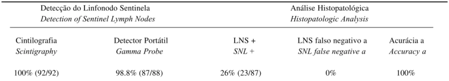
Tabela 1: Detecção e resultados da análise do LNS em pacientes com melanoma
Table 1: Detection and results of SNL analysis in melanoma patients
Detecção do Linfonodo Sentinela Análise Histopatológica
Detection of Sentinel Lymph Nodes Histopatologic Analysis
Cintilografia Detector Portátil LNS + LNS falso negativo a Acurácia a
Scintigraphy Gamma Probe SNL + SNL false negative a Accuracy a
100% (92/92) 98.8% (87/88) 26% (23/87) 0% 100%
a: calculada entre os 32 pacientes submetidos à ressecção da cadeia ganglionar / calculation based on 32 patients subjected to resection of the ganglionic chain
LNS: linfonodo sentinela; +: positivo para tumor / SNL: sentinel lymph node; +: positive for tumor
A
fonodos, ao contrário dos materiais particulados (como os colóides), que são retidos provavelmente por fagocitose. Os colóides representam a maioria dos radiofármacos empregados na detecção do linfonodo sentinela. A detec-ção do LNSé possível porque o colóide é ligado a um tra-çador radioativo, em geral o tecnécio-99m, cuja radioativi-dade pode ser detectada nas imagens obtidas pela câmara de cintilação ou pelo detector portátil.
A linfocintilografia é uma etapa essencial da pesqui-sa de LNS, pois as imagens obtidas permitem a avaliação do padrão de drenagem e a correta identificação de uma ou mais cadeias relacionadas ao sítio de injeção. O detector portátil (ou mesmo o corante) pode, dessa forma, ser utilizado de maneira mais direcional durante o tempo cirúrgico. Padrões atípicos ou inesperados de drenagem são observados fre-qüentemente, sobretudo nos pacientes com lesões do tronco. O emprego da linfocintilografia tem mostrado que as áreas com drenagem ambígua têm extensão muito maior do que a classicamente descrita na linha média da cabeça ao tronco e na transição tóraco-abdominal (áreas descritas nos estudos de Sappey).15Neste estudo, 15 pacientes (16%) apresentaram drenagem para mais de uma cadeia linfática ou para uma cadeia não esperada pelo sítio primário da lesão. Outros sete apresentaram linfonodo sentinela na região poplítea, um achado comum em casos de tumor de extremidades, porém pouco freqüentemente presente nos relatos da literatura.16
A literatura apresenta resultados relativamente homogêneos na detecção do LNScom radioisótopos, apesar de várias diferenças metodológicas. As variações mais fre-qüentes correspondem ao emprego de diferentes radiofár-macos, atividades, volumes e vias de administração. A administração de pequenos volumes (0,2ml) ao redor da lesão primária ou da cicatriz cirúrgica, por via intra ou sub-dérmica, é recomendada de forma quase consensual.
Em geral também se considera ideal o uso de partí-culas com diâmetro entre 20-500nm,17pois o emprego de partículas de maior diâmetro é limitado pela lenta progres-são linfática, enquanto as partículas de menor diâmetro não sofrem fagocitose e podem progredir para toda a cadeia. Entre os radiofármacos mais empregados encontram-se os nanocolóides de albumina na Europa e os compostos anti-moniais na Oceania, assim como o enxofre coloidal modi-ficado/filtrado nos EUA. Entretanto, os dois primeiros radiofármacos citados têm disponibilidade limitada no Brasil, havendo ressalvas quanto à não-uniformidade e dificuldades técnicas no preparo do enxofre coloidal. Provavelmente por esse motivo, vemos que os radiofárma-cos aqui mais empregados são o Dextran e o Fitato marca-do com tecnécio-99m.
Apesar de ser difícil medir diretamente o diâmetro da partícula, considera-se, há cerca de cinco anos, que o 99m Tc-Fitato poderia ser uma alternativa na detecção do LNS. Essa possibilidade foi levantada devido à similaridade na biodis-tribuição do Fitato e dos nanocolóides no sistema retículo-endotelial. O 99mTc-Fitato foi inicialmente descrito para o
nodes, as opposed to particulate materials (such as col-loids), which are retained most likely by phagocytosis. Colloids consist of the majority of radiopharmaceuticals used to detect sentinel lymph nodes. SNLdetection is made possible because a colloid is linked to a radioactive trac-er, usually the 99mTc. The radioactivity of latter may be detected in images obtained by the scintillation camera or gamma probe.
Lymphoscintigraphy is an essential step in studying
SNL. The images obtained allow an assessment of the draining pattern and accurate identification of one or more related chains at the injection site. As such, the gamma probe (or even the dye) may provide better guidance in its use during surgery. Atypical or unexpected draining pat-terns were often observed, above all in patients with trunk lesions. The use of lymphoscintigraphy has shown that areas with ambiguous draining are far more extensive than those classically described along the head to truck medial line, and in the thoracic-abdominal transition (areas described in Sappey's studies).15
In this latter study, 15 patients (16%) showed drainage in more than one lym-phatic basin, or in an unexpected basin with respect to the primary lesion site. Another seven showed sentinel lymph nodes in the popliteal region, which is a common finding in cases of tumors in the extremities, though seldom found in reports in the literature.16
The literature does show relatively homogenous results in SNLdetection with radioisotopes in spite of many methodological differences. The most frequent variations match up with the use of different radiopharmaceuticals, radioactivity, volumes and administration pathways. There is almost unanimous agreement to recommend administra-tion of small volumes (0.2 ml) around the primary lesions or surgical wound via intra- or subdermal pathways.
What is also considered ideal is the use of particles (20-500 nm in diameter),17
since the use of large diameter particles is limited by a slow lymphatic progression. On the other hand, small diameter particles do not experience phagocytosis and may engulf the entire basin. Among the most common radiopharmaceuticals, albumin nanocol-loids are used in Europe and antimony composites in Oceania, as well as modified/filtered colloidal sulfur in the
USA. Still, availability of the first two radiopharmaceuti-cals cited is very limited in Brazil. This situation highlights the lack of uniformity and the technical difficulties involved in preparing colloidal sulfur. It is most likely for this rea-son that we see radiopharmaceuticals being used here more than Dextran and 99m
Tc-Phytate.
In spite of how hard it is to measure the particle diameter correctly, about five years ago 99m
Tc-Phytate was cited as possibly becoming an alternative in SNLdetection. This suggestion was raised due especially to how phytate biodistribution resembles nanocolloids in the reticuloen-dothelial system. 99m
estudo do sistema reticuloendotelial em 1973, havendo a for-mação de colóides in vivo, após reação com o cálcio iônico.18 As medidas efetuadas após adição de cálcio em relação molar de 1:1 ou 2:1 ao 99mTc-Fitato, realizadas por microscopia ele-trônica e estudos de condutibilidade, indicam a formação de partículas com diâmetro inferior a 500nm.19,20 A captação esplênica observada após a administração intravenosa de 99mTc-Fitato em pacientes é similar à observada com
nanoco-lóides e inferior à observada com enxofre coloidal, confir-mando a formação de partículas com menor diâmetro.
O emprego do 99mTc-Fitato para linfocintilografia já foi descrito,21,22 e a utilização específica para detecção de LNSfoi relatada pelo grupo destes autores.23A rápida pro-gressão do 99mTc-Fitato na fase inicial, com a identificação do LNSainda durante a fase dinâmica nos minutos iniciais do estudo, pode estar relacionada à migração de compostos de baixo peso molecular antes da formação in vivodo
colói-de. No entanto, a retenção e concentração do radiofármaco no LNS, com baixa progressão para o restante da cadeia, indicam que há formação de colóide no percurso entre o subcutâneo e o LNS.
Impacto da biópsia do LNS no estadiamento e prognóstico do melanoma
A pesquisa intra-operatória do LNSé amplamente indi-cada para pacientes com lesões com espessura acima de 1mm e sem evidências clínicas de metástases ganglionares. Aproximadamente 20% dos pacientes com tumor apresentando Breslow de um a 4mm têm metástase no LNS, valor que sobe para 34% entre pacientes com índice > 4mm e cai para 4,7% em pacientes com índice < 1mm associado a lesões ulceradas ou com nível IV de Clark.6O
statusdo LNSé tão aceito, que foi incluído no estadiamento proposto pela American Joint Committee on Cancer for Malignant Cutaneous Melanoma.24,25 A detecção de LNSpode ser também indicada para pacientes com lesões mais superficiais (<1mm),26,27 apesar de ser evidente o fato de que tumores mais profundos apre-sentam maior probabilidade de disseminação ganglionar.28,29 O valor prognóstico da biópsia do linfonodo sentinela é comprovado para tumores com espessura acima de 4mm.30
Descreve-se a correta identificação intra-operatória do LNSem quase 98% dos casos com colóides radiomarca-dos, 75 a 80% com corantes e 98 a 99% com a combinação de ambos.25Também se descreve acurácia superior a 98% da biópsia do LNSna predição do acometimento de toda a cadeia linfática.6,12,13Em recente revisão de 1.135 casos de melanoma, o LNSfoi detectado com radioisótopos em 97% dos pacientes, com valor preditivo negativo de 100%.31Os resultados apresentados no presente trabalho são similares, com taxas de 98% e 100%, respectivamente.
A pesquisa de acometimento ganglionar pelo LNS pode ser até mesmo superior à abordagem de toda a cadeia, por permitir o estudo mais detalhado e o emprego de técnicas visando à detecção de micrometástases. A redução do núme-ro de linfonodos analisados permite que o patologista não só
a colloid formation in vivo after a calcium ion reaction. 18 The measures undertaken after adding calcium in a 1:1 or 2:1 molar relation to 99m
Tc-Phytate, and performed with the electron microscope and conductibility studies, indicate the formation of particles <500 nm in diameter.19,20
The spleen imaging observed after intravenously administering the
99m
Tc-Phytate in patients is similar to what has been observed with nanocolloids and poorer than with colloidal sulfur. These results confirm the lower-diameter particle formation.
The use of 99m
Tc-Phytate for lymphoscintigraphy has already been described.21,22
The specific use of SNL detec-tion was reported for the group by the authors.23
The fast-moving progression of 99m
Tc-Phytate in the initial phase, with SNLidentification still at the dynamic phase in the ini-tial minutes of the study, may be related to migrating low-weight molecular composites prior to the colloid's forma-tion in vivo. On the other hand, retention and concentration of radiopharmaceuticals in SNL, only slowly progressing to the rest of the basin, indicates that colloid formation occurs in the course between the subcutaneous layer and SNL.
Impact of the SNL biopsy on the staging and prognosis of the melanoma
The intraoperative mapping of SNLis widely indicated for patients with thick (>1-mm) lesions and with no clinical evidence of ganglionic metastasis. Roughly 20% of tumor patients showing a 1-4 mm Breslow have SNLmetastasis. This value rises to 34% among patients with >4 mm index, and falls to 4.7% in patients with <1 mm index in association with ulcerated lesions or level of Clark IV.6
The SNL status has achieved such widespread acceptance that it is now included in the staging proposed by the American Joint Committee on Cancer for Malignant Cutaneous Melanoma.24,25
SNLdetection may also be indicated for patients with more superficial lesions (<1mm),26,27
in spite of the obvious fact that deeper tumors are more likely to show ganglionar dissemination.28,29
The prognostic value of sentinel node biopsy is confirmed for thick (>/=4-mm) tumors.30
Proper intraoperative SNL identification is described in 98% of cases with radiomarked colloids, 75-80% with dyes and 98-99% with a combination of both.25
Also described is a greater than 98% accuracy rate from
SNLbiopsy to predict the affection of the entire lymphatic basin.6,12,13
In a recent review of 1,135 melanoma cases, SNL
was detected with radioisotopes in 97% of patients, with a negative predictive value of 100%.31
The results shown in the present study are similar, with rates of 98 and 100%, respectively.
apply more sensitive techniques, such as immunohisto-chemistry or PCR (polymerase chain reaction).32,33
Immunohistochemistry with antibodies directed at S100 and
HMB45 antigens enhances the sensitivity of metastasis detection by roughly 14%,34,35
though it was not routinely available for the study of patients at the institution.
The latest recommendations by the American Joint Committee on Cancer for Malignant Cutaneous Melanoma include SNLstatus in the staging, in addition to using the term "micrometastasis" for lymph nodes that show no macroscopic involvement.24
The clinical significance of a micrometastasis (mainly when detected only by PCR) and its impact on survival rates, however, remain controversial.32,33
The infiltration of SNLis recognized to be the most important prognostic factor for primary melanoma patients (in the absence of metastases at other sites). SNL involve-ment is a strong predictor of recurrence and survival. A mortality rate of 6% is found in SNL-negative patients com-pared to 30% in SNL-positive patients who received follow-up for an average of 37 months.2,36
Gershenwald2,36
has reported that SNLis the most important survival indicator in stage I and II melanoma patients. It is therefore more sig-nificant than Breslow thickness and presence of ulceration. The percentage of patients who are free of disease for an interval of three years dropped from 96.8 to 69.9% when receiving SNLfollow up. Even among patients with thicker melanoma, there is a significant reduction in recurrence when SNLis negative (37 vs. 73% of patients free of disease for three years).7
A negative SNLbiopsy, however, does not guarantee the patient against showing recurrence in the future. Various authors report recurrence in 1-6% of SNL-negative patients36,37,38,39
Regional recurrence among SNL-negative patients might be even less when using more sensitive tech-niques. It has been noted that the review of SNLcuts eluci-dates a large part of the recurrences in cases initially inter-preted to be SNL-negative.40
Also, it ought to be observed that 5% frequency rate of close regional recurrence is sim-ilar to what can be seen to occur following elective lym-phadenectomy,41
and that complete resection of the basin did not noticeably increase survival rates in melanoma and
SNL-negative patients.28,31
The indications for elective lymphadenectomy are still controversial. Their impact upon survival is not clear when considering a population with a roughly 20% likeli-hood of ganglionic affection. The best selection of patients for elective lymphadenectomy might perhaps be performed by means of assessing the SNL. SNL-negative patients would thus be exempted from the procedure. In this case, the main cause of morbidity in elective lymphadenectomy would be reduced, since lymphedema is described in only 1.7% of patients after SNLbiopsy.42
The fact of micrometastasis patients showing better survival than those with macroscopic involvement43
sug-gests that early removal of the affected lymph nodes may
aumente o número de secções no linfonodo como também aplique técnicas de mais sensibilidade, tais como a imuno-histoquímica ou PCR (polymerase chain reaction).32,33 A imuno-histoquímica com anticorpos dirigidos a antígenos S-100 e HMB-45 aumenta a sensibilidade na detecção de metás-tases em cerca de 14%,34,35porém não estava disponível roti-neiramente para o estudo dos pacientes na instituição.
A nova recomendação da American Joint Committee on Cancer for Malignant Cutaneous Melanomainclui o statusdo
LNSno estadiamento, além de empregar o termo micrometás-tase para linfonodos sem envolvimento macroscópico.24O sig-nificado clínico de uma micrometástase (principalmente quan-do detectada apenas pela PCR) e de seu impacto em termos de sobrevida, contudo, ainda é controverso.32,33
A infiltração do LNSé reconhecida como o fator prog-nóstico mais importante para pacientes com melanoma primá-rio (na ausência de metástases em outros sítios). O acometi-mento do LNSé forte preditor de recorrência e de sobrevida, com a mortalidade de 6% em pacientes com LNS negativo comparada a 30% nos pacientes com LNSpositivo acompa-nhados pelo período médio de 37 meses.2,36 Gershenwald6 relatou que o LNSé o mais importante indicador de sobrevida nos pacientes com melanoma nos estádios I e II, mais signifi-cativo do que a espessura de Breslow e a presença de ulcera-ção. A porcentagem de pacientes livres de doença após inter-valo de três anos caiu de 96,8% para 69,9% quando havia aco-metimento do LNS. Mesmo entre os pacientes com melanoma de maior espessura, há significativa redução de recorrência quando o LNSse apresenta negativo (37% x 73% dos pacien-tes livres de doença após três anos).7
envolvi-dos possa melhorar a evolução do paciente. Entretanto, três ensaios clínicos randomizados não foram capazes de mostrar diferenças significativas de sobrevida em pacientes com LNS positivo submetidos à complementação do esvaziamento gan-glionar.44,45,46Esses estudos foram discutidos por Balch,47 desta-cando-se o fato de a maioria dos pacientes não ter realizado a cintilografia pré-operatória para confirmar a identificação cor-reta da cadeia de drenagem. Esse dado é crucial quando se con-sidera a alta freqüência de drenagem para mais de uma cadeia linfática ou para locais inesperados. Novos estudos clínicos para a avaliação dos resultados terapêuticos da linfadenecto-mia seletiva baseada na detecção do LNSestão sendo realiza-dos.48 As mesmas considerações, sobre a necessidade de melhor estratificação dos pacientes para avaliação de resposta terapêutica, aplicam-se não só à linfadenectomia, mas também a outros protocolos, tais como o uso de interferon ou vacinas.
CONCLUSÕES
A biópsia do LNS apresenta alta acurácia no esta-diamento ganglionar de pacientes com melanoma maligno e também é um importante fator prognóstico. Uma defini-ção correta do estadiamento linfático pode ter implicações terapêuticas, incluindo a indicação mais precisa da ressec-ção de toda a cadeia linfática ou o emprego de terapias sis-têmicas.
Os resultados obtidos na detecção do LNScom 99m Tc-Fitato, um radiofármaco com ampla disponibilidade no Brasil, foram comparáveis aos descritos na literatura. q
improve the patient's course. Nevertheless, three clinical randomized trials failed to show significant differences in overall survival rates for SNL-positive patients subjected to a complementation of ganglionic emptying.44,45,46
These tri-als were discussed by Balch,47
who highlighted the fact that most patients had not undergone preoperative scintigraphy to confirm the correct identification of the draining basin. These data are crucial when considering the high frequen-cy of draining to more than one lymphatic basin or unex-pected localizations. New clinical studies meant to assess the therapeutic results of selective lymphadenectomy based on SNLdetection are being carried out.48
These considera-tions, namely on the need for better patient stratification to assess therapeutic response, are being applied not only to lymphadenectomy, but also to other protocols, such as interferon or vaccine use.
CONCLUSIONS
SNLbiopsy showed a high degree of accuracy for the ganglionic staging of patients with malignant melanoma. It has also proved to be an important prognostic factor. Correctly defining lymphatic staging, then, may have ther-apeutic implications. The latter specifically include whether the most required indication is to resection the entire lym-phatic basin, or use systemic therapies.
The results obtained in SNL detection with 99m
Tc-Phytate, which is a widely available radiopharmaceutical in Brazil, were comparable to those described in the literature. q
sis and other histologic factors that predict outcome in patients with thicker melanomas. J Am Acad Dermatol 2001;44(5):762-766. 8. Cabanas RM. An approach for the treatment of penile carcino-ma. Cancer 1977;39(2):456-466.
9. Morton DL, Wen DR, Wong JH, Economou,JS, Cagle LA, Storm FK et al. Technical details of intraoperative lymphatic map-ping for early stage melanoma. Arch Surg 1992;127(4):392-399. 10. Bostick PJ, Giuliano AE. Vital dyes in sentinel node localiza-tion. Semin Nucl Med 2000;30(1):18-24.
11. Alex JC, Weaver DL, Fairbank,JT, Rankin BS, Krag DN. Gamma-probe-guided lymph node localization in malignant melanoma. Surg Oncol 1993;2(5):303-308.
12. Reintgen D, Cruse CW, Wells K, Berman C, Fenske N, Glass F et al. The orderly progression of melanoma nodal metastases. Ann Surg 1994;220(6):759-767.
13. Thompson JF, McCarthy WH, Bosch CM, O'Brien CJ, Quinn MJ, Paramaesvaran S et al. Sentinel lymph node status as an indi-cator of the presence of metastatic melanoma in regional lymph nodes. Melanoma Res 1995;5(4):255-260.
14. Lubach D, Ludemann W, Berens VR. Recent findings on the angioarchitecture of the lymph vessel system of human skin. Br J Dermatol 1996;135(5):733-737.
15. Uren RF, Howman-Giles R, Thompson JF. Patterns of lym-phatic drainage from the skin in patients with melanoma. J Nucl Med 2003;44(4):570-582.
16. Thompson JF, Hunt JA, Culjak G, Uren RF, Howman-Giles R,
REFERÊNCIAS / REFERENCES
1. Estimativa da incidência e mortalidade por cancer no Brasil. INCA - Ministério da Saúde 2002:
2. Fife K, Thompson, JF. Lymph-node metastases in patients with melanoma: what is the optimum management? Lancet Oncol 2001;2(10):614-621.
3. Agarwala SS, Kirkwood JM. Update on adjuvant interferon therapy for high-risk melanoma. Oncology (Huntingt) 2002;16(9):1177-1187.
4. Kirkwood JM, Ibrahim JG, Sosman JA, Sondak VK, Agarwala SS, Ernstoff MS et al. High-dose interferon alfa-2b significantly prolongs relapse-free and overall survival compared with the GM2-KLH/QS-21 vaccine in patients with resected stage IIB-III melanoma: results of intergroup trial E1694/S9512/C509801. J Clin Oncol 2001;19(9):2370-2380.
5. Morton DL, Wanek L, Nizze JA, Elashoff RM, Wong JH. Improved long-term survival after lymphadenectomy of melanoma metastatic to regional nodes. Analysis of prognostic factors in 1134 patients from the John Wayne Cancer Clinic. Ann Surg 1991;214(4):491-499.
6. Gershenwald JE, Thompson W, Mansfield PF, Lee JE, Colome MI, Tseng CH et al. Multi-institutional melanoma lymphatic map-ping experience: the prognostic value of sentinel lymph node sta-tus in 612 stage I or II melanoma patients. J Clin Oncol 1999;17(3):976-983.
micrometasta-1988;12(8):612-618.
35. Baisden BL, Askin FB, Lange JR, Westra WH. HMB-45 immunohistochemical staining of sentinel lymph nodes: a specif-ic method for enhancing detection of mspecif-icrometastases in patients with melanoma. Am J Surg Pathol 2000;24(8):1140-1146. 36. Cascinelli N, Belli F, Santinami M, Fait V, Testori A, Ruka W et al.Sentinel lymph node biopsy in cutaneous melanoma: the WHO Melanoma Program experience. Ann Surg Oncol 2000;7(6):469-474.
37. Gershenwald JE, Colome MI, Lee JE, Mansfield PF, Tseng C, Lee JJ et al. Patterns of recurrence following a negative sentinel lymph node biopsy in 243 patients with stage I or II melanoma. J Clin Oncol 1998;16(6):2253-2260.
38. Landi G, Polverelli M, Moscatelli G, Morelli R, Landi C, Fiscelli O et al. Sentinel lymph node biopsy in patients with pri-mary cutaneous melanoma: study of 455 cases. J Eur Acad Dermatol Venereol 2000;14(1):35-45.
39. Statius Muller MG, Borgstein PJ, Pijpers R, Van Leeuwen PA, Van Diest PJ, Gupta A et al. Reliability of the sentinel node pro-cedure in melanoma patients: analysis of failures after long-term follow-up. Ann Surg Oncol 2000;7(6):461-468.
40. Clary BM, Brady MS, Lewis JJ, Coit DG. Sentinel lymph node biopsy in the management of patients with primary cuta-neous melanoma: review of a large single-institutional experience with an emphasis on recurrence. Ann Surg 2001;233(2):250-258. 41. Shen P, Guenther JM, Wanek LA, Morton DL. Can elective lymph node dissection decrease the frequency and mortality rate of late melanoma recurrences? Ann Surg Oncol 2000;7(2):114-119. 42. Wrone DA, Tanabe KK, Cosimi AB, Gadd MA, Souba WW, Sober AJ. Lymphedema after sentinel lymph node biopsy for cutaneous melanoma: a report of 5 cases. Arch Dermatol 2000;136(4):511-514.
43. McMasters KM, Reintgen DS, Ross MI, Gershenwald JE, Edwards MJ, Sober A et al. Sentinel lymph node biopsy for melanoma: controversy despite widespread agreement. J Clin Oncol 2001;19(11):2851-2855.
44. Balch CM, Soong S, Ross MI, Urist MM, Karakousis CP, Temple WJ et al. Long-term results of a multi-institutional ran-domized trial comparing prognostic factors and surgical results for intermediate thickness melanomas (1.0 to 4.0 mm). Intergroup Melanoma Surgical Trial. Ann Surg Oncol 2000;7(2):87-97. 45. Sim FH, Taylor WF, Pritchard DJ, Soule EH. Lymphadenectomy in the management of stage I malignant melanoma: a prospective randomized study. Mayo Clin Proc 1986;61(9):697-705.
46. Veronesi U, Adamus J, Bandiera DC, Brennhovd O, Caceres E, Cascinelli N et al. Delayed regional lymph node dissection in stage I melanoma of the skin of the lower extremities. Cancer 1982;49(11):2420-2430.
47. Balch CM. The John Wayne Clinical Research Lecture. Surgical management of melanoma: results of prospective ran-domized trials. Ann Surg Oncol 1998;5(4):301-309.
48. McMasters KM. Sentinel Lymph Node biopsy for melanoma. Melanoma Res 2001;11(suppl 1):s5-s7.
Harman CR. Popliteal lymph node metastasis from primary cuta-neous melanoma. Eur J Surg Oncol 2000;26(2):172-176. 17. Eshima D, Fauconnier T, Eshima L, Thornback JR. Radiopharmaceuticals for lymphoscintigraphy: including dosimetry and radiation considerations. Semin Nucl Med 2000;30(1):25-32. 18. Strand SE, Persson BR. Quantitative lymphoscintigraphy I: Basic concepts for optimal uptake of radiocolloids in the paraster-nal lymph nodes of rabbits. J Nucl Med 1979;20(10):1038-1046. 19. Campbell J, Bellen JC, Baker RJ, Cook DJ. Technetium-99m calcium phytate--optimization of calcium content for liver and spleen scintigraphy: concise communication. J Nucl Med 1981;22(2):157-160.
20. Galvez AJ, Garcia SC, Garcia DR, Moreno, FJ. [99mTc]Ca-phytate: some colloidal characteristics related to the optimal prepa-ration conditions. Int J Appl Radiat Isot 1983;34(12):1647-1649. 21. Alavi A, Staum MM, Shesol BF, Bloch PH. Technetium-99m stannous phytate as an imaging agent for lymph nodes. J Nucl Med 1978;19(4):422-426.
22. Kaplan WD, Davis MA, Rose CM. A comparison of two tech-netium-99m-labeled radiopharmaceuticals for lymphoscintigra-phy: concise communication. J Nucl Med 1979;20(9):933-937. 23. Tavares MG, Sapienza MT, Galeb NA, Belfort FA, Costa RR, Osorio CA et al. The use of 99mTc-phytate for sentinel node map-ping in melanoma, breast cancer and vulvar cancer: a study of 100 cases. Eur J Nucl Med 2001;28(11):1597-1604.
24. Balch CM, Buzaid AC, Soong SJ, Atkins MB, Cascinelli N, Coit DGet al. Final version of the American Joint Committee on Cancer staging system for cutaneous melanoma. J Clin Oncol 2001;19(16):3635-3648.
25. Mariani G, Gipponi M, Moresco L, Villa G, Bartolomei M, Mazzarol G et al. Radioguided sentinel lymph node biopsy in malignant cutaneous melanoma. J Nucl Med 2002;43(6):811-827. 26. Bedrosian I, Faries MB, Guerry D, Elenitsas R, Schuchter L, Mick R et al.Incidence of sentinel node metastasis in patients with thin primary melanoma (< or = 1 mm) with vertical growth phase. Ann Surg Oncol 2000;7(4):262-267.
27. Lowe JB, Hurst E, Moley JF, Cornelius LA. Sentinel lymph node biopsy in patients with thin melanoma. Arch Dermatol 2003;139(5):617-621.
28. Caggiati A, Potenza C, Gabrielli F, Passarelli F, Tartaglione G. Sentinel node biopsy for malignant melanoma: analysis of a four-year experience. Tumori 2000;86(4):332-335.
29. Gennari R, Bartolomei M, Testori A, Zurrida S, Stoldt HS,
Audisio RA et al. Sentinel node localization in primary
melanoma: preoperative dynamic lymphoscintigraphy, intraoper-ative gamma probe, and vital dye guidance. Surgery 2000;127(1):19-25.
30. Carlson GW, Murray DR, Hestley A, Staley CA, Lyles RH, Cohen C. Sentinel Lymph Node Mapping for Thick (>/=4-mm) Melanoma: Should We Be Doing It? Ann Surg Oncol 2003; 10(4):408-415.
31. Chan AD, Morton DL. Sentinel node detection in malignant melanoma. Recent Results Cancer Res 2000;157:161-177. 32. Krag DN, Weaver DL. Pathological and molecular assessment of sentinel lymph nodes in solid tumors. Semin Oncol 2002;29(3):274-279.
33. da Silva AM, Oliveira Filho RS, Ferreira LM, Saconato H. Relevance of micrometastases detected by reverse transcriptase-polymerase chain reaction for melanoma recurrence: systematic review and meta-analysis. Sao Paulo Med J 2003;121(1):24-27. 34. Cochran AJ, Wen DR, Morton DL. Occult tumor cells in the lymph nodes of patients with pathological stage I malignant melanoma. An immunohistological study. Am J Surg Pathol
ENDEREÇO PARA CORRESPONDÊNCIA: / MAILINGADDRESS:
Marcelo Tatit Sapienza
Serviço de Medicina Nuclear - Hospital Samaritano Rua Conselheiro Brotero, 1486
01232-010 São Paulo SP Tel.: (11) 3825-4433