* Corresponding author: Yusuke Inoue, Department of Diagnostic Radiology, Kitasato University School of Medicine, 1-15-1 Kitasato, Minami-ku, Sagamihara, Kanagawa 252-0374, Japan. Tel: 42-778-8111; Fax: 81-42-778-9436; E-mail: inoueys34@gmail.com
© 2014 mums.ac.ir All rights reserved.
This is an Open Access article distributed under the terms of the Creative Commons Attribution License (http://creativecommons.org/licenses/by/3.0), which permits unrestricted use, distribution, and reproduction in any medium, provided the original work is properly cited.
Phase
IIa
Clinical
Trial
of
Trans
‐
1
‐
Amino
‐
3
‐
18F
‐
Fluoro
‐
Cyclobutane
Carboxylic
Acid
in
Metastatic
Prostate
Cancer
Yusuke )noue , Yuji Asano , Takefumi Satoh , Ken‐ichi Tabata , Kei Kikuchi ,
Reiko Woodhams , Shiro Baba , Kazushige (ayakawa
Department of Diagnostic Radiology, Kitasato University School of Medicine, Sagamihara, Kanagawa, Japan Department of Urology, Kitasato University School of Medicine, Sagamihara, Kanagawa, Japan
Department of Radiology, Kitasato University (ospital, Sagamihara, Kanagawa, Japan
Department of Radiation Oncology, Kitasato University School of Medicine, Sagamihara, Kanagawa, Japan
ARTICLE INFO ABSTRACT
Articletype:
Original Article
Objective(s): We performed a phase ))a clinical trial of trans‐ ‐amino‐ ‐ F‐
fluoro‐cyclobutane carboxylic acid anti‐ F‐FACBC , a synthetic amino acid analog for PET, in patients with metastatic prostate cancer.
Methods:The study subjects consisted of untreated prostate cancer patients
having lymph node and/or bone metastasis. Five patients underwent whole‐body PET and min after intravenous injection of anti‐ F‐FACBC. The other five patients underwent min dynamic PET of the pelvis. Safety assessment was performed before and h after injection. PET/CT images were assessed visually, and time courses of anti‐ F‐FACBC uptake were evaluated from dynamic imaging.
Results: Two mild adverse events were observed and resolved without
treatment. All patients showed increased accumulation of anti‐ F‐FACBC in the primary prostate lesion. CT revealed five enlarged lymph nodes indicating metastasis, and all showed increased uptake. Additionally, anti‐ F‐FACBC PET delineated unenlarged lymph nodes as hot spots. Anti‐ F‐FACBC PET demonstrated metastatic bone lesions, similar to conventional imaging. )n one of two patients with lung metastasis, some lesions showed increased uptake. Regarding the time course, increased uptake of anti‐ F‐FACBC in the lesion was demonstrated immediately after injection, followed by gradual washout.
Conclusion:The results of this phase ))a clinical trial indicated the safety of anti‐
F‐FACBC in patients with prostate cancer and the potential of anti‐ F‐FACBC PET to delineate primary prostate lesions and metastatic lesions. This clinical trial was registered as JapicCT)‐ .
Articlehistory:
Received: Mar Revised: Apr Accepted: May
Keywords:
Anti‐ F‐FACBC Metastasis
Positron Emission Tomography Prostate cancer
►Pleasecitethispaperas:
)noueY, Asano Y, Satoh T, Tabata K, Kikuchi K, Woodhams R, Baba Sh, (ayakawa K. Phase ))a Clinical Trial of Trans‐ ‐ Amino‐ ‐ F‐Fluoro‐Cyclobutyl‐Carboxylic Acid in Metastatic Prostate Cancer. Asia Oceania J Nucl Med Biol. ;
: ‐ .
Introduction
)maging modalities play an important role in the staging and restaging of prostate cancer , and use of positron emission tomography PET has been investigated , . Although ‐ deoxy‐ ‐[ F]fluoro‐D‐glucose is used in various malignant neoplasms, it is not useful in prostate cancer because of low tumor uptake and high urinary excretion. Better diagnostic perform‐
88 Asia Oceania J Nucl Med Biol. 2014; 2(2):87-94.
Table1.Patient Characteristics
Patient.No. Group Age Sex BH(cm) BW(kg) Dose(MBq) PSA(ng/ml) Positivebiopsy Gleasonscore
1 WB M . . . . / +
2 WB M . . . . / +
3 WB M . . . . / +
4 WB M . . . . / +
5 WB M . . . . / +
6 Dyn M . . . . / +
7 Dyn M . . . . / +
8 Dyn M . . . . / +
9 Dyn M . . . . / +
10 Dyn M . . . . / +
B(: body height, BW: body weight, WB: whole‐body group, Dyn: dynamic group.
Positive biopsy indicates the number of cancer‐positive biopsy cores and total cores obtained.
The synthetic amino acid analog trans‐ ‐ amino‐ ‐ F‐fluoro‐cyclobutane carboxylic acid
anti‐ F‐FACBC is another candidate for PET imaging of prostate cancer. Rapid, high uptake of anti‐ F‐FACBC was shown in prostate cancer ‐ , and its usefulness for the assessment of
recurrent prostate cancer has been
demonstrated ‐ . The phase ) clinical trial of
anti‐ F‐FACBC development code, NMK in
Japan indicated safety, acceptable radiation dose, and favorable characteristics for imaging
brain and pelvic tumors . As a next step, we
performed a phase ))a clinical trial in patients with metastatic prostate cancer to evaluate the safety of anti‐ F‐FACBC in patients with metastatic prostate cancer and to examine the capability of anti‐ F‐FACBC PET to delineate primary and metastatic lesions of prostate cancer.
Methods
InvestigationalDrug
Anti‐ F‐FACBC was prepared by Nihon Medi‐Physics Co., Ltd. Tokyo, Japan based on
the method reported previously , and was
supplied in a vial containing ml solution with additives to ensure high stability for commercial distribution. The radiochemical purity was . ± . %.
Subjects
Ten male patients with prostate cancer aged . ± . years mean ± SD were enrolled in
this study between November and March
Table . The inclusion criteria were: a histological positivity for prostate cancer on two or more cores obtained by needle biopsy of the prostate; and b presence of lymph node
metastasis and/or bone metastasis
demonstrated on conventional imaging such as
computed tomography CT , magnetic
resonance MR imaging, and bone scintigraphy. Patients, who had undergone therapy for prostate cancer, including surgical resection,
radiation therapy, hormonal therapy, and chemotherapy, were excluded. All patients underwent anti‐ F‐FACBC PET after an interval of more than weeks after biopsy to avoid possible effects of biopsy on PET images. The study was performed after approval by the institutional review boards of the host institution and according to Good Clinical
Practice GCP Ordinance No. , , March
. Written informed consent was obtained from all patients before participation. This
clinical trial was registered as JapicCT)‐ .
PET/CTProcedures
Anti‐ F‐FACBC was injected after fasting for h or longer, followed by PET/CT imaging using a TruePoint Biograph PET/CT scanner Siemens Medical Solutions, (offman Estates, )L . For CT, mm thick contiguous slices were acquired without the administration of contrast medium. The initial five patients underwent whole‐body PET imaging whole‐body group ,
and the injected dose ranged from . to .
MBq. Whole‐body PET imaging was performed
min early and min late after injection.
Data were acquired from the mid‐thigh level to the top of the head for . min per bed position. The remaining five subjects underwent min dynamic PET imaging of the pelvic region immediately after injection dynamic group , and the injected dose was fixed at
approximately MBq . – . MBq .
)mages for visual evaluation were reconstructed using data of . min duration and centered at , , , and min after injection. Additionally, images were reconstructed at s/frame × , s/frame × , min/frame × , min/frame × , and min/frame × to assess the time course quantitatively.
SafetyAssessment
Asia Oceania J Nucl Med Biol. 2014; 2(2):87-94. 89
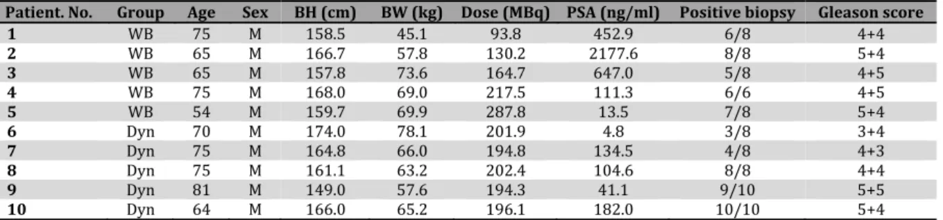
Figure1. Axial images of the pelvis in a patient with lymph node metastasis and multiple bone metastases patient no. in Table . From left to right, contrast‐enhanced thin‐slice CT images, plain CT images of anti‐ F‐FACBC PET/CT, PET images, and PET/CT fusion images are presented. )mages at a given body level are presented in a row. Contrast‐enhanced CT and PET/CT were performed on different occasions. The primary site thick arrow , a significantly enlarged lymph node in the left external iliac region thin arrow , unenlarged lymph nodes in the left external and internal iliac regions arrowheads , and multiple bone lesions showed increased uptake
interview of subjective symptoms, physical
examinations, lead electrocardiography,
measurements of blood pressure and pulse rate, hematology, blood chemistry, and urinalysis. An interview of subjective symptoms and physical examinations were also performed at the completion of PET/CT imaging.
ConventionalImaging
CT, MR imaging, and bone scintigraphy were performed within month before PET/CT with anti‐ F‐FACBC. )n CT, after contrast injection, contiguous thin slices of . mm thickness were acquired from the neck to the pelvis. )n MR imaging, T ‐weighted imaging, T ‐weighted imaging, diffusion‐weighted imaging, and
dynamic contrast‐enhanced T ‐weighted
imaging of the pelvis were performed.
Although primary lesions were
histologically proven to be prostate cancer, histological evaluation of metastatic lesions was not performed and results of these conventional imaging were used as the diagnostic standard. A lymph node with a short‐axis diameter mm on CT was judged to be significantly enlarged
and regarded as a metastatic lymph node .
The evaluation of bone metastasis was based primarily on bone scintigrams, with reference to CT and MR images. Patients were classified into three categories: multiple bone metastases >
lesions , limited bone metastasis lesions ,
and no bone metastasis. )n patients with limited bone metastasis, the locations of metastatic bone lesions were recorded. The locations were not considered in the analysis of patients with multiple bone metastases.
Judgment was performed by two board‐ certified radiologists, referring to official reports described by other board‐certified radiologists. For inconclusive judgments, a third board‐ certified radiologist made the final decision.
VisualInterpretationofPET/CTimages
90 Asia Oceania J Nucl Med Biol. 2014; 2(2):87-94.
Figure2. Axial fusion images of anti‐ F‐FACBC PET/CT in a patient with lymph node and limited bone metastasis patient no. in Table . From left to right, min, min, min, and min images are presented. On the min images, the primary site thick arrow , significantly enlarged lymph node in the left external iliac region thin arrow , and sacral bone lesion large arrowhead showed increased uptake. Activity decreased over time; however, it was still evident on the ‐min images. Urinary excretion in the bladder is noted on the min image small arrowheads , followed by a gradual increase
the results were compared with judgments of lymph node metastasis on CT. A focal area of increased anti‐ F‐FACBC accumulation in the bone was regarded as bone metastasis. Similar to judgments using conventional imaging methods, the locations of metastatic bone lesions were analyzed in detail only in patients
with less than metastatic bone lesions.
Metastasis to other organs was also evaluated.
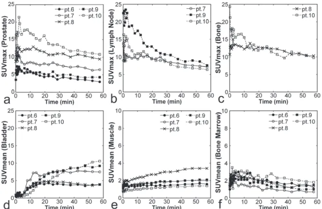
TimeCoursesofAnti‐18F‐FACBCUptake
Time courses of standardized uptake values SUVs were determined from dynamic PET imaging in five dynamic group patients. Spherical volumes of interest VO)s with approximately cm diameters were placed in
the primary prostate lesions, metastatic lymph nodes, and metastatic bone lesions, including an area of maximal activity for each lesion, and the
maximum SUV SUVmax was obtained. Activity
Asia Oceania J Nucl Med Biol. 2014; 2(2):87-94. 91
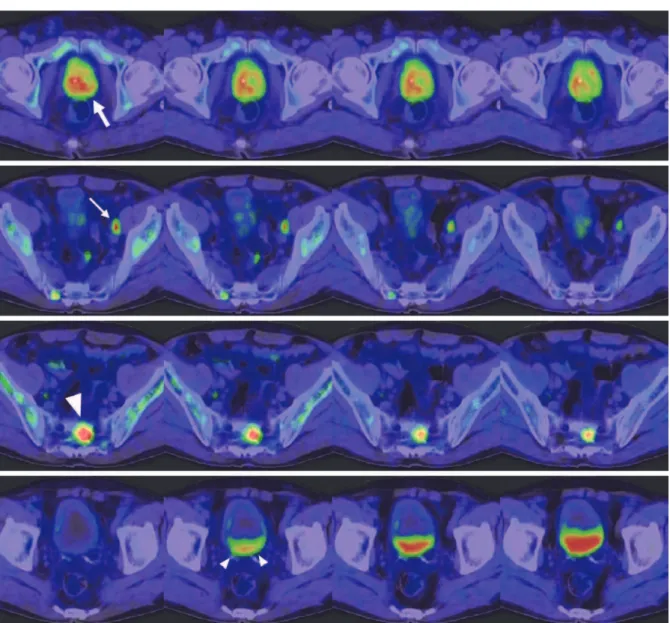
Figure3.Anterior maximum‐intensity projection image of anti‐ F‐FACBC PET a and anterior bone scintigram b in the patient presented in Figure . PET shows multiple areas of increased bone uptake, similar to bone scintigraphy. )ncreased uptake is also demonstrated in the primary site
arrow and lymph nodes arrowheads . )ntense
physiological uptake of anti‐ F‐FACBC is noted in the pancreas and liver
Results
SafetyAssessment
Two of the patients experienced mild
adverse events after anti‐ F‐FACBC injection. One patient complained of drowsiness at the completion of PET/CT imaging, which was resolved spontaneously on the following day. Lack of sleep due to pain from bone metastasis was thought to be the cause of the drowsiness. )n the other patient, occult blood in urine was noticed on the day after anti‐ F‐FACBC injection, and disappeared days later without treatment. A minor urinary tract infection was suspected in this patient based on slight increases in white blood cells and bacteria in urine, and was thought to be the cause of the urinary occult blood. These two events were judged to be clinically insignificant and to have no causal relationship with anti‐ F‐FACBC injection.
PrimaryLesion
All patients showed increased
accumulation of anti‐ F‐FACBC in the primary prostate lesion Figure , . )n the five patients
of the whole‐body group, increased
accumulation was demonstrated on both early and late images, irrespective of injected dose. (igh radioactivity in the bladder, indicating urinary excretion, was identified in two and five patients on early and late images, respectively. )ncreased accumulation in the primary lesion
was also shown in the five dynamic group
patients, except on min images in one
patient. Bladder activity was not noted on min images but was observed on min images in all the five patients Figure .
LymphNodeMetastasis
CT demonstrated two enlarged lymph nodes in two patients of the whole‐body group and three enlarged lymph nodes in three patients of the dynamic group. )n total, five enlarged lymph nodes were detected and judged as metastasis. They were all located in the pelvis and included in the field of view FOV of PET. Their short‐ axis diameters were , , , , and mm. One patient in the dynamic group was enrolled in the study based on enlargement of a lymph node; however, the short‐axis diameter of the node was measured to be < mm on the final assessment. As a result, the patient was judged to have no metastatic lesion.
)ncreased uptake of anti‐ F‐FACBC was observed in the five metastatic lymph nodes Figure , . )ncreased uptake was shown on both early and late images in the whole‐body group and at all phases in the dynamic group. Additionally, increased uptake was detected in unenlarged lymph nodes of eight patients with short‐axis diameters of – mm Figure . These lymph nodes were all located in the pelvic cavity. They were visualized during the early period, and some disappeared subsequently. )n
the whole‐body group, hot, unenlarged
lymph nodes were shown in five patients on early images and eight of them remained hot on late images. )n the dynamic group, four hot, unenlarged lymph notes were detected in three patients on and min images, and three and two of the four nodes exhibited increased uptake on and min images, respectively.
BoneMetastasis
92 Asia Oceania J Nucl Med Biol. 2014; 2(2):87-94.
Figure4. Axial images of the chest in the patient presented in Figure . From left to right, a CT image of anti‐ F‐FACBC PET/CT, PET image, and PET/CT fusion image are presented. )ncreased uptake is shown in tiny lung nodules arrows . The same scale was used for presentation of PET in this figure as in Figure
Figure5. Time courses of SUVs from dynamic )maging. SUVmax for primary prostate lesions a, n = , SUVmax for metastatic lymph nodes b, n = , SUVmax for metastatic bone lesions c, n = , SUVmean for the bladder d, n = , SUVmean for the muscle e, n = , SUVmean for the bone marrow f, n = are presented for each patient. The patient number for example, pt. corresponds to that in Table . Note that the scales of panels E and F are different from those of panels a‐d
anti‐ F‐FACBC PET demonstrated multiple sites of increased uptake indicating multiple bone metastases Figure . )n three patients, neither conventional imaging nor PET showed bone metastasis. )n the remaining three patients, both conventional imaging and PET revealed limited bone metastasis Figure . The locations of bone metastasis were concordant between conventional imaging and PET as far as the metastatic sites were within the FOV of PET. Judgments were consistent irrespective of the timing of PET imaging after injection.
LungMetastasis
Two patients in the whole‐body group showed multiple lung metastases on CT. )n one
of the two patients Figure , anti‐ F‐FACBC PET demonstrated some of the lesions, on the early and late images similarly. The long‐axis diameters of lesions visualized on PET ranged from to mm. The long‐axis diameter of the maximal invisible lung metastasis was mm in the patient with PET visible lesions and mm in the other patient. No increased activity indicating metastasis to other organs was observed.
TimeCoursesofAnti‐18F‐FACBCUptake
Asia Oceania J Nucl Med Biol. 2014; 2(2):87-94. 93
washout Figure , . A relatively rapid
washout was noted in two of the three metastatic lymph nodes. Urinary activity appeared in the bladder at about min. Uptake in the gluteal muscles was low initially but increased slightly over time. Bone marrow uptake was above the background level initially and peaked at about min, followed by a gradual decrease.
Discussion
The phase ) clinical trial of anti‐ F‐FACBC in Japan indicated its safety in six young adults . )n the present phase ))a clinical trial, safety
was tested in patients with metastatic
prostate cancer. Mild adverse events were noted in two patients, however, they resolved spontaneously without treatment, suggesting the safety of a single intravenous injection of anti‐ F‐FACBC in patients with prostate cancer. PET images of diagnostic quality were acquired after injection of anti‐ F‐FACBC ranging from . to . MBq in dose. Avidity of anti‐ F‐ FACBC to primary prostate lesions, metastatic lymph nodes, and metastatic bone lesions was indicated. The study population was small, predominantly included patients with high Gleason scores, and did not include control subjects. Despite these limitations, the results of the present study, in combination with those of
previous studies ‐ , warrant further
investigation of diagnostic performance of anti‐ F‐FACBC PET in comparison with conventional imaging.
Anti‐ F‐FACBC accumulated in primary prostate lesions in all patients during the early period. Because increased uptake areas were not correlated with histologically proven cancer tissues regarding localization within the prostate, the increased uptake may have partly represented accumulation in benign tissues. Accumulation of anti‐ F‐FACBC has been shown to be increased in benign prostatic hyperplasia as well as prostate cancer , and overlap of the
intensity of accumulation has been
demonstrated between malignant and non‐
malignant prostate tissues . Although anti‐
F‐FACBC PET is not specific for prostate cancer, it might play a role complementary to MR imaging in localizing cancer foci within the
prostate or might be used to guide biopsy to
the most aggressive lesion .
On CT, significantly enlarged lymph nodes are considered to be malignant using a
threshold short‐axis diameter of cm .
Thus, small metastatic lesions are inevitably
missed. )n the present study, all five lymph nodes judged to be metastatic on CT showed increased uptake of anti‐ F‐FACBC. Previous studies have demonstrated the usefulness of anti‐ F‐FACBC PET in the detection of lymph
node metastasis from prostate cancer ,
and our observations are consistent with the
previous reports. Additionally, unenlarged
lymph nodes were delineated as increased uptake areas. Because of the lack of histological confirmation, we cannot determine whether these increased uptakes represented true‐ positive or false‐positive findings. (owever, these observations suggest the potential of anti‐
F‐FACBC PET to detect small lymph node metastasis and warrant further investigation to define its performance for the evaluation of nodal status. Although intestinal uptake was frequent, it was mild and did not interfere with the recognition of nodal uptake. )n addition, rapid blood clearance appears to have aided the detection of accumulation in small lymph nodes. A greater detection rate of bone lesions in prostate cancer recurrence has been reported using anti‐ F‐FACBC than using C‐choline . )n the present trial, classification of skeletal status into three categories, multiple bone metastases, limited bone metastasis, and no bone metastasis, was concordant between conventional imaging and anti‐ F‐FACBC PET. The locations of bone metastasis were also concordant in patients with limited bone metastasis as far as the lesions were located within the FOV of PET. These results support avidity of anti‐ F‐FACBC to metastatic bone lesions. Metastatic bone lesions from prostate cancer can be detected effectively by bone scintigraphy because they induce strong osteoblastic activity and consequent intense uptake of a bone‐seeking agent. Sensitivity to bone metastasis should be compared between anti‐ F‐FACBC PET and bone scintigraphy in the future. Bone scintigraphy reflects bone reaction caused by a bone tumor, but does not directly visualize the bone tumor. PET with a tumor‐seeking agent including anti‐ F‐FACBC is expected, at least, to play complementary roles in the differentiation of benign and malignant bone lesions and the evaluation of viability after therapy.
94 Asia Oceania J Nucl Med Biol. 2014; 2(2):87-94.
)n detecting lung metastasis, CT is highly sensitive and the role of anti‐ F‐FACBC PET would be limited. (owever, anti‐ F‐FACBC PET may provide a clue to the detection and aid the differentiation of benign and metastatic lung nodules.
Previous studies demonstrated that accumulation of anti‐ F‐FACBC in prostate cancer lesions peaked within min ‐ . )n the present trial, high lesional uptake was observed immediately after injection, suggesting no need for a waiting time, and high activity in the lesions lasted for a long period. Whereas early imaging would be better to detect strong lesional activity and to avoid disturbance of lesion recognition by urinary excretion, the imaging time window may not be strictly restricted to an early period.
Conclusion
The present phase ))a clinical trial indicated the safety of intravenous anti‐ F‐FACBC injection in patients with metastatic prostate cancer and the capability of anti‐ F‐FACBC PET to delineate primary prostate lesions and metastatic lesions including those in the lymph nodes, bones, and lungs. The results of the present preliminary trial warrant a further clinical trial in a larger patient population and using more reliable reference standards for the diagnosis of metastatic lesions to determine the usefulness of anti‐ F‐FACBC for staging and restaging of prostate cancer.
References
. (ricak (, Choyke PL, Eberhardt SC, Leibel SA, Scardino PT. )maging prostate cancer: a multidisciplinary perspective. Radiology. ;
: ‐ .
. Jadvar (. Prostate cancer: PET with F‐FDG, F‐ or C‐acetate, and F‐ or C‐choline. J Nucl Med.
; : ‐ .
. Apolo AB, Pandit‐Taskar N, Morris MJ. Novel tracers and their development for the imaging of metastatic prostate cancer. J Nucl Med. ;
: ‐ .
. Bauman G, Belhocine T, Kovacs M, Ward A, Beheshti M, Rachinsky ). F‐fluorocholine for prostate cancer imaging: a systematic review of the literature. Prostate Cancer Prostatic Dis.
; : ‐ .
. Sörensen J, Owenius R, Lax M, Johansson S. Regional distribution and kinetics of [ F]fluciclovine anti‐[ F]FACBC , a tracer of amino acid transport, in subjects with primary
prostate cancer. Eur J Nucl Med Mol )maging. ; : ‐ .
. Schuster DM, Votaw JR, Nieh PT, Yu W, Nye JA, Master V, et al. )nitial experience with the radiotracer anti‐ ‐amino‐ ‐ F‐fluorocyclob‐ utane‐ ‐carboxylic acid with PET/CT in prostate carcinoma. J Nucl Med. ; : ‐ .
. Turkbey B, Mena E, Shih J, Pinto PA, Merino MJ, Lindenberg ML, et al. Localized prostate cancer detection with F FACBC PET/CT: Comparison with MR )maging and histopathologic analysis. Radiology. ; : ‐ .
. Schuster DM, Savir‐Baruch B, Nieh PT, Master VA, (alkar RK, Rossi PJ, et al. Detection of recurrent prostate carcinoma with anti‐ ‐amino‐
‐ F‐fluorocyclobutane‐ ‐carboxylic acid PET/CT and )n‐capromab pendetide SPECT/CT. Radiology. ; : ‐ . . Nanni C, Schiavina R, Boschi S, Ambrosini V,
Pettinato C, Brunocilla E, et al. Comparison of F‐ FACBC and C‐choline PET/CT in patients with radically treated prostate cancer and biochemical relapse: preliminary results. Eur J Nucl Med Mol )maging. ; Suppl :S ‐ . . Nanni C, Schiavina R, Brunocilla E, Borghesi M,
Ambrosini V, Zanoni L, et al. F‐FACBC compared With C‐choline PET/CT in patients with biochemical relapse after radical prostatectomy: a prospective study in patients. Clin Genitourin Cancer. ; : ‐
.
. Schuster DM, Nieh PT, Jani AB, Amzat R, Bowman FD, (alkar RK, et al. Anti‐ ‐[ F]FACBC positron emission tomography‐computerized tomography and )n‐capromab pendetide single photon emission computerized tomography‐ computerized tomography in recurrent prostate carcinoma: results of a prospective clinical trial. J Urology. ; : ‐ .
. Asano Y, )noue Y, )keda Y, Kikuchi K, (ara T, Taguchi C, et al. Phase ) clinical study of NMK : a new PET tracer with the synthetic amino acid analogue anti‐[ F]FACBC. Ann Nucl Med. ;
: ‐ .
. McConathy J, Voll RJ, Yu W, Crowe RJ, Goodman MM. )mproved synthesis of anti‐[ F]FACBC: improved preparation of labeling precursor and automated radiosynthesis. Appl Radiat )sot.
; : ‐ .
. (övels AM, (eesakkers RA, Adang EM, Jager GJ, Strum S, (oogeveen YL, et al. The diagnostic accuracy of CT and MR) in the staging of pelvic lymph nodes in patients with prostate cancer: a meta‐analysis. Clin Radiol. ; : ‐ . . Schuster DM, Taleghani PA, Nieh PT, Master VA,