P120-Catenin Regulates Early Trafficking
Stages of the N-Cadherin Precursor Complex
Diana P. Wehrendt1, Fernando Carmona1, Ana E. González Wusener1, Ángela González1,
Juan M. Lázaro Martínez2, Carlos O. Arregui1 *
1Instituto de Investigaciones Biotecnológicas, (IIB-INTECH), Universidad de San Martín, San Martín, Argentina,2Departamento de Química Orgánica, Facultad de Farmacia y Bioquímica, Universidad de Buenos Aires, CABA, Argentina
Abstract
It is well established that binding of p120 catenin to the cytoplasmic domain of surface cad-herin prevents cadcad-herin endocytosis and degradation, contributing to cell-cell adhesion. In the present work we show that p120 catenin bound to the N-cadherin precursor, contributes to its anterograde movement from the endoplasmic reticulum (ER) to the Golgi complex. In HeLa cells, depletion of p120 expression, or blocking its binding to N-cadherin, increased the accumulation of the precursor in the ER, while it decreased the localization of mature N-cadherin at intercellular junctions. Reconstitution experiments in p120-deficient SW48 cells with all three major isoforms of p120 (1, 3 and 4) had similar capacity to promote the pro-cessing of the N-cadherin precursor to the mature form, and its localization at cell-cell junc-tions. P120 catenin and protein tyrosine phosphatase PTP1B facilitated the recruitment of the N-ethylmaleimide sensitive factor (NSF), an ATPase involved in vesicular trafficking, to the N-cadherin precursor complex. Dominant negative NSF E329Q impaired N-cadherin trafficking, maturation and localization at cell-cell junctions. Our results uncover a new role for p120 catenin bound to the N-cadherin precursor ensuring its trafficking through the bio-synthetic pathway towards the cell surface.
Introduction
Cadherins belong to a superfamily of transmembrane cell–cell adhesion molecules which play important roles in development, morphogenesis, and cancer [1,2]. The function of cadherins is exerted at the cell surface, where extracellular domains of identical cadherins interact in a homophilic, Ca+2-dependent manner to form adherens junctions between adjacent cells. The intracellular domains interact with several cytoplasmic proteins, the most prominent of which are the catenins [3]. Proximal and distal regions of cadherin cytosolic domains interact directly with p120 catenin andβ-catenin (or its close relative plakoglobin), respectively. Catenins bound to surface cadherins modulate cell-cell adhesion through different mechanisms involv-ing cadherin recyclinvolv-ing, stability, and couplinvolv-ing to the actin cytoskeleton. P120 binds to a ~40 amino acids region at the juxtamembrane domain of cadherins, masking clathrin-dependent endocytic motifs [4–7]. Therefore, p120 plays a key role as an inhibitor of cadherin turnover
a11111
OPEN ACCESS
Citation:Wehrendt DP, Carmona F, González Wusener AE, González Á, Martínez JML, Arregui CO (2016) P120-Catenin Regulates Early Trafficking Stages of the N-Cadherin Precursor Complex. PLoS ONE 11(6): e0156758. doi:10.1371/journal. pone.0156758
Editor:Yulia Komarova, University of Illinois at Chicago, UNITED STATES
Received:January 19, 2016
Accepted:May 19, 2016
Published:June 2, 2016
Copyright:© 2016 Wehrendt et al. This is an open access article distributed under the terms of the
Creative Commons Attribution License, which permits unrestricted use, distribution, and reproduction in any medium, provided the original author and source are credited.
Data Availability Statement:All relevant data are within the paper and its Supporting Information files.
Funding:Funding was provided by Agencia Nacional de Promoción Científica y Tecnológica,
http://www.agencia.mincyt.gob.ar/frontend/agencia/ fondo/foncyt(PICTs 1363 and 2129), to COA; and Consejo Nacional de Investigaciones Científicas y Técnicas (CONICET),http://www.conicet.gov.ar, to COA, DPW and JMLM.
and as a "set point" for cadherin expression levels [8,9]. Most cells express multiple p120 iso-forms, and N-terminal splicing events lead to the use of four alternative start codons [10]. All isoforms contain a conserved and central Armadillo repeat domain which mediates equivalent binding to cadherin [11]. However, the efficiency in stabilizing cadherin at the plasma mem-brane differs among isoforms containing (isoform 3) or lacking (isoform 4) the N-terminal reg-ulatory domain [12]. Thus, different p120 isoforms may affect cadherin function by recruiting distinct binding partners to the cadherin complex.
Cadherins biosynthesis occurs at ER-bound ribosomes as precursors containing a pro-domain at the N-terminus that inhibits cadherin dimerization and adhesion [13–15]. In a late Golgi compartment, the pro-domain is cleaved by pro-protein convertases of the furin family [15–17]. Beta catenin and p120 bind to the cytoplasmic domain of cadherin precursors, traf-ficking as a complex towards the cell surface [17–20]. The functional significance of catenin binding at this early stage of cadherin synthesis is unclear. P120 has been implicated in post-Golgi trafficking of cadherins to the cell surface via association and recruitment of the microtu-bule-associated motor kinesin [21]. Kinesin binds to the p120 N-terminal regulatory domain. Whether p120 bound to the N-cadherin precursor plays a role at earlier stages of anterograde trafficking has not been addressed. Cells expressing N-cadherin with the p120 binding site mutated, displayed accumulation of the precursor suggesting this possibility [22]. However additional cadherin partners, such as presenilin-1, ankyrin-G and the glutamate receptor inter-acting protein (GRIP), whose binding sites overlap with that of p120, could also be implicated [23–25]. In the present paper we specifically inhibited p120 expression by shRNAi and con-firmed the role of p120 as a positive regulator of the anterograde traffic and processing of the N-cadherin precursor. We also show that p120 and protein tyrosine phosphatase PTP1B are required for recruiting N-ethylmaleimide sensitive factor (NSF) to the cadherin precursor com-plex. NSF is an essential ATPase of the vesicular trafficking machinery. It disassembles cis v/t-SNARE complexes maintaining free v- and t-v/t-SNARE pools necessary for membrane fusion events [26]. Our results suggest a novel mechanism by which p120 and PTP1B ensures the traf-ficking of nascent N-cadherin precursor through early stages of the biosynthetic pathway, likely implicating the NSF machinery.
Materials and Methods
Antibodies and reagents
Monoclonal antibody against N-cadherin was from BD Transduction laboratories (final dilu-tion 1/4000). Monoclonal anti-p120 antibodies (clones 6H11 and 15D2; final diludilu-tion 1/1000) were kindly provided by A. B. Reynolds (Vanderbilt University, Nashville, U. S.). Monoclonal antibodies against HA (clone HA-7; final dilution 1/1000) and anti-α-tubulin (clone DM1A, final dilution 1/20000), and the polyclonal antibody against calnexin (final dilution 1/1000), were from Sigma-Aldrich. Monoclonal anti-NSF was kindly provided by T. Mustelin (Burn-ham Institute, La Jolla, U. S.) or obtained from Enzo Life Sciences (clone 9G7-3) or Abcam (clone NSF-1); all used at a final dilution 1/1000. Monoclonal anti-phosphotyrosine (clone 4G10, final dilution 1/750) was from Millipore. Alexa-488, Alexa-568 and Alexa-647 conju-gated fluorescent secondary antibodies were from Molecular Probes (final dilution 1/1000). HRP-conjugated antibodies (final dilution 1/20000) were from Jackson Immunoresearch. Fluorescent secondary antibodies IR680 anti mouse and anti rabbit for infrared imaging were from LI-COR (final dilution 1/20000). Protein-G conjugated to Sepharose was from GE Healthcare and protein A/G conjugated to magnetic beads was from Thermo Scientific (final dilution 1/20000) Coverslips were from Marienfeld GmbH & Co and fibronectin was from Sigma-Aldrich.
DNA constructs
N-cadherin with EGFP added at the C-terminal and the HA-tag inserted in the propeptide (pro-WT) and N-cadherin-3A mutant with YFP at the C-terminal and the HA-tag inserted in the propeptide (pro-3A), were previously described (HA-N-cadherin-EGFP and HA-N-cad-herin-3A-YFP respectively, [22]). For colocalization studies we prepared pro-WT2 (pro-WT without EGFP) using pro-WT as PCR template (Forward primer: 5’GGGAT ATCTGCCAC
CATGTGCCGGATAG 3’; Reverse primer: 5‘GCTCTAGATCAGTCAT ACCTCCACCGTA
CATGTC 3’). The PCR product was inserted into EcoRV/XbaI sites of the pcDNA3.1 plasmid. The cDNA of p120 isoforms 1A and 3A in pms vector were kindly provided by A. B. Reynolds (Vanderbilt University, Nashville, USA). For expression in SW48 cells, the isoforms were cloned into the EcoRI/EcoRV sites of pcDNA3.1. The isoform p120-4A was obtained by PCR, using p120-1A-pms as template, and cloned into the EcoRI/EcoRV sites of pcDNA3.1 (For-ward primer 5´ CGGAATTCGTATGATTGG TGAAGAGGTCC 3´; Reverse primer: 5´ CCG ATATCGGTACCAGGTCTAG 3´). All constructs were verified by DNA sequencing. The cDNAs of NSF-WT and NSF-E329Q in pcDNA3.1 were kindly provided by M. G. Coppolino (University of Toronto, Toronto, Canada). GalNacT-DsRed was kindly provided by J. L. Daniotti (Universidad Nacional de Córdoba, Córdoba, Argentina). EGFP-ERGIC-53 was a gift from H. P. Hauri (Biozentrum, University of Basel, Switzerland).
RNA interference
For human p120 silencing by short hairpin interference RNA, an oligoduplex was obtained by hybridization of single strand oligonucleotides consisting of the sense and antisense target sequence of human p120 (underlined) separated by a 9 nucleotide loop: Sense, 5´ GATCCCC GCCAGAGGTGGTTCGGATATTCAAGAGATATCCGAACCACCTCTGGCTTTTT 3´; antisense, 3´ GGGCGGTCTCCACCAAGCCTATAAGTTCTCTATAGGC TTGGTGGAGAC CGAAAAATCGA 5´. The oligoduplex was inserted into BglII/HindIII sites of pG-Shin2 vector (kindly provided by S.I. Kojima, Gakushuin University, Japan; [27]). A control plasmid was made inserting a target sequence of mouse p120 (5´ GCCAGAGTGGTGCGAATA 3´) into the hairpin. Both, human and mouse target sequences were validated previously [8]. For colocali-zation assays, the second expression cassette of pG-Shin2 vector, which encodes for EGFP, was modified by PCR insertion of a nuclear localization signal (PKKKRKV) at the N-terminus of the EGFP cDNA. (Forward primer:
5`ATCCACCGGTGCCGTTATGGGACCCAAGAA-GAAGCGGAAAGTGAGCA AGGGCGAGGAG 3`; Reverse primer: 5’
GAGTCAGCT-GAGCGAGGAAGC 3’). The PCR product containing NLS-EGFP was cloned into the AgeI and SalI sites of pG-Shin2. Targeting EGFP to the nucleus facilitated simultaneous detection of pG-Shin2-transfected cells and cytoplasmic proteins without interference.
Knockdown of NSF was performed using ON-TARGETplus siRNA duplexes designed to a previously validated target sequence [28], and obtained from Dharmacon. The siRNA duplexes were tested in a range of 10–100 nM and used at a final concentration of 10 nM for NSF. Cells maintained in Opti-MEM were transfected with the siRNA using Lipofectamine 2000 (Invitro-gen) according to the manufacturer’s instructions, and processed after 48 h. As a control, the ON-TARGETplus non-targeting siRNA duplex #1 from Dharmacon was used.
Cell Culture and DNA Transfection
(Invitrogen) in a 5% CO2, 37°C incubator. Transient transfections were performed in 24-well tis-sue-culture plates (1–1.5μg DNA/well), in 35 mm dishes (5μg DNA/dish) or in 60 mm dishes (10μg DNA/dish) using Lipofectamine 2000 (Invitrogen) for SW48 cells, or PEI (polyethyleni-mine) 87K (25 mM ethyleneimine monomer) [30] for HeLa cells. Transfections were performed according to the manufacturer's recommendations (LF: 2μl/μg DNA; PEI: 3μl/μg DNA). After 48 h of transfection, cells were harvested for analysis.
Immunoprecipitation and Western Blotting
We followed previously described procedures [22]. Cells were lysed on ice (30 min) in a buffer containing 20 mM Tris-HCl, pH 7.4, 137 mM NaCl, 1% v/v Triton X-100, 1 mM PMSF, 10μg/ ml leupeptin, 5μg/ml aprotinin and 10 mM NaF. Dishes were scrapped, and the cell suspen-sion was centrifuged at 13.000gfor 10 min at 4°C. About 1 mg of supernatant protein was sequentially incubated at 4°C with 2μg primary monoclonal antibodies (3 h), and protein G-Sepharose or protein A/G- magnetic beads (1.5 h). Immunocomplexes were washed three times with lysis buffer and boiled 5 min in SDS-PAGE sample buffer. Supernatants were frac-tionated by SDS-PAGE and transferred to polyvinyl difluoride (PVDF) or nitrocellulose mem-branes (Millipore). Blots were probed with primary antibodies, followed by HRP-conjugated secondary antibodies and revealed by enhanced chemiluminescence, or incubated with IR-680 fluorescent secondary antibodies and scanned with the Odyssey CLx infrared imaging system (LI-COR). Quantitative analysis of the signal intensity of the bands was performed using Ima-geJ software (Wayne Rasband, National Institutes of Health). For phosphotyrosine analysis, blots were stripped [22] and probed with 4G10 monoclonal antibody.
Immunofluorescence and quantification methods
Cells were processed as described [22]. Briefly, cells attached to fibronectin-coated coverslips (20μg/ml) were sequentially fixed with 4% paraformaldehyde in PBS for 20 min, permeabilized with 0.5% Triton X-100/PBS for 5 min, and blocked in 3% BSA/PBS for 1 h. Primary and sec-ondary antibodies were incubated at room temperature for 1 h. Samples were mounted in Vec-tashield (Vector) and analyzed with a 60 x, 1.4 NA objective in a Nikon TE2000-U microscope (Nikon) coupled to an ORCA-AG cooled CCD camera (Hamamatsu) or an Olympus FV1000 confocal microscope (Olympus). To quantify signal overlapping, confocal optical sectioning of the entire cell volume was performed taking images (1024 x 1024 pixels, 16 bits) every 0.4μm intervals on the zeta axis. Image stacks were processed with ImageJ. Background subtraction was as follows: For punctate features, a rolling ball algorithm (radius size 5 pixels) was applied to images. For reticular and large pleiomorphic membranous compartments, an extracellular region of interest (ROI) was subtracted to images. The fraction of HA puncta overlapping with ERGIC-53 puncta was determined in ROIs excluding the nuclear and perinuclear compart-ments. The fraction of HA puncta overlapping with the endoplasmic reticulum (ER) calnexin was analyzed in ROIs of peripheral areas showing well defined ER tubules. Images were seg-mented based on signal intensity (default algorithm). HA and ERGIC puncta in binary images were further filtered by size (0.07–0.5μm2) and circularity (0.5–1). Binary images were multi-plied by original images and then analyzed for colocalization using the JACOP plugin, using the default settings. Manders´ colocalization coefficients were obtained. To analyze HA and GalNacT colocalization, images from confocal stacks were thresholded to detect objects with intensities of one standard deviation above the mean. Under these conditions, the Golgi area, revealed by the GalNacT label, was included in the binary images. Signal overlapping in Gal-NacT and HA images was evaluated by calculating Manders coefficients.
Statistical analysis
Statistical analyses were performed using GraphPad Prism version 5 (GraphPad Software). Two-tailed t-student and Mann-Whitney tests were used to compare two samples, and a one-way ANOVA, followed by the Dunnett´s multiple comparison post hoc test, was used for mul-tiple comparisons. p<0.05 was considered significant.
Results
Expression of p120 catenin is required for N-cadherin precursor
processing
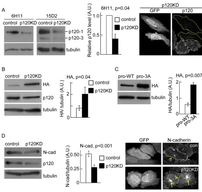
In a previous study we found that inefficient delivery of N-cadherin to the cell surface corre-lated with reduced association of p120 catenin to the N-cadherin precursor [22]. In addition, an N-cadherin mutant unable to bind p120 failed to traffic efficiently from ER to Golgi. Thus, we hypothesized that p120 catenin plays an active role promoting N-cadherin precursor trafficking at early stages of the biosynthetic pathway. To study the N-cadherin precursor traf-ficking in HeLa cells we used a previously characterized construct, HA-N-cadherin-GFP (pro-WT), which contains the hemagglutinin (HA) epitope inserted into the pro-peptide sequence at the N-terminus of N-cadherin, and EGFP at the C-terminus [22]. The relative levels and subcellular distribution of the precursor and the entire pool of N-cadherin can be determined by HA and EGFP detection, respectively. The pro-WT construct migrates slower than endoge-nous N-cadherin in SDS-PAGE and can be easily distinguished. Analysis by Western blotting probed with anti-N-cadherin, revealed that expression levels of pro-WT represented, on aver-age, ~ 12% of the endogenous N-cadherin levels (Figure A inS1 File). Since in our experimen-tal conditions about 40% of the toexperimen-tal cell population is transfected, the expression of pro-WT in transfected cells must be ~ 30% of the endogenous N-cadherin. To investigate the role of p120 in pro-WT trafficking, we knocked down p120 expression in HeLa cells by expression of a small hairpin RNAi against a validated human p120 target sequence [8]. The shRNAi vector used for this purpose co-expresses EGFP as an independent cistron, allowing an easy identifica-tion of knockdown cells by microscopy [27]. As a control, we used a shRNAi vector encoding the target sequence of mouse p120, which differs by three bases from the human p120, and did not have any effect in human cells [8] (Figure B inS1 File). The levels of p120 were analyzed by Western blotting probed with two monoclonal antibodies: clone 6H11 recognizes an N-terminal epitope and detects isoforms 1 and 2, while clone 15D2 recognizes an epitope near the C-termi-nus of p120 and detects the four main p120 isoforms [31]. Consistent with previous work, we found that control HeLa cells express prominently the p120-1 isoform, and low levels of the p120-3 isoform (Fig 1A; [32]). In cells expressing the p120 shRNAi (p120KD), the levels of both p120 isoforms were reduced in a similar proportion. Quantification of p120-1 levels showed a reduction of 70% compared to the levels in cells expressing control shRNAi. The strong inhibi-tion of p120 expression in individual EGFP positive cells was also confirmed by immunofluores-cence microscopy (Fig 1A). In cells co-expressing the p120 shRNAi and pro-WT, the levels of N-cadherin precursor, revealed by anti-HA detection, increased ~2.4-fold compared to cells expressing control shRNAi (Fig 1B). Similar results were observed in HeLa cells expressing a con-struct containing a triple alanine substitution in the juxtamembrane domain which prevents p120 binding [5,22]. This construct (pro-3A) also contained an HA epitope inserted within the pro-peptide to detect the precursor form [22]. Western blotting analysis using anti-HA revealed a ~3-fold increase in pro-3A levels compared to the control pro-WT construct (Fig 1C).
were significantly reduced in p120KD cells (Fig 1D). Immunofluorescence analysis further revealed a reduction of N-cadherin at intercellular junctions and an increase of the cytoplasmic pool (Fig 1D). Collectively, these results strongly suggest that p120 expression and binding to Fig 1. P120 catenin promotes N-cadherin precursor processing.(A) Left, Western blotting analysis of HeLa cells transfected with shRNAi targeting mouse p120 (control) or human p120 (p120KD). Specific antibodies were used for detection of endogenous levels of p120 isoforms and tubulin. Clone 15D2 recognizes the four isoforms of p120 and clone 6H11 recognizes isoforms 1 and 2. Both antibodies showed that p120-1 is the predominant isoform. The reduction of p120-1 in p120KD cells, relative to control is shown in the middle graph. Right panels are representative images of fluorescence microscopy showing the strong effect of p120KD in individual transfected cells (GFP-positive, dotted outlines). The p120 levels were detected by immunofluorescence with 6H11. (B) Western blotting of HeLa cells co-transfected with pro-WT and either, control or p120KD shRNAi. N-cadherin precursor levels, detected by HA labeling, were quantified and expressed as HA/tubulin ratios. (C) Western blotting of HeLa cells transfected with pro-WT or pro-3A. N-cadherin precursor levels, detected by HA labeling, were quantified and expressed as HA/tubulin ratios. (D) Western blotting showing
endogenous cadherin levels in control and p120KD HeLa cells and immunofluorescence showing the distribution of endogenous N-cadherin in control and p120KD HeLa cells (identified by GFP expression, left panels). Yellow arrowheads at the right panels point intercellular junctions and yellow arrows point the intracellular accumulation of N-cadherin in p120KD cells. At least three repetitions were used for quantifications of blots. A. U. (arbitrary units). Bars in graphs represent means±S.E.M. Statistical significance was
determined by two-tailed Student´st-test.
doi:10.1371/journal.pone.0156758.g001
the N-cadherin precursor is required for trafficking from the ER, to the late compartment in the secretory pathway where it is proteolytically processed.
P120 facilitates progression of the N-cadherin precursor through early
stages of the secretory pathway
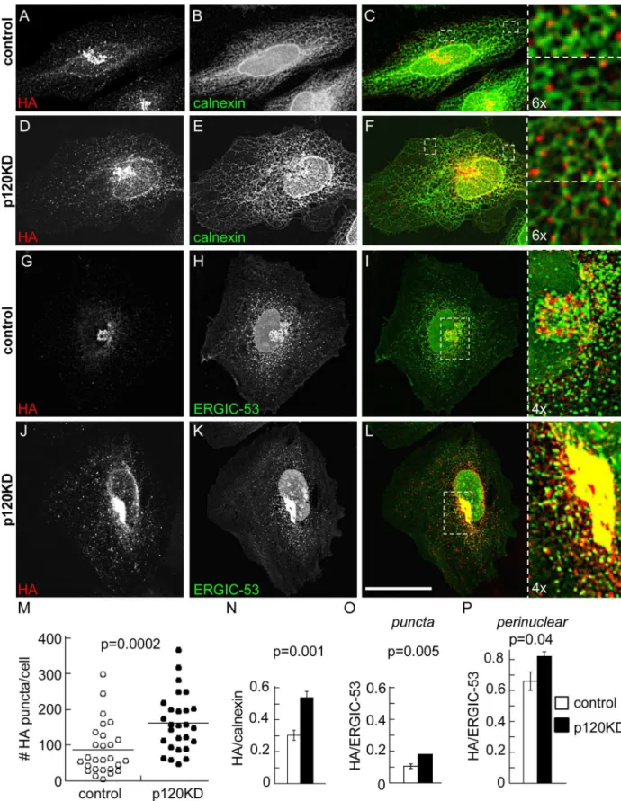
We predicted that accumulation of the N-cadherin precursor in conditions of p120 inhibition must occur in a compartment of the secretory pathway previous to the proteolytic removal of the pro-peptide at the TGN. To address this issue we performed colocalization analysis of the HA label with markers of ER-Golgi traffic compartments in stacks of optical sections obtained by confocal microscopy. For this study we used a variant of the WT construct (called pro-WT2) in which the EGFP was eliminated from the N-cadherin C-terminus. In addition, the EGFP from the shRNAi vector was modified by addition of a nuclear localization sequence that delivers the protein to the nucleus and avoids the interference of EGFP in the colocaliza-tion analysis. The distribucolocaliza-tion of pro-WT2 in control cells was prominent in the perinuclear region (Fig 2A and 2G), where we previously showed an overlapping with Golgi markers [22]. In p120KD cells the N-cadherin precursor accumulated in the perinuclear compartment, but in addition, displayed a widespread punctate distribution throughout the cell (Fig 2D and 2J). Quantification of HA puncta per cell revealed a significant difference between p120KD and control cells (mean ± S.E.M., control: 88 ± 13; p120KD: 162 ± 15;Fig 2M). We examined whether the punctate distribution overlapped with the ER marker calnexin. High resolution confocal imaging revealed that pro-WT puncta partly overlapped with tubular ER in both, con-trol and p120KD conditions (Fig 2C and 2Fand insets). Quantification of Manders´ overlap-ping coefficients determined in peripheral regions of the ER network confirmed these observations and revealed higher colocalization of HA/calnexin in the p120KD condition (mean ± S.E.M., control: 0.30 ± 0.03, n = 422 HA puncta from 9 cells; p120KD: 0.54 ± 0.04, n = 617 puncta from 7 cells;Fig 2N). We then examined the colocalization of the precursor with ERGIC-53, a marker of the intermediate compartment between ER and Golgi, which dis-tributes in punctate structures and in a perinuclear pleiomorphic compartment [33,34]. Expression of GFP-ERGIC-53 in HeLa cells displayed the typical punctate and perinuclear dis-tribution in both, control and p120KD conditions. Signal overlapping between HA and GFP-ERGIC-53 was not evident in the punctate distribution but was obvious in the perinuclear compartment (Fig 2I and 2Land insets). Quantification of the Manders´colocalization coeffi-cients in punctate and perinuclear compartments confirmed these observations and revealed higher colocalization in the p120KD condition (Fig 2O and 2P). These results suggest a higher accumulation of the N-cadherin precursor at early traffic compartments under p120
inhibition.
P120 regulates the recruitment of NSF to the N-cadherin precursor
complex
Fig 2. Distribution of N-cadherin precursor in p120KD HeLa cells.HeLa cells were co-transfected with pro-WT2 and either control (A-C, G-I) or p120KD (D-F, J-L) shRNAi. Cells were analyzed by confocal microscopy and representative images projected along the z-axis are shown. (A-F) Colocalization of N-cadherin precursor (HA, red label) with the ER marker calnexin (green label), detected by immunofluorescence. (G-L) Colocalization of the N-cadherin precursor (HA, red label) with transfected GFP-ERGIC-53 (green signal).
that PTP1B deficiency impaired ER to Golgi trafficking of the N-cadherin precursor, and this effect correlated with reduced p120 association to the precursor [22].
Thus, we sought to examine whether NSF is present in the complex of proteins associated with the N-cadherin precursor. We transfected HeLa cells with pro-WT and isolated the pre-cursor complex by HA immunoprecipitation. Western blotting probed with different monoclo-nal antibodies against NSF consistently detected a band of ~76 kDa in the complex, which is not seen in the mock immunoprecipitation (Fig 3A). Remarkably, subsequent immunoprecipi-tation to isolate mature N-cadherin did not show associated NSF (Fig 3B). To determine if p120 was involved in NSF association to the N-cadherin precursor, we analyzed the presence of NSF in HA immunoprecipitates of cells transfected with pro-WT and the mutant pro-3A which cannot bind p120. As it is shown inFig 1C, pro-3A precursor accumulated to a higher level than pro-WT; however, the amount of NSF associated with pro-3A precursor was signifi-cantly reduced compared to pro-WT (Fig 3C). Quantification of data revealed that NSF/HA ratios in pro-3A expressing cells were a third of those in pro-WT expressing cells. These results were confirmed in p120KD HeLa cells transfected with pro-WT (Fig 3D). Our data suggest that association of p120 to the cytoplasmic domain of N-cadherin precursor contributes to the recruitment of NSF to the complex. Since association of p120 with the N-cadherin precursor is impaired under PTP1B deficiency [22], we predicted that the amount of NSF co-immunopre-cipitated with the N-cadherin precursor in murine PTP1B knockout (KO) cells should be impaired compared to KO cells reconstituted with PTP1B (WT). Results from three indepen-dent experiments revealed that NSF/HA ratios were consistently higher in WT cells than in KO cells (Fig 3E). Stripping the membranes and re-probing them with anti-phosphotyrosine revealed that NSF was phosphorylated in both, KO and WT cells, although the ratio pY/NSF in KO cells was higher than in WT cells in two experiments.
NSF function is required for N-cadherin trafficking and processing
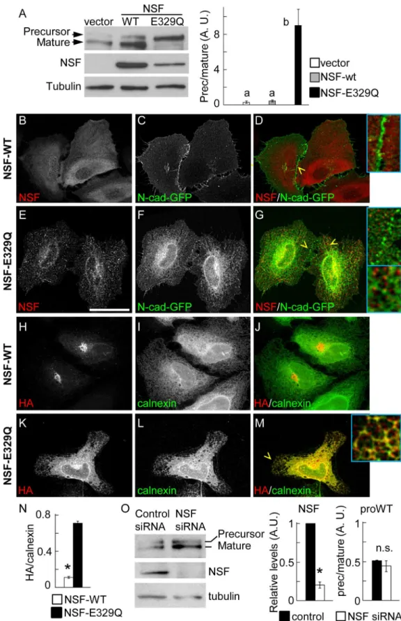
To determine the role of NSF in N-cadherin precursor trafficking and processing, HeLa cells were co-transfected with pro-WT, and either NSF-WT or NSF-E329Q constructs. NSF-E329Q is deficient in ATP hydrolysis and therefore inhibits the function of endogenous NSF, resulting in a dominant negative effect [38]. Western blotting probed with anti-N-cadherin, to simulta-neously detect the ectopic precursor and mature N-cadherin forms, revealed that in cells expressing the empty vector or overexpressing NSF-WT, more than 70% of the ectopic N-cad-herin is processed to the mature form, while in cells expressing NSF-E329Q more than 90% of the ectopic N-cadherin accumulated as a precursor form (Fig 4A). To complement these stud-ies we analyzed, by confocal microscopy, the subcellular distribution of precursor and mature forms of N-cadherin in HeLa cells expressing NSF-WT or NSF-E329Q. The distribution of over-expressed NSF-WT was diffuse in the cytosol, while that of NSF-E329Q followed a punc-tate pattern throughout the cell, in agreement with previous findings [39] (Fig 4B and 4E). The distribution of N-cadherin-GFP in cells expressing NSF-WT was prominent at intercellular junctions (Fig 4C and 4D, inset). In contrast, the distribution of N-cadherin-GFP in cells
Enlarged views from selected regions (white dotted boxes) are shown at the right. The expression of the control or p120KD shRNAi constructions in the analyzed cells is revealed by the modified nuclear-targeted GFP encoded by the vector (green channel). Scale bar in (L), 35μm. (M) Number of HA puncta per cell in control and p120KD conditions (n = 27 cells per condition). Horizontal lanes indicate the position of means (control = 87,8; p120KD = 162.1). (N) Manders colocalization coefficients of the HA puncta overlapping calnexin in peripheral regions (control, n = 9 cells, total 422 puncta; p120KD, n = 7 cells, total 617 puncta). (O, P) Manders colocalization coefficients of the HA label overlapping GFP-ERGIC-53; independent analysis of the punctate (O) and clustered perinuclear distribution (P) of the HA label was performed (n = 24 cells per condition). Bars represent means±S.E.M. Statistical significance was determined by a two-tailed Mann-Whitney test.
Fig 3. NSF association with the N-cadherin precursor.(A, B) Soluble protein extracts from HeLa cells transfected with pro-WT were sequentially immunoprecipitated with anti-HA and anti-N-cadherin, to isolate the precursor (A) and the mature (B) N-cadherin pools. Mock immunoprecipitations omitted the primary antibody. The presence of NSF in the complexes was analyzed in Western blotting probed with anti-NSF. (C)
Immunoprecipitation of N-cadherin precursor from cells transfected with pro-WT or pro-3A constructs, and analysis of NSF in the complexes. (D) Cells co-transfected with pro-WT and either control or p120shRNAi. N-cadherin precursor was isolated by HA-immunoprecipitation and the presence of NSF in the complex was detected in blots. The graphs in (C) and (D) represent quantifications of NSF bands, normalized to the HA signal.
expressing NSF-E329Q showed a discontinuous pattern at intercellular junctions and a signifi-cant accumulation at the ER, frequently overlapping with NSF-E329Q puncta (Fig 4F and 4G, inset). The distribution of the N-cadherin precursor was evaluated in cells transfected with pro-WT2. As expected, the N-cadherin precursor in cells expressing NSF-WT was visualized at the perinuclear region, in a Golgi location, and showed minimal colocalization with calnexin (Fig 4H, 4J and 4N). In contrast, in cells expressing NSF-E329Q most of N-cadherin precursor was retained at the ER, where it showed a tight colocalization with calnexin (Fig 4K and 4M, inset, 4N). Strikingly, siRNA-mediated knockdown of NSF did not affect the rate of N-cadherin precursor processing (Fig 4O).
P120 reconstitution in p120-defficient cells rescues N-cadherin
trafficking and processing
Most cell types express multiple isoforms of p120, generated by alternative splicing of a single gene [40]. The use of different translation start codons results in four isoforms, p120-1, 2, 3, and 4. All isoforms contain a central armadillo domain, which mediates binding to cadherin, but differ in the length of the N-terminal regulatory domain, being p120-1 the longest isoform, and p120-2, -3 and -4 progressively shorter at the N-terminal region. The p120 gene in the SW48 human colon carcinoma cell line contains a nonsense mutation in exon 7, yielding a pre-mature stop codon and negligible expression levels of the protein [41]. We used SW48 cells to test if different p120 isoforms were capable of promoting N-cadherin trafficking and process-ing. Western blotting analysis of SW48 cells transfected with pro-WT and probed with anti-N-cadherin, to simultaneously detect the precursor and mature N-cadherin species revealed that the precursor was the predominant form (Fig 5A). Endogenous levels of N-cadherin were neg-ligible (not shown). Using an antibody that recognizes all p120 isoforms (clone 15D2, [31]) we also confirmed the negligible expression of endogenous p120 isoforms (Fig 5A). Individual reconstitution of SW48 cells with p120-1, -3, and -4 promoted the processing of the N-cad-herin precursor and the appearance of mature N-cadN-cad-herin, visualized as a lower molecular weight band in blots (Fig 5A). Confocal analysis of cells at equatorial planes revealed that all p120 isoforms transfected in SW48 cells accumulated at intercellular junctions, as expected (Fig 5B–5H). Parental, non transfected cells did not develop conspicuous intercellular junc-tions, as revealed by F-actin labeling (Fig 5B–5D, "nt"). In addition, expression of pro-WT in
parental SW48 cells, did not show accumulation of GFP at intercellular junctions, both the HA (precursor cadherin) and GFP label (total N-cadherin) colocalized at intracellular compart-ments (Fig 5I). In contrast, cells reconstituted with different p120 isoforms showed the expected intracellular HA (and GFP) label, but a prominent and exclusive GFP signal accumu-lated at intercellular junctions, consistent with the production of mature and functional N-cad-herin (Fig 5J–5L). In addition, p120-reconstituted cells tend to form more compacted cellular aggregates than parental cells, in agreement with previous findings showing that expression of p120 in SW48 cells induced a significant compaction of epithelial colonies [41]. We confirmed the expression of p120 in reconstituted cells in separate experiments (not shown). Confocal colocalization analysis of the pro-WT2 construct (lacking the GFP at the C-terminus) revealed that in parental SW48 cells the N-cadherin precursor (HA label) colocalized with calnexin (ER
(E) Murine PTP1B KO and WT cell lines transfected with pro-WT were immunoprecipitated with anti-HA, and the protein complexes were analyzed by Western blotting probed with antibodies against HA, NSF, phosphotyrosine and tubulin. Numbers under the panels indicate the pY-NSF/NSF ratio. The graph at the right shows the quantification of NSF bands normalized to HA. In all cases, at least three independent experiments were used for quantifications. Data represent means±S.E.M. Statistical significance was determined by two-tailed Student
´st-test.
Fig 4. Function of NSF in N-cadherin trafficking.(A) Western blotting analysis of HeLa cells co-transfected with pro-WT, and empty vector or vector encoding NSF-WT or NSF-E329Q. Precursor and mature forms of pro-WT were detected with anti-N-cadherin. The lower size of the endogenous N-cadherin is not depicted. Visualization of ectopic NSF-WT and NSF-E329Q signals under non-saturating conditions did not allow
marker) and in a minor degree with GalNacT (Golgi marker, [42] (Fig 5M, 5N and 5Q). In contrast, in cells reconstituted with p120 isoforms 1, 3 and 4, the colocalization of N-cadherin precursor with calnexin was less evident, while its colocalization with GalNacT increased (Fig 5O, 5P and 5Q; only data of isoform 1 is shown). Taken together, the reconstitution of different p120 isoforms in SW48 cells confirm the results of p120 knockdown in HeLa cells, strongly indicating that p120 expression promotes the trafficking and processing of the N-cadherin pre-cursor and the surface localization of mature N-cadherin. This function of p120 can be per-formed by all isoforms tested.
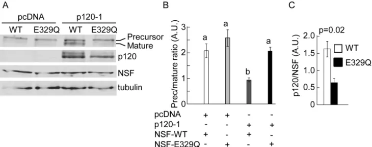
To further confirm the role of NSF and p120 in N-cadherin precursor processing, SW48 cells were co-transfected with pro-WT along with empty vector (pcDNA) or pcDNA-p120-1 in combination with either NSF-WT or NSF-E329Q. Western blotting analysis revealed that overexpression of functional NSF in parental cells was not enough to promote N-cadherin pro-cessing in absence of p120 expression (Fig 6A). Also, reconstitution of parental cells with p120 isoform 1 was not enough to promote the processing of N-cadherin if the dominant negative NSF-E329Q was co-expressed. Expression of both, NSF-WT and p120-1 was necessary for N-cadherin precursor processing to the mature form (Fig 6A and 6B). In three independent repe-titions of this experiment we consistently noticed lower levels of p120 in cells expressing NSF-E329Q, compared to those expressing NSF-WT. Quantification of p120 and NSF bands in densitometric scans, and comparison of p120/NSF ratios (or p120/tubulin ratios, not shown), confirmed the statistical significance of these observations (NSF-WT mean: 1.6 ± 0.23 vs NSF-E329Q mean: 0,65 ± 0.12) (Fig 6C).
Discussion
After biosynthesis at the ER, cadherins are delivered to the cell surface where they mediate homophilic, Ca+2-dependent intercellular adhesion. Adhesion strength is modulated by the amount of cadherin at the cell surface, which results from a complex interplay among biosyn-thesis, degradation, traffic, and anchorage to the cytoskeleton [3,43–45]. P120 andβ-catenin are major direct interacting partners of cadherins. They bind to different sites in the cyto-plasmic domain of cadherin precursors soon after their insertion at the ER membranes, traf-ficking as a complex towards the cell surface [17–19,46,47].
The present study proposes that early anterograde traffic of nascent cadherin is regulated by associated p120 catenin, suggesting a mechanism by which p120 catenin recruits NSF to the cadherin precursor complex, facilitating its movement from the ER to Golgi. So far it was well
detection of endogenous NSF (first lane). At least three independent experiments were used for quantification of precursor and mature forms of pro-WT (right graph). Bars represent means±S.E.M. of precursor/mature
ratios normalized to tubulin. Statistical significance (p<0.05) was determined using one-way ANOVA, followed by the Dunnett´s multiple comparisonpost hoctest. Different letters indicate statistically different means. (B-G) Distribution of GFP and ectopic NSF constructs. HeLa cells were co-transfected with N-cadherin-GFP and either NSF-WT (B-D) or NSF-E329Q (E-G). (H-M) Colocalization of N-cadherin precursor (HA, red label) and the ER marker calnexin (green label) in cells co-transfected with pro-WT2 and NSF-WT (H-J) or NSF-E329Q (K-M). The expression of exogenous NSF constructs was verified in triple label samples (not shown). (N) Manders colocalization coefficients of the HA/calnexin signal overlapping in NSF-WT (n = 15 cells) and NSF-E329Q (n = 23 cells) conditions. Bars represent means±S.E.M. Statistical significance was
determined by a two-tailed Mann-Whitney test. Cells were analyzed by confocal microscopy and representative images projected along the z-axis are shown. Enlarged views (4x) from selected regions (yellow arrowheads) are shown as insets. Scale bar in (E), 35μm. (O) HeLa cells were co-transfected with proWT and non-targeting siRNA duplexes (Control) or NSF siRNA duplexes (NSF siRNA). After 48 h, precursor and mature forms of the proWT construct, NSF and tubulin were analyzed by Western blotting. Quantification of NSF levels (NSF siRNA values relativized to control siRNA) and precursor/mature ratios in control and NSF siRNA conditions is shown at the right. Bars represent means±S.E.M. from three independent experiments. Statistical significance
(asterisks, p<0.05) was determined by a two-tailed Student´st-test.
Fig 5. N-cadherin distribution in SW48 colon carcinoma cells reconstituted with p120.(A) Cells were co-transfected with pro-WT and p120 isoforms 1, 3 or 4. Soluble protein extracts from parental (P) and reconstituted cells (1, 3 and 4) were analyzed in Western blotting probed with anti-N-cadherin to recognize the precursor and mature forms of pro-WT, the p120
established that p120 association to mature cadherin at the cell surface prevented its internali-zation and degradation, acting as a "set-point" for cadherin expression [48]. However, the role of p120 in cadherin traffic through the biosynthetic pathway is intriguing and less understood. Pulse-chase experiments combined with surface biotinylation performed in human cervical carcinoma A431 cells, revealed that p120 knockdown (p120KD) did not affect E-cadherin bio-synthesis and traffic to the cell surface [8]. However, other studies showed that p120 associated directly with microtubules and kinesin, and disruption of p120/kinesin interaction impaired the delivery of nascent N-cadherin to newly forming junctions [21,49–51]. It should be noted that these studies suggest a role of p120 in post-Golgi delivery of N-cadherin towards the plasma membrane. Recent work from our laboratory, however, suggested that p120 could also be implicated in ER to Golgi transport of the N-cadherin precursor [22]. We found that in fibroblasts deficient in protein tyrosine phosphatase 1B (PTP1B) expression, the passage of the N-cadherin precursor from ER to Golgi was impaired. This defect correlated with decreased association of p120 to the precursor. Additionally, expression of HA-N-cadherin-3A mutant
isoforms (clone 15D2), and tubulin. The graph shows quantification of precursor/mature ratios normalized to tubulin. (B-H) Confocal analysis of representative cells reconstituted with isoform 1 (B-D), 3 (E-G) and 4 (H). Cells were labeled for p120 (B, E, H) and F-actin (C, F). Arrows point intercellular junctions. "nt" in (B) indicates non-transfected cells. (I-L) Distribution of precursor and mature forms of N-cadherin in parental cells expressing pro-WT and empty vector (I), or vectors encoding p120-1 (J), pp120-120-3 (K) or pp120-120-4 (L). The distribution of ectopic N-cadherin precursor (red label) and total N-cadherin (green label) was detected by anti-HA and GFP fluorescence, respectively. (M-P) Colocalization of the N-cadherin precursor with ER and Golgi markers. Cells expressing pro-WT2 in parental (M) and reconstituted with p120-1 (O) conditions were double-labeled for HA (red signal) and calnexin (green signal). The expression of p120-1 construct was verified in triple label samples (not shown). Note the redistribution of the HA signal to the perinuclear compartment in p120-1 reconstituted cells. The Golgi apparatus was revealed by GalNacT-DsRed transfection in parental (N) and p120-1 reconstituted (P) cells. Note the tight overlapping of the HA label (shown in red) with GalNacT-DsRed (shown in green) in the Golgi. (Q) Manders colocalization coefficients calculated from HA and calnexin (n = 13 cells), and HA and GalNacT (n = 13 cells) signals. Bars represent means±S.E.M. Statistical significance was determined by a two-tailed Mann-Whitney test. All cell images represent equatorial
confocal planes and white arrows point intercellular junctions. Scale bar in (P), 14μm.
doi:10.1371/journal.pone.0156758.g005
Fig 6. Combinatorial analysis of p120 and NSF in N-cadherin processing.(A) SW48 cells expressing pro-WT in combination with empty vector (pcDNA) or p120-1, and NSF-WT (WT) or NSF-E329Q (E329Q). Shown is a representative blot probed with anti-N-cadherin to detect the precursor and mature form of N-cadherin, anti-p120, anti-NSF, and anti-tubulin. (B, C) Quantification of N-cadherin precursor/ mature ratio (B) or p120/ NSF ratio (C) from blot scans expressed in arbitrary units (A. U.). Graph bars represent means±S.E.M. from three independent experiments. Statistical significance (p<0.05) was determined using one-way ANOVA, followed by the Dunnett´s multiple comparisonpost hoctest (B), or a two-tailed Student´st-test (C). Different letters in (B) indicate statistically different means.
unable to bind p120, was more sensitive to endoglycosidase H than wild type N-cadherin, sug-gesting a defect in transport of the precursor from ER to Golgi [22]. These results, however, do not rule out the possibility that other partners, which bind to overlapping sites within the juxta-membrane region, such as presenilin-1, ankyrin-G and GRIP-1 could also be implicated [23–
25]. Thus, in the present study we specifically inhibited p120 expression in HeLa cells by shRNAi, and provided a direct evidence of a p120 function in the ER-Golgi traffic of N-cad-herin precursor. High resolution confocal analysis of p120KD cells revealed a punctate distri-bution of the N-cadherin precursor, which significantly overlapped with ER marker calnexin. In control conditions, however, most of the N-cadherin precursor accumulated at the Golgi apparatus and puncta distribution is rare. Additionally, in p120KD cells the N-cadherin pre-cursor colocalized more tightly with the intermediate compartment marker ERGIC-53 than in control conditions. These results suggest that, under p120 inhibition, the N-cadherin precursor is able to aggregate at the ER, but its export and transport through the intermediate compart-ment is impaired. In normal conditions, and following arrival of the cadherin precursor to the
trans-Golgi network (TGN), the propeptide is cleaved by furin proteases [15–17]. Consistent
with an impaired flow of N-cadherin precursor from the ER to TGN in p120KD cells, we found a significant accumulation of precursor levels compared to control conditions. These observations were confirmed in HeLa cells transfected with the N-cadherin mutant unable to bind p120. In addition, the reconstitution of p120 expression in the p120-defficient SW48 cell line, promoted N-cadherin traffic and processing. We found that isoforms p120-1, -3 and -4 have an equivalent function. These isoforms share the conserved cadherin-binding domain and the C-terminal region, but differ in the length and presence of regulatory motifs localized in the terminal region [40]. Isoform-1 is the longest protein and contains the full length N-terminal domain, bearing a putative coiled-coil motif and a phosphorylatable regulatory domain. Isoform-3 lacks the coiled-coil motif but retains the regulatory domain, and isoform-4 lacks the entire N-terminal domain (~300 amino acids). Our results suggest that the capacity to promote trafficking and processing of the N-cadherin precursor does not reside in the N-ter-minal region of p120. However, we cannot rule out a modulatory effect of this region, as it has been reported for p120 function controlling cadherin dynamics [12].
Depletion of p120 could alter the anterograde traffic of the N-cadherin precursor through an indirect effect, for example impairing the association ofβ-catenin. Indeed, previous work in MDCK cells showed that expression of E-cadherin mutants with deletions or with inactivating amino acid substitutions in theβ-catenin binding region, were mostly retained at the ER [47], or accumulated in Golgi and post-Golgi compartments [52]. It is not clear, however, if these mutants still associate efficiently with p120. Beta catenin could facilitate the export of N-cad-herin from ER through binding to a PX-RICS-14-3-3z/θ-dynein-dynactin complex, since RNAi depletion of these components lead to N-cadherin retention at the ER [53]. Moreover, recent structural analysis revealed a novel functional consequence ofβ-catenin association to cadherin [54]. Beta catenin could contribute to strengthen cadherin-p120 association by recruitingα-catenin to the complex, and promoting additional contacts between p120 andα -catenin. However, most short term experiments inhibiting p120 expression or association with cadherin did not show a correlative effect onβ-catenin. It was noted previously that p120 knockdown in SK-CO15 colon carcinoma cells did not affectβ-catenin expression levels [35]. Similarly, we did not observe reduction ofβ-catenin levels by p120 knockdown in HeLa cells (results not shown). In addition, we and others clearly showed thatβ-catenin association with N-cadherin precursor was not coupled to p120 dissociation [7,22,55]. Thus, our present data on N-cadherin precursor traffic likely reflect the direct consequence of loosing p120 function. What is the mechanism by which p120 associated with the N-cadherin precursor promotes ER to Golgi traffic? A major finding of the present work is that NSF is recruited to the
cadherin precursor complex in a p120- and PTP1B-dependent manner. Previously we found that binding of p120 to the N-cadherin precursor, and ER to Golgi traffic, was impaired in PTP1B deficient cells [22]. Together, these results suggest a functional interplay among PTP1B, p120 and NSF, with consequences in N-cadherin traffic. PTP1B localization at the cytosolic face of ER membranes [56] may facilitate dephosphorylation of proteins associated with the cytoplasmic domain of the N-cadherin precursor, such as p120 and beta catenin [17, 20,22]. Besides promoting the association of p120 with the N-cadherin precursor, and indi-rectly the recruitment of NSF to the complex, PTP1B may also have a direct effect on NSF activity. Indeed, three laboratories have independently shown a positive effect of PTP1B in membrane fusion, likely by dephosphorylation and activation of NSF [28,36,37]. Our results showing higher retention of phosphotyrosine in the NSF fraction associated with the N-cad-herin precursor in KO cells compared to WT cells suggest this possibility.
As a general regulator of membrane fusion, NSF is likely required not only for ER-Golgi traffic but also for post-Golgi events. However, we were unable to detect NSF in complexes of mature N-cadherin, suggesting that different mechanisms regulate the composition of N-cad-herin complexes at specific subcellular locations and functional stages. Regulation of protein trafficking to the plasma membrane in complexes with NSF has recently been described for transmembrane receptors, including theα-amino-3-hydroxy-5-methyl-4-isoxazolepropionic acid (AMPA) subtype of glutamate receptors [26]. These complexes could be linked with cad-herins by multi-PDZ domain scaffold proteins such as the glutamate receptor interacting pro-tein (GRIP), which interacts with the p120 relativeδ-catenin [57]. GRIP also links AMPA receptors and N-cadherin with the anterograde motor protein KIF5C, ensuring the proper co-deliver of N-cadherin and AMPA receptors to dendritic spines [25]. Interestingly,δ-catenin binds to GRIP through a C-terminus motif. This is relevant to our finding that all isoforms of p120 tested have equivalent capacity to promote N-cadherin trafficking, suggesting that the molecular determinant implicated in NSF recruitment must reside in the armadillo and/or the C-terminal domain. Both mechanisms, the association of N-cadherin to KIF5C through GRIP and the association of NSF to the N-cadherin precursor (described in this work), could work together to ensure the proper delivery of N-cadherin to the cell surface.
Our results showed that impairing NSF function with the dominant negative construct NSF-E329Q led to a dramatic retention of N-cadherin precursor at the ER, impairing its pro-cessing to the mature form, and leading to a significant reduction of N-cadherin at intercellular junctions. Unexpectedly, NSF depletion by siRNA did not affect the processing of the N-cad-herin precursor. Although we cannot completely exclude that a residual fraction of NSF may still fulfill the requirements for the N-cadherin precursor traffic, these results suggest a non essential role of NSF in the ER-Golgi movement of the N-cadherin precursor. In agreement with this, recent studies in NSF-depleted SK-CO15 colonic epithelial cells showed a lack of effect on the distributions of KDEL-R and GM130 [58]. However, depletion of the NSF homo-log, p97, caused a redistribution of these markers from a well defined Golgi localization in con-trol cells to a punctate distribution scattered throughout the cell, suggesting an impairment of ER-Golgi traffic [58]. Thus, the lack of effect of NSF siRNA depletion in our experiments may reflect compensatory functions performed by p97. The strong effect of the dominant negative NSF construct, observed in the present work, may reflect a "substrate trap" activity, likely sequestering the adaptor proteinαSNAP [39,58]. Recent work showed that RNAi depletion of
αSNAP in SK-CO15 cells impaired epithelial junctions, and downregulated p120 catenin and E-cadherin, but notβ-catenin expression [35]. Strikingly,αSNAP knockdown also impaired ER-Golgi transport, and produced accumulation of ER-glycosylated forms of theβ1 integrin precursor, effects that are not mimicked by NSF depletion [35,58]. The mechanistic aspects of
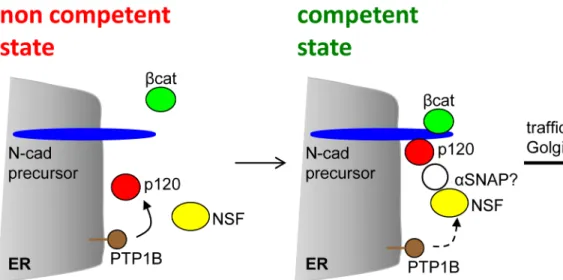
suggests a role in pre-docking/docking steps during the membrane fusion process [59]. Our results expressing the dominant negative NSF construct in HeLa and SW48 cells recreate sev-eral effects of theαSNAP knockdown, including the impairment of intercellular junctions and ER-Golgi transport, as well as the downregulation of p120 levels [35,58]. Thus, we cannot rule out that the dominant negative NSF-E329Q effects on N-cadherin traffic may be related to indirect inhibition ofαSNAP. It remains to be determined ifαSNAP, which recruits NSF to membranes, is present in the complex of proteins associated with the N-cadherin precursor, and the potential p120 dependency. We envisage a scenario in which ER-bound PTP1B func-tion ensures p120-dependent recruitment of NSF to the N-cadherin precursor complexes. NSF activation, likely by PTP1B, facilitates membrane fusion events associated with vesicular traffic initiated at the ER (Fig 7). In the absence of NSF, its homolog p97 likely compensates NSF function.
Supporting Information
S1 File. Figure A: Expression levels of pro-WT. The HA-N-cadherin-GFP (pro-WT) construct
was transfected in HeLa cells and the expression levels were analyzed by Western blotting with anti-N-cadherin, to allow for the direct comparison with endogenous N-cadherin levels. Due to the GFP fusion, pro-WT migrates slower than endogenous N-cadherin. Under our detection conditions essentially all ectopic and endogenous N-cadherin correspond to mature, processed proteins. Representative blots of two independent experiments are shown at the left. Quantifi-cation of the bands is shown in the graph. Bars represent means ± S.E.M. from five experi-ments. Figure B: Effect of p120 shRNAi targeting. (A) Western blotting analysis of HeLa cells transfected with empty pG-Shin2 vector (ve), the vector encoding the human p120 targeting sequence (hu), or the equivalent mouse p120 targeting sequence (mo). Blots were probed with anti-p120 (clone 6H11), anti-N-cadherin, and anti-tubulin. Only the human p120 targeting sequence showed a marked effect on p120 expression. A representative blot of 3 independent experiments is shown. (B-D) HeLa cells expressing the control, mouse p120 targeting sequence. Fig 7. Model representing the complex assembly required for N-cadherin precursor traffic at the ER. N-cadherin precursor (blue) is co-translationally inserted at the ER membrane, with the C-terminus localized in the cytoplasm. Binding of p120 (red) andβ-catenin (green) to the precursor cytoplasmic domain occurs at the ER. PTP1B (brown) localized at the ER dephosphorylates p120 and stabilize its association with the complex [22]. Perturbation of these events impairs traffic. In a traffic competent state, p120 recruits NSF (yellow) to the complex, where is likely dephosphorylated and activated by PTP1B. The recruitment ofαSNAP (white) to the N-cadherin precursor complex, and its role in ER-Golgi traffic, remains to be confirmed.
doi:10.1371/journal.pone.0156758.g007
Immunofluorescence with anti-p120 reveals similar p120 levels (red signal) in transfected (GFP positive cells) and non transfected cells. Scale bar in (D), 35μm.
(PDF)
Acknowledgments
We thank all colleagues that generously shared cell lines, antibodies and plasmids used in this work.
Author Contributions
Conceived and designed the experiments: COA DPW. Performed the experiments: DPW FC AEGW AG. Analyzed the data: DPW COA. Contributed reagents/materials/analysis tools: JMLM. Wrote the paper: DPW COA.
References
1. Berx G, Van Roy F (2009) Involvement of members of the cadherin superfamily in cancer. Cold Spring Harb. Perspect. Biol. 1, a003129. doi:10.1101/cshperspect.a003129PMID:20457567
2. Stepniak E, Radice GL, Vasioukhin V (2009) Adhesive and signaling functions of cadherins and cate-nins in vertebrate development. Cold Spring Harb. Perspect. Biol. 1, a002949. doi:10.1101/ cshperspect.a002949PMID:20066120
3. Pokutta S, Weis WI (2007) Structure and mechanism of cadherins and catenins in cell-cell contacts. Annu. Rev. Cell Dev. Biol. 23, 237–261. PMID:17539752
4. Ohkubo T, Ozawa M (1999) p120 (ctn) binds to the membrane-proximal region of the E-cadherin cyto-plasmic domain and is involved in modulation of adhesion activity. J. Biol. Chem. 274, 21409–21415. PMID:10409703
5. Thoreson MA, Anastasiadis PZ, Daniel JM, Ireton RC, Wheelock MJ, Johnson KR, et al. (2000) Selec-tive uncoupling of p120(ctn) from E-cadherin disrupts strong adhesion. J. Cell Biol. 148, 189–202. PMID:10629228
6. Ishiyama N, Lee SH, Liu S, Li GY, Smith MJ, Reichardt LF, et al. (2010) Dynamic and static interactions between p120 catenin and E-cadherin regulate the stability of cell-cell adhesion. Cell. 141, 117–128. doi:10.1016/j.cell.2010.01.017PMID:20371349
7. Nanes BA, Chiasson-MacKenzie C, Lowery AM, Ishiyama N, Faundez V, Ikura M, et al. (2012) p120-catenin binding masks an endocytic signal conserved in classical cadherins. J. Cell Biol. 199, 365– 380. doi:10.1083/jcb.201205029PMID:23071156
8. Davis MA, Ireton RC, Reynolds AB (2003) A core function for p120-catenin in cadherin turnover. J. Cell Biol. 163, 525–534. PMID:14610055
9. Xiao K, Allison DF, Buckley KM, Kottke MD, Vincent PA, Faundez V, et al. (2003) Cellular levels of p120 catenin function as a set point for cadherin expression levels in microvascular endothelial cells. J. Cell Biol. 163, 535–545. PMID:14610056
10. Keirsebilck A, Bonné S, Staes K, van Hengel J, Nollet F, Reynolds A, et al. (1998) Molecular cloning of the human p120 ctn catenin gene (CTNND1): expression of multiple alternatively spliced isoforms. Genomics. 50, 129–146. PMID:9653641
11. Reynolds AB, Daniel JM, Mo YY, Wu J, Zhang Z (1996) The novel catenin p120 cas binds classical cadherins and induces an unusual morphological phenotype in NIH3T3 fibroblasts. Exp. Cell Res. 225, 328–337. PMID:8660921
12. Fukumoto Y, Shintani Y, Reynolds AB, Johnson KR, Wheelock MJ (2008) The regulatory or phosphory-lation domain of p120 catenin controls E-cadherin dynamics at the plasma membrane. Exp. Cell Res. 314, 52–67. PMID:17719574
13. Ozawa M, Kemler R (1990) Correct proteolytic cleavage is required for the cell adhesive function of uvomorulin. J. Cell Biol, 111, 1645–1650. PMID:2211831
14. Ozawa M (2002) Lateral dimerization of the E-cadherin extracellular domain is necessary but not suffi-cient for adhesive activity. J. Biol. Chem. 277, 19600–19608. PMID:11916976
16. Posthaus H, Dubois CM, Laprise MH, Grondin F, Suter MM, Müller E (1998) Proprotein cleavage of E-cadherin by furin in baculovirus over-expression system: potential role of other convertases in mamma-lian cells. FEBS Lett. 438, 306–310. PMID:9827567
17. Wahl JK, Kim YJ, Cullen JM, Johnson KR, Wheelock MJ (2003) N-cadherin-catenin complexes form prior to cleavage of the proregion and transport to the plasma membrane. J. Biol. Chem. 278, 17269– 17276. PMID:12604612
18. Ozawa M, Kemler R (1992) Molecular organization of the uvomorulin-catenin complex. J. Cell Biol. 116, 989–996. PMID:1734027
19. Hinck L, Näthke IS, Papkoff J, Nelson WJ (1994) Dynamics of cadherin/catenin complex formation: novel protein interactions and pathways of complex assembly. J. Cell Biol. 125, 1327–1340. PMID: 8207061
20. Curtis MW, Johnson KR, Wheelock MJ (2008) E-cadherin/catenin complexes are formed cotranslation-ally in the endoplasmic reticulum/Golgi compartments. Cell Commun. Adhes. 15, 365–378. doi:10. 1080/15419060802460748PMID:18937087
21. Chen X, Kojima S, Borisy GG, Green KJ (2003) P120 catenin associates with kinesin and facilitates the transport of cadherin-catenin complexes to intercellular junctions. J. Cell Biol. 163, 547–557. PMID: 14610057
22. Hernandez MV, Wehrendt DP, Arregui CO (2010) The Protein Tyrosine Phosphatase PTP1B Is Required for Efficient Delivery of N-Cadherin to the Cell Surface. Mol. Biol. Cell. 21, 1387–1397. doi: 10.1091/mbc.E09-10-0880PMID:20181825
23. Baki L, Marambaud P, Efthimiopoulos S, Georgakopoulos A, Wen P, Cui W, et al. (2001) Presenilin-1 binds cytoplasmic epithelial cadherin, inhibits cadherin/p120 association, and regulates stability and function of the cadherin/catenin adhesion complex. PNAS. 98, 2381–2386. PMID:11226248
24. Kizhatil K, Davis JQ, Davis L, Hoffman J, Hogan BL and Bennett V (2007) Ankyrin-G is a molecular part-ner of E-cadherin in epithelial cells and early embryos. J. Biol. Chem, 282, 26552–26561. PMID: 17620337
25. Heisler FF, Lee HK, Gromova KV, Pechmann Y, Schurek B, Ruschkies L, et al. (2014) GRIP1 interlinks N-cadherin and AMPA receptors at vesicles to promote combined cargo transport into dendrites. PNAS. 111, 5030–5035. doi:10.1073/pnas.1304301111PMID:24639525
26. Zhao C, Slevin JT, Whiteheart SW (2007) Cellular functions of NSF: Not just SNAPs and SNAREs. FEBS lett. 581, 2140–2149. PMID:17397838
27. Kojima S-I, Vignjevic D, Borisy GG (2004) Improved silencing vector co-expressing GFP and small hair-pin RNA. Biotechniques 36, 74–79. PMID:14740488
28. Sangwan V, Abella J, Lai A, Bertos N, Stuible M, Tremblay ML, et al. (2011) Protein Tyrosine Phospha-tase PTP1B Modulates Early Endosome Fusion and Trafficking of Met and EGF Receptor Tyrosine Kinases. J. Biol. Chem. 286, 45000–45013. doi:10.1074/jbc.M111.270934PMID:22045810 29. Haj FG, Verveer PJ, Squire A, Neel BG, Bastiaens PI (2002) Imaging sites of receptor
dephosphoryla-tion by PTP1B on the surface of the endoplasmic reticulum. Science 295, 1708–1711. PMID: 11872838
30. Lázaro-Martínez JM, Rodríguez-Castellón E, Vega D, Monti GA, Chattah AK (2015) Solid-state Studies of the Crystalline/Amorphous Character in Linear Poly (ethylenimine hydrochloride) (PEIHCl)
Poly-mers and Their Copper Complexes. Macromolecules. 48, 1115–1125.
31. Wu J, Mariner MA, Thoreson MA, Reynolds AB (1998) Production and characterization of monoclonal antibodies to the catenin p120ctn. Hybridoma 17, 175
–183. PMID:9627058
32. Mo YY, Reynolds AB (1996) Identification of murine p120cas isoforms and heterogeneous expression of p120cas isoforms in human tumor cell lines.Cancer Research, 56, 2633–2640. PMID:8653709 33. Appenzeller-Herzog C, Hauri HP (2006) The ER-Golgi intermediate compartment (ERGIC): in search
of its identity and function. J. Cell Sci. 119, 2173–2183. PMID:16723730
34. Ben-Tekaya H, Miura K, Pepperkok R, Hauri HP (2005) Live imaging of bidirectional traffic from the ERGIC. J. Cell Sci. 118, 357–367. PMID:15632110
35. Naydenov NG, Brown B, Harris G, Dohn MR, Morales VM, Baranwal S, et al. (2012) A membrane fusion proteinαSNAP is a novel regulator of epithelial apical junctions. PloS One. 7, e34320. doi:10. 1371/journal.pone.0034320PMID:22485163
36. Zarelli VE, Ruete MC, Roggero CM, Mayorga LS, Tomes CN (2009) PTP1B dephosphorylates N-ethyl-maleimide-sensitive factor and elicits SNARE complex disassembly during human sperm exocytosis. J. Biol. Chem. 284, 10491–10503. doi:10.1074/jbc.M807614200PMID:19208619
37. Shi L, Zhang Q, Jiang X, Dai Y, Zhang CY, Zen K (2014) Sustained high protein-tyrosine phosphatase 1B activity in the sperm of obese males impairs the sperm acrosome reaction. J. Biol. Chem. 289, 8432–8441. doi:10.1074/jbc.M113.517466PMID:24519936
38. Coppolino MG, Kong C, Mohtashami M, Schreibe AD, Brumell JH, Finlay BB, et al. (2001) Requirement for N-Ethylmaleimide-sensitive Factor Activity at Different Stages of Bacterial Invasion and Phagocyto-sis. J. Biol. Chem. 276, 4772–4780. PMID:11092884
39. Dalal S, Rosser MFN, Cyr DM, Hanson PI (2004) Distinct Roles for the AAA ATPases NSF and p97 in the Secretory Pathway. Mol. Biol. Cell. 15, 637–648. PMID:14617820
40. Anastasiadis PZ, Reynolds AB (2000) The p120 catenin family: complex roles in adhesion, signaling and cancer. J. Cell Sci. 113, 1319–1334. PMID:10725216
41. Ireton RC, Davis M, van Hengel J, Mariner DJ, Barnes K, Thoreson MA, et al. (2002) A novel role for p120 catenin in E-cadherin function. J. Cell Biol. 159, 465–476. PMID:12427869
42. Giraudo CG, Rosales Fritz VM, Maccioni HJ (1999) GA2/GM2/GD2 synthase localizes to the trans-golgi network of CHO-K1 cells. Biochem. J. 342, 633–640. PMID:10477274
43. Meng W, Takeichi M (2009) Adherens junction: molecular architecture and regulation. Cold Spring Harb. Perspect. Biol. 1, a002899. doi:10.1101/cshperspect.a002899PMID:20457565
44. Baum B, Georgiou M (2011) Dynamics of adherens junctions in epithelial establishment, maintenance, and remodeling. J. Cell Biol. 192, 907–917. doi:10.1083/jcb.201009141PMID:21422226
45. Kowalczyk AP, Nanes BA (2012) Adherens Junction Turnover: Regulating Adhesion Through Cadherin Endocytosis, Degradation, and Recycling. (T. Harris, Ed.) Subcell. Biochem. 60, 197–222.
46. Shore EM, Nelson WJ (1991) Biosynthesis of the Cell Adhesion Molecule Uvomorulin (E-Cadherin) in Madin-Darby Canine Kidney Epithelial Cells. J. Biol. Chem. 266, 19672–19680. PMID:1918074 47. Chen YT, Stewart DB, Nelson WJ (1999) Coupling assembly of the E-cadherin/beta-catenin complex
to efficient endoplasmic reticulum exit and basal-lateral membrane targeting of E-cadherin in polarized MDCK cells. J. Cell Biol. 144, 687–699. PMID:10037790
48. Kowalczyk AP, Reynolds AB (2004) Protecting your tail: regulation of cadherin degradation by p120-catenin. Curr. Opin. Cell Biol. 16, 522–527. PMID:15363802
49. Roczniak-Ferguson A, Reynolds AB (2003) Regulation of p120-catenin nucleocytoplasmic shuttling activity. J. Cell. Sci. 116, 4201–4212. PMID:12953069
50. Yanagisawa M, Kaverina IN, Wang A, Fujita Y, Reynolds AB, Anastasiadis PZ (2004) A novel interac-tion between kinesin and p120 modulates p120 localizainterac-tion and funcinterac-tion. J. Biol. Chem. 279, 9512– 9521. PMID:14676216
51. Teng J, Rai T, Tanaka Y, Takei Y, Nakata T, Hirasawa M, et al. (2005) The KIF3 motor transports N-cadherin and organizes the developing neuroepithelium. Nat. Cell Biol. 7, 474–482. PMID:15834408 52. Miyashita Y, Ozawa M (2007) A dileucine motif in its cytoplasmic domain directsβ-catenin-uncoupled
E-cadherin to the lysosome. J. Cell Sci. 120, 4395–4406. PMID:18057030
53. Nakamura T, Hayashi T, Mimori-Kiyosue Y, Sakaue F, Matsuura K, Iemura S, et al. (2010) The PX-RICS-14-3-3zeta/theta complex couples N-cadherin-beta-catenin with dynein-dynactin to mediate its export from the endoplasmic reticulum. J. Biol. Chem. 285, 16145–16154. doi:10.1074/jbc.M109. 081315PMID:20308060
54. Troyanovsky RB, Klingelhöfer J, Troyanovsky SM (2011)α-Catenin contributes to the strength of E-cadherin–p120 interactions. Mol. Biol. Cell. 22, 4247–4255. doi:10.1091/mbc.E11-03-0250PMID: 21937720
55. Arregui CO, Pathre P, Lilien J, Balsamo J (2000) The nonreceptor tyrosine kinase fer mediates cross-talk between N-cadherin and beta1-integrins. J. Cell Biol. 149, 1263–1274. PMID:10851023 56. Frangioni JV, Beahm PH, Shifrin V, Jost CA, Neel BG (1992) The nontransmembrane tyrosine
phos-phatase PTP-1B localizes to the endoplasmic reticulum via its 35 amino acid C-terminal sequence. Cell. 68, 545–560. PMID:1739967
57. Silverman JB, Restituito S, Lu W, Lee-Edwards L, Khatri L, Ziff EB (2007) Synaptic anchorage of AMPA receptors by cadherins through neural plakophilin-related arm protein AMPA receptor-binding protein complexes. J. Neurosci. 27, 8505–8516. PMID:17687028
58. Naydenov NG, Harris G, Brown B, Schaefer KL, Das SK, Fisher PB, et al. (2012) Loss of soluble N-ethylmaleimide-sensitive factor attachment proteinα(αSNAP) induces epithelial cell apoptosis via down-regulation of Bcl-2 expression and disruption of the Golgi. J. Biol. Chem. 287, 5928–5941. doi: 10.1074/jbc.M111.278358PMID:22194596