Iranian Journal of Basic Medical Sciences
ijbms.mums.ac.ir
Protective effects of fractions from
Artemisia biennis
hydro-ethanolic extract against doxorubicin-induced oxidative stress
and apoptosis in PC12 cells
Mahdi Mojarrab
1, Mehran Mehrabi
2, Farahnaz Ahmadi
1, Leila Hosseinzadeh
1*
1 Pharmaceutical Sciences Research Center, School of Pharmacy, Kermanshah University of Medical Sciences, Kermanshah, Iran 2 Students Research Committee, Kermanshah University of Medical Sciences, Kermanshah, Iran
A R T I C L E I N F O A B S T R A C T Article type:
Original article
Objective(s): This study was designed to indicate whether different fractions from Artemisia biennis hydroethanolic extract could provide cytoprotection against oxidative stress and apoptosis induced by doxorubicin (DOX) in rat pheochromocytoma cell line (PC12).
Material and Methods:Cell viability was determined by MTT assay. Also, activation of caspase-3 and superoxide dismutase were evaluated by spectrophotometry. Detection of reactive oxygen species (ROS) and measurement of mitochondrial membrane potential (MMP) were performed by flowcytometry.
Results: Treatment of PC12 cells with DOX reduced viability dose dependently. For evaluation of the effect of fractions (A-G) on DOX-induced cytotoxicity, PC12 cells were pretreated for 24 hr with the A. biennis fractions and then cells were treated with DOX. The fractions C and D increased PC12 cells viability significantly compared to DOX treated cells. Moreover, pretreatment with fractions C and D for 24 hr attenuated DOX-mediated apoptosis and the anti-apoptotic action of A. biennis fractions was partially dependent on inhibition of caspase 3 activity and also increasing the mitochondrial membrane potential (MMP). Selected A. biennis fractions also suppressed the generation of ROS and increased superoxide dismutase (SOD) activity.
Conclusion: Taken together our observation indicated that subtoxic concentration of aforementioned fractions of A. biennis hydroetanolic extract has protective effect against apoptosis induced by DOX in PC12 cell. The results highlighted that fractions C and D may exert cytoprotective effects through their antioxidant actions.
Article history: Received: Aug 26, 2015 Accepted: Jan 7,2016 Keywords:
Apoptosis Artemisia biennis Doxorubicin Oxidative stress PC12 cell line
►
Please cite this article as:Mojarrab M, Mehrabi M, Ahmadi F, Hosseinzadeh L.Protective effects of fractions from Artemisia biennis hydroethanolic extract against doxorubicin- induced oxidative stress and apoptosis in PC12 cells. Iran J Basic Med Sci 2016; 19:503-510.
Introduction
Oxidative stress results from an imbalance between the generations of oxygen derived radicals and the antioxidant potential of the organism (1). Reactive oxygen species (ROS) mediate most of the reactions leading to oxidative stress. The effects of ROS are exerted through reactions with a large variety of easily oxidizable cellular components such as NADH and NADPH (2). It has been reported that many anti-neoplastic agents induce oxidative stress in biological systems. This can have a considerable impact on the conditions and treatment outcome of patients undergoing chemotherapy (3).
Doxorubicin (DOX) is one of the most effective chemotherapeutic agents and has been used in cancer therapy for over thirty years (4). For a long time, DOX was considered to act as a pro-oxidative
acting agent but an increasing amount of
experimental data has shown that this anticancer
drugcan induceconsiderable oxidative stress inside
the cell (5, 6). This stress can lead to a number of unwanted side effects on nonspecific organs such as the heart and brain (7, 4). Despite the well-known side effects of DOX treatment on the heart, little is known about its effects in the brain. The penetration ability of DOX into the brain is low, but if the blood brain barrier is temporarily opened by use of manitol, morphine, dexamethasone or ondansetron,
it’s penetration will be enhanced into the brain. Therefore the toxicity of DOX is expressed in a more diffuse manner with respect to injuries of the neurons in the cortex and subcortical nuclei of the brain (8). Furthermore, some evidence indicates that
DOX reduces hippocampal neurogenesis and volume (9, 10).
Artemisia biennis Willd. (Compositae) is one of
the 34 Artemisia species growing in Iran (11).
Camphor has been reported as the main component of the essential oil of A. biennis from
Iran(12). Themajor volatile compound identified
*Corresponding author:Leila Hosseinzadeh. Pharmaceutical Sciences Research Center, School of Pharmacy, Kermanshah University of Medical Sciences,
in the aerial parts of A. biennis in Western Canada is (E)-beta-farnesene (13). Dichloromethane, ethyl acetate and ethanolic fractions of A. biennis were reported to exhibit cytotoxicity on the cervical cancer cell line (14) and potential antimalarial effect (15) as well as leishmanicidal activity (16), respectively while no sesquiterpene lactone has been detected in the terpenoid extract of the
species (17). Leaf decoction of A. biennis has
traditionally been used for gastric troubles and stomach pain in Western Himalaya (18).
In our previous study we evaluated the antioxidant activity and total phenolic content of fractions from hydro-ethanolic extract of A. biennis
using cell free systems (19). The purpose of the present study was to determine whether the fractions derived from reversed – phase vacuum liquid chromatography of A. bienni hydroethanolic extract(A- G) could provide neuroprotection against cytotoxicity induced by DOX in the PC12 cell line as a widely accepted model of neuronal cells (20, 21). Efforts were also made to investigate the underlying mechanism of protection.
Materials and Methods
Reagents and chemicals
Fluorescent probe 2,7-dichlorofluorescein diace-tate (DCF-DA), 3- (4,5-dimethylthiazol-2-yl)-2,5-diphenyl tetrazolium (MTT), Triton X-100, FBS and Rhodamine-123 were purchased from Sigma( St
Louis, MO, USA . Dulbecco’s modified Eagle’s
medium (DMEM-F12) was purchased from Gibco (Gibco, Grand Island, NY, USA). Caspase-3 Detection Kit was provided from Sigma. Super Oxide Dismutase Assay Kits were purchased from Cayman (Ann Arbor,
MI, USA). LiChroprep® RP-18 (15- μm were
purchased from Merck (Darmstadt, Germany) and all the solvents used for extraction from Scharlau and Caledon (Sentmenate, Spain).
Plant materials
Aerial parts of A. biennis Willd. were collected from Zoshk (Razavi Khorasan province, Iran) in September 2010. Sample was identified by Dr Valiollah Mozaffarian (Research Institute of Forest and Rangelands, Tehran, Iran). The voucher specimen (No. 12570) has been deposited in the Herbarium of School of Pharmacy, Mashhad University of Medical Sciences, Mashhad, Iran.
Preparation of extracts and fractions
The dried powdered aerial parts (80 g) of
A.biennis were extracted with petroleum ether (40-60), dichloromethane, ethyl acetate, ethanol and ethanol-water (1:1 v/v) respectively (Sequential maceration with ca. 3× 0.8 l of each solvent). The extracts were filtrated with filter paper and dried using rotary evaporator at a reduced pressure at a
temperature below 45 °C to yield 4.2, 5.8, 0.4, 1.1 and 7.9 g of each extract, respectively. Five g of the most
active extract (hydroethanolic) in cell free
antioxidant assays (19) was subjected to a vacuum liquid chromatography (VLC) system (reversed-phase RP-18 (25-40 µm), 25 g) with H2O containing increasing amounts of MeOH (5%, 10%, 20%, 40%, 60%, 80% and 100%) to give 2.2, 0.74, 0.21, 0.43, 0.36, 0.2 and 0.18 g of each fraction (A-G) respectively.
Cell culture and treatment
Rat pheochromocytoma-derived cell line PC-12 was obtained from Pasteur Institute (Tehran, Iran) and maintained at 37ºC in a humidified atmosphere (90%) containing 5% CO2. Cells were cultured in DMEM-F12 with 10% (v/v) heat-inactivated fetal bovin serum, 100 U/ml penicillin and 100 mg/ml streptomycin. The medium was changed every two days and cells were plated at an appropriate density according to each experimental scale.
Cell viability assay
Four sets of experiments were performed at standard culture conditions: (1) untreated control cells, (2) cells were treated with different concentrations of A. biennis (0-125 �g/ml), (3) cells were treated with different concentrations of DOX (0-33 �M), and (4) cells were pretreated with different concentrations of fractions for 24 hr, then medium was changed and cells were treated with IC50 concentration of DOX for another 24 hr.Viability of PC-12 cells were measured using MTT method. Briefly, after treatment, the medium was removed
and replaced by μl of . mg/ml of MTT in
growth medium and then the plates transferred to a 37 °C incubator for 3 hr. Then, the medium was removed, and the purple formazan crystals were
dissolved in DMSO (200 μl/well . Absorbance was
determined on an ELISA plate reader (Biotek, H1M) with a test wavelength of 570 nm and a reference wavelength of 630 nm to obtain sample signal (OD570–OD630). Morphological alterations and cell damage were investigated using a phase contrast
inverted microscope (Motic, China) at 10x
magnification.
Measurement of intracellular ROS
to reflect the level to which ROS are present (8). Briefly cells pretreated with fractions C and D were then treated with DOX for an additional 24 hr. After washing with PBS, the cells were incubated with
μl of DCF-DA at 37 °C for 30 min. After incubation, cells were lysed with Triton X-100. The fluorescence
was measured by flowcytometery using a PartecTM
cytometer (Germany) with standard Argon laser for 480-nm excitation and 530-nm band pass (FL1) filter.
Determination of superoxide dismutase (SOD) activity
SOD are metallo enzymes that catalyze the dismutation of superoxide anion to molecular oxygen and hydrogen proxide and thus form a crucial part of the cellular antioxidant defense mechanism (22). The SOD activity was measured using commercial SOD
assay kit Cayman (USA), following the
manufacturer’s protocol. Cayman superoxide dismutase assay kit utilizes a tetrazolium salt for detection of superoxide radicals generated by xanthine oxidase and hypoxanthine (23). One unit of SOD is defined as the amount of enzyme needed to exhibit 50% dismutation of the superoxide radicals. Values were expressed as U/mg protein.
Measurement of mitochondrial membrane
potential (MMP)
In this study MMP was measured by using rhodamine 123 fluorescent dye. Depolarization of MMP during cell apoptosis results in the loss of rhodamine 123 from the mitochondria and a decrease in intracellular fluorescence intensity (22). Cells were incubated with rhodamine 123 for 30 min at 37 °C. The fluorescence was measured by
flow-cytometery using a PartecTM cytometer (Germany)
with standard Argon laser for 488-nm excitation and 520-nm band pass (FL1) filter.
Determination of Caspase-3 activity
The caspase-3 activity was measured using commercial caspase-3 assay kit Sigma (USA),
according to the manufacturer’s protocol. The kit is based on the hydrolysis of the peptide substrate
acetyl-Asp-Glu-val-Asp p-nitroanilide
(Ac-DEVD-pNA) by caspase 3, resulting in the release of the
p-nitroaniline (pNA) moiety. Briefly, 1×10⁶ cells were
collected and lysed with μl of chilled lysis buffer
and incubated on ice for 10 min. Cell lysates were centrifuged at maximum speed for 5 min at 4 °C,
μl of cell lysate was combined with an equal amount of substrate reaction buffer containing a caspase-3 colorimetric substrate. This mixture was incubated for 2 hr at 37 °C, and then the pNA light emission was quantified using a microplate reader at 400 or 405 nm (BioTek, H1M.). Comparison of the
absorbance of pNA from an apoptotic sample with an
un-induced control allowed determination of the fold
increase in caspase-3 activity. The protein content was determined by the Bradford method using the bovine serum albumin as a standard.
Statistical analysis
Each experiment was performed at least three times and the results were presented as mean±SEM. One-way analysis of variance (ANOVA) followed by
Tukey’s test was used to compare the differences
between means. A probability value of �<0.05 was considered to be statistically significant.
Results
Effect of the fractions on cytotoxicity induced by DOX
Cells were treated with fractions of A. biennis
hydroethanolic extract at concentrations between 0 and 125 µg/ml. Fractions A to E had no significant effect on the PC12 cell viability up to 125 µg/ml (relative MTT activity > 80%) (Figure 1b). Moreover, the viability of PC12 cells was evaluated after 24 hr of exposure to different concentrations of DOX using MTT method. Our results showed that DOX induced cytotoxicity in a concentration dependent manner. The mean IC50 value± SEM for DOX was 2.5±0.098
μM (Figure 1a). Using the non-toxic concentrations
of fractions, another experiment was performedto
Figure 1. The effects of a) DOX (0-33.3 µM) and b) A-G fractions (0-125 �g/ml) of A. biennis hydroetanolic extract on the viability of the PC-12 cells. The cell viability was determined by MTT assay after 24 hr exposure as described in materials and methods. Data are expressed as the mean±SEM of three separate experiments. ** P<0.01, ***P<0.001 vs. Control
a
Figure 2. Representative photomicrograph shows morphological changes of the PC12 cells. Cells were pretreated with fractions C and D of aerial parts of Artemisia biennis hydroethanolic extract for (25 µg/ml) 24 hr before exposure to 2.5 µM of DOX and imaged by inverted phase contrast microscope
evaluate the effects of these fractions on cytotoxicity induced by DOX. As shown in the table 1, fractions C and D (25 µg/ml) proved to have outstandingly protective effects against DOX-induced toxicity in PC12 cells as demonstrated by the IC50 values. The other fractions exhibited different sequences of potency (B > E > A > F). Moreover, after incubation
with 2.5 µM of DOX the occurrence of various
morphological abnormalities was recorded and a large number of cells became rounded and ongoing to detach from the flasks. Compared with the group treated with DOX alone, the cells number had increased in the groups that had been pretreated with fractions D and C (25 µg/ml). They showed no significant difference compared with control PC12 cells (Figure 2). Based on this information 24 hr pretreatment with fractions C and D from hydroethanolic extract of A. biennis was used for subsequent studies.
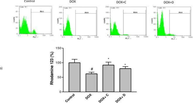
Effects of the selected fractions on DOX- induced mitochondrial membrane potential (MMP) collapse
MMP was evaluated using a cell permeable cationic fluorescent dye. Depolarization of mito-chondrial membrane potential caused by the DOX-induced damage of the outer membrane resulted in the loss of the dye from the mitochondria and a decrease in intracellular fluorescence, so that DOX
. ± . significantly decreased MMP to . %
of control in PC12 cells. Pretreatment of cells with
the active fractions was able to inhibit reduction of MMP induced by DOX (Figure 3).
Effect of the selected fractions on caspase-3 activity
Activation of caspase cascade is critical in the initiation of apoptosis in various biological systems. Caspase-3 has been shown to be an important regulator of apoptosis (24). The obtained results showed that DOX significantly increased caspase-3 activation in PC12 cells to 137.70±5.88% compared to the control cells. Pretreatment with fractions C and D decreased significantly caspase-3 activation to 81.16±3.64% and 71.42±2.91%, respectively (Figure 4).
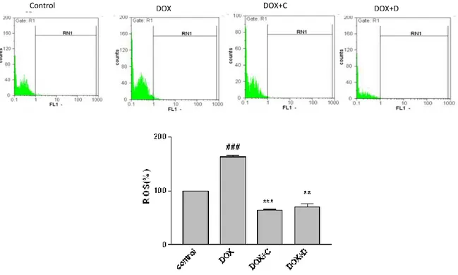
Effect of the selected fractions on DOX-induced ROS generation
Figure 3. Effect of fractions C and D (25 µg/ml) of aerial parts of Artemisia biennis hydroethanolic extract on DOX (2.5 µg/ml) -induced mitochondrial membrane potential (MMP) collapse as detected by Rhodamine 123 using flowcytometry. a) Representative of flow cytometry with rhodamine 123 plots of PC12 cells in different groups. b) Column bar graph of mean cell florescent for rhodamine 123. Data are expressed as the mean± SEM of three separate experiments. # P <0.05 vs. Control, * P<0.05 vs. DOX treated cells
Figure 4. Effect of fractions C and D on caspase-3 activity. Cell pretreated with fractions (25 µg/ml) 24 hr before exposure to 2.5 µM of DOX. Caspase-3 activity was measured by colorimetric detection of p-nitroaniline and expressed as percent of control. Data are expressed as the mean± S.E.M of three separate experiments. ## P<0.01 vs. Control, *** P<0.001 vs. DOX treated cells
Effect of the selected fractions on the activity of SOD
Reactive oxygen species (ROS) has important role in the activation of apoptosis pathway. SOD as one of the components of intrinsic antioxidant defense
system, is responsible for dissemination of
superoxide anion radicals (22). Therefore, we next tested the activity of SOD in the PC12 cells. As shown in Figure 5, after exposure to DOX, the activity of SOD decreased significantly (P<0.05) when compared with the control. Treatment with two selected fractions markedly increased the activity of SOD when compared with DOX-incubated cells under our
experimental conditions. This result demonstrates
that fractions C and D improve the activity of SOD leading to suppression of apoptosis of PC12 cells in the presence of DOX (Figure 6).
Discussion
Evidence from the current investigation suggested that DOX could decrease the cell viability in the PC12 cells. DOX significantly increased intracellular ROS, and also inhibited SOD activity which may eventually lead to cytotoxicity in PC12 cells. Additionally, our results
indicated that DOX- induced cytotoxicity is mainly
executed by apoptosis. The involvement of mito-chondria in DOX-induced apoptosis was investigated by evaluating the loss of the mitochondrial membrane potential. Results indicated that the stimulation of the intrinsic mitochondrial apoptotic pathway by ROS causes an increase in outer membrane permeabiliza-tion and mitochondria-to-cytosol trans-locapermeabiliza-tion of cytochrome c, AIF, or Smac/Diablo which trigger caspase-dependent or caspase-independent cytosolic signaling events (25). Caspases play an important role in the apoptotic process via two pathways: the death receptor pathway and the mitochondrial pathway. Regardless of the pathway involved, caspase-3 acts as an apoptotic executor. Free radical-mediated oxidative stress has been suggested as a main mechanism causing DOX- induced apoptosis in the brain (22). Previous studies show that DOX treatment promotes lipid peroxidation in the brain, heart, liver, lung, and kidney tissues (3, 26). A similar study using rat brain
mitochondria demonstrated the role of oxidative
Control DOX DOX+C DOX+D
Cont
rol DOX
DOX+
C
DOX+
D
0 50 100 150
#
R
h
o
d
am
in
e
12
3
(%
)
*
* b)
Figure 5. Effect of fractions C and D of aerial parts of Artemisia biennis hydroethanolic extract on DOX–induced ROS overproduction as detected by DCF using flowcytometry. Cells were pretreated with fractions C and D (25 µg/ml) 24 hr before exposure to 2.5 µM of DOX. a) Representative of flowcytometry with DCF-DA plots of PC12 cells in different groups. b) Column bar graph of mean cell florescent for DCF- DA. Data are expressed as the mean± SEM of three separate experiments. ### P<0.0_1 vs. Control, ** P <0.01, *** P <0.001 vs. DOX treated cells
stress in DOX-induced toxicity andsuggested that DOX treatment increases the brain mitochondria permeability to Ca2+ thereby predis-posing brain cells to degeneration and death (7). In another study, the administration of DOX decreased MMP, disturbed the balance of Bcl-2 family protein, increased cytochrome c release and ultimately led to apoptotic cell death in the brain tissue (27). Furthermore, Oz and Ilhan (2006) reported that DOX-treated rats undergoing co-treatment with melatonin, a free radical scavenger and antioxidant compound, showed lower levels of lipid peroxidation in the kidney, lung, liver, and brain (28). Accordingly, evidence from the current investigation suggested that ROS plays a significant role in DOX-induced apoptosis in this model. When the protective effect of seven fractions on DOX-induced cytotoxicity in PC12 cells was examined, it was observed that the pretreatment of PC12 cells with sub-toxic
concentration of fractions C and D markedly protected the cells from DOX-induced cytotoxicity. A marked increase in the SOD activity and significant reduction of intracellular ROS indicated that fractions C and D may protect the PC12 cells from oxidative injury by preventing increased oxidative stress. Moreover, our results indicated that selected fractions prevent DOX-induced collapse of mito- chondrial membrane potential in PC12 cells. It is not
Figure 6.Effect of fractions C and D of aerial parts of Artemisia
biennis hydroethanolic extract on the activity of SOD. Cells were pretreated with fractions C and D (25 µg/ml) 24 hr before exposure to 2.5 µM of DOX. SOD activity was measured using a colorimetric assay kit, and activity was represented as the percent inhibition of the superoxide anions. ## P<0.01 vs. Control, **P<0.01, *** P <0.001 vs. DOX treated cells
surprising that in our experiments, pretreatment
with potent fractions was also associated with
the inhibition of downstream apoptosis signaling
pathways, ultimately reducing effector caspase-3 activity. It was previously reported that the fraction D of A. biennis hydroethanolic extract had the highest total phenolic content compared with other fractions . The same fraction showed the most potent antioxidant effects using free radical scavenging methods while chelating activity of fraction C for
Control DOX+C
con trol
DO X
DO X+
C
DO X+
D
0 10 20 30
*** **
##
S
O
D
a
c
ti
v
it
y
(
%
)
ferrous ions was superior to that of fraction D (19). Naturally occurring phenolic compounds are
regarded as potential neuroprotective agents. New isolated phenolic compounds from the rhizomes of
Gastrodia elata exhibited potent neuroprotective activity against the H2O2-induced PC12 cell damage (29). Two well-known flavonoids, baicalein
and daidzein, have been reported to exert
neuroprotective effects against β-amyloid Aβ via inhibitory effects on in-vitro Aβ aggregation and Aβ -induced cytotoxicity in PC12 neuronal cells (30). Isolated coumarin glycosides from the stems of
Hydrangea paniculata presented neuroprotective activity against the serum deprivation-induced PC12 cell damage (31). In summary, our findings, which agree with previous results for the same samples (19), imply that some fractions of the hydroethanolic extract of A. biennis attenuate oxidative stress injury and apoptosis induced by DOX in the PC12 cells. However, further studies are necessary to isolate and elucidate the structures of the active components and to determine explicit neuroprotective mechanisms before definite conclusions can be drawn.
Conflict of Interests
The authors declare that there is no conflict of interests regarding the publication of this paper.
Acknowledgment
The results presented here are extracted from the Pharm. D. thesis of M Mehrabi. This study was financially supported by the Research Council of
Kermanshah University of Medical Sciences,
Kermanshah, Iran.
References
1. Herrera B, Murillo MM, Alvarez-Barrientos A, Beltrn J, Fernndez M, Fabregat I. Source of early reactive oxygen species in the apoptosis induced by transforming growth factor-(beta) in fetal rat hepatocytes. Free Rad Biol Med 2004; 36:16-26. 2. Circu ML, Aw TY Reactive oxygen species, cellular redox systems, and apoptosis. Free Rad Biol Med 2008; 48:749-762.
3. Conklin KA. Chemotherapy-associated oxidative stress: impact on chemotherapeutic effectiveness. Integr Cancer Ther 2004; 3:294-300.
4. Hosseinzadeh L, Behravan J, Mosaffa F, Bahrami G, Bahrami A, Karimi G. Curcumin potentiates doxorubicin-induced apoptosis in H9c2 cardiac muscle cells through generation of reactive oxygen species.
Food Chem Toxicol 2011; 49:1102–1109.
5. Lopes AM, Meisel A, Dirnagl U, Carvalho FD, Bastos Mde L. Doxorubicin induces biphasic neurotoxicity to rat cortical neurons. Neurotoxicology 2008; 29:286-293. 6. Tangpogon J, Miriyala S, Noel T, Sinthupibulyakit C, Jungsuwadee P, St Clair DK. Doxorubicin induced central nevous system toxicity and protection by
xanthone derivatives of Garcinia mangostana.
Neuroscience 2011; 175:292–299.
7. Cardoso S, Santos RX, Carvalho C, Correia S, Pereira G, Pereira S, Oliveira P, Santos MS, Proença T. Doxorubicin increases the susceptibility of brain
mitochondria to Ca2+-induced permeability
transition and oxidative damage. Free Radic Biol
Med 2008; 45:1395–1402.
8. Karlsson M, Kurz T, Brunk UT, Nilsson SE,
Frennesson C). What does the commonly used DCF
test for oxidative stress really show? Biochem J
2010; 428:183–190.
9. Janelsins MC, Roscoe JA, Berg MJ, Thompson BD,
Gallagher MJ, Morrow GR, et al. IGF-1 partially
restores chemotherapy-induced reductions in neural cell proliferation in adult C57BL/6 mice. Cancer
Invest 2010; 28:544–553.
10. Kesler s, Janelsins M, Koovakkattu D, Palesh O,
Mustian K, Morrow G, et al. Reduced hippocampal
volume and verbal memory performance associated with interleukin-6 and tumor necrosis factor-alpha levels in chemotherapy-treated breast cancer
survivors. Brain Behav Immun 2013; 30:S109–S116.
11. Mozaffarian V. A Dictionary of Iranian Plant Names. Tehran: Farhang Moaser Publishers; 1978. 12. Nematollahi F, Rustaiyan A, Larijani K, Nadimi M.
Essential oil composition of Artemisia biennis Willd
and Pulicariaundulata (L.) C.A. Mey., two Compositae herbs growing wild in Iran. J Essent Oil Res 2006;
18:339–341.
13. Lopes-Lutz D, Alviano DS, Alviano CS,
Kolodziejczyk PP. Screening of chemical composition, antimicrobial and antioxidant activities of Artemisia essential oils. Phytochemistry 2008; 69:1732-1738. 14. Emami A, Zamani Taghizadeh Rabe SH, Ahi A, Mahmoudi M. Study on toxic effects of Artemisia spp. fractions from Iran on human cancer cell lines. J Zanjan Univ Med Sci 2010; 18:58-67.
15. Mojarrab M, Naderi R, Heshmati Afshar F. Screening of different extracts from Artemisia species for their potential antimalarial activity. Iran J Pharm Res 2015: 14:603-608.
16. Emami A, Zamani Taghizadeh Rabe SH, Ahi A, Mahmoudi M. Inhibitory activity of eleven Artemisia species from Iran against Leishmania major parasites. Iran J Basic Med Sci 2012; 15: 807-811. 17. Iranshahi M, Emami SA, Mahmoud-Soltani M. Detection of sesquiterpene lactones in ten Artemisia species population of Khorasan provinces. Iran J Basic Med Sci 2007; 10:183-188.
18. Singh KN, Lal B. Ethnomedicines used against four common ailments by the tribal communities of Lahaul-Spiti in Western Himalaya. J Ethnopharmacol 2008; 4; 115:147-159.
19. Hatami T, Emami SA, Miraghaee SS, Mojarrab M. Total phenolic contents and antioxidant activities of different extracts and fractions from the aerial parts
of Artemisia biennis Willd. Iran J Pharm Res 2014;
13:551–559.
20. Hatanaka H. Nerve growth factor-mediated stimulation of tyrosine hydroxylase activity in a clonal rat pheochromocytoma cell line. Brain Res
1981; 222:225–233.
21. Rebois RV, Reynolds E, Toll L, Howard BD. Storage of dopamine and acetylcholine in granules of PC12, a clonal pheochromocytoma cell line.
. Klaassen CD. Casarett & Doull’s Toxicology. th ed. McGraw Hill; 2007.p.30- 31.
23. Malstorm B, Andreasson L, Reinhammer B. The enzymeas. Boyer; 1975.p.533.
24. Hosseinzadeh L, Khorand A, Aliabadi A.
Discovery of 2- Phenyl –N-(5-(trifluoromethyl) – 1, 3,
4- thiadiazol-2-yl) acetamid derivatives as apoptosis inducer via caspase pathway whit potential anticancer activity. Arch Pharm Chem Life 2013;
346:812–818.
25. Shokoohinia Y, Hosseinzadeh L, Alipour M, Mostafaie A, Mohammadi-Motlagh HR. Comparative evaluation of cytotoxic and apoptogenic effects of several coumarins on human cancer cell lines: osthole induces apoptosis in p53-deficient H1299 cells. Adv Pharmacol Sci 2014; 2014:847574. 26. Shokoohinia Y, Hosseinzadeh L, Moieni-Arya M, Mostafaie A, Mohammadi-Motlagh HR. Osthole attenuates doxorubicin-Induced apoptosis in PC12 cells through inhibition of mitochondrial dysfunction and ROS production. Biomed Res Int 2014; 2014:156848. 27. Pal S, Ahir M, Sil PC. Doxorubicin-induced
neurotoxicity is attenuated by a 43-kD protein from
the leaves of Cajanus indicus L. via NF-κB and
mitochondria dependent pathways. Free Radic Res 2012; 46:785-798.
28. Oz E, Ilhan. Effects of melatonin in reducing the toxic effects of doxorubicin. Mol Cell Biochem 2006; 286:11-15.
29. Zhang ZC, Su G, Li J, Wu H, Xie XD. Two new
neuroprotective phenolic compounds from
Gastrodiaelata. J Asian Nat Prod Res 2013; 15:619-623.
30. Choi RC, Zhu JT, Yung AW, Lee PS, Xu SL, Guo AJ,
et al. Synergistic action of flavonoids, baicalein, and
daidzein in estrogenic and neuroprotective effectspp A development of potential health products and therapeutic drugs against alzheimer's disease. Evid Based Complementary Altern Med 2013; 63:56-94. 31. Shi J, Li CJ, Yang JZ, Yuan YH, Chen NH, Zhang DM. Coumarin glycosides and iridoid glucosides with
neuroprotective effects from Hydrangea paniculata.