Article
J. Braz. Chem. Soc., Vol. 22, No. 7, 1279-1285, 2011. Printed in Brazil - ©2011 Sociedade Brasileira de Química
0103 - 5053 $6.00+0.00
A
*e-mail: [email protected]
Metabolites from Roots of
Colubrina greggii
var.
yucatanensis
and Evaluation of
their Antiprotozoan, Cytotoxic and Antiproliferative Activities
Dafne B. Domínguez-Carmona,a Fabiola Escalante-Erosa,a Karlina García-Sosa,a Grace Ruiz-Pinell,b David Gutierrez-Yapu,b Manuel J. Chan-Bacab,c Rosa E. Moo-Puc,d
Nigel C. Veitch,e Alberto Giménez-Turbab and Luis M. Peña-Rodríguez*,a
aUnidad de Biotecnología, Centro de Investigación Cientíica de Yucatán.
Calle 43, N. 130, Col. Chuburná, Mérida, Yucatán, 97200 México
bInstituto de Investigaciones Fármaco-Bioquímicas, Universidad Mayor de San Andrés,
Av. Saavedra 2224, La Paz, Bolivia
cDepartamento de Microbiología Ambiental y Biotecnología, Universidad Autónoma de Campeche,
Agustín Melgar s/n, Campeche, Campeche, México
dUnidad de Investigación Médica Yucatán, Unidad Médica de Alta Especialidad, Centro Médico
Ignacio García Téllez IMSS, Calle 41, N. 439, Col. Industrial, Mérida, Yucatán, 97150 México
eJodrell Laboratory, Royal Botanic Gardens Kew, Richmond, Surrey, TW9 3AB UK
A puriicação do extrato da raiz de Colubrina greggii var. yucatanensis levou ao isolamento e identiicação do ácido 3-O-acetil ceanótico, um novo triterpeno natural, juntamente com os metabólitos já descritos: ácido ceanótico, ácido cenoténico, ácido betulínico, discarina B e crisofaneína. Os produtos naturais e os derivados semi-sintéticos éster de acetil dimetil ceanotato, dimetil ceanotato e peracetato de crisofaneína mostraram moderada a baixa atividade leishmanicida e tripanocida. Nenhum dos metabólitos mostrou ser citotóxicos ou ter atividade antiproliferativa. Os resultados também sugerem que o ácido betulínico contribui para a atividade antiplasmódica inicialmente detectada na raiz do extrato bruto de C. greggii var. yucatanensis.
Puriication of the root extract of Colubrina greggii var. yucatanensis resulted in the isolation and identiication of 3-O-acetyl ceanothic acid as a new natural ceanothane triterpene, together with the known metabolites ceanothic acid, cenothenic acid, betulinic acid, discarine B and chrysophanein. The natural products and the semisynthetic esters acetyl dimethyl ceanothate, dimethyl ceanothate and chrysophanein peracetate showed moderate to low leishmanicidal and trypanocidal activities. None of the metabolites showed cytotoxic or antiproliferative effects. The results also suggested that betulinic acid contributes to the antiplasmodial activity originally detected in the crude root extract of C. greggii var. yucatanensis.
Keywords:Colubrina greggii var. yucatanensis, Rhamnaceae, antiprotozoan, cytotoxic ceanothane
Introduction
Leishmaniosis, trypanosomiosis and malaria are a group of protozoan diseases considered of signiicant importance due to their incidence and rate of mortality in developing countries.1 The low effectiveness, limited availability and high
toxicity of existing treatments for these illnesses emphasize
the importance of continuing the search for new antiprotozoan pharmaceuticals.2 Plants are considered an important source
of biologically active natural products,3 including a number
of them with antiprotozoan activity.4-6 During the screening
Metabolites from Roots of Colubrina greggii var. yucatanensis J. Braz. Chem. Soc.
1280
trypanocidal, antimalarial, leishmanicidal and cytostatic activity.8,9 Previous phytochemical studies of the genus
Colubrina (Rhamnaceae), which include 31 species,10
reported the presence of a wide structural diversity of metabolites, including ansa macrolides, saponins, aporphinic alkaloids, phenolic acids, lavones and triterpenoid acids.11-18
To date, only chrysophanol, an anthraquinone with antimicrobial activity, has been reported as a metabolite from C. greggii.19 In the present study we report the leishmanicidal,
trypanocidal, antiplasmodial, cytotoxic and antiproliferative activity of the crude extract, semi-puriied fractions and isolated metabolites from the root extract of C. greggii var. yucatanensis.
Results and Discussion
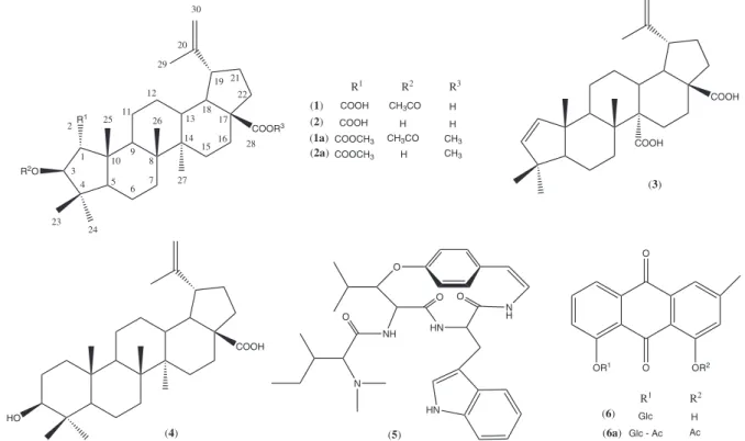
Fractionation of the bioactive root extract of C. greggii var. yucatanensis yielded a low polarity fraction with leishmanicidal, trypanocidal and antiplasmodial activities and a medium-polarity fraction with leishmanicidal activity. Successive puriication of the medium-polarity fraction resulted in the isolation of 3-O -acetyl-ceanoth-20(30)-en-1,28-dioic acid (3-O-acetyl ceanothic acid) (1) as a new natural ceanothane triterpene, together with the known metabolites ceanothic acid (2), discarine B (5) and chrysophanein (6). Similarly, puriication of the low-polarity fraction led to the isolation and identiication of ceanothenic acid (3) and betulinic acid (4) (Figure 1).
The FTIR spectrum of 1 showed absorption bands at 3073 (CH=C), 1731 (ester) and 1681 cm-1 (carboxyl). The pseudo
molecular ion peak [M + Na]+ at m/z 551.3524 in HRMS (high
resolution mass spectrometry) indicated a molecular formula C32H48O6, implying nine degrees of unsaturation. Carbon
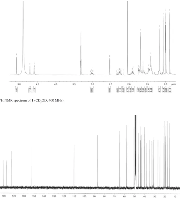
multiplicity, deduced from HMQC (heteronuclear multiple quantum coherence) and DEPT (distortionless enhancement by polarization transfer) experiments, indicated the presence of seven methyl groups, nine methylene, seven methine, and nine quaternary carbons. The 1H NMR spectrum of 1 (Table S1,
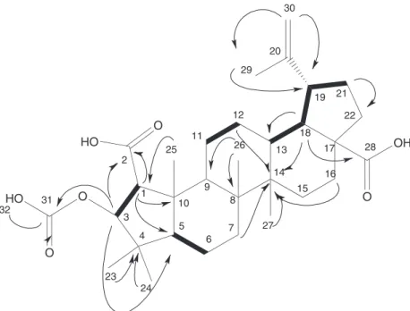
Figure S1) displayed seven three-proton singlets at d 0.88, 0.98, 0.99, 1.06, 1.16, 1.68 and 2.03 consistent with methyl groups attached to quaternary carbons. The presence of an acetyl group was conirmed by the HMBC (heteronuclear multiple bond coherence) correlation of the methyl at d 2.03 with the carbonyl at d 172.5, whereas an isopropenyl group was assigned from NMR signals corresponding to a methyl group (dΗ1.68) attached to a sp
2-carbon (d
C 152.0) showing
HMBC correlations with two vinylic protons at dΗ 4.58
and d4.70. The double bond and three carbonyl groups observed in the 13C NMR of 1 (d 172.5, 177.4 and 180.1)
accounted for four degrees of unsaturation thus indicating that the ive remaining unsaturation sites corresponded to a pentacyclic structure. The spectroscopic data of 1 proved to be very similar to those reported for ceanothic acid (2), a ceanothane triterpene also known as emmolic acid, originally isolated from Ceanothus americanus20 and later identiied
from Colubrina granulosa.13 However, the presence of a
Figure 1. Structures of natural products 1-6 isolated from C. greggii, and semisynthetic derivatives 1a, 2a and 6a.
COOR3
R2O R1
(2) (1) (1a) (2a)
COOH CH3CO COOH
H COOCH3 CH3CO COOCH3 H
H H
CH3
R1 R2 R3
CH3 1
3 4 2
10 9 8
7 6 5 11 12 23 24 25 26 27 13 14 15 16 17 18 19 20 29 30 21 22 28 COOH COOH
(3)
COOH
HO
(4)
O N H HN O HN NH N O O
(5)
OR1 O
O
OR2
(6) Glc
Glc - Ac (6a)
R1 R2
H
low-ield carbinol proton (d 5.07) and an acetyl methyl singlet (d 2.03) in the 1H NMR spectrum of 1, both showing strong
HMBC correlations with the ester carbonyl carbon (d 172.5), suggested that 1 was the 3-O-acetyl derivative of ceanothic acid, which was conirmed when acetylation of 2 produced
1 as the only product. The presence of 1 in the original root extract of C. greggii, as detected by TCL (Figure S2), ruled out its being an artifact of the isolation procedure.
Discarine B (5), chrysophanein (6) and ceanothenic acid (3) were identified from comparison of their spectroscopic data (Tables S1, S2 and S3) with those reported in literature.21-23 Betulinic acid (4) was identiied
by comparison with an authentic sample.24
All of the isolated metabolites and the semisynthetic esters acetyl-dimethyl ceanothate (1a), dimethyl ceanothate (2a) and chrysophanein peracetate (6a) were evaluated for their in vitro antiprotozoan (leishmanicidal, trypanocidal, and antiplasmodial), cytotoxic and antiproliferative activities (Table 1). The results showed a moderate leishmanicidal activity (IC50 values of 20-28 µg mL
-1) for
natural ceanothanes 1 and 3 and semisynthetic derivatives
2a and 6a, whereas a low trypanocidal activity (IC50 of
30-70 µg mL-1) was observed for the natural products 3-5
and the semisynthetic esters 1a and 6a. Betulinic acid (4) appeared to contribute to the antiplasmodial activity of the crude extract of C. greggii, with an IC50 of 9.7 µg mL
-1.25,26
Although the crude root extract, together with the low and medium polarity fractions showed cytotoxic activity against HEp-2 cells, none of the isolated metabolites displayed this type of activity (Table S4). Furthermore, none of the metabolites tested, with the exception of the peracetylated chrysophanein (6a), showed antiproliferative activity against KB cells (Table S5). It is interesting to point out that the cytotoxic activity of C. macrocarpa and C. texensis has been attributed to the presence of colubrinol and its acetate,11,27 however these metabolites were not
detected in the root extract of C. greggii.
To date, ceanothane triterpenes have only been reported to occur in species of the Rhamnaceae family,20,23,28-36 and
particularly in those belonging to the ziziphoids in the tribal classiication reported by Richardson et al.38 These
results, together with our inding of ceanothanes in the root extract of C. greggii, support the possible use of this class of triterpenes as chemotaxonomic markers for a classiication of Rhamnaceae based on a phylogenetic analysis.
Experimental
General
Analytical TLC (thin layer chromatography) was carried out on aluminum-backed silica gel (60F254) plates
Table 1.Leishmanicidal, trypanocidal, antiplasmodial and cytotoxic activity [IC50 (µg mL-1)] of crude extract, semipuriied fractions, natural products 1-6
and semisynthetic derivatives 1a, 2a and 6a
Extract/ Compound
Antiprotozoan activity
L. amazonensis T. cruzi tulahuen P. falciparum VERO
IC50 SI IC50 SI IC50 IC50
CG-1 32.4 - > 100 - 8.0 NT
CG-2A < 25 - 73.9 - 4.5 NT
CG-2B < 25 - > 100 - > 10 NT
1 28.2 ± 2.7 3.6 > 100 - > 10 103.1 ± 1.8
2 46.4 ± 8.4 2.8 > 100 - NT 131.2 ± 3.2
1a 40.2 ± 9.8 - 56.2 ± 5.2 - NT NT
2a 20.6 ± 6.8 - > 100 - NT NT
3 22.1 ± 2.7 4.4 64.0 ± 4.3 1.5 NT 98.6 ± 1.2
4 > 100 - 34.2 ± 11.1 4.2 9.7 145.0 ± 2.9
5 > 100 - 56.2 ± 3.2 3.5 > 10 199.1 ± 2.7
6 > 100 - 56.7 ± 19.5 9.1 > 10 521.0 ± 6.3
6a 12.7 ± 1.1 9.7 65.3 ± 9.0 1.8 > 10 123.8 ± 4.2
PTM 10.0 ± 0.8 - - - - NT
BZD - - 7.4 ± 0.5 - - NT
CLQ - - - - 0.1 ± 0.02 NT
Metabolites from Roots of Colubrina greggii var. yucatanensis J. Braz. Chem. Soc.
1282
(E.M. Merck, 0.2 mm thickness) and the chromatograms visualized using a solution of phosphomolybdic acid (20 g) and ceric sulfate (2.5 g) in 500 mL of sulfuric acid (5%). Flash chromatography puriications were performed using silica gel (Aldrich, 200-400 mesh), while TLC-grade silica gel 60GF254
(E.M. Merck) was used for vacuum liquid chromatography (VLC). Prep-TLC puriications were carried out using glass plates impregnated with silica gel 60 F254 (E.M. Merck,
0.25 mm thickness, 20 × 20 cm). Melting points (uncorrected) were determined from a Mel-Temp II apparatus (Laboratory Devices Inc.). The optical rotations were measured in CHCl3
using a Perkin Elmer 341 polarimeter. FTIR (Fourier transform infrared) spectra were recorded in CHCl3 or MeOH (ilm)
using an FT-Nicolet Magna Protégé 460 spectrophotometer.
1H NMR (400 and 600 MHz) and 13C NMR (100 and
150 MHz) spectra were acquired on a Bruker Avance 400 spectrometer or a Bruker Avance 600 spectrometer with CHN cryoprobe, using the residual solvent resonances as internal references, calibrated to TMS. Electrospray high-resolution mass spectra (ESI-HRMS) were determined by direct injection on a Waters Q-TOF microsystem (using 0.1% phosphoric acid in a 1:1 water/acetonitrile mixture as reference), or using an Orbitrap MS (Thermo) connected to a Surveyor HPLC (high-performance liquid chromatography, Thermo) for sample injection.
Plant material
Roots of Colubrina greggii S. Watson var. yucatanensis M. C. Johnst. were collected in Abalá, Yucatán, México. A voucher specimen (P. Simá-D. Domínguez 2916) was deposited in the Herbarium of Centro de Investigación Cientíica de Yucatán.
Extraction of plant material and puriication of crude extract
Dry roots of C. greggii (365 g) were extracted three times with ethanol (4 L) at room temperature. Evaporation of the solvent yielded the corresponding crude extract (CG-1, 54.5 g, 14.9%), which was suspended in 1.8 L of a H2O/MeOH
(3:2, v/v) mixture. The suspension was fractionated by successive liquid-liquid partition with hexane (three times; 2:1, 1:1, 1:1; v:v of solvent:aqueous suspension), ethylacetate (three times; 2:1, 1:1, 1:1) and water-saturated butanol (1:2; v:v solvent:aqueous suspension) to yield the corresponding low (CG-2A), medium (CG-2B) and high (CG-2C) polarity fractions. Puriication of the medium polarity fraction (CG-2B, 9.6 g) by VLC, using a gradient elution with CH2Cl2/Me2CO (99:1 to 94:6) followed
by CHCl3/hexane/MeOH (70:25:5), yielded fractions
CG-5A-N. Fraction CG-5E (550 mg) was puriied by lash
chromatography using an isocratic elution with hexane/ Me2CO 8:2, to produce pure 3-O-acetyl-ceanothic acid (1,
172.1 mg), and fraction CG-6I (50 mg) which was further puriied by column chromatography [ether/hexane (1:1)] to yield ceanothic acid (2, 16.5 mg). Additional puriication of fraction CG-5C (414 mg) by lash chromatography, using a gradient elution with hexane/Me2CO (8:2-7:3), resulted in the
isolation of discarine B (5,108 mg). Finally, chrysophanein (6, 168.5 mg) was collected as a yellow precipitate by iltration from fraction CG-5G. Puriication of the low polarity fraction (CG-2A, 1.5 g) by VLC, using a gradient elution with mixtures of hexane/Me2CO/MeOH (95:3:2 to
60:38:2), produced eleven fractions (CG-3A-K). Fractions CG-3E-F were combined (500 mg) and puriied by lash chromatography (hexane/ether 7:3), to yield ceanothenic acid (3, 20.8 mg) and betulinic acid (4, 20.4 mg).
3-O-acetyl-ceanothic acid (1)
White amorphous solid; mp: 271.5-273.1 °C; [α]
D
20 +33.7° (c 0.01, Me
2CO); FTIR (ilm) νmax cm -1: 3073
(CH=C), 1731 (ester), 1685 (carboxyl); 1H NMR (CD 3OD,
400 MHz) and 13C NMR (CD
3OD, 100 MHz) data: see
Table S1; ESI-HRMS m/z 551.3524 [M + Na]+ (calc. for
C32H48NaO6: 551.3584).
Ceanothic acid (2)
White amorphous solid; [α]D20 + 30.8° (c 0.003,
MeOH); FTIR (ilm) ν
max cm
-1: 3411 (OH), 1697 (>C=O);
1H NMR (CD
3OD, 400 MHz): d 0.88 (s, 3H, H-24), 0.90
(s, 3H, H-26), 0.97 (s, 3H, H-27), 1.01 (s, 3H, H-25), 1.35 (s, 3H, H-23), 1.67 (s, 3H, H-29), 2.45 (s, 1H, H-1), 3.06 (m, 1H, H-19), 4.06 (s, 1H, H-3), 4.57 (br s, 1H, H-30a), 4.69 (br s, 1H, H30-b); ESI-HRMS m/z 487.3418 [M + H]+
(calc. for C30H47O5: 487.3423).
3-O-acetyl-dimethyl-ceanothate (1a)
A mixture of 1 (5.1 mg), K2CO3 (80 mg), CH3I
(300 µL) and acetone (1 mL) was stirred for 72 h at room temperature. The reaction mixture was poured over distilled water (14 mL) and the resulting suspension was extracted twice with EtOAc (4:1, v/v). The organic layer was dried over anhydrous Na2SO4 and evaporated to produce 4.6 mg
of the crude esteriied product, which was puriied by column chromatography (hexane/Me2CO 9:1) to give 1a
(4.2 mg, 91.3% yield) as a white powder. 1H NMR (CDCl 3,
600 MHz) and 13C NMR (CDCl
3, 150 MHz) data: see
Table S1; LR-MS m/z 557 [M + H]+ .
Dimethyl-ceanothate (2a)
(800 µL) and acetone (1 mL), and then stirred for 72 h at room temperature. The reaction mixture was poured over distilled water (13 mL) and the resulting suspension was extracted twice with EtOAc (4:1, v/v). The organic phase was dried over anhydrous Na2SO4 and evaporated to
produce 9.6 mg of the crude esteriied product, which was puriied by multiple-elution (5 ×) prep-TLC (hexane/ether 7:3) to give 5.4 mg of 4 (56.2%) as a white solid. 1H NMR
(CD3OD, 400 MHz) and 13C NMR (CDCl3, 100 MHz) data:
see Table S1; ESI-HRMS m/z 515.3731 [M + H]+ (calc. for
C32H51O5: 515.3736).
Ceanothenic acid (3)
White powder; FTIR (ilm) ν max/cm
-1:3067 (CH=C),
1721 (carboxyl), 1685 (carboxyl); 1H NMR (CDCl 3/
CD3OD 9:1, 400 MHz) and
13C NMR (CDCl
3/CD3OD 9:1,
100 MHz) data: see Table S1; ESI-HRMS m/z 455.3156 [M + H]+ (calc. for C
29H43O4: 455.3161).
Betulinic acid (4)
Colorless needles; ESI-HRMS m/z 440.3690
[M-H2O + 2H]
.+ (calc. for C
30H48O2: 440.3654).
Discarine B (5)
White amorphous solid; νmax/cm-1:3267 (NH), 3027
(CH=C), 1634 (>C=O); 1H NMR (CD
3OD, 30 °C,
400 MHz) and 13C NMR (CD
3OD, 30 °C, 100 MHz) data:
see Table S3; ESI-HRMS m/z 574.3393 [M + H].+ (calc.
for C33H44N5O4: 574.7336).
Chrysophanein (6)
Yellow powder; FTIR (ilm) ν max/cm
-1:3344 (OH), 1634
(>C=O); 1H NMR (DMSO-d
6, 400 MHz) and 13C NMR
(DMSO-d6, 100 MHz) data: see Table S2; ESI-HRMS m/z 255.2302 [M-C6H10O5 + H]+ (calc. for C15H11O4: 255.0657).
Chrysophanein peracetate (6a)
A mixture of 6 (10 mg), acetic anhydride (1 mL) and pyridine (0.5 mL) was stirred at room temperature for 72 h. The reaction mixture was poured over distilled water (20 mL) and the resulting suspension was extracted twice with ethylacetate (2:1 v/v). The organic layer was washed successively with equal volumes of HCl (5%), NaOH (3%), H2O, and NaCl saturated, and then dried over anhydrous
MgSO4. Filtration and evaporation of the solvent yielded
13.9 mg (92.4%) of crude acetylated product obtained as a yellow powder; FTIR (ilm) νmax cm-1:3021 (CH=C),
1757 (ester), 1680 (>C=O); 1H NMR (CDCl
3, 400 MHz)
and 13C NMR (CDCl
3, 100 MHz) data: see Table S2;
ESI-HRMS m/z 649.1499 [M + Na]+ (calc. for C
31H30NaO14:
649.1533).
Bioassays
Leishmanicidal assay
The growth inhibition of promastigotes was carried out following the procedure previously reported by Muñoz et al.,39 and Inchausti et al.40 Briely, a strain of L.
amazonensis (IFLA/BR/75/PH8) was grown in Schneider culture medium with 10% fetal bovine serum (FBS), penicillin (100 IU mL-1) and streptomycin (100 mg mL-1) at 25 °C;
parasites in the log phase of their growth cycle were then transferred to a microplate (96 wells; 1 × 10⁵ parasites/well).
Stock solutions of DMSO (blank), pentamidine (positive control), crude extract, semipurified fractions and pure metabolites were diluted in Schneider medium at ≤ 100 µg mL-1, added to the plate, and incubated for 72 h.
The percentages of inhibition were obtained by directed observation of each well with an inverted phase microscope. All the assays were carried out in triplicate.
Trypanocidal assay
Epimastigotes of Trypanosoma cruzi strain Tulahuen parasites were maintained in liver infusion tryptose (LIT) medium supplemented with 5% FBS, following the procedure modiied by Chataing et al.41 Briely, parasites in the log phase
of growth cycle were transferred to a microplate (96 wells; 1 × 10⁶ parasites/well) together with stock solutions of
benznidazole, DMSO (positive control and blank respectively), extract, semipuriied fractions or pure metabolites prepared at different concentrations (≤ 100 µg mL-1). The microplates
were incubated at 26 °C for 72 h.
Antiplasmodial assay
Plasmodium falciparum strain F32 was grown at 37 °C in RPMI medium with 10% of human serum and 4% of hematocrit (O, Rh+), under anaerobic conditions, according to a reported method.41 Cultures with 1% parasitemic and
2% hematocrite (100 µL) were transferred to a 96 well plate. Stock solutions of chloroquine (positive control), DMSO (blank), extract, semipuriied fractions or pure metabolites were diluted in RPMI medium to a concentration of < 10 µg mL-1 and added to each well. The plate was then
incubated at 37 °C for 48 h.
Cytotoxicity assay
Human laryngeal carcinoma (HEp-2), human cervical adenocarcinoma (HeLa), human nasopharyngeal carcinoma (KB), and green monkey Vero kidney cells (VERO) were grown in DMEM (Gibco) media supplemented with 10% (v/v) FBS (Gibco), penicillin (100 IU mL–1),
and streptomycin (100 mg mL-1). All the cell lines were
Metabolites from Roots of Colubrina greggii var. yucatanensis J. Braz. Chem. Soc.
1284
humidity. The cytotoxicity assay was performed according to a method described by Rahman et al.43 Briely, cell lines
were transferred to a microplate (1.5 × 10⁴ viable cells of
each cell line) and incubated at 37 °C, with 95% humidity
and 5% CO2 in DMEM medium supplemented with 10%
of FBS, penicillin (10000 IU), streptomycin (10 mg mL-1),
and amphotericine B (5 mg mL-1). After 24 h, the medium
was replaced by fresh medium with 0.05% DMSO (blank) or different concentrations of docetaxel (positive control, Sigma), crude extract, semipuriied fractions or pure metabolites dissolved in DMSO (100, 50, 25, 12.5 and 6.25 µg mL-1), and the cells were incubated for 72 h
under the conditions already described. The medium was removed and 200 µL of a 0.5% MTT (Sigma) solution in PBS (pH 7.2) were added to each well, and left to stand for 4 h at 37 °C. Then 100 µL of acidiied isopropanol (0.4 mol L-1 HCl) were added to each well and the optical
density (OD) measured at 540 nm using a Bioassay reader (Bio-Rad). The experiment was carried out in triplicate and each concentration was tested in duplicate.
Antiproliferative assay
The sulforhodamine B (SRB) assay was carried out according to the method reported by Rahman et al.,43
using DMEM medium with 10% FBS to induce cell proliferation. After 48 h of incubation, the medium was discarded and 100 µL of ice-cold 40% trichloroacetic acid (TCA, Aldrich) were added to fix the cells, incubating for 1 h at 4 °C. The cells were washed ive times with water, left to dry, and then 50 µL of SRB stain (10 mg 1% acetic acid, Sigma) were added to each well and left to stand for 30 min. Finally, the cells were washed with 50 mL 1% acetic acid, and rinsed four times with water. The OD was measured at 540 nm using an ELISA reader (Bio-Rad model 450). The experiment was carried out in triplicate.
Statistical analysis
Data were analyzed with commercial software (GraphPad 4.0, Software Inc., San Diego, CA). The dose– response curves (variable slope) to obtain the inhibitory concentration (in µg mL-1) of 50% of parasites (IC
50), the
growth inhibition of 50% of cells (IG50), and the cytotoxic
concentration of 50% of cells (CC50), were itted to the
algorithm: Y = Emin + [(Emax −Emin)/(1 + 10(log ED50−
log D) hill slope)].
Supplementary Information
Supplementary data (Figures S1-S2, Tables S1-S5) are available free of charge at http://jbcs.sbq.org.br as PDF ile.
Acknowledgements
The authors wish to thank to Paulino Simá for the identiication of plant material. This work was supported by Program CYTED (Projects X.5 and RIBIOFAR) and Project FOMIX-Yucatán (66262).
References
1. World Health Organization; The Global Burden Disease: 2004 update, WHO Library Cataloguing-in-publication data, 2004. http://www.who.int/healthinfo/global_burden_disease/2004_ report_update/en/index.html.
2. Ioset, J. -R.; Curr. Org. Chem.2008, 12, 643.
3. Fabricant, D. S.; Farnsworth, N. R.; Env. Health Perspect.2001,
109, 69.
4. Sepúlveda-Boza, S.; Cassels, B. K.; Planta Med.1996, 62, 98. 5. Chan-Bacab, M. J.; Peña-Rodríguez L. M.; Nat. Prod. Rep.
2001, 18, 674.
6. Saxena, S.; Pant, N.; Jain, D. C.; Bhakuni, R. S.; Curr. Sci.
2003, 85, 1314.
7. Mendieta, R. M.; Del Almo, S.; Plantas Medicinales del Estado de Yucatán,1st ed., Compañía Editorial Continental: Xalapa, Mexico, 1981.
8. Vera-Kú, B. M.; Evaluación de la Actividad Biológica en Plantas Medicinales Nativas de la Península de Yucatán,MSc Dissertation, Centro de Investigación Cientíica de Yucatán: Mérida, Yucatán, México, 2004.
9. Getti, G.; Durgadoss, P.; Dominguez-Carmona, D.; Quintal-Martin, Z.; Peraza-Sanchez, S.; Peña-Rodriguez, L. M.; Humber, D.; J. Parasitol.2009, 95, 456.
10. Johnston, M. C.; Brittonia1971, 23, 2.
11. Wani, M. C.; Taylor, H. L.; Wall, M. E.; J. Chem. Soc., Chem. Comm.1973, 390.
12. Wani, M. C.; Taylor, H. L.; Wall, M. E.; Tetrahedron Lett.1973, 4675. 13. Roitmain, J. N.; Jurd, L.; Phytochemistry1978, 17, 491. 14. Guinaudeau, H.; Seligmann, O.; Wagner, H.; Neszmely, A.;
Phytochemistry 1981, 20, 1113.
15. Baxter, R. L.; Walkinshaw, D.; Phytochemistry1988, 27, 2350. 16. Oulad-Ali, A.; Guillaume, D.; Weniger, B.; Jiang, Y.; Anton, R.;
Phytochemistry1994, 36, 445.
17. Elsohly, H. N.; Danner, S.; Li, X. C.; Nimrod, A. C.; Clark, A. M.;
J. Nat. Prod.1999, 62,1341.
18. Xing-Gong, L.; ElSohly, H. N.; Nimrod, A. C.; Clark, M. A.;
J. Nat. Prod.1999, 62, 674.
19. García-Sosa, K.; Villarreal-Alvarez, N.; Lübben, P.; Peña-Rodríguez, L. M.; J. Mex. Chem. Soc.2006, 50, 76.
20. Julian, P. L.; Pikl, J.; Dawson, R.; J. Am. Chem. Soc.1938, 60, 77. 21. Herz, W.; Falk, H.; Kirby, G. W.; Moore, R. E.; Tamm, C;
22. Kubo, I.; Murai, Y.; Soediro, I.; Soetarno, S.; Sastrodihardjo, S.; Phytochemistry 1992, 31, 1063.
23. De Mayo, P.; Starratt, A. N.; Can. J. Chem.1962, 49, 1632. 24. Mahato, S. B.; Kundu, A. P.; Phytochemistry1994, 37, 1517. 25. Duker-Eshun, G.; Jaroszewski, J.W.; Asomaning, W.A.;
Phytother. Res.2004, 18, 128.
26. Ziegler, H. L.; Franzyk, H.; Sairaianpour, M.; Tabatabai, M.; Tehrani, M. D.; Bagherzadeh, K.; Hägerstrand, H.; Stærk, D.; Jaroszewski, J. W.; Bioorg. Med.Chem.2004, 12, 119. 27. Suksamrarn, S.; Panseeta, P.; Kunchanawatta, S.; Distaporn, T.;
Ruktasing, S.; Suksamrarn, A.; Chem. Pharm. Bull.2006, 54, 535.
28. Popoca, J.; Aguilar, A.; Alonso, D.; Villarreal, M. L.;
J. Ethnopharmacol.1998, 59, 173.
29. Lee, S. -S.; Su, W. -C.; Liu, K. C.; J. Nat. Prod.1991, 54, 615 30. Li, X. C.; Cai, L.; Wu, C. D.; Phytochemistry1997, 46, 97. 31. Lee, S. –S.; Chen, W. –C; Huang, C. -F.; Su, Y.; J. Nat. Prod.
1998, 61, 1343.
32. Popoca, J.; Aguilar, A.; Alonso, D.; Villarreal, M. L.;
J. Ethnopharmacol.1998, 59, 173.
33. Eade, R. A.; Ellis, J.; Harper, P.; Simes, J. J. H.; Aust. J. Chem.
1973, 26, 831.
34. Lee, S. -S.; Lin, B. F.; Liu, K. C. S.; Phytochemistry1996, 43, 847.
35. Lee, S. -S.; Shy, S. N.; Liu, K. C. S.; Phytochemistry1997, 46, 549.
36. Lee, S. M.; Min, B. S.; Lee, C. G.; Kim, K. S.; Kho, Y. H.;
Planta Med.2003, 69, 1051.
37. Giacomelli, S. R.; Maldener, G.; Stücker, C.; Marasclulo, C.; Schmidt, J.; Wessjohann, L.; Dalcol, I. I.; Morel, A. F.; Planta Med.2007, 73, 488.
38. Richardson, J. E.; Fay, M. F.; Cronk, Q. C. B.; Bowman, D.; Chase, M. W.; Am. J. Bot.2000, 87, 1309.
39. Muñoz, V.; Moretti, C.; Sauvain, M.; Caron, C.; Porzel, A.; Massiot, G.; Richard, B.; Le Men-Olivier, L.; Planta Med.
1994, 60, 455.
40. Inchausti, A.; Yaluff, G.; Arias, A. R.; Torres, S.; Ferreira, M. E.; Nakayama, H.; Schinini, A.; Lorenzen, K.; Anke, T.; Fournet, A.;
Phytother. Res.1997, 11, 193.
41. Chataing, B.; Concepción, J. L.; Lobaton, R.; Usubillaga, A.;
Planta Med. 1998, 64, 31.
42. Trager, W.; Jensen, J. B.; Science1976, 193, 673.
43. Rahman, A.; Choudhary, M. I.; Thomsen, W. J.; Manual of Bioassay Techniques for Natural Products Research, Harwood Academic Publishers: Netherlands, 2001.
Supplementary Information
S
I
J. Braz. Chem. Soc., Vol. 22, No. 7, S1-S6, 2011. Printed in Brazil - ©2011 Sociedade Brasileira de Química 0103 - 5053 $6.00+0.00
*e-mail: [email protected]
Metabolites from Roots of
Colubrina greggii
var.
yucatanensis
and Evaluation of
their Antiprotozoan, Cytotoxic and Antiproliferative Activities
Dafne B. Domínguez-Carmona,a Fabiola Escalante-Erosa,a Karlina García-Sosa,a Grace Ruiz-Pinell,b David Gutierrez-Yapu,b Manuel J. Chan-Bacab,c Rosa E. Moo-Puc,d
Nigel C. Veitch,e Alberto Giménez-Turbab and Luis M. Peña-Rodríguez*,a
aUnidad de Biotecnología, Centro de Investigación Cientíica de Yucatán.
Calle 43, N. 130, Col. Chuburná, Mérida, Yucatán, 97200 México
bInstituto de Investigaciones Fármaco-Bioquímicas, Universidad Mayor de San Andrés,
Av. Saavedra 2224, La Paz, Bolivia
cDepartamento de Microbiología Ambiental y Biotecnología, Universidad Autónoma de Campeche,
Agustín Melgar s/n, Campeche, Campeche, México
dUnidad de Investigación Médica Yucatán, Unidad Médica de Alta Especialidad, Centro Médico
Ignacio García Téllez IMSS, Calle 41, N. 439, Col. Industrial, Mérida, Yucatán, 97150 México
eJodrell Laboratory, Royal Botanic Gardens Kew, Richmond, Surrey, TW9 3AB UK
1 2
3
4 5 10
6 7
8 9 11
12
13
14
15 16 17 18
19 20
21
22
23 24
25 26
27
28 29
30
31 32
O HO
O
O
HO OH
O
Figure S2. TLC analyses of 3-O-acetyl ceanothic acid (1) in the crude extract of root from C. greggii. 1) crude extract of root from C. greggii; 2) Co-chromatography of crude extract and 3-O-acetyl ceanothic acid (1); 3) 3-O-acetyl ceanothic acid (1); (A) hexane/Me2CO 8:2; (B) ether/hexane 1:1.
Figure S3.1H NMR spectrum of 1 (CD
3OD, 400 MHz).
Figure S4.13C NMR spectrum of 1 (CD
Domínguez-Carmona et al. 3 Vol. 22, No. 7, 2011
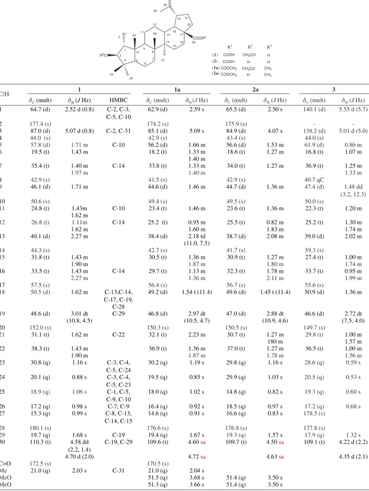
Table S1. 13C and 1H NMR data (d in ppm) for 3-O-acetyl ceanothic acid(1), dimethyl ceanothate (2a), ceanothenic acid (3) (400 MHz and 100 MHz,
respectively) and acetyl-dimethyl ceanothate (1a) (600 MHz and 150 MHz, respectively)
COOR3
R2O R1
1 3 2
4 5
6 7 8 9 10
11 12
13 14
15 16 17 18
19 20
21
22
23 24
25 26
27
28 29
30
(2) (1)
(1a) (2a)
COOH CH3CO COOH
H COOCH3 CH3CO CH3 COOCH3 H CH3 H H
R1 R2 R3
C/H 1 1a 2a 3
dC (mult) dH (J Hz) HMBC dC (mult) dH (J Hz) dC (mult) dH (J Hz) dC (mult) dΗ (J Hz)
1 64.7 (d) 2.52 d (0.8) C-2, C-3,
C-5, C-10
62.9 (d) 2.59 s 65.5 (d) 2.50 s 140.1 (d) 5.55 d (5.7)
2 177.4 (s) 174.2 (s) 175.9 (s) -
-3 87.0 (d) 5.07 d (0.8) C-2, C-31 85.1 (d) 5.09 s 84.9 (d) 4.07 s 138.2 (d) 5.01 d (5.0)
4 44.0 (s) 42.9 (s) 43.4 (s) 44.0 (s)
5 57.8 (d) 1.71 m C-10 56.2 (d) 1.66 m 56.6 (d) 1.53 m 61.9 (d) 0.86 m
6 19.5 (t) 1.43 m 18.2 (t) 1.33 m 18.6 (t) 1.27 m 16.8 (t) 1.07 m
1.40 m
7 35.4 (t) 1.40 m C-14 33.8 (t) 1.33 m 34.0 (t) 1.27 m 36.9 (t) 1.25 m
1.97 m 1.40 m 1.33 m
8 42.9 (s) 41.5 (s) 42.9 (s) 40.7 qC
9 46.1 (d) 1.71 m 44.6 (d) 1.46 m 44.7 (d) 1.36 m 47.4 (d) 1.48 dd
(3.2, 12.3)
10 50.6 (s) 49.4 (s) 49.5 (s) 50.0 (s)
11 24.8 (t) 1.43m C-10 23.4 (t) 1.46 m 23.6 (t) 1.36 m 22.3 (t) 1.20 m
1.62 m
12 26.8 (t) 1.11m C-14 25.2 (t) 0.95 m 25.5 (t) 0.82 m 25.2 (t) 1.30 m
1.62 m 1.60 m 1.83 m 1.74 m
13 40.1 (d) 2.27 m 38.4 (d) 2.18 td 38.7 (d) 2.08 m 39.0 (d) 2.02 m
(11.0, 7.5)
14 44.3 (s) 42.7 (s) 41.7 (s) 59.3 (s)
15 31.8 (t) 1.43 m 30.5 (t) 1.36 m 30.9 (t) 1.27 m 27.4 (t) 1.00 m
1.90 m 1.87 m 1.80 m 1.74 m
16 33.5 (t) 1.43 m C-14 29.7 (t) 1.13 m 32.3 (t) 1.78 m 33.7 (t) 0.95 m
2.27 m 1.36 m 2.11 m 1.99 m
17 57.5 (s) 56.4 (s) 56.7 (s) 55.6 (s)
18 50.5 (d) 1.62 m C-13,C-14, C-17, C-19,
49.2 (d) 1.54 t (11.4) 49.6 (d) 1.45 t (11.4) 50.9 (d) 1.36 m
C-28
19 48.6 (d) 3.01 dt C-29 46.8 (d) 2.97 dt 47.0 (d) 2.88 dt 46.6 (d) 2.72 dt
(10.8, 4.5) (10.5, 4.7) (10.9, 4.6) (7.5, 4.0)
20 152.0 (s) 150.3 (s) 150.5 (s) 149.7 (s)
21 31.1 (t) 1.62 m C-22 32.1 (t) 2.23 m 30.7 (t) 1.27 m 29.8 (t) 1.00 m
180 m 1.57 m
22 38.3 (t) 1.43 m 36.9 (t) 1.36 m 37.0 (t) 1.27 m 36.5 (t) 1.00 m
1.90 m 1.87 m 1.78 m 1.56 m
23 30.8 (q) 1.16 s C-3, C-4,
C-5, C-24
30.2 (q) 1.19 s 29.8 (q) 1.16 s 28.6 (q) 0.59 s
24 20.1 (q) 0.88 s C-3, C-4,
C-5, C-23
19.5 (q) 0.85 s 29.9 (q) 1.03 s 20.5 (q) 0.53 s
25 18.9 (q) 1.06 s C-1, C-5, C-9, C-10
18.0 (q) 1.02 s 14.8 (q) 0.82 s 19.3 (q) 0.60 s
26 17.2 (q) 0.98 s C-7, C-9 16.4 (q) 0.92 s 18.5 (q) 0.97 s 17.2 (q) 0.68 s
27 15.3 (q) 0.99 s C-8, C-13,
C-14, C-15
14.6 (q) 0.91 s 16.6 (q) 0.83 s 178.5 (s)
28 180.1 (s) 176.6 (s) 176.8 (s) 177.8 (s)
29 19.7 (q) 1.68 s C-19 19.4 (q) 1.67 s 19.3 (q) 1.57 s 17.9 (q) 1.32 s
30 110.3 (t) 4.58 dd
(2.2, 1.4)
C-19, C-29 109.6 (t) 4.60 sa 109.7 (t) 4.50 sa 109.1 (t) 4.22 d (2.2)
4.70 d (2.0) 4.72 sa 4.63 sa 4.35 d (2.1)
C=O 172.5 (s) 170.5 (s)
Me 21.0 (q) 2.03 s C-31 21.0 (q) 2.04 s
MeO 51.5 (q) 3.68 s 51.4 (q) 3.50 s
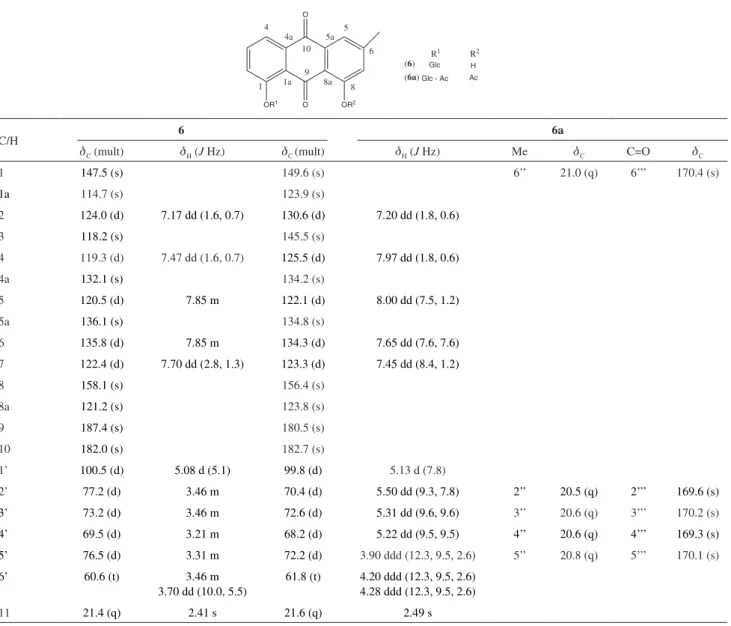
Table S2. 13C and 1H NMR data for compounds 6 and 6a
OR1 O O
OR2
1a 1
4 4a
10 5a
5
6
8 8a
9 (
6) Glc Glc - Ac
(6a)
R1 R2 H Ac
C/H 6 6a
dC (mult) dH (J Hz) dC (mult) dH (J Hz) Me dC C=O dC
1 147.5 (s) 149.6 (s) 6’’ 21.0 (q) 6’’’ 170.4 (s)
1a 114.7 (s) 123.9 (s)
2 124.0 (d) 7.17 dd (1.6, 0.7) 130.6 (d) 7.20 dd (1.8, 0.6)
3 118.2 (s) 145.5 (s)
4 119.3 (d) 7.47 dd (1.6, 0.7) 125.5 (d) 7.97 dd (1.8, 0.6)
4a 132.1 (s) 134.2 (s)
5 120.5 (d) 7.85 m 122.1 (d) 8.00 dd (7.5, 1.2)
5a 136.1 (s) 134.8 (s)
6 135.8 (d) 7.85 m 134.3 (d) 7.65 dd (7.6, 7.6)
7 122.4 (d) 7.70 dd (2.8, 1.3) 123.3 (d) 7.45 dd (8.4, 1.2)
8 158.1 (s) 156.4 (s)
8a 121.2 (s) 123.8 (s)
9 187.4 (s) 180.5 (s)
10 182.0 (s) 182.7 (s)
1’ 100.5 (d) 5.08 d (5.1) 99.8 (d) 5.13 d (7.8)
2’ 77.2 (d) 3.46 m 70.4 (d) 5.50 dd (9.3, 7.8) 2’’ 20.5 (q) 2’’’ 169.6 (s)
3’ 73.2 (d) 3.46 m 72.6 (d) 5.31 dd (9.6, 9.6) 3’’ 20.6 (q) 3’’’ 170.2 (s)
4’ 69.5 (d) 3.21 m 68.2 (d) 5.22 dd (9.5, 9.5) 4’’ 20.6 (q) 4’’’ 169.3 (s)
5’ 76.5 (d) 3.31 m 72.2 (d) 3.90 ddd (12.3, 9.5, 2.6) 5’’ 20.8 (q) 5’’’ 170.1 (s)
6’ 60.6 (t) 3.46 m
3.70 dd (10.0, 5.5)
61.8 (t) 4.20 ddd (12.3, 9.5, 2.6)
4.28 ddd (12.3, 9.5, 2.6)
Domínguez-Carmona et al. 5 Vol. 22, No. 7, 2011
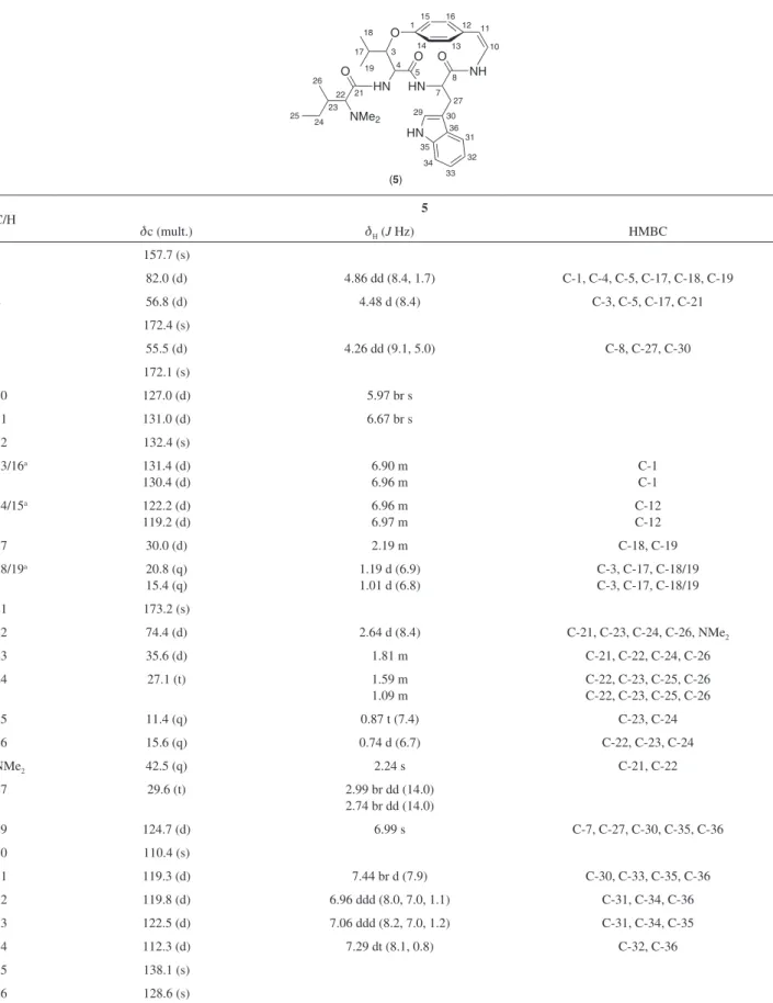
Table S3.13C and 1H NMR data for compound 5 in CD
3OD at 30 °C (400 MHz)
O
HN HN O
NH O
HN O
NMe2
18
19
17 3
1 12
10
8
7 27 29 30
36 31
32
33 34 35 25
24 23 26
22 21 5 4
11
13 14
15 16
(5)
C/H 5
dc (mult.) dH (J Hz) HMBC
1 157.7 (s)
3 82.0 (d) 4.86 dd (8.4, 1.7) C-1, C-4, C-5, C-17, C-18, C-19
4 56.8 (d) 4.48 d (8.4) C-3, C-5, C-17, C-21
5 172.4 (s)
7 55.5 (d) 4.26 dd (9.1, 5.0) C-8, C-27, C-30
8 172.1 (s)
10 127.0 (d) 5.97 br s
11 131.0 (d) 6.67 br s
12 132.4 (s)
13/16a 131.4 (d)
130.4 (d)
6.90 m 6.96 m
C-1 C-1
14/15a 122.2 (d)
119.2 (d)
6.96 m 6.97 m
C-12 C-12
17 30.0 (d) 2.19 m C-18, C-19
18/19a 20.8 (q)
15.4 (q)
1.19 d (6.9) 1.01 d (6.8)
C-3, C-17, C-18/19 C-3, C-17, C-18/19
21 173.2 (s)
22 74.4 (d) 2.64 d (8.4) C-21, C-23, C-24, C-26, NMe2
23 35.6 (d) 1.81 m C-21, C-22, C-24, C-26
24 27.1 (t) 1.59 m
1.09 m
C-22, C-23, C-25, C-26 C-22, C-23, C-25, C-26
25 11.4 (q) 0.87 t (7.4) C-23, C-24
26 15.6 (q) 0.74 d (6.7) C-22, C-23, C-24
NMe2 42.5 (q) 2.24 s C-21, C-22
27 29.6 (t) 2.99 br dd (14.0) 2.74 br dd (14.0)
29 124.7 (d) 6.99 s C-7, C-27, C-30, C-35, C-36
30 110.4 (s)
31 119.3 (d) 7.44 br d (7.9) C-30, C-33, C-35, C-36
32 119.8 (d) 6.96 ddd (8.0, 7.0, 1.1) C-31, C-34, C-36
33 122.5 (d) 7.06 ddd (8.2, 7.0, 1.2) C-31, C-34, C-35
34 112.3 (d) 7.29 dt (8.1, 0.8) C-32, C-36
35 138.1 (s)
36 128.6 (s)
Table S5. Inhibition of the growth [IG50 (µg mL-1)] in HeLa, KB, HEp-2 and VERO cells of compounds 1-6 from C. greggii, and derivative 6a
Compound
Antiproliferative activity
HeLa KB HEp-2 VERO
1 192.3 ± 2.4 45.0 ± 4.5 70.2 ± 4.5 146.8 ± 6.7
2 141.9 ± 1.6 55.4 ± 5.6 53.6 ± 5.6 189.7 ± 5.2
3 48.8 ± 3.1 33.8 ± 3.1 89.8 ± 4.9 89.2 ± 2.1
4 107.7 ± 1.2 46.0 ± 4.5 152.4 ± 3.2 221.5 ± 5.6
5 125.4 ± 2.3 56.9 ± 3.4 140.0 ± 2.5 201.4 ± 3.8
6 73.9 ± 2.9 66.6 ± 7.8 98.4 ± 2.8 351.0 ± 8.9
6a 55.9 ± 2.5 19.22 ± 2.3 44.7 ± 5.6 143.5 ± 6.2
Docetaxel 0.03 ± 0.01 0.05 ± 0.04 0.06 ± 0.02 0.11 ± 0.02
Table S4. Cytotoxic activity [CC50 (µg mL-1)] in HeLa, KB, HEp-2 and VERO cells of organic crude extract from C. greggii, low and medium polarity
fractions, compounds 1-6, and semisynthetic derivatives 1a, 2a and 6a
Extract/ fraction/ compund
Cytotoxic activity
HeLa KB HEp-2 VERO
CC50 SI CC50 SI CC50 SI CC50
CG-1 249.8 - 533.3 - 8.9 - NT
CG-2A 20.6 - 19.6 - 6.9 - NT
CG-2B 136.9 - 140.4 - 13.1 - NT
1 36.2 ± 5.1 2.8 46.9 ± 9.2 2.1 389.0 ± 10.9 0.2 103.1 ± 1.8
2 NA - 56.0 ± 2.1 2.3 68.7 ± 3.4 1.9 131.2 ± 3.2
1a NT NT NT NT
2a NT NT NT NT
3 67.7 ± 10.2 1.4 35.2 ± 3.2 2.8 54.5 ± 3.6 1.8 98.6 ± 1.2
4 15.5 ± 4.7 9.3 43.3 ± 4.2 3.3 174.6 ± 2.1 0.8 145.0 ± 2.9
5 43.9 ± 3.9 4.5 66.1 ± 7.2 3.0 179.5 ± 2.1 1.1 199.1 ± 2.7
6 69.3 ± 4.2 7.5 86.8 ± 11.3 6.0 102.7 ± 4.1 5.0 521.0 ± 6.3
6a 120.5 ± 9.1 1.0 46.0 ± 2.1 2.6 65.7 ± 3.4 1.8 123.8 ± 4.2
Docetaxel 0.20 ± 0.01 5.5 0.23 ± 0.03 4.7 0.08 ± 0.01 13.7 1.1 ± 0.05
![Table 1. Leishmanicidal, trypanocidal, antiplasmodial and cytotoxic activity [IC 50 (µg mL -1 )] of crude extract, semipuriied fractions, natural products 1-6 and semisynthetic derivatives 1a, 2a and 6a](https://thumb-eu.123doks.com/thumbv2/123dok_br/18995311.462018/3.892.90.824.690.1071/leishmanicidal-trypanocidal-antiplasmodial-cytotoxic-semipuriied-fractions-semisynthetic-derivatives.webp)
![Table S5. Inhibition of the growth [IG 50 (µg mL -1 )] in HeLa, KB, HEp-2 and VERO cells of compounds 1-6 from C](https://thumb-eu.123doks.com/thumbv2/123dok_br/18995311.462018/13.892.72.797.245.589/table-inhibition-growth-hela-hep-vero-cells-compounds.webp)
