Moderate Exercise Attenuates
Lipopolysaccharide-Induced Inflammation
and Associated Maternal and Fetal
Morbidities in Pregnant Rats
Karina T. Kasawara1,2☯, Tiziana Cotechini1☯, Shannyn K. Macdonald-Goodfellow1‡
, Fernanda G. Surita2‡, João L. Pinto e Silva2‡, Chandrakant Tayade1‡, Maha Othman1‡,
Terence R. S. Ozolinš1‡
, Charles H. Graham1☯*
1Department of Biomedical and Molecular Sciences, Queen’s University, Kingston, ON, Canada,
2Department of Obstetrics and Gynaecology, University of Campinas, Campinas, SP, Brazil
☯These authors contributed equally to this work.
‡These authors also contributed equally to this work. *[email protected]
Abstract
Fetal growth restriction (FGR) and coagulopathies are often associated with aberrant mater-nal inflammation. Moderate-intensity exercise during pregnancy has been shown to
increase utero-placental blood flow and to enhance fetal nutrition as well as fetal and pla-cental growth. Furthermore, exercise is known to reduce inflammation. To evaluate the effect of moderate-intensity exercise on inflammation associated with the development of maternal coagulopathies and FGR, Wistar rats were subjected to an exercise regime before and during pregnancy. To model inflammation-induced FGR, pregnant rats were adminis-tered daily intraperitoneal injections ofE.colilipopolysaccharide (LPS) on gestational days
(GD) 13.5–16.5 and sacrificed at GD 17.5. Control rats were injected with saline. Maternal hemostasis was assessed by thromboelastography. Moderate-intensity exercise prevented LPS-mediated increases in white blood cell counts measured on GD 17.5 and improved maternal hemostasis profiles. Importantly, our data reveal that exercise prevented LPS-induced FGR. Moderate-intensity exercise initiated before and maintained during preg-nancy may decrease the severity of maternal and perinatal complications associated with abnormal maternal inflammation.
Introduction
Fetal growth restriction (FGR) affects 5–10% of clinically recognized pregnancies [1–3] and is often associated with aberrant maternal inflammation. Although normal pregnancy is consid-ered to be a state of low-grade inflammation [4,5], there is strong evidence that adverse preg-nancy complications, including FGR and pre-eclampsia (PE), are linked to aberrant maternal inflammation [6,7]. Women affected by FGR/PE have a heightened inflammatory state
a11111
OPEN ACCESS
Citation:Kasawara KT, Cotechini T, Macdonald-Goodfellow SK, Surita FG, Pinto e Silva JL, Tayade C, et al. (2016) Moderate Exercise Attenuates Lipopolysaccharide-Induced Inflammation and Associated Maternal and Fetal Morbidities in Pregnant Rats. PLoS ONE 11(4): e0154405. doi:10.1371/journal.pone.0154405
Editor:Victor Sanchez-Margalet, Virgen Macarena University Hospital, School of Medicine, University of Seville, SPAIN
Received:December 31, 2015
Accepted:April 13, 2016
Published:April 28, 2016
Copyright:© 2016 Kasawara et al. This is an open access article distributed under the terms of the
Creative Commons Attribution License, which permits unrestricted use, distribution, and reproduction in any medium, provided the original author and source are credited.
Data Availability Statement:All relevant data are within the paper and a previous publication from our group (Cotechini et al. J. Exp. Med. 211 (2014), 165-79).
Funding:KTK received scholarships from the Canadian Bureau for International Education–
Department of Foreign Affairs and International Trade (CBIE/DFAIT) and Coordination for the Improvement of Higher Education Personnel–The Ministry of
characterised by increased levels of pro-inflammatory cytokines and chemokines such as tumour necrosis factor alpha (TNF), interleukin 6, and the macrophage chemoattractant che-mokine ligand 2 (CCL2), systemically and locally in the placenta [8–12]. This abnormal inflammatory response often leads to oxidative and nitrosative stress [10,13,14].
Normal pregnancy is associated with a shift in maternal hemostasis towards a pro-throm-botic state [15,16], and pregnancy-associated coagulopathies have been implicated in the pathophysiology of complications including PE, fetal loss and FGR [17–20]. While it is unclear whether disruptions in maternal hemostasis are causally linked to the deficient utero-placental perfusion that characterises these pregnancy disorders, there is recent evidence that antenatal anti-thrombotic therapy reduces the risk of poor pregnancy outcomes, including FGR, in women at risk of placental dysfunction [21]. Moreover, thrombophilia is commonly associated with fetal demise when utero-placental insufficiency is present [22].
There is substantial cross talk between inflammatory and hemostasis pathways, and dysre-gulation of both systems is implicated in the pathophysiology of pregnancy complications [20]. Previous work from our laboratory revealed that inflammation-induced coagulopathies in a rat model are associated with altered utero-placental hemodynamics and fetal death [18,19,23]. Importantly, our data revealed that inhibition of TNF successfully prevented inflammation-induced hemostatic alterations, normalized utero-placental perfusion and prevented fetal loss [18,23].
Exercise has immunomodulatory effects. Specifically, exercise reduces the release of pro-inflammatory cytokines, promotes the release of anti-pro-inflammatory cytokines, decreases vis-ceral fat mass and reduces the expression of Toll-Like Receptor 4 (TLR-4) on monocytes and macrophages [24,25]. The latter is a cell surface protein that plays a fundamental role in the release of pro-inflammatory cytokines and activation of innate immunity. Moderate exercise also regulates hemostasis by decreasing platelet reactivity and fibrin formation [26,27].
There is evidence revealing the benefits of exercise on achieving optimal vascular function [28]. In particular, moderate-intensity exercise has been shown to promote beneficial vascular adaptations and reduce cardiovascular risks [29]. Additionally, moderate-intensity exercise during pregnancy increases utero-placental blood flow, enhances fetal nutrition and improves fetal and placental growth [30,31]. In the present study we tested the hypothesis that exercise attenuates fetal and maternal morbidities associated with aberrant inflammation. To the best of our knowledge, this is the first study revealing a beneficial effect of an exercise protocol initi-ated before and during pregnancy in the attenuation of maternal inflammation, systemic coa-gulopathies and FGR.
Materials and Methods
Animals
All procedures for animal experimentation were undertaken in accordance with the principles and guidelines of the Canadian Council on Animal Care and were approved by the Queen's University Animal Care Committee (Permit Number: 2010-012-R3-A8). Virgin female Wistar rats (3–4 months old; Charles River Laboratories, St-Constant, QC, Canada) were housed in a light- and humidity-controlled facility with free access to food and water. All animals were evaluated daily in order to ensure health and well-being.
Exercise Protocol
Exercise under regulated conditions (i.e. constant time and intensity) was achieved using a rodent treadmill (LE 8700 series, Panlab, Harvard Apparatus, Barcelona, Spain). Belt speed was
Awards–Joint Research Projects (2011-2012) and
from FAEPEX (University of Campinas). TC was a recipient of a recipient of a CIHR Doctoral Award-Frederick Banting and Charles Best Canada Graduate Scholarship and an Ontario Graduate Scholarship. This research was supported by a grant from the Canadian Institutes of Health Research (CIHR; grant number MOP 119496) awarded to CHG. The TEG studies were supported by a grant from the Canadian Haemophilia Society awarded to MO.
adjusted and individualized according to the maximal exercise ability (MEA; described below) determined for each rat during a training phase.
Evaluation of Maximal Exercise Ability
Evaluation of maximal exercise ability (MEA) for each rat was performed as previously pub-lished by Jiaoet al. [32]. Briefly, belt inclination was set at 0◦and belt speed was set to 30 cm/ sec. Every 30 sec, belt speed was increased 2 cm/sec until rats were unable to run regularly, or until they rested on the shock grid (delivering a shock of<1mA) more than three times. The
final belt speed at which each animal was able to run was set as the MEA for that subject. This MEA assessment was repeated three times for each rat (with one-hour rest times between tri-als) and the average MEA over the three trials was calculated. Mean MEA was calculated both before and after pre-pregnancy exercise training (described below) in order to evaluate the change in each rat’s aerobic capacity prior to mating.
Pre-Pregnancy Exercise Protocol
The protocol for exercise training was based on the MEA for each rat and was established from an exercise training protocol for pregnant rats published by Amorim,et al. [33] and modified by us. Briefly, all rats were subjected to four weeks of pre-pregnancy exercise training. Each training week consisted of five consecutive days of training followed by two days of rest. The daily pre-pregnancy exercise program was divided in three stages consisting of 1) warm-up; 2) training and; 3) cool-down. During the warm-up stage (lasting five minutes), belt speed inten-sity was set to 40% of the subject’s MEA. During the training phase, belt intensity and duration of exercise were modulated based upon the week of training. Specifically, rats ran at 40% of their MEA for 20 min during week one, 50 min during week two, 60 min during week three and 60 min at 65% of their MEA during week four. During the cool-down period (five min-utes), belt speed was slowly decreased 2–3 cm/sec every 30 sec until the treadmill was stopped.
Mating
Following four weeks of pre-pregnancy exercise training, virgin female rats were co-housed overnight with a male rat (at a 2:1 ratio). The detection of sperm in the vaginal lavage the fol-lowing morning represented gestational day (GD) 0.5. Pregnant rats were then subjected to the pregnancy exercise protocol (described below) the same day.
Pregnancy Exercise Protocol
Throughout pregnancy, rats were subjected to a moderate-intensity exercise program. Preg-nant rats ran at 65% of their MEA for various durations over gestation (cycle one: 50 min/day; cycle two: 30 min/day; and cycle three: 20 min/day). Each cycle consisted of five consecutive days of exercise followed by one day of rest until study endpoint on GD 17.5.
Complete Blood Cell Count Analysis
Inflammation-Induced Rat Model of FGR
We used our previously established model of LPS-induced FGR [7]. Pregnant Wistar rats received daily intraperitoneal (i.p.) injections of low-dose lipopolysaccharide (LPS; 10μg/kg on GD 13.5 followed by 40μg/kg on GD 14.5, 15.5 and 16.5) or saline (0.1 ml/100 g) during the second half of gestation, and were euthanized on GD 17.5. To assess whether exercise attenu-ates LPS-induced inflammation and associated maternal and fetal morbidities, we utilized results from previously published work in which rats were treated with saline (Se + saline) or LPS (Se + LPS) as sedentary control data [7]. In this previous study, FGR was defined as fetal weight falling below the 10thpercentile for gestational age. Specifically, the threshold of FGR was determined by evaluating the distribution of all fetal weights from the saline-treated con-trol cohort (n = 22 dams; n = 305 fetuses; mean fetal weight = 0.9244 ± 0.007 g) and fetuses with weights below 0.8071 g (lower 10th percentile) were designated as FGR. Treatment of sed-entary rats with LPS in our previous study resulted in a mean fetal weight = 0.8421 ± 0.006 g (n = 28 dams; n = 258 fetuses) [7].
To evaluate the effect of exercise on LPS-induced FGR, exercised rats (as described in the above sections) received daily i.p. injections of saline or LPS (Escherichia coliserotype 0111:B4; Sigma-Aldrich, Oakville, ON, Canada) according to the same protocol established for the sed-entary group during pregnancy [7]. Fetal weights were measured on GD 17.5 and were normal-ized to litter size (fetal weight/number of fetuses in litter) to account for alterations in fetal weight attributable to variations in litter size as has been previously described [34].
Thromboelastography (TEG)
Prior to euthanasia on GD 17.5, pregnant rats were anaesthetized using 40–50 mg/kg sodium pentobarbital (CEVA Santé Animale, Rutherford, NJ, USA). Maternal blood was collected via cardiac puncture, using a 26’-gauge needle, and placed into a tube pre-filled with trisodium cit-rate. Thromboelastography (TEG) was performed on citrated blood as previously described [18,
19,23] using a TEG1
5000 Haemostasis System and TEG1
Haemostasis Analyzer software Version 4.2 (Haemoscope Corporation, Skokie, IL, USA). Prior to each analysis, an electronic quality test was performed on the TEG1
5000 Haemostasis System by a trained operator. Blood (340μl) was re-calcified by adding 20μl of 0.2 M calcium chloride and loaded into a disposable plastic cuvette for analysis. Data were collected for 75–90 min and the following parameters were evaluated: time to clot formation (R), speed of clot propagation (α), rate of clot formation (K), strength/stability of clot (MA), clotting index (CI; a value that is based on the four parame-ters above,) and LY30 (percent clot dissolution in 30 min). Our previously published data col-lected from the sedentary cohort of saline- and LPS-treated rats (n = 9 and 13, respectively) [19] were used as the reference range and controls. Exercised rats from the current study were con-sidered to exhibit hemostatic alterations if two or more parameters fell beyond reference ranges previously established during normal pregnancy in saline-treated, sedentary rats.
Statistical Analysis
All statistical analyses were performed using GraphPad Prism 6.0 Software (GraphPad Soft-ware Inc., La Jolla, CA, USA). Data are presented as mean ± standard error of the mean (SEM). Student’s t-test was used to compare means between two groups. To evaluate whether exercise ameliorates LPS-induced outcomes, data were analysed using two-way ANOVA and the Bon-ferroni correction was applied to all row and column comparisons to determine significant dif-ferences between comparison groups. Effect size was measured by Eta Squared and appear in the text asη2xwhere x refers to the main effect being tested. Differences between groups were
Results
Exercise Training Increased the Aerobic Capacity of Rats Prior to
Pregnancy
All trained rats (n = 10) increased their aerobic capacity following four weeks of pre-pregnancy exercise. Specifically, MEA significantly increased from 63.7 cm/sec ± 8.6 cm/sec prior to train-ing, to 79.1 cm/sec ± 6.4 cm/sec following training (p<0.001). Since all rats achieved a similar
final belt speed following the training phase, animals were randomly assigned to the saline (Ex +Saline; n = 5) or LPS (Ex+LPS; n = 5) group. Five rats were unable to start the pre-pregnancy training phase due to persistent refusal to run on the treadmill. These rats did not exhibit any obvious physical condition suggestive of inability to run. An additional rat was diagnosed with hip displacement. These six rats were excluded from the study.
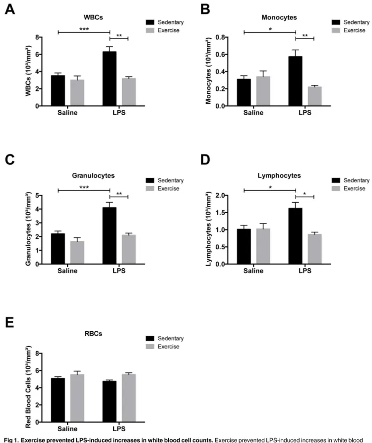
Exercise Prevented LPS-Induced Increases in White Blood Cell Counts
WBC counts measured from Ex+LPS-treated animals were significantly reduced compared with counts measured from Se+LPS-treated rats (Fig 1A;η2Inflammation= 0.144;η2Exercise=0.217;η2Interaction= 0.111); our previous study had revealed that WBC counts in this latter
group of rats were significantly increased compared with sedentary control (saline-treated) rats [7]. Differential WBC analysis (Fig 1) also revealed that exercise significantly prevented LPS-induced increases in the number of circulating monocytes (Fig 1B;η2Inflammation= 0.025;η2 Exer-cise= 0.127;η2Interaction= 0.178), granulocytes (Fig 1C;η2Inflammation= 0.201;η2Exercise= 0.237; η2Interaction= 0.075) and lymphocytes (Fig 1D;η2Inflammation= 0.045;η2Exercise= 0.126;η2 Interac-tion= 0.006). Exercise did not affect red blood cell counts in LPS-treated animals (p = 0.12;Fig
1E).
Exercise Attenuated Inflammation-Induced FGR
Our previously published data revealed that administration of LPS to sedentary pregnant rats induced FGR [7] (Fig 2A and 2B). This LPS-induced reduction in fetal weights observed in sed-entary rats was not observed in exercised rats (Fig 2A). Moreover, weights of fetuses from Ex +LPS dams trended (p = 0.07) towards being significantly increased compared with weights of fetuses from Se+LPS dams (Fig 2A;η2Inflammation<0.001;η2Exercise= 0.004;η2Interaction=
0.006). Exercise did not significantly alter the of proportion of growth restricted fetuses (Fig 2B), and we observed no differences in total litter size (all implantation sites) or fetal viability (number of live pups in a litter) when all treatment groups were compared (data not shown).
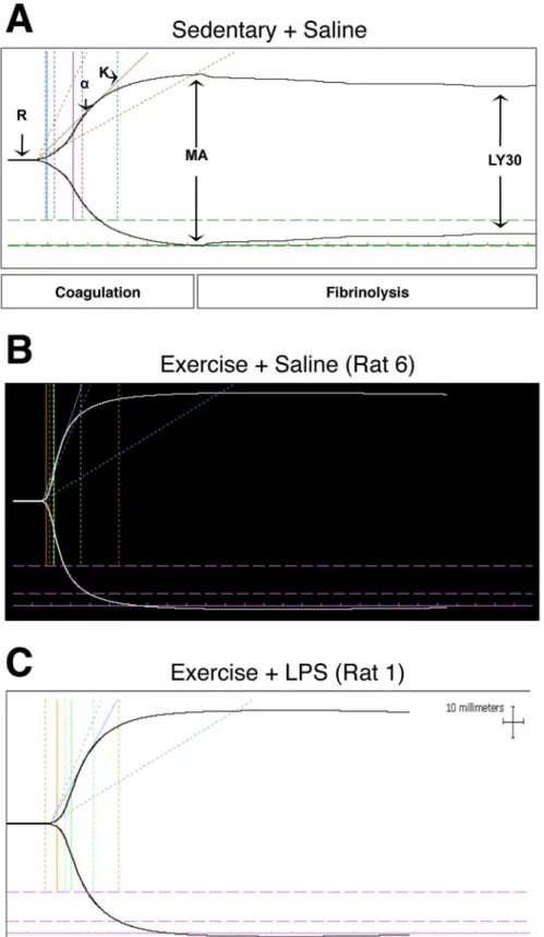
Exercise Normalized Coagulation Parameters Measured by TEG
TEG coagulation parameters obtained from exercised rats (Saline and LPS) were compared with our previously published coagulation parameters obtained from sedentary rats (Saline and LPS) [19] (Table 1). Our data here reveal that exercise normalized coagulation parameters (Fig 3). Of the four rats in the Ex+Saline group, three had normal TEG parameters when com-pared with Se+Saline rats (Fig 3A and 3B). One rat (Rat 9) had TEG parameters indicative of a hypocoagulable state with more than two parameters falling beyond the reference ranges for normal pregnant rats, including increased K and decreased alpha angle and MA (Table 1).The TEG parameters assessed from blood collected from four of the five rats from the Ex +LPS group were not significantly different from TEG parameters assessed from blood samples collected from the Se+Saline group (Table 1;Fig 3A and 3C). For the remaining animal (Rat 3;
Fig 1. Exercise prevented LPS-induced increases in white blood cell counts.Exercise prevented LPS-induced increases in white blood cell (leukocyte) counts measured in blood samples collected on GD 17.5 (A). Exercise significantly abrogated LPS-induced increases in the number of circulating monocytes (B), granulocytes (C) and lymphocytes (D), but did not significantly alter the red blood cell count (E). Ex+LPS, n = 5; Ex+Saline, n = 5; Se+LPS, n = 11; Se+Saline, n = 10.
Fig 2. Exercise prevented LPS-induced fetal growth restriction.LPS significantly reduced fetal weights/litter size in sedentary rats (Se+LPS), whereas fetal weights in exercised rats treated with LPS (Ex+LPS) trended (p = 0.07) towards being significantly higher than in the Se+LPS cohort (A). Although the proportion of growth restricted fetuses trended towards being increased in Se+LPS rats than in Se+Saline rats [7], exercise did not significantly alter this proportion in the present study (B). FGR, fetal growth restriction. Ex+LPS (number of pups = 41, litter size varied 5 to 11), Ex+Saline (number of pups = 63; range in litter size = 4–19), Se+LPS (number of pups = 258), Se+Saline (number of pups = 305) (A); Ex+LPS (n = 5), Ex+Saline (n = 5), Se+LPS (n = 28), Se+Saline (n = 22) (B).
doi:10.1371/journal.pone.0154405.g002
Table 1. Thromboelastography parameters assessed on GD 17.5.
R K αangle MA CI LY30 Coagulopathy#
Exercise + LPS(n = 5) 10.78 (7.5–
13.8)
3.68 (2.3–
7.2)
50.86 (31.8–
61.5)
62.7 (48.1–71.9) 1.54 (-1.1–
3.1)
0.34 (0–
1.7)
Rat 1 10.4 2.8 53.7 71.9 3.1 0.0 Normal
Rat 2 9.9 2.6 56.6 59.0 1.0 0.0 Normal
Rat 3 13.8 7.2 31.8 48.1 -1.1 1.7 Hypocoagulable
Rat 4 12.3 3.5 50.7 71.4 2.6 0.0 Normal
Rat 5 7.5 2.3 61.5 63.1 2.1 0.0 Normal
Exercise + Saline(n = 4) 9.8 (8.6–10.9) 3.98 (2.1–
8.1)
46.28 (29.2–
62.9)
59.55 (52.8–
71.8)
1.5 (0.8–3.3) 1.15 (0–
2.6)
Rat 6 8.6 2.1 62.9 71.8 3.3 0.0 Normal
Rat 7 10.9 2.8 36.6 56.1 0.8 2.6 Normal
Rat 8 10.8 4.4 42.1 57.5 0.9 2.0 Normal
Rat 9 8.9 8.1 29.2 52.8 1.0 0.0 Hypocoagulable
Sedentary + LPSa,[19] (n = 13)
8.9 (2.4–21.5) 2.5 (0.8–7.4) 60.6 (25.2–78.4) 67.4 (36.9–82.3) 3 (-3.3–5.8) 5.4 (0–
47.5)
Sedentary + Salinea,[19] (n = 9)
11.6 (7.9–18.2) 3.6 (1.8–5.9) 51.6 (33.2–65) 62.5 (56.7–71.5) 1.2 (-0.9–3.4) 5.1 (0–
13.1)
a
Historical data from previoulsy published work [19]. All values presented as mean (minimum—maximum); R, time for clot formation;α, speed of clot propagation; K, rate of clot formation; MA, strength/Stability of clot; CI, clotting index; LY30, percent of clot dissolution in 30 min (%).
#
Coagulopathy was compared with Sedentary + Saline group.
Fig 3. Exercise normalized thromboelastography parameters and tracings.Representative TEG trace revealing TEG parameters measured from blood collected from a Se+Saline control dam on GD 17.5 (A). TEG tracing from an Ex+Saline dam on GD 17.5 (B). TEG tracing from an Ex+LPS dam on GD 17.5 (C). R, time to clot formation;α, speed of clot propagation; K, rate of clot formation; MA, strength/stability of clot; CI, clotting index; LY30, percent clot dissolution in 30 min (%).
hypocoagulable state. Overall, exercise normalized LPS-induced coagulation parameters as evaluated by TEG.
Exercise Did Not Affect Maternal Weight Gain
Exercise did not affect maternal weight gain over the course of saline or LPS treatment when compared with sedentary rats. Two-way ANOVA comparing the change in maternal weight measured between GD 13.5 (onset of treatment) and GD 17.5 (endpoint) revealed that varia-tions in maternal weight at endpoint are attributable to LPS treatment and not exercise (data not shown).
Discussion
Here we describe the effect of moderate-intensity maternal exercise before and during preg-nancy on preventing LPS-induced FGR and decreasing the intensity of the associated maternal inflammation and hemostatic alterations. The results of this study are in agreement with similar data reported in mouse studies in which voluntary exercise performed before and during preg-nancy revealed positive effects on maternal outcomes. In those studies, voluntary maternal exer-cise was associated with better control of blood pressure during pregnancy, reduced proteinuria (albumin/creatinine ratio), decreased oxidative stress increasing placental antioxidant capacity, and reduced maternal and placental circulating sFlt-1 levels [35–37]. Despite the beneficial effects of exercise reported, voluntary exercise can result in intervention bias as the intensity of exercise experienced by each animal may differ. There is evidence that animals voluntarily reduce their physical activity later in gestation and that this reduction in activity varies between individuals [35,36]. Though the reason for this variance is not well described, it is possible that physiological and biomechanical changes during pregnancy (i.e. weight gain and cardiovascular adaptations) play a role. In consideration of such inter-animal variability, we designed our experiments such that all rats were exposed to the same exercise regime throughout the study.
In our study, seven weeks of moderate-intensity exercise (including the pre-pregnancy training phase and exercise during gestation) prevented LPS-induced increases in white blood cell counts, normalized coagulation parameters and attenuated the development of LPS-induced coagulopathies. In addition, we observed a beneficial effect of maternal exercise on fetal outcomes such that exercise prevented LPS-induced FGR. Our data align with previous studies revealing that voluntary exercise ameliorates poor fetal growth [35,36]. However, work from Rocha and colleagues revealed that maternal exercise (swimming) did not increase fetal weight in spontaneously hypertensive (SHR) rats [38]. In contrast to our model, in that study, rats began exercise (20 minutes of daily swimming initially; increased gradually to one hour per day until GD 20) on GD 7 and exercise promoted (rather than attenuated) FGR when com-pared with sedentary control rats [38]. Whereas these data suggest that exercise initiated dur-ing an established pregnancy may be detrimental as a result of increased maternal
physiological demands and cardiovascular overload [36], our current study reveals that exer-cise initiated prior to pregnancy and consistently maintained throughout gestation may be beneficial.
While exercise promotes positive physiological adaptations during pregnancy [35,36], forced exercise could also induce physiological stress [39]. We designed our study with a regi-mented protocol in order to control exercise intensity and duration. Moreover, we chose to model moderate-intensity exercise (65% of maximum exercise ability) because this level of exercise has been associated with maternal benefits and, importantly, because it has been rec-ommended for pregnant women by the American College of Obstetrics and Gynecology [33,
completely prevent LPS-induced maternal and fetal alterations. Therefore, it is possible that exercise at a different level of intensity (i.e. high or low) may be a more effective option with broader outcomes. Indeed, there is evidence that differing exercise intensities during pregnancy have variable effects on maternal and neonatal health outcomes [41,42].
To the best of our knowledge, this study provides the first evidence that moderate-intensity maternal exercise before and during pregnancy attenuates inflammation and its associated hemostatic alterations in a rat model. TEG is an effective tool for the evaluation of global hemostatic changes associated with both normal pregnancy [43] and adverse pregnancy out-comes [44]. Moreover, we previously demonstrated that inflammation-induced maternal hemostatic alterations, detected systemically using TEG, are comparable to hemostatic alter-ations detected locally at the utero-placental interface in a model of inflammation-induced fetal demise [18]. Though some studies have described a pro-thrombotic effect of high intensity exercise (>75% of VO2max) [45,46], there is evidence that exercise prevents thrombosis during
hospitalization and prolonged bedtime periods in non-pregnant individuals, and that exercise improves the hemostatic profile [47,48]. In the present study, exercise normalized LPS-induced alterations in TEG parameters such that the values were not different from those of sedentary, saline-treated, control rats. The use of TEG has been an important tool for the evalu-ation of the coagulevalu-ation index in rats [18,19] and in this study TEG was used to assess individ-ual hemodynamic changes in response to LPS and exercise. Moreover, our findings are consistent with previous publications that report a beneficial effect of exercise on coagulation parameters and an overall enhanced fibrinolytic state [47,49].
The causative link between exaggerated maternal inflammation and development of preg-nancy complications is becoming prominent [50] despite the fact that the initiating factors pre-cipitating inflammation have yet to be conclusively identified. We chose to model
inflammation-induced pregnancy complications using systemic LPS exposure in pregnant rats. Our model is based, in part, on studies by Faas and colleagues who first described the use of low-dose LPS infusion to model a PE-like syndrome in pregnant rats [51,52]. Though infection with LPS-positive bacteria is relatively uncommon in pregnant women, it is well established that infection is associated with the onset of pre-term labour in approximately 40% of cases [53]. Moreover, there is evidence that periodontal disease, in which bacterial LPS is the most prominent pro-inflammatory factor, is associated with miscarriage [54], low birthweight [55] and PE [56] as assessed using both human observational data andin vivoanimal models [57]. Exercise is known to reduce the release of pro-inflammatory cytokines, stimulate the release of anti-inflammatory cytokines and diminish the physiological consequences of exaggerated inflammation. In light of accumulating evidence for a role of abnormal inflammation in the development of pregnancy complications [50], including FGR [7] and spontaneous pregnancy loss [18,19,23], the results of our study warrant further investigation of the potential benefits of exercise for women with clearly identified complications (e.g. recurrent pregnancy loss) associated with aberrant inflammation.
Acknowledgments
We wish to thank the following individuals for technical help: Bruno Zavan, Maggie Chasmar and Kevin Robb for assistance with animal care.
Author Contributions
References
1. Gardosi J, Chang A, Kalyan B, Sahota D, Symonds EM. Customised antenatal growth charts. Lancet. 1992; 339(8788):283–7. PMID:1346292.
2. Cetin I, Foidart JM, Miozzo M, Raun T, Jansson T, Tsatsaris V, et al. Fetal growth restriction: a work-shop report. Placenta. 2004; 25(8–9):753–7. Epub 2004/09/29. doi:10.1016/j.placenta.2004.02.004
S014340040400058X [pii]. PMID:15450396.
3. Pollack RN, Divon MY. Intrauterine growth retardation: definition, classification, and etiology. Clin Obstet Gynecol. 1992; 35(1):99–107. Epub 1992/03/01. PMID:1544253.
4. Redman CW, Sargent IL. Preeclampsia and the systemic inflammatory response. Semin Nephrol. 2004; 24(6):565–70. PMID:15529291.
5. Tosun M, Celik H, Avci B, Yavuz E, Alper T, Malatyalioglu E. Maternal and umbilical serum levels of interleukin-6, interleukin-8, and tumor necrosis factor-alpha in normal pregnancies and in pregnancies complicated by preeclampsia. J Matern Fetal Neonatal Med. 2010; 23(8):880–6. doi:10.3109/ 14767051003774942PMID:20441409.
6. Elfayomy AK, Habib FA, Almasry SM, Safwat MD, Eldomiaty MA. Serum levels of adrenomedullin and inflammatory cytokines in women with term idiopathic intrauterine growth restriction. J Obstet Gynae-col. 2013; 33(2):135–9. doi:10.3109/01443615.2012.721821PMID:23445133.
7. Cotechini T, Komisarenko M, Sperou A, Macdonald-Goodfellow S, Adams MA, Graham CH. Inflamma-tion in rat pregnancy inhibits spiral artery remodeling leading to fetal growth restricInflamma-tion and features of preeclampsia. J Exp Med. 2014; 211(1):165–79. doi:10.1084/jem.20130295PMID:24395887; PubMed Central PMCID: PMC3892976.
8. Szarka A, Rigo J Jr., Lazar L, Beko G, Molvarec A. Circulating cytokines, chemokines and adhesion molecules in normal pregnancy and preeclampsia determined by multiplex suspension array. BMC Immunol. 2010; 11:59. Epub 2010/12/04. 1471-2172-11-59 [pii] doi:10.1186/1471-2172-11-59PMID:
21126355; PubMed Central PMCID: PMC3014878.
9. LaMarca BD, Ryan MJ, Gilbert JS, Murphy SR, Granger JP. Inflammatory cytokines in the pathophysi-ology of hypertension during preeclampsia. Curr Hypertens Rep. 2007; 9(6):480–5. Epub 2008/03/28. PMID:18367011.
10. Borzychowski AM, Sargent IL, Redman CW. Inflammation and pre-eclampsia. Semin Fetal Neonatal Med. 2006; 11(5):309–16. Epub 2006/07/11. S1744-165X(06)00043-6 [pii] doi:10.1016/j.siny.2006.04. 001PMID:16828580.
11. Redman CW, Sacks GP, Sargent IL. Preeclampsia: an excessive maternal inflammatory response to pregnancy. Am J Obstet Gynecol. 1999; 180(2 Pt 1):499–506. 1479. PMID:9988826
12. Jain K, Kavi V, Raghuveer CV, Sinha R. Placental pathology in pregnancy-induced hypertension (PIH) with or without intrauterine growth retardation. Indian J Pathol Microbiol. 2007; 50(3):533–7. Epub 2007/09/22. PMID:17883125.
13. Roggensack AM, Zhang Y, Davidge ST. Evidence for peroxynitrite formation in the vasculature of women with preeclampsia. Hypertension. 1999; 33(1):83–9. PMID:9931086.
14. Lowe DT. Nitric oxide dysfunction in the pathophysiology of preeclampsia. Nitric Oxide. 2000; 4 (4):441–58. 1185. PMID:10944429
15. Chen H, Zhou L, Meng L, Liu M, Tan J, Gao L, et al. Coagulation and prothrombotic state parameters: a clinical analysis during early pregnancy. Ir J Med Sci. 2011; 180(4):813–7. doi: 10.1007/s11845-011-0737-xPMID:21809018.
16. Koh MB, Lao ZT, Rhodes E. Managing haematological disorders during pregnancy. Best Pract Res Clin Obstet Gynaecol. 2013; 27(6):855–65. doi:10.1016/j.bpobgyn.2013.08.002PMID:24060203.
17. Han L, Liu X, Li H, Zou J, Yang Z, Han J, et al. Blood coagulation parameters and platelet indices: changes in normal and preeclamptic pregnancies and predictive values for preeclampsia. PLoS One. 2014; 9(12):e114488. doi:10.1371/journal.pone.0114488PMID:25464515; PubMed Central PMCID: PMC4252147.
18. Falcon BJ, Cotechini T, Macdonald-Goodfellow SK, Othman M, Graham CH. Abnormal inflammation leads to maternal coagulopathies associated with placental haemostatic alterations in a rat model of foetal loss. Thromb Haemost. 2012; 107(3):438–47. doi:10.1160/TH11-09-0626PMID:22234563.
19. Cotechini T, Othman M, Graham CH. Nitroglycerin prevents coagulopathies and foetal death associ-ated with abnormal maternal inflammation in rats. Thromb Haemost. 2012; 107(5):864–74. doi:10. 1160/TH11-10-0730PMID:22274747.
21. Dodd JM, McLeod A, Windrim RC, Kingdom J. Antithrombotic therapy for improving maternal or infant health outcomes in women considered at risk of placental dysfunction. Cochrane Db Syst Rev. 2013;( 7). Artn Cd006780 doi:10.1002/14651858.Cd006780.Pub3WOS:000322568300005.
22. Silver RM. Fetal death. Obstet Gynecol. 2007; 109(1):153–67. Epub 2007/01/02. 109/1/153 [pii] doi:
10.1097/01.AOG.0000248537.89739.96PMID:17197601.
23. Renaud SJ, Cotechini T, Quirt JS, Macdonald-Goodfellow SK, Othman M, Graham CH. Spontaneous pregnancy loss mediated by abnormal maternal inflammation in rats is linked to deficient uteroplacental perfusion. J Immunol. 2011; 186(3):1799–808. Epub 2010/12/29. jimmunol.1002679 [pii] doi:10.4049/ jimmunol.1002679PMID:21187445.
24. Gleeson M, Bishop NC, Stensel DJ, Lindley MR, Mastana SS, Nimmo MA. The anti-inflammatory effects of exercise: mechanisms and implications for the prevention and treatment of disease. Nat Rev Immunol. 2011; 11(9):607–15. doi:10.1038/nri3041PMID:21818123.
25. Petersen AM, Pedersen BK. The anti-inflammatory effect of exercise. J Appl Physiol (1985). 2005; 98 (4):1154–62. doi:10.1152/japplphysiol.00164.2004PMID:15772055.
26. Keating FK, Schneider DJ, Savage PD, Bunn JY, Harvey-Berino J, Ludlow M, et al. Effect of exercise training and weight loss on platelet reactivity in overweight patients with coronary artery disease. J Car-diopulm Rehabil Prev. 2013; 33(6):371–7. doi:10.1097/HCR.0000000000000015PMID:24126611.
27. Kumar A, Kar S, Fay WP. Thrombosis, physical activity, and acute coronary syndromes. J Appl Physiol (1985). 2011; 111(2):599–605. doi:10.1152/japplphysiol.00017.2011PMID:21596926; PubMed Cen-tral PMCID: PMC3154689.
28. La Gerche A, Prior DL. Exercise—is it possible to have too much of a good thing? Heart Lung Circ. 2007; 16 Suppl 3:S102–4. doi:10.1016/j.hlc.2007.03.014PMID:17606401.
29. Durand MJ, Gutterman DD. Exercise and vascular function: how much is too much? Can J Physiol Pharmacol. 2014; 92(7):551–7. doi:10.1139/cjpp-2013-0486PMID:24873760.
30. Weissgerber TL, Wolfe LA, Davies GA, Mottola MF. Exercise in the prevention and treatment of mater-nal-fetal disease: a review of the literature. Applied physiology, nutrition, and metabolism = Physiologie appliquee, nutrition et metabolisme. 2006; 31(6):661–74. doi:10.1139/h06-060PMID:17213880.
31. Clapp JF 3rd, Kim H, Burciu B, Lopez B. Beginning regular exercise in early pregnancy: effect on feto-placental growth. Am J Obstet Gynecol. 2000; 183(6):1484–8. doi:10.1067/mob.2000.107096PMID:
11120515.
32. Jiao Q, Bai Y, Akaike T, Takeshima H, Ishikawa Y, Minamisawa S. Sarcalumenin is essential for main-taining cardiac function during endurance exercise training. Am J Physiol Heart Circ Physiol. 2009; 297 (2):H576–82. doi:10.1152/ajpheart.00946.2008PMID:19502553; PubMed Central PMCID:
PMCPMC2724216.
33. Amorim MF, dos Santos JA, Hirabara SM, Nascimento E, de Souza SL, de Castro RM, et al. Can physi-cal exercise during gestation attenuate the effects of a maternal perinatal low-protein diet on oxygen consumption in rats? Exp Physiol. 2009; 94(8):906–13. doi:10.1113/Expphysiol.2009.047621
WOS:000267967700006. PMID:19482898
34. Romero A, Villamayor F, Grau MT, Sacristan A, Ortiz JA. Relationship between fetal weight and litter size in rats: application to reproductive toxicology studies. Reprod Toxicol. 1992; 6(5):453–6. PMID:
1463926.
35. Genest DS, Falcao S, Michel C, Kajla S, Germano MF, Lacasse AA, et al. Novel role of the renin-angio-tensin system in preeclampsia superimposed on chronic hypertension and the effects of exercise in a mouse model. Hypertension. 2013; 62(6):1055–61. doi:10.1161/HYPERTENSIONAHA.113.01983
PMID:24101664.
36. Falcao S, Bisotto S, Michel C, Lacasse AA, Vaillancourt C, Gutkowska J, et al. Exercise training can attenuate preeclampsia-like features in an animal model. J Hypertens. 2010; 28(12):2446–53. doi:10. 1097/HJH.0b013e32833e97d0PMID:20811291.
37. Gilbert JS, Banek CT, Bauer AJ, Gingery A, Needham K. Exercise training attenuates placental ische-mia-induced hypertension and angiogenic imbalance in the rat. Hypertension. 2012; 60(6):1545–51. doi:10.1161/HYPERTENSIONAHA.112.202275PMID:23090773; PubMed Central PMCID: PMC3909775.
38. Rocha R, Peracoli JC, Volpato GT, Damasceno DC, de Campos KE. Effect of exercise on the maternal outcome in pregnancy of spontaneously hypertensive rats. Acta Cir Bras. 2014; 29(9):553–9. PMID:
25252201.
40. Committee on Obstetric P. ACOG committee opinion. Exercise during pregnancy and the postpartum period. Number 267, January 2002. American College of Obstetricians and Gynecologists. Int J Gynae-col Obstet. 2002; 77(1):79–81. PMID:12053898.
41. Oliveira AO, Fileto C, Melis MS. Effect of strenuous maternal exercise before and during pregnancy on rat progeny renal function. Braz J Med Biol Res. 2004; 37(6):907–11. /S0100-879X2004000600017. PMID:15264035.
42. Szymanski LM, Satin AJ. Strenuous exercise during pregnancy: is there a limit? Am J Obstet Gynecol. 2012; 207(3):179.e1–6. doi:10.1016/j.ajog.2012.07.021PMID:22939718; PubMed Central PMCID: PMC3464969.
43. Miall FM, Deol PS, Barnes TA, Dampier K, Watson CC, Oppenheimer CA, et al. Coagulation status and complications of pregnancy. Thromb Res. 2005; 115(6):461–7. Epub 2005/03/29. S0049-3848(04) 00529-8 [pii] doi:10.1016/j.thromres.2004.09.019PMID:15792676.
44. Rai R, Tuddenham E, Backos M, Jivraj S, El'Gaddal S, Choy S, et al. Thromboelastography, whole-blood haemostasis and recurrent miscarriage. Hum Reprod. 2003; 18(12):2540–3. PMID:14645169.
45. Wang JS. Exercise prescription and thrombogenesis. J Biomed Sci. 2006; 13(6):753–61. doi:10.1007/ s11373-006-9105-7PMID:16933027.
46. Posthuma JJ, van der Meijden PE, Ten Cate H, Spronk HM. Short- and Long-term exercise induced alterations in haemostasis: a review of the literature. Blood Rev. 2014. doi:10.1016/j.blre.2014.10.005
PMID:25467962.
47. Nascimento Dda C, Neto FR, de Santana FS, da Silva RA, Dos Santos-Neto L, Balsamo S. The interac-tions between hemostasis and resistance training: a review. Int J Gen Med. 2012; 5:249–54. doi:10. 2147/IJGM.S29197PMID:22419885; PubMed Central PMCID: PMC3302766.
48. Kahn SR, Shrier I, Kearon C. Physical activity in patients with deep venous thrombosis: a systematic review. Thromb Res. 2008; 122(6):763–73. doi:10.1016/j.thromres.2007.10.011PMID:18078981.
49. Kupchak BR, Creighton BC, Aristizabal JC, Dunn-Lewis C, Volk BM, Ballard KD, et al. Beneficial effects of habitual resistance exercise training on coagulation and fibrinolytic responses. Thromb Res. 2013; 131(6):e227–34. doi:10.1016/j.thromres.2013.02.014PMID:23510721.
50. Cotechini T, Graham CH. Aberrant maternal inflammation as a cause of pregnancy complications: A potential therapeutic target? Placenta. 2015; 36(8):960–6. doi:10.1016/j.placenta.2015.05.016PMID:
26094029.
51. Faas MM, Schuiling GA, Baller JF, Bakker WW. Glomerular inflammation in pregnant rats after infusion of low dose endotoxin. An immunohistological study in experimental pre-eclampsia. Am J Pathol. 1995; 147(5):1510–8. PMID:7485413; PubMed Central PMCID: PMC1869514.
52. Faas MM, Schuiling GA, Baller JF, Visscher CA, Bakker WW. A new animal model for human pre-eclampsia: ultra-low-dose endotoxin infusion in pregnant rats. Am J Obstet Gynecol. 1994; 171 (1):158–64. 1513. PMID:8030692
53. Lettieri L, Vintzileos AM, Rodis JF, Albini SM, Salafia CM. Does "idiopathic" preterm labor resulting in preterm birth exist? Am J Obstet Gynecol. 1993; 168(5):1480–5. PMID:8498431.
54. Xiong X, Buekens P, Vastardis S, Yu SM. Periodontal disease and pregnancy outcomes: state-of-the-science. Obstetrical & gynecological survey. 2007; 62(9):605–15. doi:10.1097/01.ogx.0000279292. 63435.40PMID:17705886.
55. Vergnes JN, Sixou M. Preterm low birth weight and maternal periodontal status: a meta-analysis. Am J Obstet Gynecol. 2007; 196(2):135.e1–7. doi:10.1016/j.ajog.2006.09.028PMID:17306654.
56. Kunnen A, van Doormaal JJ, Abbas F, Aarnoudse JG, van Pampus MG, Faas MM. Periodontal disease and pre-eclampsia: a systematic review. J Clin Periodontol. 2010; 37(12):1075–87. doi:10.1111/j. 1600-051X.2010.01636.xPMID:21070324.
57. Kunnen A, van Pampus MG, Aarnoudse JG, van der Schans CP, Abbas F, Faas MM. The effect of Por-phyromonas gingivalis lipopolysaccharide on pregnancy in the rat. Oral Dis. 2014; 20(6):591–601. doi: