VIEWS AND REVIEWS
Neurosarcoidosis: guidance for the general
neurologist
Neurosarcoidose: orientações para o neurologista geral
Lívia Almeida Dutra, Pedro Braga-Neto, Ricardo Araújo Oliveira, José Luiz Pedroso, Agessandro Abrahão, Orlando Graziani Povoas Barsottini
Sarcoidosis is an inlammatory multisystem disorder of unknown etiology with a worldwide distribution that can af-fect lungs, lymphatic system, skin, liver, eyes, and nervous sys-tem1. We owe the irst recorded and illustrated description of
sarcoidosis to Jonathan Hutchinson in 1878, when he report-ed a patient with a dermatologic disease, which comprisreport-ed purplish, symmetric, and non-tender skin plaques, initially considered a manifestation of gout2. he term sarcoidosis
comes from histological studies performed by Cesar Boeck, in 1899, who described pathological indings of skin lesions that resembled those of sarcoma, which he called ‘sarkoid’ or ‘sarcoma-like’3. Involvement of the nervous system was irst
recognized, in 1905, by Winkler, and, in 1909, Heerfordt de-scribed three patients in the context of facial nerve palsies, uveitis, parotid enlargement, and fever1,3.
Neurosarcoidosis (NS) may involve any part of the ner-vous system with acute and chronic courses1,4.Cranial
nerves, hypothalamus, and pituitary gland are the most commonly regions involved, but meninges, parenchyma, brainstem, and spinal cord may also be afected4. Clinical
diferentiation from other neurological diseases may be dif-icult when an isolated involvement of the nervous system is present3,4. In this review, we highlighted its clinical forms,
pathogenesis, and treatment guidelines for NS, as guidance for general neurologists.
NEUROLOGICAL MANIFESTATIONS OF SARCOIDOSIS
Neurologic manifestations are found in 5 to 20% of cases and symptoms may be mild or even severe, requiring a more aggressive intervention. About one-half of patients with NS can develop neurologic manifestations before systemic sar-coidosis is evident5.
Division of General Neurology, Department of Neurology and Neurosurgery, Universidade Federal de São Paulo, São Paulo SP, Brazil.
Correspondence: Lívia Almeida Dutra; Rua Pedro de Toledo 650; 04039-000 São Paulo SP - Brasil; E-mail: [email protected]
Conflict of interest: There is no conflict of interest to declare.
Received 02 October 2011; Received in final form 11 November 2011; Accepted 18 November 2011
ABSTRACT
Neurosarcoidosis (NS) more commonly occurs in the setting of systemic disease. The diagnosis is based on a clinical history suggestive of NS, presence of noncaseating granulomas, and supportive evidence of sarcoid pathology, laboratory, and imaging studies. NS could involve any part of the nervous system and often demands high doses of steroids for symptom control. It presents low response to isolated steroids administration and frequently requires immunosuppressive agents. In NS, lymphocytes are polarized toward an excessive Th1 response, leading to overproduction of TNF-alpha and INF-gama, as well as lL-2 and IL-15. Infliximab, a chimeric monoclonal antibody that neutralizes the biological activity of TNF-alpha, is a new option in the NS treatment. We revised pathophysiology, clinical manifestations, diagnostic work up, and treatment of NS as guidance for the general neurologist.
Key words: sarcoidosis, neurosarcoidosis, methotrexate, azathioprine, cyclophosphamide, infliximab.
RESUMO
A neurosarcoidose (NS) ocorre frequentemente no contexto de doença sistêmica. O diagnóstico é baseado na história clínica sugestiva de NS, presença de granulomas não-caseosos e achados anatomopatológicos, laboratoriais e radiológicos de sarcoidose. A NS causa manifestações neurológicas variadas, que apresentam, em geral, baixa resposta ao corticoide isoladamente e, portanto, necessitam uso de imunossupressores. Na NS, os linfócitos estão polarizados para resposta Th1 excessiva, levando à produção aumentada de TNF-alfa e IFN-gama, assim como IL-2 e IL-15. Infliximabe, um anticorpo monoclonal quimérico que neutraliza a atividade biológica do TNF-alfa, é uma nova opção no tratamento da NS. Revisou-se a fisiopatologia, as manifestações clínicas, o diagnóstico e o tratamento da NS para orientar neurologistas gerais.
Hydrocephalus and chronic meningitis
Meningitis may present as a single episode or as a recur-rent or chronic course. Cerebrospinal luid (CSF) pleocyto-sis often occur in the setting of other manifestations, such as cranial neuropathy, myelopathy, mass lesions, or other focal neurologic manifestations. Granulomata in the fourth ven-tricle could be responsible for hydrocephalus, which may re-quire urgent treatment6.
Central nervous system
NS patients with headache may present meningitis, in-creased intracranial pressure or granulomatous mass lesions, which may mimic demyelination or neoplasm lesions7. In
pa-tients with intraxial lesions, seizures are found in 5 to 22%, including generalized, complex partial and myoclonic sei-zures8. Hypothalamic and pituitary involvement is a
char-acteristic feature of NS, and the posterior pituitary is more commonly afected than the anterior. Polydipsia and polyuria may be the presenting symptoms of NS secondary to diabe-tes insipidus. Other manifestations include hypothyroidism, amenorrhea, galactorrhea, sleep disorders, weight gain, tem-perature dysregulation, and personality changes. he possi-bility of endocrine dysfunction should always be investigated in the context of nonspeciic complaints such as weight loss, fatigue and generalized weakness9.
Patients with NS may present white matter abnormalities on the magnetic resonance imaging (MRI) (Fig 1) and pro-gressive multifocal leukoencephalopathy should be consid-ered as a diferential diagnosis10. Myelopathy in NS is
unusu-al, often evolving over weeks to months, with radiculopathy
or long tract dysfunction. Intramedullary sarcoidosis may present focal enlargement of the cord with gadolinium en-hancement. Progressive motor dysfunction may even mimic anterior horn syndrome10.
Involvement of basal ganglia may lead to movement dis-orders, with rare reports linking sarcoidosis to Parkinsonism and generalized chorea11. Stroke, subarachnoid
hemor-rhage, or intraparenchymal hemorrhage are quite rare in NS and may result from blood vessels involvement by sarcoid granulomas9,11. Most NS patients with increased
intracrani-al pressure present mass lesions, meningitis, or venous sinus thrombosis. In despite of that, few reports described pseudo-tumor cerebri due to NS12.
Neuropsychiatric manifestations
Patients with sarcoidosis and extensive central ner-vous system (CNS) involvement may develop dementia, psychoses, depression, poor concentration, hallucina-tions, and encephalopathy. Neuropsychiatry symptoms (NPS) develop months, sometimes presenting a relapse-remitting course. They can be found in sarcoid patients without evident CNS involvement. However, when NPS are found, CSF analysis is frequently abnormal, disclosing pleocytosis or increased protein13.
Cranial and peripheral neuropathy
he most common cranial neuropathy in NS is facial pal-sy, which may represent an isolated manifestation or which can occur in the context of multiple cranial nerve involve-ment. NS should be considered when multiple cranial nerves are afected or in patients with bilateral facial palsy. he optic nerve is involved in up to 38% of NS patients and some may present a progressive and painless monocular visual loss, sometimes with papilledema or optic atrophy. Less often, an acute or subacute visual loss may occur. Retrobulbar optic neuritis mimics multiple sclerosis. Approximately 20% of pa-tients have uveitis, usually bilateral, granulomatous that may be anterior (more common) or posterior14,15.
Sarcoid patients may present hearing loss or vestibu-lar dysfunction due to acoustic neuropathy. Lower cranial nerves are more afected than those responsible for extraoc-ular movements (III, IV, and VI), determining dysphagia or dysarthria16.
Involvement of the peripheral nerves is a well-recognized feature of NS, occurring in up to 20% of patients. Most of them present depressed tendon relexes or minor sensory changes, although severe weakness may be found. he clinical presenta-tions mainly include symmetrical distal neuropathy, monon-europathy, mononeuritis multiplex, and acute or chronic de-myelinating polyneuropathy. he mechanism of nerve injury is unclear, with evidence for both axonal injury and demyeli-nation process. Considering that some NS patients have pre-sented chronic inlammatory demyelinating polyneuropathy,
some authors believe that cytokines and immune factors may also play a role in nerve injury17. Peripheral neuropathy may be
associated with myositis or CNS involvement.
Myopathy
Asymptomatic involvement of the skeletal muscle by sar-coid granulomas may occur in more than 50% of patients with sarcoidosis. Only 1–2% of the patients present symp-tomatic muscle disease. hree distinct myopathy clinical pre-sentations are recognized: acute myositis, nodular form, and chronic form. he most common of them is the chronic form, which presents slowly progressive weakness and normal cre-atine phosphokinase levels, whereas acute myositis is rarely found. Patients with the nodular form may present palpable nodules. MRI and muscle imaging with gallium scanning pro-vide more information for establishing diagnosis; however, muscle biopsy may be required for diagnosis9,11.
PATHOPHYSIOLOGY
he etiology of NS is multifactor, involving genetic predis-position, environmental and individual factors. he ACCESS study demonstrated that agricultural employment, mold or mildew, musty odors at work, and pesticide-using industries are modestly associated with sarcoidosis risk (OR=1.5)18-20.
On the other hand, tobacco was negatively associated with sarcoidosis18-20. Research into speciic microbial etiologies
showed that mycobacterial DNA and protein antigens are present in sarcoidosis tissues and are the target of T and B cell responses, suggesting some important etiologic factors18.
It is speculated that deposition of microbial antigens and in-soluble particules aggregates with speciic host proteins in granulomas, generating an antigenic stimulus, followed by T-cell and macrophage activation via a classic major histo-compatibility complex (MHC) II-mediated pathway21.
he lymphocytes are polarized toward an excessive h1 re-sponse, leading to overproduction of TNF-alpha and INF-gama, as well as lL-2 and IL-15. he production of IL-12 and 18 also is increased, which enhances IFN-gama production and induces diferentiation of h0 precursors into active h1 lymphocytes, perpetuating the response21. he CD4+ lymphocytes are central
to the pathogenesis of sarcoidosis, triggering the granuloma for-mation18. hese basic immunologic features (h1 polarization,
oligoclonal T-cell expansions) are found in patients with sarcoi-dosis, despite clinical manifestations.
Genetic linkage studies have identiied the butyrophi-lin-like 2 gene (BTNL2), localized in the MHC region and involved in T-lymphocyte activation and regulation, as a susceptibility factor for the development of sarcoidosis18,19.
Human leukocyte antigens (HLAs) DR subtype of class 2 antigens is associated with many inlammatory conditions, including sarcoidosis. DRB1*03, DRB1*11, HLA-DRB1*12, HLA-DRB1*14, and HLA-DRB1*15 have been as-sociated with an increased risk of developing the disease. HLA-DRB1*03 has been found to be associated with Löfgren syndrome, an acute form of sarcoidosis19.
DIAGNOSTIC WORK UP
NS should be suspected in patients with documented sar-coidosis that present neurologic complaints. In 50% of the cas-es, neurological symptoms begin at the irst signs of systemic disease22.he diagnosis is based on evidence of a clinical
his-tory suggestive of NS, exclusion of other conditions capable of producing noncaseating granulomas, presence of noncaseat-ing granulomas, and supportive evidence of sarcoid pathology, laboratory and imaging studies15. Zajicek et al. established a
classiication to the diagnosis, according to Table 115,22.
It is important to determine whether sarcoidosis occurs as a systemic disease or if it is restricted to the CNS. For that purpose, one might use the Kveim-Siltzbach procedure, rare-ly available in Brazil, which consists of an intradermal injec-tion of heat-treated homogenate derived from sarcoid tissue. he development of a noncaseating granuloma at the injec-tion site constitutes a positive response, but the sensitivity is limited, between 60 to 90%3. Chest radiography, thoracic
computer tomography scan, and pulmonary function tests may reveal intra-thoracic involvement, which is found in up to 90% of the patients1. Routine laboratory tests may show
hyperglobulinemia, hypercalcemia or elevation of alkaline phophatase3.Elevated levels of serum
angiotensin-convert-ing-enzyme (ACE) are present in 24 to 76% of the patients23.
Despite its poor predictive value, ACE remains the only se-rological marker of sarcoidosis. ACE activity in bronchoalve-olar lavage (BAL) is generally considered a good marker of sarcoidosis severity with higher prognostic value than serum
Possible: the clinical syndrome and neurorradiologic evaluation are suggestive of NS. Infection and malignancy were not rigorously excluded or there is no pathologic confirmation of systemic sarcoidosis
Probable: the clinical syndrome and neurorradiologic evaluation are suggestive of NS and differential diagnoses have been made, especially malignancy and infection. There is pathologic evidence of sarcoidosis
Definite: Probable diagnosis + supportive nervous system pathology or response to therapy for NS over a one- to two-year observation period
NS: neurosarcoidosis.
ACE24. In addition, BAL can be used as an adjunctive
mea-sure to support the diagnosis of sarcoidosis, by demonstrat-ing a cell pattern typical of lymphocytic alveolitis (lympho-cyte count greater than 15%) and a T-cell CD4+/CD8+ ratio
over 3.5, although it is present in 50% of patients22,24.
Whole body gallium scan, muscle magnetic resonance imaging, and whole body luorodeoxyglucose positron emis-sion tomography (PET) are useful approaches to search for systemic sarcoidosis when initial investigation is unreveal-ing25. Usually, obtaining tissue specimens that show
noncase-ating granulomas best supports the diagnosis of sarcoidosis. Endobronchial ultrasound guided transbronchial needle as-piration of mediastinal lymph nodes has facilitated diagno-sis, often eliminating the need for more invasive procedures, such as mediastinoscopy22.
For evaluating neurologic manifestations, the preferred imaging technique is MRI. T1-weighted images are useful to evaluate the optic chiasm, hydrocephalus, and spinal cord enlargement. Areas of increased intensity in T2-weighted im-ages and FLAIR can be seen especially in the periventricu-lar distribution. Gadolinium is useful to evaluate leptomen-ingeal involvement, parenchymal abnormalities (nodular or visual pathway enhancement), and cranial nerve lesions22.
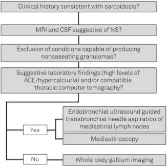
Fluorodeoxyglucose PET imaging may reveal areas of hyper or hypometabolism within the CNS, which combined with areas of hypermetabolism on systemic PET may be elucida-tive to NS diagnosis22,24. Fig 2 shows an algorithm to
investi-gate NS suspected cases.
CSF analysis can be helpful, although not speciic. he majority of patients, particularly those with meningeal in-volvement, present mononuclear pleocytosis ranging from 10 to 100 cells/mm3, glucose as low as 30 mg/dL, and elevated
opening pressure. Elevated CSF IgG titers and oligoclonal bands may be observed in some patients. Although ACE in the CSF is not speciic of NS, it is most probably synthesized within the nervous system and seems to be especially useful in the monitoring of disease activity or treatment response1.
Increased CSF lymphocytes CD4+: CD8+ ratio has also been
observed in NS. However, it requires large numbers of viable lymphocytes, which are usually unavailable5.
Potential biomarkers to detect disease activity are under investigation. More recently, chitotriosidase, an enzyme in-volved in the degradation of chitin, may induce overexpres-sion of h2 cytokines and forecast disease activity and prog-nosis in sarcoidosis, with sensitivity over 90%5.IL-2 receptor
may also be elevated in BAL of sarcoidosis patients with prognostic value and even higher levels in patients with ex-trapulmonary involvement5,19.
DIFFERENTIAL DIAGNOSIS
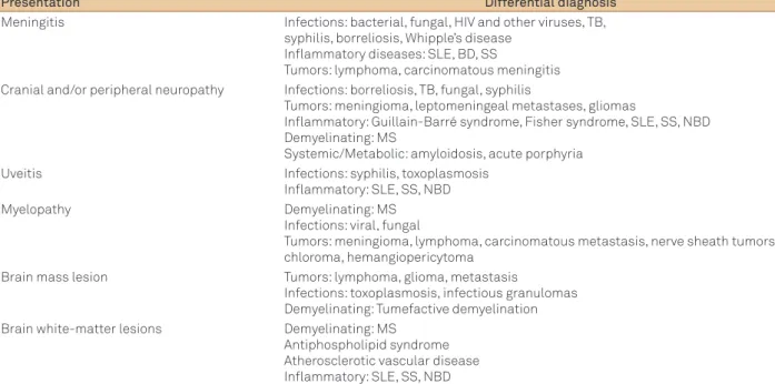
Diferential diagnosis in NS depends on the clinical pre-sentation. he presence of multisystem disease usually helps establishing the diagnosis, especially when patholog-ical analyses showing sarcoid features are available at least from one site. It is important to rule out infectious diseases, neoplasms, and other autoimmune inlammatory disorders. Clinical manifestations and also MRI-based indings may guide the investigation (Table 2).
In the setting of subacute or chronic meningitis with headache, cranial neuropathies and constitutional symp-toms, CSF analysis is mandatory. PCR for herpes family vi-ruses have high sensitivity and speciicity26. On the other
hand, PCR for tuberculosis have a variable and otherwise low sensitivity, but high speciicity27,28. Fungal and tuberculosis
(TB) stains and cultures, immunologic analysis for fungal an-tigens, and oncotic cytology are also important. hickening and enhancement of the leptomeninges of patients with NS are virtually indistinguishable from those seen in tuberculo-sis or lymphoma29. If dural involvement occurs, one should
consider meningioma, dural metastasis, lymphoma, and id-iopathic hypertrophic cranial pachymeningitis.
TB and sarcoid meningitis classically have predilection for basal involvement and may cause multiple cranial neu-ropathies. However, syphilis, lymphoma, viral and fungal meningitis may also involve such region4,30,31. When facial
pal-sy, either uni or bilaterally, is present, borreliosis, Fisher and Guillain-Barré syndrome should be considered, as well as HIV infection, mononucleosis, postinluenza, syphilis, acute porphyria, amyloidosis, and multiple sclerosis4,32. he
difer-ential diagnosis of isolated optic neuropathy includes optic nerve glioma, optic neuritis or involvement of dural nerve sheath by meningioma15. Sarcoidosis may also afect orbital
fat, extrinsic ocular muscles or lacrimal glands mimicking or-bital pseudotumor33.
Fig 2. Algorithm for investigation of suspected neurosarcoidosis.
MRI: magnetic resonance image; CSF: cerebral spinal fluid; NS: neurosarcoidosis; ACE: angiotensin converting-enzyme.
Clinical history consistent with sarcoidosis?
MRI and CSF suggestive of NS?
Exclusion of conditions capable of producing noncaseating granulomas?
Suggestive laboratory findings (high levels of ACE/hypercalciuria) and/or compatible
thoracic computer tomography?
Endobronchial ultrasound guided transbronchial needle aspiration of
mediastinal lymph nodes Mediastinoscopy
Whole body gallium imaging Yes
Involvement of hypothalamic and pituitary dysfunction in NS may mimic pituitary adenoma, Rathke cleft cyst, and craniopharyngioma. However, polyuria and polydipsia due to diabetes insipidus or evidence of dysfunction of both ante-rior and posteante-rior pituitary lobes are less frequently associ-ated with tumors32. Isolated infundibulum involvement seen
as thickening and enhancement on T1-weighted MRI images may also be found in histiocytosis29.
Intracranial mass lesions, presenting seizures or focal deicits, should be diferentiated from primary and second-ary tumors or tumecfative demyelination. In sarcoidosis, en-hancing mass lesions are usually associated with adjacent leptomeningeal involvement. Hypointensity on NS lesions T2-weighted images may also be found in lymphoma or hy-percellular metastases. Central necrosis is more commonly seen in neoplasms4,9,29.
TREATMENT
Isolated cranial nerve abnormalities and aseptic menin-gitis are frequent monophasic presentations and are at low risk for progression, with the exception of being occasional cases of progressive optic neuropathy34,35. Such
manifesta-tions usually present good response to short steroid course. However, patients who have parenchymal disease, seizures, mass lesions, leptomeningeal involvement with multiple cra-nial nerve abnormalities, spinal cord disease, and hydroceph-alus often experience a chronic remitting-relapsing course36.
he development of hydrocephalus due to chronic meningi-tis may be an ominous sign37. hese severe manifestations
of-ten require high dose and prolonged course of steroids38.
Although steroids have been the initial option, many pa-tients require alternative treatments due to steroid intoler-ance or persistent disease activity. hree studies that as-sessed the response to steroids alone showed that less than 40% of the patients stabilized or improved5,15,37,38. he report
by Scott et al.37 selected patients for early aggressive
immu-nosuppressive therapy if they ‘presented with severe CNS (in-tracranial lesions, hydrocephalus, myelopathy, seizures, or encephalopathy).’ Among the patients selected to receive ste-roids alone, 35% improved on this regimen, whereas 69% im-proved with immunosuppressive agents. Given the reports of treatment failure with steroid therapy in many patients and the toxic efects associated with long-term steroid use, cen-ters with experience in treating NS often select patients with severe disease for treatment with steroids and alternative im-munosuppressive agents (combination therapy) at the time of initial diagnosis36-38.
We reviewed the most common prescribed agents for the treatment of NS and proposed a treatment strategy based on current studies from diferent centers specialized in sarcoi-dosis and on our own experience (Fig 3).
Steroids
Steroids remain the mainstay of treatment for NS5,15,36,37,39.
hey are most useful early in the treatment since they can lead to rapid reduction of the inlammation and the mass efect36. For severe manifestations, steroids are started at
1 mg/kg or as a pulse of methylprednisolone 1,000 mg/day for three days, followed by tapering. For myopathy, facial palsy, and neuropathy, they are started at 0.5 mg/kg and maintained from two to four weeks, followed by tapering. Patients should be monitored for development of diabetes,
Presentation Differential diagnosis
Meningitis Infections: bacterial, fungal, HIV and other viruses, TB, syphilis, borreliosis, Whipple’s disease
Inflammatory diseases: SLE, BD, SS
Tumors: lymphoma, carcinomatous meningitis Cranial and/or peripheral neuropathy Infections: borreliosis, TB, fungal, syphilis
Tumors: meningioma, leptomeningeal metastases, gliomas
Inflammatory: Guillain-Barré syndrome, Fisher syndrome, SLE, SS, NBD Demyelinating: MS
Systemic/Metabolic: amyloidosis, acute porphyria Uveitis Infections: syphilis, toxoplasmosis
Inflammatory: SLE, SS, NBD Myelopathy Demyelinating: MS
Infections: viral, fungal
Tumors: meningioma, lymphoma, carcinomatous metastasis, nerve sheath tumors, chloroma, hemangiopericytoma
Brain mass lesion Tumors: lymphoma, glioma, metastasis
Infections: toxoplasmosis, infectious granulomas Demyelinating: Tumefactive demyelination Brain white-matter lesions Demyelinating: MS
Antiphospholipid syndrome Atherosclerotic vascular disease Inflammatory: SLE, SS, NBD
Table 2. Differential diagnosis of NS based on clinical and imaging manifestations.
Fig 3. Suggested treatment strategy for neurosarcoidosis.
NS: neurosarcoidosis; PRED: prednisone; MTX: methotrexate; CFA: cyclophosphamide.
Treatment algorithm for Neurosarcoidosis
Mild NS
PRED
Tapper PRED <10 mg/day
Add MTX Unable/ Relapse
PRED + MTX or
Tapper <10 mg/day
Infliximab or PRED + CFA
Unable/ Relapse Moderate/Severe NS
glucose intolerance, dislipemia, hypertension, and osteo-porosis and frequently low steroids doses are required for more than one year. he goal is to maintain the patients with doses <10 mg/day40.
Immunosuppressive agents
Methotrexate (MTX), azathioprine (AZA), and myco-phenolate (MMF) are immunosuppressive agents common-ly prescribed and equalcommon-ly efective. here are no randomized trials deining the optimal treatment for NS. MTX is the most common prescribed immunosuppressant, usually at 7.5 to 15 mg/kg per week, followed by folic acid supplemen-tation and requires bimonthly evaluation of liver function and blood cell counts with diferential. AZA is prescribed at 150 to 200 mg/day, and requires monthly liver function tests and blood cell counts.
MMF is efective in treating cutaneous and renal sarcoi-dosis. It is associated with less incidence of neutropenia than other cytotoxic agents; however, nausea and diarrhea can be dose-limiting adverse efects40. It is prescribed at 1,000 mg
twice daily and it is occasionally used in association with inf-liximab41. Although it is considered as a therapeutic option in
NS, MMF is not efective in all NS manifestations, presenting low response for myopathy21.
Cyclophosphamide (CFA) is reserved for refractory cases, according to previous studies36,38,42,43. It is started at
0.5 mg/m2 until a maximum of 1,000 mg in monthly pulses
for 6 to 12 months. Liver, renal function, blood cells count should be monitored monthly. CFA is associated with op-portunistic and fungal infections, hemorrhagic cystitis, bone marrow aplasia, and higher cancer rates.
Anti-TNF agents
Inliximab is a chimeric monoclonal antibody that neu-tralizes the biological activity of TNF-alpha by binding to its soluble and transmembrane forms and inhibiting receptor
binding44. Evidence for its use came from the inding of
el-evated TNF-alpha levels in lymph nodes and BAL of patients with sarcoidosis39 and by the demonstration that it is a
cru-cial cytokine in the establishment and maintenance of in-lammation in several autoimmune disorders44. here are 23
reports on the use of inliximab for refractory NS, sometimes in association with other immunosuppressive agents, such as CFA or MMF36,41,45,46. It is prescribed at 5 mg/kg at zero, two
and six weeks and then repeated every eight weeks. Because inliximab may be associated with reactivation of tuberculo-sis, chest X-rays and tuberculin tests are required before ini-tiating therapy. Inliximab may also precipitate oncological and autoimmune complications, and it was also associated with high mortality rates in patients with advanced conges-tive heart failure40,47.
Etarnecept, which is another TNF-alpha antagonist, pres-ents lower response than inliximab for the treatment of sar-coidosis. he reason for the enhanced eicacy of inliximab is not well understood, but it is believed that inliximab is highly speciic for TNF-alpha, whereas etanercept also binds a related molecule, lymphotoxin-α ( formerly known as TNF-beta). he molecular structure (an IgG antibody) of inlix-imab allows it to initiate the classical complement pathway and causes cell lysis, whereas etanercept, a recombinant molecule attached to the Fc portion of human IgG1, cannot. Moreover, inliximab has been demonstrated to induce T-cell apoptosis, and etanercept has not47.
Inliximab is a rescue option for patients resistant to CFA and there is evidence that it could be more efective than CFA42. For that reason, some specialists initiate inliximab
before CFA for severe manifestations, which have already failed one immunosuppressive agent36,40,44.
PROGNOSIS
Although treatment with steroids can improve MRI, clini-cal responses may not correlate with radiologiclini-cal improve-ment36. Mortality rates were 18% in a series of 37 patients and
31% developed steroid side efects, related to the high dose of steroids and longer duration of therapy48. In another series
of 54 patients, 10% developed progressive symptoms despite immunosuppressive treatment49.
CONCLUSIONS
1. Hoitsma E, Faber CG, Drent M, Sharma OP. Neurosarcoidosis: a clinical dilemma. Lancet Neurol 2004;3:397-407.
2. Sharma OP, Shigemitsu H. A historical sketch: life and time of Jonathan Hutchinson (1828-1913), the first sarcoidologist. Sarcoidosis Vasc Diffuse Lung Dis 2008;25:71-75.
3. Vinas FC, Rengachary S. Diagnosis and management of neurosarcoidosis. J Clin Neurosci 2001;8:505-513.
4. Nowak DA, Widenka DC. Neurosarcoidosis: a review of its intracranial manifestation. J Neurol 2001;248:363-372.
5. Lower EE, Broderick JP, Brott TG, Baughman RP. Diagnosis and management of neurological sarcoidosis. Arch Intern Med 1997;157:1864-1868.
6. Whelan MA, Stern J. Sarcoidosis presenting as a posterior fossa mass. Surg Neurol 1981;15:455-457.
7. Tsao CY, Lo WD, Rusin JA, Henwood MJ, Boue DR. Isolated neurosarcoidosis presenting as headache and multiple brain and spinal cord lesions mimicking central nervous system metastases. Brain Dev 2007;29:514-518.
8. Sponsler JL, Werz MA, Maciunas R, Cohen M. Neurosarcoidosis presenting with simple partial seizures and solitary enhancing mass: case reports and review of the literature. Epilepsy Behav 2005;6:623-630.
9. Chapelon C, Ziza JM, Piette JC, et al. Neurosarcoidosis: signs, course and treatment in 35 confirmed cases. Medicine (Baltimore) 1990;69:261-276.
10. Lawrence WP, el-Gammal T, Pool WH, Jr, Apter L. Radiological manifestations of neurosarcoidosis: report of three cases and review of literature. Clin Radiol 1974;25:343-348.
11. Oksanen V. Neurosarcoidosis: clinical presentations and course in 50 patients. Acta Neurol Scand 1986;73:283-290.
12. Byrne JV, Lawton CA. Meningeal sarcoidosis causing intracranial hypertension secondary to dural sinus thrombosis. Br J Radiol 1983;56:755-757.
13. Gilmore K, Rudden M, Kalman TP. Psychiatric manifestations of sarcoidosis. Can J Psychiatry 1980;25:329-331.
14. Scott TF. Neurosarcoidosis: progress and clinical aspects. Neurology 1993;43:8-12.
15. Zajicek JP, Scolding NJ, Foster O, et al. Central nervous system sarcoidosis-diagnosis and management. QJM 1999;92:103-117. 16. Delaney P. Neurologic manifestations in sarcoidosis: review of the
literature, with a report of 23 cases. Ann Intern Med 1977;87:336-345. 17. Vital A, Lagueny A, Ferrer X, Louiset P, Canron MH, Vital C. Sarcoid
neuropathy: clinico-pathological study of 4 new cases and review of the literature. Clin Neuropathol 2008;27:96-105.
18. Chen ES, Moller DR. Etiology of sarcoidosis. Clin Chest Med 2008;29:365-377.
19. Morgenthau AS, Iannuzzi MC. Recent advances in sarcoidosis. Chest 2011;139:174-182.
20. Newman LS, Rose CS, Bresnitz EA, et al. A case control etiologic study of sarcoidosis: environmental and occupational risk factors. Am J Respir Crit Care Med 2004;170:1324-1330.
21. Gerke AK, Hunninghake G. The immunology of sarcoidosis. Clin Chest Med. 2008;29:379-390.
22. Stern BJ. Neurological complications of sarcoidosis. Curr Opin Neurol 2004;17:311-316.
23. Lynch JP 3rd. Neurosarcoidosis: how good are the diagnostic tests? J Neuroophthalmol 2003;23:187-189.
24. Bargagli E, Mazzi A, Rottoli P. Markers of inflammation in sarcoidosis: blood, urine, BAL, sputum, and exhaled gas. Clin Chest Med. 2008;29:445-458.
25. Dubey N, Miletich RS, Wasay M, Mechtler LL, Bakshi R. Role of fluorodeoxyglucose positron emission tomography in the diagnosis of neurosarcoidosis. J Neurol Sci 2002;205:77-81.
26. Lakeman FD, Whitley RJ. Diagnosis of herpes simplex encephalitis: application of polymerase chain reaction to cerebrospinal fluid from brain-biopsied patients and correlation with disease. National Institute of Allergy and Infectious Diseases Collaborative Antiviral Study Group. J Infect Dis 1995;171:857-863.
27. Donald PR, Victor TC, Jordaan AM, Schoeman JF, van Helden PD. Polymerase chain reaction in the diagnosis of tuberculous meningitis. Scand J Infect Dis 1993;25:613-617.
28. Nguyen LN, Kox LF, Pham LD, Kuijper S, Kolk AH. The potential contribution of the polymerase chain reaction to the diagnosis of tuberculosis meningitis. Arch Neurol 1996;53:771-776.
29. Smith JK, Matheus MG, Castillo M. Imaging manifestations of neurosarcoidosis. Am J Roentgenol 2004;182:289-295.
30. Stern BJ, Krumholz A, Johns C, Scott P, Nissim J. Sarcoidosis and its neurological manifestations. Arch Neurol 1985;42:909-917.
31. Ranoux D, Devaux B, Lamy C, Mear JY, Roux FX, Mas JL. Meningeal sarcoidosis, pseudo-meningioma, and pachymeningitis of the convexity. J Neurol Neurosurg Psychiatry 1992;55:300-303. 32. Hoitsma E, Drent M, Sharma OP. A pragmatic approach to diagnosing
and treating neurosarcoidosis in the 21st century. Curr Opin Pulm Med 2010;16:472-479.
33. Carmody RF, Mafee MF, Goodwin JA, Small K, Haery C. Orbital and optic pathway sarcoidosis: MR findings. AJNR Am J Neuroradiol 1994;15:775-783.
34. Ferriby D, de Seze J, Stojkovic T, et al. Long-term follow-up of neurosarcoidosis. Neurology 2001;57:927-929.
35. Chapelon-Abric C, Ziza JM, Piette JC, et al. Neurosarcoidosis. Ann Med Interne (Paris) 1991;142:601-608.
36. Lower EE, Weiss KL. Neurosarcoidosis. Clin Chest Med 2008;29:475-492. 37. Scott TF, Yandora K, Valeri A, Chieffe C, Schramke C. Aggressive therapy
for neurosarcoidosis: long-term follow-up of 48 treated patients. Arch Neurol 2000;64:691-696.
38. Agbogu BN, Stern BJ, Sewell C, Yang G. Therapeutic considerations in patients with refractory neurosarcoidosis. Arch Neurol 1995;52:875-879. 39. Hunninghake GW, Costabel U, Ando M, et al. ATS/ERS/WASOG statement
on sarcoidosis. American Thoracic Society/European Respiratory Society/World Association of Sarcoidosis and other Granulomatous Disorders. Sarcoidosis Vasc Diffuse Lung Dis 1999;16:149-173. 40. Baughman RP, Costabel U, du Bois RM. Treatment of sarcoidosis. Clin
Chest Med 2008;29:533-548.
41. Moravan M, Segal BM. Treatment of CNS sarcoidosis with infliximab and mycophenolate mofetil. Neurology 2009;72:337-340.
42. Doty JD, Mazur JE, Judson MA. Treatment of corticosteroid-resistant neurosarcoidosis with a short-course cyclophosphamide regimen. Chest 2003;124:2023- 2026.
43. Vargas DL, Stern BJ. Neurosarcoidosis: diagnosis and management. Semin Respir Crit Care Med 2010;31:419-427.
44. Santos E, Shaunak S, Renowden S, Scolding NJ. Treatment of refractory neurosarcoidosis with Infliximab. J Neurol Neurosurg Psychiatry 2010;81:241-246.
45. Pettersen JA, Zochodne DW, Bell RB, Martin L, Hill MD. Refractory neurosarcoidosis responding to infliximab. Neurology 2002;59:1660-1661. 46. Pereira J, Anderson NE, McAuley D, Bergin P, Kilfoyle D, Fink J.
Medically refractory neurosarcoidosis treated with infliximab. Intern Med J 2011;41:354-357.
47. Doty JD, Mazur JE, Judson MA. Treatment of sarcoidosis with infliximab. Chest 2005;127:1064-1071.
48. Om P. Sharma. Neurosarcoidosis: A Personal Perspective Based on the Study of 37 Patients. Chest 1997;112:220-228.