CLINICAL SCIENCE
Intestinal microsporidiosis: a hidden risk in rheumatic
disease patients undergoing anti-tumor necrosis
factor therapy combined with disease-modifying
anti-rheumatic drugs?
Nadia Emi Aikawa,I,IIAline de Oliveira Twardowsky,IIJoze´lio Freire de Carvalho,IClovis A. Silva,I,II Ivan Leonardo Avelino Franc¸a e Silva,IIIAna Cristina de Medeiros Ribeiro,ICarla Gonc¸alves Schain Saad,I Julio Ce´sar Bertacini Moraes,IRoberto Acayaba de Toledo,IVEloı´sa Bonfa´I
IDivision of Rheumatology, CEDMAC Unit, Faculdade de Medicina da Universidade de Sa˜o Paulo, Sa˜o Paulo, SP/Brazil.IIPediatric Reumatology Unit,
Faculdade de Medicina da Universidade de Sa˜o Paulo, Sa˜o Paulo, SP/Brazil.IIIDivision of Infectology, Faculdade de Medicina da Universidade de Sa˜o Paulo,
Sa˜o Paulo, SP/Brazil.IVDivision of Rheumatology, CEDMAC Unit, Faculdade de Medicina de Sa˜o Jose´ do Rio Preto, SP/Brazil.
OBJECTIVE: Immunosuppressed patients are at risk of microsporidiosis, and this parasitosis has an increased rate of dissemination in this population. Our objective was to evaluate the presence of microsporidiosis and other intestinal parasites in rheumatic disease patients undergoing anti-tumor necrosis factor/disease-modifying anti-rheumatic drug treatment.
METHODS:Ninety-eight patients (47 with rheumatoid arthritis, 31 with ankylosing spondylitis and 11 with psoriatic arthritis) and 92 healthy control patients were enrolled in the study. Three stool samples and cultures were collected from each subject.
RESULTS: The frequency of microsporidia was significantly higher in rheumatic disease patients than in control subjects (36 vs.4%, respectively;p,0.0001), as well as in those with rheumatic diseases (32 vs.4%, respectively;
p,0.0001), ankylosing spondylitis (45vs.4%, respectively;p,0.0001) and psoriatic arthritis (40vs.4%, respectively;
p,0.0001), despite a similar social-economic class distribution in both the patient and control groups (p= 0.1153). Of note, concomitant fecal leukocytes were observed in the majority of the microsporidia-positive patients (79.5%). Approximately 80% of the patients had gastrointestinal symptoms, such as diarrhea (26%), abdominal pain (31%) and weight loss (5%), although the frequencies of these symptoms were comparable in patients with and without this infection (p.0.05). Rheumatoid arthritis, ankylosing spondylitis and psoriatic arthritis disease activity parameters were comparable in both groups (p.0.05). The duration of anti-tumor necrosis factor/disease-modifying anti-rheumatic drugs and glucocorticoid use were also similar in both groups.
CONCLUSION:We have documented that microsporidiosis with intestinal mucosa disruption is frequent in patients undergoing concomitant anti-tumor necrosis factor/disease-modifying anti-rheumatic drug therapy. Impaired host defenses due to the combination of the underlying disease and the immunosuppressive therapy is the most likely explanation for this finding, and this increased susceptibility reinforces the need for the investigation of microsporidia and implementation of treatment strategies in this population.
KEYWORDS: Microsporidia; Parasitosis; Anti-TNF; Rheumatoid arthritis; Ankylosing spondylitis.
Aikawa NE, Twardowsky AO, Carvalho JF, Silva CA, Franc¸a e Silva ILA, Ribeiro ACM, et al. Intestinal microsporidiosis: a hidden risk in rheumatic disease patients undergoing anti-tumor necrosis factor therapy combined with disease-modifying anti-rheumatic drugs? Clinics. 2011;66(7):1171-1175.
Received for publication onJanuary 12, 2011;First review completed onFebruary 14, 2011;Accepted for publication onMarch 21, 2011 E-mail: [email protected]
Tel.: 005511-3069-8563 Fax: 005511-3069-8503
INTRODUCTION
Microsporidia has emerged in recent years as an opportu-nistic infectious agent with a ubiquitous distribution.1-4The clinical manifestations of this infection vary widely, from none (asymptomatic) to non-bloody watery diarrhea, abdom-inal cramps, weight loss, and disseminated disease, espe-cially in immunosuppressed subjects.2-4
This parasitological disease primarily affects immuno-suppressed patients,1-4 particularly those with human immunodeficiency virus (HIV), malignancies and diabetes mellitus, as well as patients post heart-lung, liver, and renal transplantations.1,2In addition, this infection also occurs in patients undergoing immunosuppressive drug treatment.2,4
In fact, patients with rheumatic diseases who are taking disease-modifying anti-rheumatic drugs (DMARD) and anti-tumor necrosis factor (TNF) therapy have a high risk for general infections,5-7 including intestinal pathogenic
parasite infestations such as Strongyloides stercoralis.8,9 However, to our knowledge, there are no data regarding the risk of microsporidiosis in these patients.10-13
Therefore, the objective of this study was to evaluate the frequency and clinical significance of microsporidiosis in patients with rheumatic diseases who are undergoing anti-TNF/DMARD treatment compared to an age- and socio-economic condition-matched healthy population.
MATERIALS AND METHODS
We evaluated 89 consecutive patients at our hospital who were diagnosed with rheumatoid arthritis (RA) (American College of Rheumatology criteria),14ankylosing spondylitis
(AS) (New York criteria),15 or psoriatic arthritis (PsA) (European spondyloarthropathy Study Group – ESSG criteria and Moll and Wright classification).16 All patients
were undergoing anti-TNF therapy (adalimumab, etaner-cept, or infliximab) combined with DMARDs. The control group included 92 healthy employees of our hospital who were matched by age and socio-economic status. Prior to this study, no specific routine existed in our Infusion Center with regard to stool examination for parasites and fecal leukocytes before anti-TNF therapy. The prophylactic use of anti-helminthic drugs was recommended for patients under concomitant glucocorticoid therapy.
This study was approved by the local ethics committee, and informed consent was obtained from each participant or his or her legal guardian.
Demographic data, the number of school years attended, and the socio-economic status (according to Associac¸a˜o Brasileira dos Institutos de Pesquisa de Mercados)17 were recorded. Background information and parasitosis symp-toms in patients and control participants were acquired via interview and included the following: general (loss of appetite, weight loss, and adynamia), gastrointestinal (abdominal pain, abdominal distension, flatulence, diarrhea, dysentery, tenesmus, obstipation, nausea, vomiting, hema-tochezia, and worm elimination), cutaneous (exanthema), and pulmonary manifestations (wheezing, thoracic pain, and hemoptysis).
Disease activity and laboratory evaluation of rheumatic diseases
Disease activity was evaluated by the following: the duration of morning stiffness, the number of swollen and/ or painful joints, and the disease activity score (DAS28) for RA patients18 and the number of swollen and/or painful joints and the Bath Ankylosing Spondylitis Disease Activity Index (BASDAI)19for AS and PsA patients.
The laboratory parameters assessed included the erythro-cyte sedimentation rate (ESR) according to Westergreen (mm/1st hour) and C-reactive protein (CRP) level
deter-mined by nephelometry (mg/L). Data concerning current
treatment with prednisone, DMARDs, anti-TNF and immu-nosuppressive drugs (methotrexate, azathioprine, lefluno-mide, chloroquine, sulfasalazine and/or cyclosporine) were collected.
Parasitological assessment
Three stool samples were collected from rheumatic disease patients and control subjects on different days and placed into plastic vials without preservatives. The stool samples were microscopically analyzed for the detection of protozoan oocysts, cysts, helminth eggs, and larvae by a qualified technician blinded to the groups using the techniques described by Faust et al, Rugai and Lutz (modified), and Hoffman, Pons and Janer.20
Microsporidia positivity was defined as the isolation of at least one parasite using the GRAM-Chromotrope21 technique.
Intestinal coccidia were evaluated by the GRAM-Chromotrope21 technique; Leishman staining22 was used
to identify Blastocystis hominis and fecal leukocytes; the Kinyoun procedure23and capture enzyme-linked
immuno-sorbent assay (capture ELISA) were used to detect Cryptosporidium sp.,Cyclospora cayetanensis,andIsospora belli; and Kato-Katz was used to detect Schistosoma mansoni.24
Coproculture was also performed using SS agar, MacConkey agar or Karmali plate agar.25
Giardia lamblia, Strongyloides stercoralis, Ancilostoma duode-nali, Ascaris lumbricoides, Entamoeba histolytica, Microsporidia (positive stool leukocytes), Entamoeba dispar, Dientamoeba fragilis, Cryptosporidium parvum, Cyclospora cayetanensis, Isospora belli, andBlastocystis hominis(positive stool leuko-cytes) were defined as pathogenic parasites in immuno-compromised patients.1 All participants with positive
pathogenic parasites were treated with the appropriate recommended antiparasitic drugs.26
Statistical analysis
Results are presented as the mean¡standard deviation
or the median (range) for continuous variables and the number (%) for categorical variables. Continuous variables were compared using thettest and the Mann Whitney test to evaluate differences between patients with and without intestinal microsporidia and other parasitoses. For catego-rical variables, differences in proportions were assessed by Fisher’s exact test. For all statistical tests, significance was set at ap-value,0.05.
RESULTS
A total of 89 patients with rheumatic diseases (47 RA, 31 AS and 11 PsA) and 92 healthy control subjects were included in this study. The two groups were comparable with regard to the mean current age (47.2¡10.9 vs.
43.8¡14.3 years, respectively; p= 0.094), percentage who
Intestinal parasites
The overall frequency of pathogenic parasites was similar in both the patient and control groups (63 vs. 58%, respectively;p= 0.54). Positive tests for microsporidia were significantly higher in all types of rheumatic disease patients compared with the control subjects (38 vs. 4%,
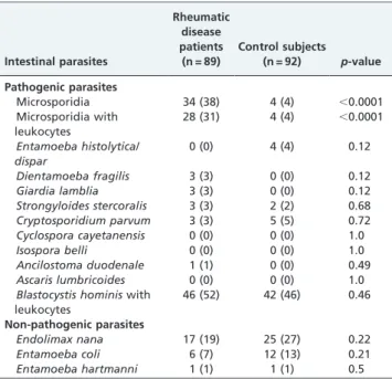
respectively; p,0.0001), and microsporidia plus positive fecal leukocytes were also more frequently detected in patients than in control subjects (31 vs. 4%, respectively; p,0.0001). Of note, 81.6% of the positive microsporidia patients had concomitant fecal leukocytes. In contrast, no differences were detected for other pathogenic parasites (Entamoeba histolytica/dispar, Dientamoeba fragilis, Giardia lamblia, Cryptosporidium parvum, Cyclospora cayetanensis, Isospora belli, Ancilostoma duodenale, Ascaris lumbricoides, and Blastocystis hominis with leukocytes) and non-patho-genic parasites (Endolimax nana, Entamoeba coli, and Entamoeba hartmanni) (p.0.05; Table 1). Among the 28 patients with microsporidia and positive stool leukocytes, 36% had other concomitant pathogenic parasites, particu-larlyBlastocystis hominis[8/28 (29%) patients]; however, no association of microsporidia and Blatocystis hominis was observed (p= 0.28). Regarding the prevalence of pathogenic parasites in each disease compared to the control group, microsporidiosis was higher in RA (32vs.4%, respectively; p,0.0001), AS (45vs. 4%, respectively;p,0.0001) and PsA patients (40vs.4%, respectively;p,0.0001). Moreover, there were no statistical differences in microsporidia positivity between the specific diseases (RA vs. AS and PsA; AS vs. PsA; all p.0.05). Giardia lamblia was significantly more common in AS patients vs. control subjects (10% vs. 0%, respectively; p= 0.0149), whereas no differences were observed for the other pathogenic parasites (p.0.05).
Disease activity parameters at the study onset were alike in patients with and without microsporidia infections for all diseases (RA, AS, and PsA) (p.0.05). The analysis of the duration of anti-TNF/DMARD therapy showed no statistical difference between patients with and without microsporidia infection (p= 0.55). The frequencies of glucocorticoid use in patients with and without microsporidiosis [44% (95% IC 0.27-0.62)vs.64% (95% IC 0.49-0.76);p= 0.08] were alike, as was the number of DMARDs used (Table 2).
Table 1 -Intestinal parasites in rheumatic disease patients undergoing anti-TNF/DMARD therapies and healthy controls.
Intestinal parasites
Rheumatic disease patients (n = 89)
Control subjects (n = 92) p-value Pathogenic parasites
Microsporidia 34 (38) 4 (4) ,0.0001
Microsporidia with leukocytes
28 (31) 4 (4) ,0.0001
Entamoeba histolytica/ dispar
0 (0) 4 (4) 0.12
Dientamoeba fragilis 3 (3) 0 (0) 0.12
Giardia lamblia 3 (3) 0 (0) 0.12
Strongyloides stercoralis 3 (3) 2 (2) 0.68
Cryptosporidium parvum 3 (3) 5 (5) 0.72
Cyclospora cayetanensis 0 (0) 0 (0) 1.0
Isospora belli 0 (0) 0 (0) 1.0
Ancilostoma duodenale 1 (1) 0 (0) 0.49
Ascaris lumbricoides 0 (0) 0 (0) 1.0
Blastocystis hominiswith
leukocytes
46 (52) 42 (46) 0.46
Non-pathogenic parasites
Endolimax nana 17 (19) 25 (27) 0.22
Entamoeba coli 6 (7) 12 (13) 0.21
Entamoeba hartmanni 1 (1) 1 (1) 0.5
Results are presented as n (%); DMARDs - disease-modifying anti-rheumatic drugs.
Table 2 -Disease activity parameters and treatments in patients with rheumatic diseases undergoing anti-TNF and DMARD therapies with and without microsporidia infection.
Parameters Patients with microsporidia (n = 34) Patients without microsporidia (n = 55) p-value Rheumatoid arthritis (n = 47)
Morning stiffness, minutes 13¡17.9 26.9¡43.2 0.24
Number of painful joints 6.5¡5.8 11¡10 0.12
Number of swollen joints 4.8¡5.1 5.1¡4.1 0.83
DAS28 3.8¡1.6 4.6¡1.3 0.10
ESR, mm/1sthour 26.6
¡26.1 27.1¡15.5 0.94
CRP, mg/L 8.49¡9.5 15.4¡17.9 0.19
Ankylosing spondylitis (n = 31)
Number of painful joints 5.6¡13.4 3.6¡4.2 0.59
Number of swollen joints 1.4¡1.4 1.7¡2.3 0.68
BASDAI 3.48¡2.01 3.47¡2.43 0.99
ESR, mm/1sthour 23¡27 14¡16 0.27
CRP, mg/L 12.6¡14.5 13.2¡21.1 0.94
Psoriatic arthritis (n = 11)
Morning stiffness (minutes) 0 (0-20) 17.5 (0-120) 0.14
Number of painful joints 5 (0-8) 3.5 (0-12) 0.93
Number of swollen joints 1 (0-2) 1 (0-2) 0.77
BASDAI 2.33 (0-6.98) 4.06 (0.27-5.16) 0.48
ESR, mm/1sthour 11.5 (3-76) 23(1-46) 0.73
CRP, mg/L 2 (0.2-19.6) 6.8 (1.5-32.5) 0.55
Rheumatic patients treated (n = 89)
Anti-TNF and DMARD therapy duration (months)
15¡11.7 13.6¡9.7 0.55
Glucocorticoid use 15 (44) 35 (64) 0.08
Methotrexate use 26 (76) 33 (60) 0.17
No differences were detected in the frequencies of other infections that required antibiotic therapy in patients with and without microsporidiosis, such as acute lower respira-tory infection (2.9 vs.14.5%, respectively; p= 0.14), urinary tract infection (29.4 vs. 30.9%, respectively; p= 1.0) cuta-neous infection (20.6 vs. 23.6%, respectively; p= 0.8) and tuberculosis (2.9vs.1.8%, respectively;p= 1.0).
The comparison of rheumatic disease patients with and without microsporidiosis revealed that 85.4% of the patients had gastrointestinal symptoms such as diarrhea (19%), abdominal pain (22%), and weight loss (11%), but these symptoms occurred just as frequently in patients with and without this infection (p.0.05). Other signs and symptoms related to parasitoses were also comparable in the two groups, with the exception of abdominal distension (p= 0.013) and adynamia (p= 0.009), both of which occurred less often in patients with microsporidiosis (Table 3). No patient or control group had a disseminated infection.
DISCUSSION
This study is the first to identify a high frequency of microsporidia infections associated with intestinal mucosa disruption in rheumatic disease patients undergoing anti-TNF and DMARD therapies.
The study design has several advantages, including the inclusion of patients with well-established rheumatic disease criteria.14-16In addition, the Gram-Chromotrope stain meth-odology applied in this study is considered to be one of the most specific detection techniques with the best sensitivity for detecting microsporidia in fluids, including feces.2 Moreover, external factors such as socio-economic conditions may influence parasitosis prevalence. For example, the
frequency of microsporidia in HIV-infected patients was higher in Venezuela (13.6%), an underdeveloped country, than in Italy (1.8%).27-28 This potential bias was greatly reduced by the matching of our patients with a control group with the same socio-financial distribution. One limitation of our study was the absence of a control group of rheumatic disease patients treated only with DMARDs, due to the impossibility of achieving an adequate matching for disease duration and severity in these patients. In fact, anti-TNF therapy is now being indicated very early for patients with these diseases. There are, however, no data on the frequency of microsporidia infection in rheumatic disease patients without anti-TNF therapy.
Microsporidia has emerged as an important cause of infectious complications in severely immunocompromised patients with HIV, recipients of solid organ transplant and patients with hematological malignancies.1-3 Cell-mediated immunity appears to be critical for protection against microsporidia through the T helper cell 1 (Th1) cytokine response.3 The importance of a Th1 response in the resistance to microsporidial infection has been demon-strated byin vitro studies showing that knockout animals for Th1 cytokines such as interferon and interleukin-12 could not clear microsporidia infections.29 In fact, more severe microsporidia infections were observed in HIV-infected patients with declining CD4+ and CD8+ T-cell numbers.29 Accordingly, the inhibition of TNF-alpha, a
cytokine well known to be related to the Th1 response,30 could possibly facilitate the microsporidia infestation observed in the present study. Moreover, experimental studies have reported a decrease in the specific protective IgG against microsporidia in animals treated with immu-nosuppressive drugs.31We have confirmed that microspor-idia infestation is more prevalent in immunosuppressed patients1-4 and have now extended this observation to
rheumatic disease patients undergoing anti-TNF/DMARD treatment.
Microsporidiosis and intestinal parasitosis may present with diverse clinical manifestations, depending on the host immune status and the microsporidium species.1-4 Diarrhea and wasting syndromes are the most common complaints;1-4 however, these parasite infections can be asymptomatic. In fact, the parasitosis in our study was frequently associated with gastrointestinal manifestations and concomitant stool leukocytes, indicating a possible intestinal mucosa disruption,32 which is a known risk for intestinal dissemination.33
Infection is a major co-morbidity in rheumatic conditions, and therapies such as conventional DMARDs and anti-TNF are known to enhance the risk of infection.34,35In this regard, previous reports have suggested that susceptibility to infection may be distinct in different underlying rheumatic diseases. The main explanation for this finding seems to be a disease-associated genetic background and immunosuppres-sive therapy.36,37However, in the present study, no
differ-ence was observed in microsporidiosis frequency between the specific rheumatic diseases, suggesting that anti-TNF/ DMARD treatment is a more relevant risk factor for this infestation than is the specific rheumatic disease itself.
We have identified microsporidiosis to be a frequent infection that is associated with mucosal lesions in patients undergoing concomitant anti-TNF/DMARD treatment. This finding supports the notion that the recommendation for the prophylactic use of anti-helminthic drugs in patients on
Table 3 -Clinical manifestations in patients with rheumatic diseases undergoing anti-TNF and DMARD therapies with and without microsporidia infection.
Clinical manifestations
Patients with microsporidia
(n = 34)
Patients without microsporidia
(n = 55) p-value
Abdominal pain 10 (29) 10 (18) 0.296
Diarrhea 10 (29) 7 (13) 0.094
Weight loss 2 (6) 8 (15) 0.306
Nausea 8 (24) 14 (25) 1.0
Vomit 6 (18) 8 (15) 0.768
Flatulence 6 (18) 19 (35) 0.096
Dysentery 2 (6) 0 (0) 0.143
Tenesmus 0 (0) 4 (7) 0.294
Obstipation 6 (18) 9 (16) 1.0
Loss of appetite 7 (21) 6 (11) 0.231
Hematochezia 2 (6) 5 (9) 0.704
Worm elimination 0 (0) 5 (9) 0.152
Wheezing 8 (24) 5 (9) 0.072
Thoracic pain 8 (24) 6 (11) 0.139
Hemoptysis 2 (6) 2 (4) 0.635
Exanthema 2 (6) 10 (18) 0.121
Abdominal distension
2 (6) 15 (27) 0.013
Adynamia 11 (32) 34 (62) 0.009
Any gastrointestinal symptom
29 (85) 47 (85) 1.0
Systemic dissemination
0 (0) 0 (0) 1.0
glucocorticoid therapy could be extended to those initiating anti-TNF therapy.
Competing interest:This study was supported by grants from FAPESP (2009/51897-5 to EB), CNPQ (3300665/2009-1 to JFC, 300248/2008-3 to CAS and 301411/2009-3 to EB), Federico Foundation (to JFC, CAS and EB) and Wyeth (to NEA).
REFERENCES
1. Stark D, Barratt JL, van Hal S, Marriott D, Harkness J, Ellis JT. Clinical significance of enteric protozoa in the immunosuppressed human population. Clin Microbiol Rev. 2009;22:634-50, doi: 10.1128/CMR. 00017-09.
2. Didier ES. Microsporidiosis: an emerging and opportunistic infection in humans and animals. Acta Trop. 2005;94:61-76, doi: 10.1016/j.actatro-pica.2005.01.010.
3. Didier ES, Weiss LM. Microsporidiosis: current status. Curr Opin Infect Dis. 2006;19:485-92, doi: 10.1097/01.qco.0000244055.46382.23.
4. Franzen C, Mu¨ller A. Cryptosporidia and microsporidia–waterborne diseases in the immunocompromised host. Diagn Microbiol Infect Dis. 1999;34:245-62, doi: 10.1016/S0732-8893(99)00003-6.
5. Rychly DJ, DiPiro JT. Infections associated with tumor necrosis factor-alpha antagonists. Pharmacotherapy. 2005;25:1181-92, doi: 10.1592/phco. 2005.25.9.1181.
6. Bernatsky S, Hudson M, Suissa S. Anti-rheumatic drug use and risk of serious infections in rheumatoid arthritis. Rheumatology. (Oxford) 2007;46:1157-60, doi: 10.1093/rheumatology/kem076.
7. Furst DE. The risk of infections with biologic therapies for rheumatoid arthritis. Semin Arthritis Rheum. 2010;39:327-46, doi: 10.1016/j.semar-thrit.2008.10.002.
8. Krishnamurthy R, Dincer HE, Whittemore D. Strongyloides stercoralis hyperinfection in a patient with rheumatoid arthritis after anti-TNF-alpha therapy. J Clin Rheumatol. 2007;13:150-2, doi: 10.1097/RHU. 0b013e3180690933.
9. Boatright MD, Wang BW. Clinical infection with Strongyloides sterocor-alis following etanercept use for rheumatoid arthritis. Arthritis Rheum. 2005;52:1336-7, doi: 10.1002/art.20882.
10. Lipsky PE, van der Heijde DM, St Clair EW, Furst DE, Breedveld FC, Kalden JR, et al. Infliximab and methotrexate in the treatment of rheumatoid arthritis. Anti-Tumor Necrosis Factor Trial in Rheumatoid Arthritis with Concomitant Therapy Study Group. N Engl J Med. 2000;343:1594-602.
11. Keystone EC, Kavanaugh AF, Sharp JT, Tannenbaum H, Hua Y, Teoh LS, et al. Radiographic, clinical, and functional outcomes of treatment with adalimumab (a human anti-tumor necrosis factor monoclonal antibody) in patients with active rheumatoid arthritis receiving concomitant methotrexate therapy: a randomized, placebo-controlled, 52-week trial. Arthritis Rheum. 2004;50:1400-11, doi: 10.1002/art.20217.
12. Punzi L, Podswiadek M, Sfriso P, Oliviero F, Fiocco U, Todesco S. Pathogenetic and clinical rationale for TNF-blocking therapy in psoriatic arthritis. Autoimmun Rev. 2007;6:524-8, doi: 10.1016/j.autrev.2006.12. 003.
13. Lovell DJ, Giannini EH, Reiff A, Cawkwell GD, Silverman ED, Nocton JJ, et al. Etanercept in children with polyarticular juvenile rheumatoid arthritis. Pediatric Rheumatology Collaborative Study Group. N Engl J Med. 2000;342:763-9.
14. Arnett FC, Edworthy SM, Bloch DA, McShane DJ, Fries JF, Cooper NS, et al. The American Rheumatism Association 1987 revised criteria for the classification of rheumatoid arthritis. Arthritis Rheum. 1988;31:315-24, doi: 10.1002/art.1780310302.
15. Bennett PH, Wood PHN. Population Studies of the Rheumatic Diseases. New York, Excerpta Medica. 1968, p 456.
16. Dougados M, van der Linden S, Juhlin R, Huitfeldt B, Amor B, Calin A, et al. The European Spondylarthropathy Study Group preliminary criteria for the classification of spondylarthropathy. Arthritis Rheum. 1991;34:1218-27, doi: 10.1002/art.1780341003.
17. Almeida PM, Wickerrhauser H. Crite´rio de classe econoˆmica da Associac¸a˜o Brasileira de Anunciantes (ABA) e Associac¸a˜o Brasileira dos Institutos de Pesquisa de Mercado (ABIPEME). 1991, pp 1-129. 18. Prevoo ML, van ’t Hof MA, Kuper HH, van Leeuwen MA, van de
Putte LB, van Riel PL. Modified disease activity scores that include twenty-eight-joint counts. Development and validation in a prospective longitudinal study of patients with rheumatoid arthritis. Arthritis Rheum. 1995;38:44-8, doi: 10.1002/art.1780380107.
19. Garrett S, Jenkinson T, Kennedy LG, Whitelock H, Gaisford P, Calin A. A new approach to defining disease status in ankylosing spondylitis: the Bath Ankylosing Spondylitis Disease Activity Index. J Rheumatol. 1994;21:2286-91.
20. Koga K, Kasuya S, Khamboonruang C, Sukavat K, Nakamura Y, Tani S, et al. An evaluation of the agar plate method for the detection of Strongyloides stercoralis in northern Thailand. J Trop Med Hyg. 1990;93:183-8.
21. Ryan NJ, Sutherland G, Coughlan K, Globan M, Doultree J, Marshall J, et al. A new trichrome-blue stain for detection of microsporidial species in urine, stool, and nasopharyngeal specimens. J Clin Microbiol. 1993;31:3264-9.
22. Price DL. Comparison of three collection-preservation methods for detection of intestinal parasites. J Clin Microbiol. 1981;14:656-60. 23. Henriksen SA, Pohlenz JF. Staining of cryptosporidia by a modified
Ziehl-Neelsen technique. Acta Vet Scand. 1981;22:594-6.
24. Pereira DB Jr. Utilization of the Kato-Katz-method (thick-smear technique) the diagnosis of Isospora (author’s transl). Rev Bras Pesqui Med Biol. 1979;12:351-2.
25. Oyarzabal OA, Macklin KS, Barbaree JM, Miller RS. Evaluation of agar plates for direct enumeration of Campylobacter spp. from poultry carcass rinses. Appl Environ Microbiol. 2005;71:3351-4, doi: 10.1128/ AEM.71.6.3351-3354.2005.
26. Farthing MJ. Treatment options for the eradication of intestinal protozoa. Nat Clin Pract Gastroenterol Hepatol. 2006;3:436-45, doi: 10.1038/ ncpgasthep0557.
27. Chacin-Bonilla L, Panunzio AP, Monsalve-Castillo FM, Parra-Cepeda IE, Martinez R. Microsporidiosis in Venezuela: prevalence of intestinal microsporidiosis and its contribution to diarrhea in a group of human immunodeficiency virus-infected patients from Zulia State. Am J Trop Med Hyg. 2006;74:482-6.
28. Marangi A, Maggi P, Panaro MA, Angarano G, Pastore G, Lisi S, et al. Intestinal microsporidiosis in AIDS patients with diarrhoeal illness in Apulia (south Italy) New Microbiol. 1995;18:435-9.
29. Khan IA, Didier ES. Insights into the immune responses to microspor-idia. In: Lindsay DS, Weiss LM. World Class Parasites. Vol. 9. Toxoplasma, sarcocystis, and microsporidia. Boston MA: Kluwer Academic Publishers. 2004. 135-157.
30. Elenkov IJ. Glucocorticoids and the Th1/Th2 balance. Ann N Y Acad Sci. 2004;1024:138-46, doi: 10.1196/annals.1321.010.
31. Galva´n AL, Agudelo Sdel P, Restrepo JG, Toro F, Galviz LA, Botero J. Cyclosporine A effect in mice C57BL/6 infected with Encephalitozoon intestinalis. Biomedica. 2006;26:126-37.
32. Jindal N, Arora S. Role of faecal leucocytes in the diagnostic evaluation of acute diarrhoea. Indian J Med Sci. 1991;45:261-4.
33. Gupta DN, Saha DR, Sengupta PG, Mondal SK, Ghosh S, Saha MR, et al. Value of faecal leucocyte count as an indicator of invasiveness in mucoid diarrhoea. J Commun Dis. 1997;29:329-32.
34. Haroon N, Inman RD. Infectious complications of biological therapy. Curr Opin Rheumatol. 2009;21:397-403, doi: 10.1097/BOR.0b013e32832c792d. 35. Furst DE. The risk of infections with biologic therapies for rheumatoid
arthritis. Semin Arthritis Rheum. 2010;39:327-46, doi: 10.1016/j.semar-thrit.2008.10.002.
36. Burmester GR, Mease P, Dijkmans BA, Gordon K, Lovell D, Panaccione R, et al. Adalimumab safety and mortality rates from global clinical trials of six immune-mediated inflammatory diseases. Ann Rheum Dis. 2009;68:1863-9, doi: 10.1136/ard.2008.102103.