Visualization of the Epiblast and Visceral
Endodermal Cells Using
Fgf5-P2A-Venus
BAC
Transgenic Mice and Epiblast Stem Cells
Le Tran Phuc Khoa1☯, Takuya Azami2,3☯
*, Tomoyuki Tsukiyama3, Jun Matsushita3, Setsuko Tsukiyama-Fujii3, Satoru Takahashi2,4,5, Masatsugu Ema3,6*
1Department of Anatomy and Embryology, Ph.D. Program in Human Biology, School of Integrative and Global Majors, University of Tsukuba, Tsukuba, Ibaraki, Japan,2Department of Anatomy and Embryology, Faculty of Medicine, University of Tsukuba, Tsukuba, Ibaraki, Japan,3Department of Stem Cells and Human Disease Models, Research Center for Animal Life Science, Shiga University of Medical Science, Otsu, Shiga, Japan,4International Institute for Integrative Sleep Medicine, Life Science Center, University of Tsukuba, Tsukuba, Ibaraki, Japan,5Animal Resource Center, University of Tsukuba, Tsukuba, Ibaraki, Japan,6PRESTO, Japan Science and Technology Agency, Kawaguchi, Saitama, Japan
☯These authors contributed equally to this work.
*[email protected](TA);[email protected](ME)
Abstract
Fibroblast growth factor 5(Fgf5) has been widely used as a marker for the epiblast in the postimplantation embryo and epiblast stem cells (mEpiSCs) in the mouse, making it valu-able for study of differentiation of various tissues and epiblast cellsin vivoandin vitro. Here, we report for the first time the generation ofFgf5-P2A-VenusBAC transgenic (Tg) mice and show that the BAC Tg can recapitulate endogenousFgf5expression in epiblast and visceral endodermal cells of E6.5 and 7.5 embryos. We also show thatFgf5-P2A-VenusBAC Tg mEpiSCs in the undifferentiated state expressed abundant Venus, and upon reprogram-ming into naïve state, Venus was suppressed. Furthermore, while most Tg mEpiSCs expressed Venus abundantly, surprisingly the Tg mEpiSCs contained a minor subpopula-tion of Venus-negative cells that were capable of conversion to Venus-positive cells, indicat-ing that evenFgf5expression shows dynamic heterogeneity in mEpiSCs. Taken together,
Fgf5-P2A-VenusBAC Tg mice and mEpiSCs generated in this study will be useful for devel-opmental biology as well as stem cell biology research.
Introduction
Mouse embryonic stem cells (mESCs) are the first pluripotent stem cell type that was derived from the inner cell mass of the developing blastocyst [1,2]. Self-renewal and pluripotency are the defining features of mESCs, meaning that these cells can be maintained indefinitely in cul-ture while retaining their ability to differentiate into all cell lineages of an adult organism. It is well-known that the core pluripotency transcription factor network formed byOct3/4,Sox2, andNanogis connected with extracellular signaling pathways, such as leukemia inhibitory fac-tor (LIF), bone morphogenetic protein, and Wnt, which shields mESCs from differentiating
a11111
OPEN ACCESS
Citation:Khoa LTP, Azami T, Tsukiyama T, Matsushita J, Tsukiyama-Fujii S, Takahashi S, et al. (2016) Visualization of the Epiblast and Visceral Endodermal Cells UsingFgf5-P2A-VenusBAC Transgenic Mice and Epiblast Stem Cells. PLoS ONE 11(7): e0159246. doi:10.1371/journal.pone.0159246
Editor:Qiang Wu, National University of Singapore, SINGAPORE
Received:April 30, 2016
Accepted:June 29, 2016
Published:July 13, 2016
Copyright:© 2016 Khoa et al. This is an open access article distributed under the terms of the
Creative Commons Attribution License, which permits unrestricted use, distribution, and reproduction in any medium, provided the original author and source are credited.
Data Availability Statement:All relevant data are within the paper and its Supporting Information files.
Funding:This work was supported in part by a grant from PRESTO, Japan Science and Technology Agency (JST) (to M.E.) and a Grant-in-Aid for JSPS Fellows (to T.A.). The funders had no role in study design, data collection and analysis, decision to publish, or preparation of the manuscript.
stimuli [3–5]. mESCs can be grown either in conventional medium supplemented with LIF and serum or in serum-free medium containing dual inhibitors (known as 2i) for mitogen acti-vated protein kinase (Mapk) and glycogen synthase kinase-3 (Gsk3) [6].
Subsequent studies led to the establishment of another pluripotent stem cell type, termed epiblast stem cells (mEpiSCs), which are isolated from the postimplantation mouse epiblast [7,8]. Unlike mESCs whose pluripotency relies on LIF/Janus-associated kinase-signal trans-ducer and activator of transcription 3 (Jak-Stat3) signaling, mEpiSC self-renewal is dependent on basic fibroblast growth factor (bFGF) and Activin/transforming growth factor beta (TGFβ) signaling. In addition, when injected back into the host blastocyst, mESCs highly contribute to chimera formation while only a very small fraction of mEpiSCs analogous to the early postim-plantation epiblast can do so [9]. However, a recent study reported that mEpiSCs could readily form chimeras including germ cell lineage provided they were grafted to gastrulating embryos that retained pluripotency of the postimplantation epiblast [10]. Thus, the inherent discrepan-cies in colony morphology, molecular and epigenetic status and chimera formation support the notion that mESCs and mEpiSCs are representatives of distinct pluripotent states termed naïve and primed pluripotency, respectively [11]. Interestingly, these naïve and primed pluripotent states can be interconverted in defined culture conditions. Naïve mESCs can achieve a primed-like state by stimulating bFGF and Activin/TGFβsignaling while mEpiSCs can be repro-grammed back into a naïve-like state by a combination of 2i/LIF and forced expression of plur-ipotency-related factors, such asNanog,Esrrb,Klf2,Klf4orKlf5[12–16].
Heterogeneity is an inherent feature of mESCs when grown in the conventional culture con-dition containing LIF and serum [17–20]. mEpiSCs also exhibit heterogeneous expression of
Oct3/4andT(also known asBrachyury), resulting in differences in differentiation potential of subpopulations [9,21].Oct3/4-negative mEpiSCs could not incorporate into the host blastocyst for chimera contribution, whereas a very small fraction ofOct3/4-positive mEpiSCs harboring distal enhancer activity ofOct3/4could efficiently form chimeras [9]. Furthermore, whileT -positive mEpiSCs were prone to differentiate towards mesoderm and endoderm fates, a feature similar to that ofin vivoepiblast cells that ingress through the primitive streak during gastrula-tion process,T-negative mEpiSCs had a propensity to give rise to the neuroectoderm cell line-age [21].
Fibroblast growth factors (FGFs) are structurally related proteins comprising 22 members in mammals [22]. The interactions between FGFs and FGF receptors (FGFRs) play important roles in regulating a wide variety of biological processes, ranging from modulation of tissue repair, inflammation, cell proliferation, survival and differentiation [23] to pluripotency and lineage specification [24] and regulation of energy expenditure [25]. Among the FGF family members,Fgf5is transiently expressed at different stages of the developing embryo [26]. Subse-quent studies proposed a potential role ofFgf5in the process of gastrulation through stably maintaining the mobility of cells subjected to become the prospective embryonic germ layers [27–30].Fgf5has since been used as a marker for epiblasts in pre-streak and streak stages of mouse embryos [31–33].Fgf5is also strongly expressed in mEpiSCs [7,15,34,35], whereas it is hardly detectable in mESCs [36]. These findings implicatedFgf5as a valuable marker for differ-entiation study of various tissues and epiblast cellsin vivoandin vitro. Therefore, generation of an animal model mimickingFgf5expressionin vivoandin vitrowould be useful for a better understanding of epiblast cells and other biological events occurring during development as well as cell fate decision made by mEpiSCs.
Here we report for the first time the generation ofFgf5-P2A-VenusBAC (bacterial artificial chromosome) transgenic (Tg) mice to traceFgf5expression during early embryonic develop-ment. Our results show the recapitulation of endogenousFgf5expression governed by
and E7.5 embryos as well as in mEpiSCs. Furthermore, surprisingly, while most Tg mEpiSCs expressed Venus abundantly, Tg mEpiSCs contained a minor subpopulation of Venus-negative cells that were capable of conversion to Venus-positive cells. This observation indicates that evenFgf5expression shows dynamic heterogeneity in mEpiSCs. These will serve as valuable tools for markingFgf5-expressing cells during development and studying lineage commitment initiated by mEpiSCs.
Material and Methods
Construction of the
Fgf5-P2A-Venus
BAC Tg
The BAC clone (RP23-153I24) harboring theFgf5gene was purchased from Invitrogen (Carls-bad, CA, USA). For generation of the reporter cassette, thePGK-gb2-neosequence flanked by FRT sites was ligated to a DNA fragment encodingP2A-Venus-ipacpA(Gene Bridges, Heidel-berg, Germany). The cassette was then inserted into the first or third exon ofFgf5by PCR amplification. The resulting BAC targeting vector and RED/ET expression plasmid (Gene Bridges) were co-transformed intoEscherichia coli. After screening with Kanamycin, recombi-nants were identified by PCR analysis.
Generation of Tg mice
Recombinant BAC DNAs were purified with a NucleobondXtra BAC Kits (Macherey-Nagel, Düren, Germany) and then linearized by Pl-SceI digestion. Pronuclear injection was performed in fertilized eggs isolated from C57B6/J females, followed by transplantation into pseudo-preg-nant ICR females (SLC Inc., Shizuoka, Japan). Tg mice were confirmed by PCR genotyping with the following primer sequences: 50-TTCAAGGACGACGGCAACTACAAGAC-30and 50-GCTTCTCGTTGGGGTCTTTCTCAG-30. The Tg mice were maintained on an ICR or B6 genetic background. This study was approved and conducted in accordance with the Regula-tions for Animal Experimentation of Shiga University of Medical Sciences.
Immunohistochemical analysis
Mice were sacrificed by cervical dislocation. Embryos were then dissected, staged in accordance with Downs and Davies [37], and fixed in 4% paraformaldehyde for 30 min at 4°C. After wash-ing twice in PBS, embryos were permeabilized in 0.5% TritonX-100 (Sigma-Aldrich, St. Louis, MO, USA) in PBS (0.5% TPBS) for 30 min at 4°C. Permeabilized embryos were then blocked in blocking solution containing 10% donkey serum (Immuno Bioscience, Mukilteo, WA, USA), 0.1% bovine serum albumin (Sigma-Aldrich) and 0.01% PBST (0.01% Tween20 in PBS, Nacalai Tesque, Inc., Kyoto, Japan) for 1 h at 4°C, followed by incubation overnight at 4°C with anti-Oct3/4 rabbit polyclonal antibody (1:300; Cat #Ab19857, Abcam, Cambridge, UK) and anti-Gata4 goat polyclonal antibody (1:300; Cat #sc-1237, Santa Cruz Biotechnology, Dal-las, TX, USA) or anti-T goat polyclonal antibody (1:200; Cat #AF2085, R&D Systems, Minne-apolis, MN, USA). After three washes with 0.5% TPBS, embryos were incubated with donkey anti-goat IgG Alexa-Fluor633-conjugated secondary antibody (1:500; Cat #A21082, Molecular Probes Inc., Eugene, OR, USA) and donkey anti-rabbit IgG Cy3-conjugated antibody (1:500; Cat #711-165-152, Jackson ImmunoResearch, West Grove, PA, USA) for 3 h at 4°C. Embryos were then washed three times with 0.5% TPBS and incubated with anti-GFP rabbit polyclonal antibody Alexa-Fluor488 conjugate (1:300; Cat #A21311, Molecular Probes Inc.) for 3 h at 4°C. Nuclei were stained with Hoechst33342 (2μg/ml; Cat #H3570, Molecular Probes Inc.) for 20
Immunohistochemical analysis for mESCs and mEpiSCs was performed as previously described [16].
Whole-mount
in situ
hybridization
Fluorescent mRNA labeling by cytoplasmic fluorescencein situhybridization (FISH) was per-formed as described previously [38]. The full-length coding region of mouseFgf5was amplified by PCR from EpiSC cDNA using following primers: mFgf5 fwd, 5’-ATGAGCCTGTCCTTGC TCTTCCTC-3’and mFgf5 rev, 5’-TCATCCAAAGCGAAACTTCAGTCTG-3’. Digoxigenin (DIG)-11-UTP (Cat# 11209256910, Roche) -labeled antisense RNA probes were generated by T7 RNA polymerase using SP6/T7 transcription kit (Cat# 10999644001, Roche). Hybridization with DIG-labeled probes was performed overnight at 65°C. The embryos were then incubated with a peroxidase conjugated anti-DIG antibody (Cat# 11207733910, Roche, Basel, Switzer-land) for 1 h at room temperature. Fluorescent staining was carried out with a Tyramide signal amplification cyanine 3 system (TSA-Cy3) kit (Code# NEL704A, Perkin-Elmer, Waltham, MA, USA) according to the manufacturer’s recommendations. To amplify the fluorescence sig-nal, a TSA-biotin amplification kit (Code# NEL700A, Perkin-Elmer) was used.
Establishment of
Fgf5-P2A-Venus
BAC Tg mEpiSCs
Fgf5-P2A-VenusBAC Tg mEpiSCs were derived from E6.5 embryos (Fgf5-P2A-VenusBAC Tg male line #571 x ICR female) as described [8] with a minor modification in culture medium. We used NDiff227 (StemCells Inc., Newark, CA, USA) medium containing human Activin A (20 ng/ml; R&D Systems) and bFGF (12 ng/ml; Wako Pure Chemical Industries, Osaka, Japan).
Cell culture
mESCs were maintained in ESC medium (DMEM supplemented with 10% fetal bovine serum (FBS), 1 mM sodium pyruvate, 0.1 mM 2-mercaptoethanol, 1X nonessential amino acids, 1 mM L-glutamine, 100 u/ml penicillin/streptomycin and 1000 U LIF per ml (prepared in house) on 0.1% gelatin-coated dishes and passaged every two days using 0.25% trypsin-EDTA as previously described [39].
Cellular Reprogramming
To overexpressNanogorKlf5inFgf5-P2A-VenusBAC Tg mEpiSCs, we used piggyBac trans-poson and a transposase system. The pPB-CAG-Flox-Nanog/Klf5-dsRedT4-iresHygroR plas-mid was generated by combination of the PB-CAG backbone and pPyCAG-Flox-Nanog/Klf5 -dsRedT4-iresHygroR, both of which were kindly provided by Dr. Hitoshi Niwa (Kumamoto University, Japan). Plasmids were then co-transfected with piggyBac transposase into the Tg mEpiSCs as previously described [16]. After 7 days of selection with 250μg/ml Hygromycin B
(InvivoGen, San Diego, CA, USA), colonies were picked for stableKlf5andNanog -overexpres-sing Tg mEpiSC lines.
For reprogramming experiments, 2–4 × 10⁴cells were seeded onto fibronectin-coated 6-well plates in EpiSC culture conditions. After 24 h, the medium was switched to 2i/LIF condi-tions; the 2i inhibitors included 1μM Mek inhibitor PD0325901 (Wako Pure Chemical
Indus-tries) and 3μM Gsk3 inhibitor CHIR99021 (Wako Pure Chemical Industries). After 7 days,
Reverse transcription (RT)-quantitative (q) PCR analysis
Total RNAs were extracted using the RNeasy Micro Kit (Qiagen, Hilden, Germany), followed by cDNA synthesis using the ReverTra Ace (TOYOBO CO., LTD, Osaka, Japan) according to the manufacturer’s instructions. Real-time PCR was performed with the Thermal Cycler Dice Real Time System (Takara Bio Inc., Otsu, Shiga, Japan) and SYBR Premix EX Taq II (Takara Bio Inc.). Data were normalized against the expression ofβ-actingene. Primer sequences are listed inS1 Table.
Cell sorting
Cells were dissociated by 0.25% trypsin-EDTA and resuspended in DMEM supplemented with 10% FBS. To exclude dead cells, the single cell suspension was incubated with propidium iodide (Cat #P3566, Molecular Probes Inc.) for 10 min on ice. Flow cytometry analysis was per-formed with FACSCalibur (Becton Dickinson Biosciences, San Jose, CA, USA). Cell sorting was performed with a FACSAria Fusion (Becton Dickinson Biosciences).
Statistical analysis
Student’s t-test was applied for statistical analysis. Data are presented as means and standard errors. Statistical significance was determined atP<0.05.
Results
Generation of
Fgf5-P2A-Venus
BAC Tg mice
To generate transgenic (Tg) mice recapitulating endogenousFgf5expression, we took advan-tage of the enhanced yellow fluorescence protein Venus, which possesses valuable features for visualization, such as quick maturation and resistance to acidosis [40]. As the first trial, a BAC clone was used to cover the entire genomic region ofFgf5with a modification at the first exon in which an in-frame fusion Venus was implemented right after the start codon (S1 Fig). Although we obtained six Tg lines, Venus expression was not found in the epiblast of E6.5 and 7.5 embryos (data not shown). Because we anticipated that the reporter cassette perturbed potential regulatory regions located around the first exon and intron, we inserted the P2A (por-cine teschovirus-1 self-cleaving peptide)-Venus reporter cassette into the third exon ofFgf5in the BAC clone (Fig 1A), and generated six lines ofFgf5-P2A-VenusBAC Tg mice. To check Venus expression, we dissected embryos at E6.5 and 7.5 from 6 Tg lines, and found strong Venus expression in the epiblast of all Tg lines (Fig 1B and 1C, data not shown), indicating that
Fgf5-P2A-VenusBAC Tg construct can efficiently direct expression in the epiblast.
Venus expression in
Fgf5-P2A-Venus
BAC Tg embryos
Fgf5-P2A-VenusBAC Tg construct recapitulated endogenousFgf5expression in Tg embryos at E6.5. In Tg embryo at E7.5, Venus and Oct3/4 expression was found to be overlapping in the epi-blast regions while Venus was also detected abundantly in the anterior visceral endoderm layer (Fig 3A), consistent with previous reports [28,41]. We also confirmed endogenousFgf5mRNA expression in the epiblast and visceral endodermal layer of Tg embryos at E7.5 (Fig 3B). Collec-tively, these results demonstrated thatFgf5-P2A-VenusBAC Tg is capable of recapitulating endogenousFgf5expression in the postimplantation epiblast and visceral endodermal layer.
While epiblast cells that ingress through the primitive streak can form the mesoderm and endoderm, epiblast cells that do not traverse the primitive streak can give rise to the ectoderm [42,43]. It is of note that at E8.25, we observed Venus expression in the neuroepithelium of the forebrain (S2 Fig), consistent with Venus expression in the anterior epiblast at E7.5.
Derivation and characterization of
Fgf5-P2A-Venus
BAC Tg mEpiSCs
mEpiSCs represent a primed pluripotent state that can be utilized as a useful model for study-ing biological events that take place durstudy-ing the transition from the primed to naïve state and vice versa. To confirm theFgf5-P2A-VenusBAC Tg expressionin vitro, we established mEpiSCs, by culturing the epiblast of E6.5Fgf5-P2A-VenusBAC Tg embryos in NDiff227Fig 1. Generation ofFgf5-P2A-VenusBAC Tg mice.(A) Construction of theFgf5-P2A-VenusBAC Tg. TheFgf5BAC clone (RP23-153I24) covering 72 kb upstream and 112 kb downstream ofFgf5gene was used. Note that thePGK-gb2-neocassette was removed from the BAC construct prior to generation of Tg mice. P2A: porcine teschovirus-1 self-cleaving peptide; ipac: ires (internal ribosome entry site)-puromycin resistant gene. (B, C) Venus expression in WT and Tg embryos at E6.5 (line #2 and #571). Ex: extraembryonic region; Em: embryonic region. Scale bar: 100μm.
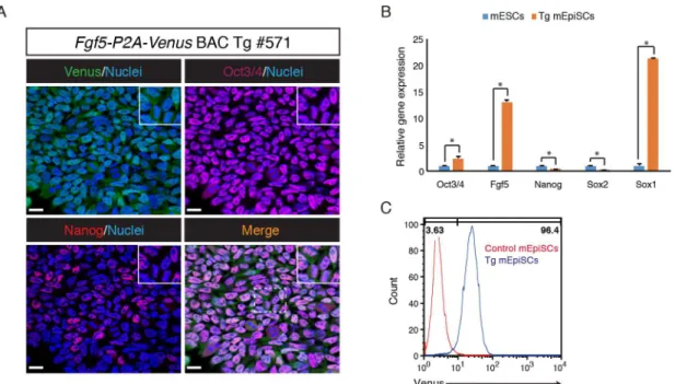
medium supplemented with bFGF and Activin. Immunofluorescence analysis revealed that the Tg mEpiSCs expressed pluripotency markers Oct3/4 and Nanog (Fig 4A). RT-qPCR analysis showed thatOct3/4expression is significantly higher in the Tg mEpiSCs than in mESCs, while expression levels ofNanogandSox2were lower in the Tg mEpiSCs than in mESCs. Notably,
Fgf5andSox1expression were detected at much higher levels in the Tg mEpiSCs than in mESCs (Fig 4B). Flow cytometric analysis showed that most Tg mEpiSCs, if not all, expressed Venus, consistent with uniform Venus expression in the epiblast of the E6.5 Tg embryo (Fig 4C). Taken together, these results indicated thatFgf5-P2A-VenusBAC Tg mEpiSCs have simi-lar properties to bona fide mEpiSCs.
Fig 2. Venus expression inFgf5-P2A-VenusBAC Tg embryo at E6.5.(A) Immunofluorescence analysis of the Tg embryo at E6.5 for Oct3/4 (red), Venus (anti-GFP, green), Gata4 (purple) and Nuclei
(Hoechst33342, blue). Higher magnification of optical sections is shown in panels X and Y. Note that Oct3/4 and Venus were co-expressed in the epiblast of the Tg embryo, while Gata4 expression was specifically observed in the endoderm regions of the Tg embryo. Venus expression was also seen in visceral endodermal layer. Open and closed arrowheads indicate endodermal and epiblast cells, respectively. Ex: extraembryonic region; Em: embryonic region; Epi: epiblast; En: endoderm. Scale bar: 50μm. All images were captured by a Leica TCS-SP8 confocal microscope using a 40x/1.25 oil objective lens. (B) Whole-mount fluorescencein situhybridization ofFgf5in the Tg embryo at E6.5. Open arrowheads indicate cytoplasmic localization of endogenousFgf5mRNA. Scale bar: 20μm. Images were captured by a Leica TCS-SP8 confocal microscope using a 40x/1.25 oil objective lens.
Reprogramming of
Fgf5-P2A-Venus
BAC Tg mEpiSCs into miPSCs
Reprogramming of mEpiSCs into miPSCs can be accomplished by ectopic expression ofNanog,Esrrb,Klf2,Klf4orKlf5cultured in the presence of 2i/LIF. [12–16]. The reprogramming process upregulates many naïve state-associated markers containingNanog,Esrrb,Tfcp2l1,
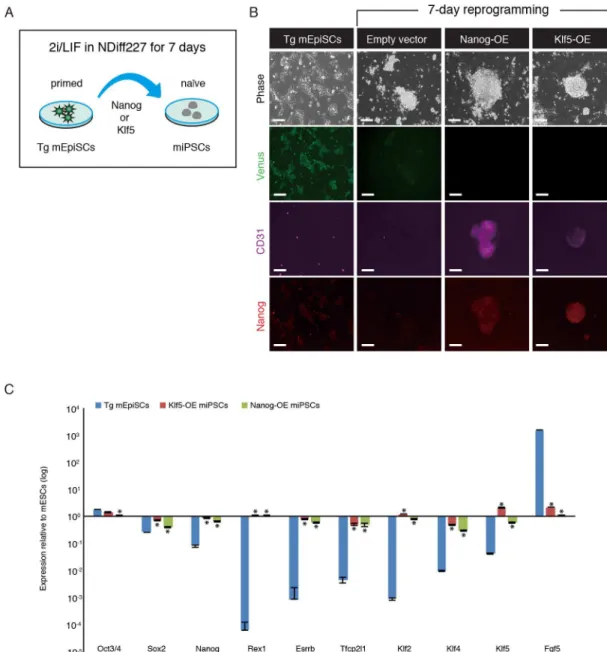
Cd31,Rex1,Stella,Nr0b1,Prdm14,Nr5a2,Tbx3,Klf2,Klf4andKlf5, with a parallel rapid reduc-tion in the expression of primed state-associated markers such asFgf5. Next, we asked whether Venus fluorescence is suppressed during the reprogramming process toward miPSC state. After ectopic expression ofNanogorKlf5in the Tg mEpiSCs, the culture medium was changed
Fig 3. Venus expression inFgf5-P2A-VenusBAC Tg embryo at E7.5.(A) Immunofluorescence images of the Tg embryo at E7.5 for Oct3/4 (red), Venus (anti-GFP, green), T (purple) and Nuclei (Hoechst33342, blue). Higher magnification of optical sections is shown in panels X and Y. Note that Oct3/4 and Venus were co-expressed in the epiblast of the Tg embryo, while T expression was observed in the mesodermal layer of the Tg embryo. Venus expression was also seen in visceral endodermal layer. Open and closed arrowheads indicate mesodermal and epiblast cells, respectively. Ex: extraembryonic region; Em: embryonic region; Epi: epiblast; Me: mesoderm; T: Brachyury/T. Scale bar: 50μm. All images were captured by a Leica TCS-SP8 confocal microscope using a 20x/0.7 dry objective lens (projection images) and 40x/1.25 oil objective lens (section, X and Y images). (B) Whole-mount fluorescencein situhybridization ofFgf5in the Tg embryo at E7.5. Open arrowheads indicate cytoplasmic localization of endogenousFgf5mRNA. Scale bar: 50μm.
from bFGF and Activin to 2i/LIF (Fig 5A). We found that ES-like colonies emerged in theKlf5 -andNanog-overexpressing Tg mEpiSCs within 5–7 days after addition of reprogramming medium. Immunofluorescence analysis revealed that overexpression ofKlf5orNanogcould reactivate the expression of CD31 (also known as PECAM-1: platelet endothelial cell adhesion molecule-1), a useful marker of inner cell mass cells, and increase Nanog expression in the miPSCs (Fig 5B). Importantly, Venus expression in the Tg mEpiSCs was markedly reduced in the miPSCs (Fig 5B). To further validate the characteristics of the miPSCs, several miPSC colo-nies were randomly picked to generate miPSC lines. These miPSC lines were maintained in 2i/ LIF conditions and used for further experiments. RT-qPCR analysis showed upregulation of pluripotency factorsNanog,Rex1,Esrrb,Tfcp2l1,Klf2,Klf4,and Klf5, and downregulation of lineage commitment factorFgf5in miPSCs (Fig 5C). Taken together, these results demon-strated that Venus expression can be used as an indicator when the Tg mEpiSCs are forced to reprogram into miPSCs.
Dynamic heterogeneity of
Fgf5
expression in
Fgf5-P2A-Venus
BAC Tg
mEpiSCs
mEpiSCs consist of several subpopulations:T-positive and -negative populations, and also
Sox1-positive and -negative populations; these positive/negative populations are interconverted [21]. Although we found thatFgf5-P2A-VenusBAC Tg embryo showed uniform Venus expres-sion in the epiblast and most Tg mEpiSCs expressed Venus abundantly, we investigated whether the Tg mEpiSCs contained a Venus-negative population. Careful examination of flow cytometry revealed that a minor fraction of the Tg mEpiSCs was Venus-negative (about 4%) (Fig 4C). To further explore this phenomenon, we purified Venus-positive and -negative mEpiSCs by cell sorting (Fig 6A) and cultured each cell fraction in mEpiSC growth conditions
Fig 4. Derivation and characterization ofFgf5-P2A-VenusBAC Tg mEpiSCs.(A) Immunofluorescence analysis of the Tg mEpiSCs for Venus (anti-GFP, green), Oct3/4 (purple), Nanog (red) and Nuclei (Hoechst33342, blue). Scale bar: 20μm. All images were captured by a Leica TCS-SP8 confocal microscope using a 63x/1.4 oil objective lens. (B) RT-qPCR analysis of genes associated with pluripotency and lineage commitment in the Tg mEpiSCs and mESCs.β-actin
was used as endogenous control for normalization. The mean and SD of three independent experiments are shown. *P<0.05. (C) Venus expression in control and the Tg mEpiSCs was analyzed by flow cytometry.
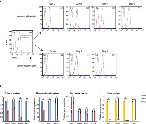
to investigate the ability to re-establish heterogeneity from each subpopulation. Interestingly, Venus-positive cells quickly emerged from the sorted Venus-negative cells, and the sorted Venus-negative cells could re-establish the original cell state within 4 days in culture (Fig 6A). Similarly, the purified Venus-positive cells also generated Venus-negative cells, although the re-establishment of heterogeneity from Venus-positive cells occurred more slowly compared with that from Venus-negative cells (Fig 6A). These results indicated that the Tg mEpiSCs con-tain at least two subpopulations that can be interconverted. We also examined gene expression patterns in Venus-positive and -negative mEpiSCs by RT-qPCR analysis (Fig 6B). We
Fig 5. Reprogramming ofFgf5-P2A-VenusBAC Tg mEpiSCs into miPSCs.(A) Experimental scheme for reprogramming of the Tg mEpiSCs into miPSCs. The Tg mEpiSCs stably expressingKlf5orNanogwere cultured in NDiff227 medium supplemented with the Mek inhibitor (PD0325901), Gsk3 inhibitor (CHIR99021) and LIF. After 7 days, miPSC colonies were subjected to immunostaining. (B) Immunostaining for Venus (anti-GFP, green), CD31 (purple) and Nanog (red) in untransfected, vector control and miPSCs. OE: overexpression. Scale bar: 100μm. (C) RT-qPCR analysis of Tg mEpiSCs and miPSCs. The mean and SD of two independent experiments are shown.*P<0.05.
confirmed thatFgf5mRNA expression was enriched in Venus-positive cells (Fig 6Ba). Impor-tantly, we found that the expression levels of other epiblast markers,Oct3/4,Nanog, andSox2, were predominantly detectable in Venus-positive cells (Fig 6Ba). Furthermore, both subpopu-lations expressed very low levels of naïve pluripotency markersRex1,Esrrb,Tfcp2l1, andKlf2
compared with mESCs (Fig 6Bd). Interestingly, we found that mesendodermal markersT,
Sox17andFoxa2were expressed at significantly higher levels in Venus-positive cells than in Venus-negative cells (Fig 6Bb). On the other hand, among the tested ectodermal markers, only
Sox1expression was enriched in Venus-negative cells relative to Venus-positive cells (Fig 6Bc). Taken together, while it was thought thatFgf5marks mEpiSCs uniformly, our experiments clearly demonstrated that evenFgf5expression shows dynamic heterogeneity in mEpiSCs.
Discussion
In this study, we demonstrated thatFgf5-P2A-VenusBAC Tg mice can recapitulate endoge-nousFgf5expression in the postimplantation epiblast and visceral endoderm. To the best of our knowledge, this is the first Tg mouse model allowing for the visualization of endogenous
Fig 6. Dynamic heterogeneity ofFgf5expression inFgf5-P2A-VenusBAC Tg mEpiSCs.(A) Venus-positive and -negative populations were purified, cultured and subjected to flow cytometry analysis at indicated days. Note that Venus-positive and -negative cells were interchangeable within 48 h post culture, and Venus-positive and -negative cells could re-establish the original cell state within 6 and 4 days, respectively. (B) Gene expression was examined by RT-qPCR in Venus-positive and -negative cells. The mean and SD of three independent experiments are shown. *P<0.05.
Fgf5expression during early embryonic development. Initial Tg lines carrying Venus at the first exon failed to drive epiblast Venus expression while Tg lines carrying P2A-Venus in the place of the stop codon drove strong Venus expression in the epiblast of all Tg lines. The exact reason underlying these observations is currently not clear. Because many genes harbor impor-tant regulatory elements around the first intron, the insertion of the Venus-pA reporter cas-sette into the first exon could have potentially abolished transcription by perturbing the epiblast promoter/enhancer elements.
Previous studies showed that primitive streak formation in the posterior portion of epiblast is a crucial event through which the body plan is established during the gastrulation process [44]. More specifically, while epiblast cells that ingress through the primitive streak can form the mesoderm and endoderm, epiblast cells that do not traverse the primitive streak can give rise to the ectoderm [42,43]. Future work will delineate the actual cell fates ofFgf5-positive epi-blast cells, which would provide important insights into how the ectoderm lineage is estab-lished and regulated in the gastrulating mouse embryo.
Gastrulation begins when a population of epiblast cells is triggered to move to the primitive streak located in the posterior epiblast while the other cells remain in the epiblast. This cell movement leads to the formation of the primary germ layers, namely the ectoderm, mesoderm, and endoderm [43,44]. However, how epiblast cell movement is regulated and which factors stimulate and determine the fates of cell populations at the onset of gastrulation is not fully understood. Therefore, a detailed analysis ofFgf5andTexpression pattern will be required to understand the molecular basis of epiblast cell behavior during gastrulation.
mESCs consist of several subpopulations; the subpopulations expressing either PECAM1, Rex1 or Stella efficiently form chimeric animals when injected into blastocysts. mEpiSCs are also heterogeneous in terms of gene expression:T-positive cells are primed to differentiate into mesoderm and endoderm lineages, whileT-negative cells are primed to ectoderm [21]. Our data showed thatFgf5overall uniformly marks mEpiSCs, consistent with the previous report [21]. However, surprisingly, there is a small subpopulation ofFgf5-negative cells in mEpiSCs. Our gene expression analysis revealed that, whileFgf5-positive cells predominantly expressed important mesendodermal markers such asT,Sox17andFoxa2,Fgf5-negative cells exhibited a high expression level of the ectodermal markerSox1. Currently, the actual cell type ofFgf5 -Venus-negative cells is unknown, but these results suggest the possibility that the heteroge-neous expression ofFgf5observed in our study may be the foundation for the distinct differen-tiation biases of subpopulations in mEpiSCs. Thus, investigation of the differendifferen-tiation
propensity ofFgf5-negative and -positive mEpiSCs in response to differentiation stimuli will be of a great interest in future studies.
Previous studies indicated potential roles ofFgf5in the progression of hepatic fibrosis [45], the process of gastrulation [27,28] and hair growth cycle [46], but the molecular basis of how
Fgf5manifests its functions has not been clearly understood. In addition, the impact of biologi-cal events on lineage commitment initiated by mEpiSCs is not known. Taken together,
Fgf5-P2A-VenusBAC Tg mice and mEpiSCs established in our study may be used to investi-gate novel functions ofFgf5as well as to unravel molecular mechanisms underlying lineage specificationin vivoandin vitro.
Supporting Information
S1 Fig. Construction of the first exonFgf5-VenusBAC Tg.TheFgf5BAC clone (RP23-153I24) covering 72 kb upstream and 112 kb downstream ofFgf5gene was used. Note that Venus was fused in frame after the start codon.
S2 Fig. Venus expression inFgf5-P2A-VenusBAC Tg embryos at E8.25. Immunofluores-cence staining of the Tg embryo at E8.25 for Venus (green). Note that Venus was expressed in the neuroepithelium of the Tg embryo. Ne: neuroepithelium. Scale bar: 100μm.
(TIF)
S1 Table. Primers used for RT-qPCR analysis. (TIF)
Acknowledgments
We would like to thank Ms. Keiko Amagai for preparation of BAC Tg DNA. This work was supported in part by a grant from PRESTO, Japan Science and Technology Agency (JST) (to M. E.) and a Grant-in-Aid for JSPS Fellows (to T. A.)
Author Contributions
Conceived and designed the experiments: ME. Performed the experiments: LTPK TA TT JM STF. Analyzed the data: LTPK TA TT JM STF ST ME. Wrote the paper: LTPK TA ME.
References
1. Evans MJ, Kaufman MH. Establishment in culture of pluripotential cells from mouse embryos. Nature. 1981; 292: 154–156. doi:10.1038/292154a0PMID:7242681
2. Martin GR. Isolation of a pluripotent cell line from early mouse embryos cultured in medium conditioned by teratocarcinoma stem cells. Proc Natl Acad Sci U S A. 1981; 78: 7634–7638. doi:10.1073/pnas.78. 12.7634PMID:6950406
3. Loh Y-H, Wu Q, Chew J-L, Vega VB, Zhang W, Chen X, et al. The Oct4 and Nanog transcription net-work regulates pluripotency in mouse embryonic stem cells. Nat Genet. 2006; 38: 431–440. doi:10. 1038/ng1760PMID:16518401
4. Chen X, Xu H, Yuan P, Fang F, Huss M, Vega VB, et al. Integration of External Signaling Pathways with the Core Transcriptional Network in Embryonic Stem Cells. Cell. 2008; 133: 1106–1117. doi:10. 1016/j.cell.2008.04.043PMID:18555785
5. Ng H-H, Surani MA. The transcriptional and signalling networks of pluripotency. Nat Cell Biol. 2011; 13: 490–496. doi:10.1038/ncb0511-490PMID:21540844
6. Ying Q-L, Wray J, Nichols J, Batlle-Morera L, Doble B, Woodgett J, et al. The ground state of embryonic stem cell self-renewal. Nature. 2008; 453: 519–523. doi:10.1038/nature06968PMID:18497825 7. Brons IGM, Smithers LE, Trotter MWB, Rugg-Gunn P, Sun B, Chuva de Sousa Lopes SM, et al.
Deriva-tion of pluripotent epiblast stem cells from mammalian embryos. Nature. 2007; 448: 191–195. doi:10. 1038/nature05950PMID:17597762
8. Tesar PJ, Chenoweth JG, Brook FA, Davies TJ, Evans EP, Mack DL, et al. New cell lines from mouse epiblast share defining features with human embryonic stem cells. Nature. 2007; 448: 196–199. doi: 10.1038/nature05972PMID:17597760
9. Han DW, Tapia N, Joo JY, Greber B, Araúzo-Bravo MJ, Bernemann C, et al. Epiblast stem cell subpop-ulations represent mouse embryos of distinct pregastrulation stages. Cell. 2010; 143: 617–627. doi: 10.1016/j.cell.2010.10.015PMID:21056461
10. Huang Y, Osorno R, Tsakiridis A, Wilson V. In Vivo Differentiation Potential of Epiblast Stem Cells Revealed by Chimeric Embryo Formation. Cell Rep. 2012; 2: 1571–1578. doi:10.1016/j.celrep.2012. 10.022
11. Nichols J, Smith A. Naive and Primed Pluripotent States. Cell Stem Cell. 2009; 4: 487–492. doi:10. 1016/j.stem.2009.05.015PMID:19497275
12. Silva J, Nichols J, Theunissen TW, Guo G, van Oosten AL, Barrandon O, et al. Nanog is the Gateway to the Pluripotent Ground State. Cell. 2009; 138: 722–737. doi:10.1016/j.cell.2009.07.039PMID: 19703398
14. Gillich A, Bao S, Grabole N, Hayashi K, Trotter MWB, Pasque V, et al. Epiblast stem cell-based system reveals reprogramming synergy of germline factors. Cell Stem Cell. 2012; 10: 425–439. doi:10.1016/j. stem.2012.01.020PMID:22482507
15. Guo G, Yang J, Nichols J, Hall JS, Eyres I, Mansfield W, et al. Klf4 reverts developmentally pro-grammed restriction of ground state pluripotency. Development. 2009; 136: 1063–1069. doi:10.1242/ dev.030957PMID:19224983
16. Jeon H, Waku T, Azami T, Khoa LTP, Yanagisawa J, Takahashi S, et al. Comprehensive Identification of Krüppel-Like Factor Family Members Contributing to the Self-Renewal of Mouse Embryonic Stem Cells and Cellular Reprogramming. PLoS One. 2016; 11: e0150715. doi:10.1371/journal.pone. 0150715PMID:26943822
17. Furusawa T, Ohkoshi K, Honda C, Takahashi S, Tokunaga T. Embryonic stem cells expressing both platelet endothelial cell adhesion molecule-1 and stage-specific embryonic antigen-1 differentiate pre-dominantly into epiblast cells in a chimeric embryo. Biol Reprod. 2004; 70: 1452–1457. doi:10.1095/ biolreprod.103.024190PMID:14736812
18. Chambers I, Silva J, Colby D, Nichols J, Nijmeijer B, Robertson M, et al. Nanog safeguards pluripotency and mediates germline development. Nature. 2007; 450: 1230–1234. doi:10.1038/nature06403PMID: 18097409
19. Toyooka Y, Shimosato D, Murakami K, Takahashi K, Niwa H. Identification and characterization of sub-populations in undifferentiated ES cell culture. Development. 2008; 135: 909–918. doi:10.1242/dev. 017400PMID:18263842
20. Hayashi K, Lopes SMCDS, Tang F, Surani MA. Dynamic Equilibrium and Heterogeneity of Mouse Plu-ripotent Stem Cells with Distinct Functional and Epigenetic States. Cell Stem Cell. 2008; 3: 391–401.
doi:10.1016/j.stem.2008.07.027PMID:18940731
21. Tsakiridis A, Huang Y, Blin G, Skylaki S, Wymeersch F, Osorno R, et al. Distinct Wnt-driven primitive streak-like populations reflect in vivo lineage precursors. Development. 2014; 141: 1209–1221. doi: 10.1242/dev.101014PMID:24595287
22. Ornitz DM, Itoh N. The fibroblast growth factor signaling pathway. Rev Dev Biol. 2015; 4: 215–266. doi: 10.1002/wdev.176
23. Mason J. The Ins and Outs of Fibroblast Growth Factors. Cell. 1994; 79: 547–552.
24. Lanner F, Rossant J. The role of FGF/Erk signaling in pluripotent cells. Development. 2010; 137: 3351–3360. doi:10.1242/dev.050146PMID:20876656
25. Straub L, Wolfrum C. FGF21, energy expenditure and weight loss–how much brown fat do you need?
Mol Metab. 2015; 4: 605–609. doi:10.1016/j.molmet.2015.06.008PMID:26413466
26. Haub O, Goldfarb M. Expression of the fibroblast growth factor-5 gene in the mouse embryo. Develop-ment. 1991; 112: 397–406. PMID:1794310
27. Hébert JM, Basilico C, Goldfarb M, Haub O, Martin GR. Isolation of cDNAs encoding four mouse FGF family members and characterization of their expression patterns during embryogenesis. Dev Biol. 1990; 138: 454–463. doi:10.1016/0012-1606(90)90211-ZPMID:2318343
28. Hébert JM, Boyle M, Martin GR. mRNA localization studies suggest that murine FGF-5 plays a role in gastrulation. Development. 1991; 112: 407–415. PMID:1794311
29. Yeom YI, Ha HS, Balling R, Schöler HR, Artzt K. Structure, expression and chromosomal location of the Oct-4 gene. Mech Dev. 1991; 35: 171–179. doi:10.1016/0925-4773(91)90016-YPMID:1768618 30. Pelton TA, Sharma S, Schulz TC, Rathjen J, Rathjen PD. Transient pluripotent cell populations during primitive ectoderm formation: correlation of in vivo and in vitro pluripotent cell development. J Cell Sci. 2002; 115: 329–39. PMID:11839785
31. Di-Gregorio A, Sancho M, Stuckey DW, Crompton LA, Godwin J, Mishina Y, et al. BMP signalling inhib-its premature neural differentiation in the mouse embryo. Development. 2007; 134: 3359–3369. doi: 10.1242/dev.005967PMID:17699604
32. Li L, Liu C, Biechele S, Zhu Q, Song L, Lanner F, et al. Location of transient ectodermal progenitor potential in mouse development. Development. 2013; 140: 4533–43. doi:10.1242/dev.092866PMID: 24131634
33. Guzman-Ayala M, Sachs M, Koh FM, Onodera C, Bulut-Karslioglu A, Lin C-J, et al. Chd1 is essential for the high transcriptional output and rapid growth of the mouse epiblast. Development. 2014; 118–
127. doi:10.1242/dev.114843PMID:25480920
35. Kojima Y, Kaufman-Francis K, Studdert JB, Steiner KA, Power MD, Loebel DAF, et al. The transcrip-tional and functranscrip-tional properties of mouse epiblast stem cells resemble the anterior primitive streak. Cell Stem Cell. 2014; 14: 107–120. doi:10.1016/j.stem.2013.09.014PMID:24139757
36. Trott J, Arias AM. Single cell lineage analysis of mouse embryonic stem cells at the exit from pluripo-tency. Biol Open. 2013; 2: 1049–1056. doi:10.1242/bio.20135934PMID:24167715
37. Downs KM, Davies T. Staging of gastrulating mouse embryos by morphological landmarks in the dis-secting microscope. Development. 1993; 118: 1255–1266. PMID:8269852
38. Gasnier M, Dennis C, Vaurs-Barrière C, Chazaud C. Fluorescent mRNA labeling through cytoplasmic FISH. Nat Protoc. 2013; 8: 2538–47. doi:10.1038/nprot.2013.160PMID:24263093
39. Ema M, Mori D, Niwa H, Hasegawa Y, Yamanaka Y, Hitoshi S, et al. Krüppel-like factor 5 is essential for blastocyst development and the normal self-renewal of mouse ESCs. Cell Stem Cell. 2008; 3: 555–
67. doi:10.1016/j.stem.2008.09.003PMID:18983969
40. Nagai T, Ibata K, Park ES, Kubota M, Mikoshiba K, Miyawaki A. A variant of yellow fluorescent protein with fast and efficient maturation for cell-biological applications. Nat Biotechnol. 2002; 20: 87–90. doi: 10.1038/nbt0102-87PMID:11753368
41. Mesnard D, Donnison M, Fuerer C, Pfeffer PL, Constam DB. The microenvironment patterns the plurip-otent mouse epiblast through paracrine furin and Pace4 proteolytic activities. Genes Dev. 2011; 25: 1871–1880. doi:10.1101/gad.16738711PMID:21896659
42. Lawson KA. Fate mapping the mouse embryo. Int J Dev Biol. 1999; 43: 773–775. doi:10.1387/IJDB. 10668985PMID:10668985
43. Tam PP, Loebel DA. Gene function in mouse embryogenesis: get set for gastrulation. Nat Rev Genet. 2007; 8: 368–381. doi:10.1038/nrg2084PMID:17387317
44. Tam PP, Behringer RR. Mouse gastrulation: The formation of a mammalian body plan. Mech Dev. 1997; 68: 3–25. doi:10.1016/S0925-4773(97)00123-8PMID:9431800
45. Hanaka H, Hamada T, Ito M, Nakashima H, Tomita K, Seki S, et al. Fibroblast growth factor-5 partici-pates in the progression of hepatic fibrosis. Exp Anim. 2014; 63: 85–92. doi:10.1538/expanim.63.85
PMID:24521867