and In Silico Assessment of Antileishmanial Activities
Eduardo Sobarzo-Sa´nchez1,2, Pablo Bilbao-Ramos3, Maria Dea-Ayuela4, Humberto Gonza´lez-Dı´az5¤a¤b, Matilde Yan˜ez6, Eugenio Uriarte7, Lourdes Santana7, Victoria Martı´nez-Serna´ndez5, Francisco Bola´s-Ferna´ndez3, Florencio M. Ubeira5*
1Departamento de Tecnologı´a Farmace´utica, Facultad de Farmacia, Universidad de Santiago de Compostela, Santiago de Compostela, Spain,2Facultad de Ciencias de la Salud, Universidad Auto´noma de Chile, Santiago, Chile,3Departamento de Parasitologı´a, Facultad de Farmacia, Universidad Complutense de Madrid, Madrid, Spain, 4Departamento de Farmacia, Universidad Cardenal Herrera, Valencia, Spain,5Departamento de Microbiologı´a y Parasitologı´a, Facultad de Farmacia, Universidad de Santiago de Compostela, Santiago de Compostela, Spain,6Departamento de Farmacologı´a, Facultad de Farmacia, Universidad de Santiago de Compostela, Santiago de Compostela, Spain,7Departamento de Quı´mica Orga´nica, Facultad de Farmacia, Universidad de Santiago de Compostela, Santiago de Compostela, Spain
Abstract
Leishmaniasis is a growing health problem worldwide. As there are certain drawbacks with the drugs currently used to treat human leishmaniasis and resistance to these drugs is emerging, there is a need to develop novel antileishmanial compounds, among which isoquinoline alkaloids are promising candidates. In this study, 18 novel oxoisoaporphine derivatives were synthesized and their possible antileishmanial activity was evaluated. The in vitro activity of these derivatives againstLeishmania amazonensisaxenic amastigotes was first evaluated, and the selected compounds were then tested in an inhibition assay with promastigotes ofL. infantum,L. braziliensis,L. amazonensisandL. guyanensis,and with intracellular amastigotes ofL. infantumand L. amazonensis. Finally, the most active compounds, OXO1(2,3-dihydro-7H -dibenzo[de,h]quinolin-7-one) and OXO13(2,3,8,9,10,11-hexahydro-7H-dibenzo[de,h]quinolin-7-one), were tested in BALB/c mice infected withL. infantum. Treatment of mice at a dose of 10 mg/kg with OXO1yielded significant reductions (p,0.05) in parasite burden in liver and spleen (99% and 78%, respectively) whereas with OXO13were not significant. Although previous reports suggest that this family of molecules displays inhibitory activity against monoamine oxidase A and acetylcholinesterase, these enzymes were not confirmed as targets for antileishmanial activity on the basis of the present results. However, after development of a new bioinformatics model to analyze theLeishmaniaproteome, we were able to identify other putative targets for these molecules. The most promising candidates were four proteins: two putative pteridine reductase 2 (1MXF and 1MXH), oneN-myristoyltransferase (2WUU) and one type I topoisomerase (2B9S).
Citation:Sobarzo-Sa´nchez E, Bilbao-Ramos P, Dea-Ayuela M, Gonza´lez-Dı´az H, Yan˜ez M, et al. (2013) Synthetic Oxoisoaporphine Alkaloids: In Vitro, In Vivo and In Silico Assessment of Antileishmanial Activities. PLoS ONE 8(10): e77560. doi:10.1371/journal.pone.0077560
Editor:Abhay R. Satoskar, The Ohio State University, United States of America
ReceivedApril 26, 2013;AcceptedSeptember 3, 2013;PublishedOctober 29, 2013
Copyright:ß2013 Sobarzo-Sa´nchez et al. This is an open-access article distributed under the terms of the Creative Commons Attribution License, which permits unrestricted use, distribution, and reproduction in any medium, provided the original author and source are credited.
Funding:This work was partially financed by grants AGL2011-30563-C03-01 from the Spanish Ministerio de Ciencia e Innovacio´n and A-024457/09 from the Spanish Agency for International Cooperation and Development (AECID). Eduardo Sobarzo-Sa´nchez is grateful to the Xunta de Galicia (Spain) for the ‘‘Isidro Parga Pondal’’ fellowship. Pablo Bilbao-Ramos is thankful to the MAEC-AECID for a PhD fellowship. The funders had no role in study design, data collection and analysis, decision to publish, or preparation of the manuscript.
Competing Interests:The authors have declared that no competing interests exist.
* E-mail: [email protected]
¤a Current address: Departamento de Quı´mica Orga´nica II, Facultad de Ciencia y Tecnologı´a, Universidad del Paı´s Vasco UPV/EHU, Bizkaia, Paı´s Vasco, Spain ¤b Current address: IKERBASQUE, Fundacio´n Vasca para la Ciencia, Bilbao, Spain
Introduction
Leishmaniasis is a vector-borne disease caused by an obligate intra-macrophage protozoan parasite. The disease, which is endemic in large areas of tropical and subtropical countries, is caused by more than 20 species ofLeishmaniaand transmitted to humans by more than 30 different species of phlebotomine sandflies. The clinical manifestations of leishmaniasis largely depend on complex interactions between the virulence of the infectingLeishmaniaspecies and the immune responses of the host. The three major clinical syndromes that are recognized in human disease are visceral, cutaneous and muco-cutaneous leishmaniasis [1].
Leishmaniasis is a major public health problem and the burden is increasing. An estimated 2 million new cases (1.5 million cases of cutaneous leishmaniasis and 500,000 of visceral leishmaniasis)
occur annually, and about 12 million people are currently infected [2]. This epidemiological scenario has worsened because dogs are the principal reservoirs as well as suffers of the disease, for which fully successful treatment is still lacking [3].
At present, the following drugs are used to treat human leishmaniasis: pentavalent antimonials, paromomycin, amphoter-icin, miltefosine and pentamidine. However, there is an urgent need to develop new antileishmanial drug candidates to overcome problems such as toxic side effects, route of administration, long-term treatment and the generation of resistance mechanisms to current drugs [4].
Annonaceae, Magnoliaceae, Menispermaceae, Monimiaceae and Rutaceae [5], and which has been shown to be active againstL. amazonensis, L. braziliensis, L. donovani, L. guyanensis and L. major
[6,7,8,9]. Liriodenine is a potent inhibitor of topoisomerase II and also displays other types of activity, such as antibacterial, antifungal, antitumor and antiviral activity [5,10]. Other oxoa-porphines reported to possess antileishmanial activity include dicentrinone, obtained from the stem bark of Duguetia furfuracea, and N-methylliriodendronine, isolated from Stephania dinklagei; these have been shown to be active against L. braziliensis [10] and L. donovani[7], respectively. In addition to oxoaporphines, a small group of isomers (oxoisoaporphines) that occur in some Chinese medicinal plants, such as the roots of Menispermum dauricum, may exert leishmanicidal activity. However, these isomers have not been well studied, mainly because of the low concentrations at which they occur in the plants. Given the potential interest in these compounds for treating leishmaniasis, the aim of the present study was to investigate, for the first time, the in vitro and in vivo leishmanicidal activity of several novel synthetic oxoisoaporphine compounds. In addition, we have also developed a new bioinformatics tool for predicting putative targets of these molecules in the parasite proteome.
Materials and Methods
Ethics Statement
All procedures for animal manipulations were approved by the Institutional Animal Care and Use Committee of the Complutense University of Madrid, following Spanish law.
Synthesis of Oxoisoaporphine Derivatives (OXO 1–18)
The compounds used in the present study were synthesized according to a previously described general procedure [11–13].
Reference Drugs
Miltefosine and rasagiline were purchased from Sigma-Aldrich (Madrid, Spain).
In vitro Assays with LeishmaniaAxenic Amastigotes
For preliminary screening of potential active compounds, axenic
amastigotes of L. amazonensis (strain MHOM/BR/76/LTB-012)
were arbitrarily chosen and cultured according to Estevez et al. [14]. The axenic amastigotes were cultured in medium supple-mented with 20% foetal bovine serum (FBS) (Sigma-Aldrich) and incubated at 32uC with 5% CO2 in 25 cm
2
culture bottles. In order to determine the activity of the compounds, a colorimetric method with 3-(4,5-dimethylthiazol-2-yl)-2,5-diphenyltetrazolium bromide (MTT; Sigma-Aldrich) was used, as previously described [15]. The compounds were dissolved in dimethylsulfoxide (DMSO) and added to wells of microtiter plates containing 100ml of cultivated amastigotes (in logarithmic growth phase) to
final concentrations ranging from 0.05mg/ml to 50mg/ml. The
plates were incubated for 72 h, and 10ml of the MTT solution (10 mg/ml in PBS) were then added to each well and incubated for a further 4 h. The reaction was stopped by addition of 100ml
of isopropanol-sodium dodecyl sulphate (SDS). Finally, the optical density (OD) was read at 570 nm. All experiments were carried out in triplicate.
In vitro Assays with LeishmaniaPromastigotes
For these assays, the followingLeishmaniaspecies were used: an
autochthonous isolate of L. infantum (MCAN/ES/92/BCN83)
obtained from an asymptomatic dog in the Priorat region of Catalunya (Spain) and kindly donated by Prof. Montserrat Portu´s
(University of Barcelona); and L. braziliensis 2903, L. amazonensis
(MHOM/Br/79/Maria) andL. guyanensis141/93, kindly provided by Prof. Alfredo Toran˜o (Instituto del Salud Carlos III, Madrid). The promastigotes were cultured at 26uC in Schneider’s Insect Medium (Sigma-Aldrich) supplemented with 10% heat-inactivated FBS and 100 U/ml of penicillin plus 100mg/ml of streptomycin
(Sigma-Aldrich), in 25 ml culture flasks.
To test the oxoisoaporphine compounds, promastigotes of each species (2.56105parasites/well) were cultured in 96-well micro-titer plates. The compounds were dissolved in DMSO and diluted in the culture medium at concentrations ranging from 0.8 to 100mg/ml in a final volume of 200ml. Miltefosine was used as the reference drug. After incubation for 48 h at 26uC, 20ml of
2.5 mM resazurin (Sigma–Aldrich) solution were added to each well, and the fluorescence intensity (535 nm -excitation wave-length- and 590 nm -emission wavelength) was measured in a fluorometer (Infinite 200, Tecan Group Ltd, Ma¨nnedorf, Swit-zerland). All tests were carried out in triplicate.
In vitro Assays withLeishmaniaIntracellular Amastigotes
The fluorometric assay for drug screening against intracellular amastigotes was carried out as described by Bilbao-Ramos et al. [16]. Briefly, 56104macrophages and stationary promastigotes at a 1:10 ratio in 200ml/well of culture medium were seeded and the plates were incubated for 24 h at 33uC, 5% CO2 in humidity
chamber. The temperature was then increased to 37uC for
another 24 h. The cells were then washed several times to remove free non-infective promastigotes; the final washing medium was replaced with 200ml/well of culture medium containing 2-fold
serial dilutions of the test compounds in a triplicate assay. Miltefosine was included as reference drug. After incubation of the plates for 48 h at 37uC, 5% CO2, the culture medium was replaced with an equal volume of the lysis solution (RPMI-1640 with 0.048% HEPES and 0.006% SDS) and maintained at room temperature for 20 min. The lysis solution was then replaced with Schneider’s medium followed by incubation at 26uC for another 3 days to allow transformation of viable amastigotes into promas-tigotes and subsequent proliferation. Aliquots of 20ml of 2.5 mM
resazurin were added to each well and the plates were incubated for 3 h. Finally, fluorescence emission was measured as described above.
Macrophage Cytotoxicity Assays
Toxicity cell assays were carried out as described elsewhere [17]. To test the oxoisoaporphine compounds, the J774.2 cells (EACC 80011428) were seeded (56104cells/well) in 96-well flat bottomed microplates and allowed to adhere for 24 h at 37uC in
5% CO2. The medium was then replaced with different
concentrations of the test compounds, followed by incubation for another 24 h. Each concentration was assayed three times and growth controls were also included. Thereafter, 20ml of a 2.5 mM resazurin solution were added to each well and the plates were incubated for 3 h. The fluorescence emission was measured as indicated above.
In vivo Assays
animals were anaesthetized with sodium pentobarbital and then infected with 107promastigotes per animal, administered via the intracardiac route.
Treatment. The treatments began on day 35 post-infection and were administered on 3 continuous days. The compounds were first dissolved in DMSO and diluted in physiological saline to provide doses of 2.5, 5 and 10 mg/kg, which were administered daily by the intraperitoneal route in a final volume of 0.1 ml. One group administered with the vehicle alone was used as an untreated control and another group was treated with miltefosine as reference drug. Miltefosine was administered intraperitoneally for purposes of comparison with test compounds. Five days later, the mice were killed and the parasitic burden was estimated.
Estimation of parasite burden. The liver and spleen were removed from each animal and homogenised. After eliminating cell debris, the suspension was centrifuged, the supernatants were discarded and the pellets were resuspended as previously described [18]. Aliquots (200ml) of the suspension were transferred to each well of 96-well microtiter plates containing NNN medium supplemented with antibiotics. The parasite burden was estimated by the limit dilution assay, according to Hill et al. [19] and Titus et al. [20].
Statistical Analysis
For in vitro assays, the antiparasitic activity and cytotoxic effect of compounds, expressed as IC50(or IC90) and CC50, respectively, were assessed by multinomial probit analysis. For in vivo assays, the data were analyzed by Tukey’s HSD post-hoc test. Differences were considered significant atp,0.05. SPSS v20.0 and Microsoft Excel 2007 software were used for all analyses.
Determination of the Inhibitory Activity Against the Monoamine Oxidase (MAO) A and B Enzymes
The potential effects of the tested drugs on hMAO activity were previously investigated [21] using the AmplexHRed MAO assay kit (Molecular Probes, Inc., Eugene, Oregon, USA) and micro-somal MAO isoforms prepared from insect cells (BTI-TN-5B1-4) infected with recombinant baculovirus containing cDNA inserts for hMAO-A or hMAO-B [22].
Determination of Inhibition of the Cholinesterase Enzyme
The cholinesterase assay method of Ellman [23] was used to determine the in vitro cholinesterase activity. The assay medium contained 50 mM PBS, pH 8.0, 20 mM dithiobisnitrobenzoate (DTNB; Sigma-Aldrich), 0.165 U/ml of recombinant acetylcho-linesterase (AChE) expressed in HEK 293 cells (Sigma-Aldrich) and 0.75mM substrate (acetylthiocholine iodide; Sigma-Aldrich). The activity was determined by measuring the increase in absorbance at 412 nm in a FLUOstar Optima microplate reader (BMG Labtech GmbH, Offenburg, Germany), at 1 min intervals, for 10 min at 37uC. In dose-dependent inhibition studies, the substrate was added to the assay medium containing enzyme, buffer, and DTNB with inhibitor after incubation for 10 min. All experiments were carried out in duplicate and the results were expressed as means6SEM. The relative activity is expressed as the percentage ratio of enzyme activity in the absence of inhibitor.
Bioinformatics Analysis
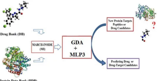
The model was constructed with protein data obtained from the Protein Data Bank (PDB) [24]. All 128LeishmaniaProteins with known ligands (LPs), including both single proteins and protein complexes, were downloaded. Separation of the protein complexes yielded 624 single LPs in total (protein chains). The option known as ligand report was used to collect all Ligand-LP Interactions pairs (LLPIs) reported in the PDB. Only organic drugs and metabolites were considered. The final dataset contains 563 LLPIs, including 122 different ligands and 3,823 non-interacting Ligand-LPs pairs (nLLPIs). The nLLPIs are pairs formed between the 122 ligands and LPs that do not interact with them. The MARCH-INSIDE (MI) software was then used to calculate the values of spectral momentspk(Lm) andpk(Pn)for both ligands and LPs sequences, respectively [25]. These molecular descriptors were used as inputs to search for a linear model. The Linear Discriminant Analysis (LDA) method, implemented in STATIS-TICA version 6.0 (ST) [26], was used to develop a simple linear classifier with the following general formula:
SmnðLLPIÞpred~
X5
k~0
ak:kpðLmÞz
X5
k~0
bk:kpðPnÞzc0 ð1Þ
This equation has only two additive terms; however difference or interaction terms can also be used in non-linear analysis. The model deals with the classification of ligands into two sub-sets: with or without affinity against many different proteins expressed in the proteome ofLeishmania sp. A dummy variable Ligand-Leishmania
Protein Interaction (LLPImn) was used as input to codify the affinity. This variable indicates either high (LLPImn= 1) or low (LLPImn= 0) affinity of a given ligandLmby different LPsPn.The predicted scoreSmn(LLPI)predis the output and it is a continuous dimensionless score that classifies ligands from low to high affinity to the protein target. In the model, ak, bk, and c0represent the coefficients of the classification function. The statistical significance of the model was determined by calculating the p-value (p) with the Chi-square test. The specificity, sensitivity and total accuracy were also checked to determine the quality-of-fit to data in training. The canonical regression coefficient Rc was used to determine the strength of the linear correlation between the independent inputs (pk) and dependent variable LLPImn. The model validation was corroborated with external prediction series. A diagram of the general workflow used to develop and apply this and other models is shown in Figure 1.
Results
In vitro Assays
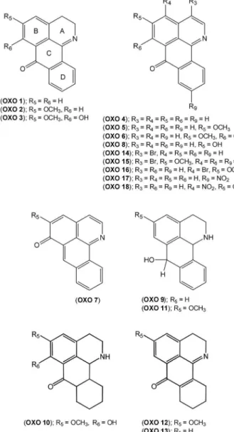
Eighteen different oxoisoaporphine derivatives, OXO 1–18 (Figure 2), were evaluated in vitro in axenic cultures of amastigotes ofL. amazonensis, to test for possible antileishmanial activity. The inhibitory activity of these compounds was tested at several concentrations ranging from 0.05–50mg/ml. The results showed
that only OXO1and OXO13rendered maximal inhibition at
the lowest concentration of 0.05mg/ml (Table 1). These
pharmacological results appear to indicate that although all tested compounds are chemically related structures, the presence of either a certain type of carbon framework or moieties are important for the in vitro anti-Leishmania activity of oxoisoapor-phines.
Compounds OXO 1 and OXO 13 were then selected to
determine the in vitro effectiveness against promastigotes of four species ofLeishmaniarepresentative of the main clinical forms of the disease: L. infantum (as a model of visceral leishmaniasis) andL. amazonensis,L. braziliensis, andL. guyanensis (as models of cutaneous or mucocutaneous forms). The cytotoxicity of both molecules against the J774.2 macrophage cell line was also tested. Both compounds displayed antileishmanial activity against the 4 species tested with the highest activity recorded against L. guyanensis
(Table 2). Comparatively, compound OXO 13 was the most
active (IC50lower than that recorded for the reference compound, miltefosine), but this compound also displayed a high degree of toxicity (CC50= 31.4mg/ml compared with 55.4mg/ml for miltefosine). By contrast, compound OXO1showed an interme-diate level of activity against allLeishmaniaspecies, and it was not toxic for the J774.2 cell line. In order to simulate natural physiological conditions in the vertebrate host, the selected compounds were tested against intracellular amastigotes in a newly developed fluorometric assay using L. infantum and L. amazonensis. Both compounds displayed significant antileishmanial activity, relative to that of the reference drug, against both species.
OXO13was most active, almost 5 times as active as miltefosine
(IC50 4.4960.64mg/ml and 4.8360.61mg/ml versus
20.961.47mg/ml and 23.761.78mg/ml against L. amazonensis
and L. infantum, respectively) (Table 3). Both products also exhibited good selectivity indexes in comparison with the reference drug.
In vivo Assays
Finally, both in vitro active compounds were tested in vivo in BALB/c mice infected withL. infantum. The results, expressed as mean values (number of parasites6106) 6 standard deviation (SD) of individual data, as assessed by limit dilution assay taken from two independent experiments, are shown in Table 4.
Compound OXO 13 was not effective, as the percentage
reduction of Leishmania amastigotes in liver and spleen did not differ from that obtained for the untreated control. In contrast, compound OXO1caused a significant reduction in the parasite burden in spleen and livers of treated mice (Table 4). The reduction of parasite with OXO 1 in livers reached 9962% Figure 2. Chemical structures of oxoisoaporphine derivatives (OXO 1–18).
(p,0.05) for a dose of 10 mg/kg, whereas the reduction for the
same dose in spleens was slightly lower 78633% but also
significant (p,0.05). Considering the toxicity, relative to the reference drug miltefosine, which caused 12.5% mortality at a dose of 5 mg/kg, OXO1was not toxic, even at the maximal dose of 10 mg/kg.
Binding of Oxoisoaporphine Compounds to MAO and AChE Enzymes
To investigate possible enzyme targets that may explain the different capacity of oxoisoaporphine compounds to inhibit the
growth of Leishmania parasites, we compared the ability of
compounds OXO 1 and OXO 13 to inhibit the MAO and
AChE enzymes. The enzymes were chosen on the basis of the results of previous studies, which showed that several molecules chemically related to oxoisoaporphines display inhibitory activity against AChE and MAO-A [21,27,28]. The data in Table 5 show that both compounds were good inhibitors of MAO-A (data taken
from Prado-Prado et al. [21]) and display a significant degree of inhibitory activity against AChE; however, there were no differences in the inhibitory activity of these compounds against MAO-A or AChE enzymes.
Bioinformatic Analysis of Other PutativeLeishmania
Targets for Oxoisoaporphine Compounds
Taking into consideration that the in vitro inhibition assays with the MAO and AChE enzymes did not indicate any differences
between OXO 1 and OXO 13 compounds, we carried out a
predictive study of putative targets among LPs. As there are no previous reports of any predictive method for drug-target interactions developed specifically for LPs, we developed a new model. The best LDA model found was as follows:
SmnðLLPIÞpred~0:50308:
5
pðPnÞz0:12527:
5
pðLmÞ{4:82360
N~3294 Rc~0 :7 x
2
~1964
:436 pv0:001
ð2Þ Table 1.In vitro assays of axenic amastigotes fromLeishmania amazonensiswith oxoisoaporphine derivatives 1–18.
Inhibition (%) at concentrations tested Leishmanicidal activity
Compound 50mg/ml 5mg/ml 0.5mg/ml 0.05mg/ml IC90 IC50
OXO 1 107.8 107.4 105.7 100.6 ,0.05 ,0.05
OXO 2 93.9 84.8 0 0 16.4 0.2
OXO 3 98.1 70.8 0 2.00 19.5 5.5
OXO 4 97.5 20.3 10.1 2.3 31.1 4.9
OXO 5 65.5 0 1.0 3.6 134.6 40.9
OXO 6 100.0 88.8 15.2 7.9 9.7 1.5
OXO 7 103.4 94.7 58.6 10.3 3.6 0.34
OXO 8 98.9 87.4 5.4 3.0 12.8 2.6
OXO 9 98.0 7.3 15.9 3.9 31.1 13.3
OXO 10 101.5 23.8 10.7 2.8 19.5 3.8
OXO 11 100.8 0.1 1.7 1.0 29.438 15.2
OXO 12 101.1 101.8 14.1 8.5 2.3 0.5
OXO 13 106.9 103.2 108.0 106.7 ,0.025 ,0.025
OXO 14 95.2 0 0 2.0 19.9 10.4
OXO 15 95.7 0 2.3 0 19.8 10.8
OXO 16 88.0 0 4.1 1.7 27.0 13.3
OXO 17 98.3 0 0.5 0.5 18.2 10.6
OXO 18 103.3 99.4 0 0 1.6 0.8
IC90: 90% inhibitory concentration, IC50: 50% inhibitory concentration. doi:10.1371/journal.pone.0077560.t001
Table 2.In vitro antileishmanial activity against promastigotes and cytotoxic activity of oxoisoaporphine derivatives (OXO 1 and 13).
IC50(mg/ml) CC50(mg/ml)
Compound L. infantum L. amazonensis L. braziliensis L. guyanensis J774.2
OXO 1 42.163.5 31.862.2 48.362.7 16.361.4 .100
OXO 13 4.460.2 3.660.3 4.460.2 1.860.1 31.463.2
Miltefosine 7.260.6 12.560.4 7.260.2 7.960.5 55.464.2
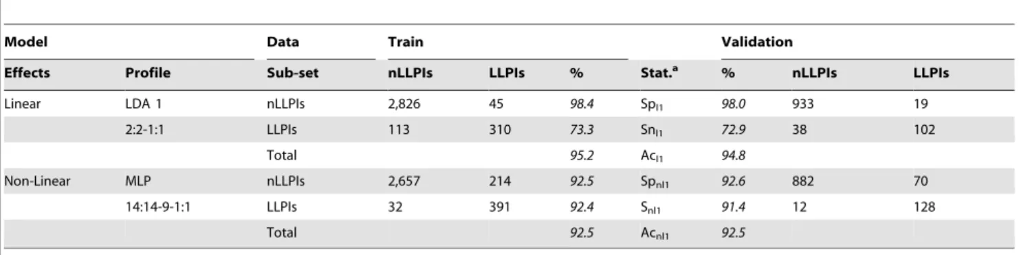
where N is the number of cases (LLPIs and nLLPIs) used to train the model. This model has a Rc = 0.7 and an accuracy of 95.2% in training and of 94.8% in external validation series (Table 6). These results indicate the development of an accurate model according to previous reports on the use of LDA in drug discovery [25]. Nonetheless, the LDA model is unbalanced because only correctly classified < 70% of LLPIs and 98% of nLLPIS. Therefore, the
Artificial Neural Network (ANN) module of ST was used to search for a non-linear model [26]. The best ANN model was a Multi Layer Perceptron with 3 layers of neurons (MLP3). The MLP3 classifier presented an overall Ac = 92.5% in both training and external validation series (Table 6). In contrast to the LDA model, the ANN model is not unbalanced and correctly classified<92%
of both LLPIs and nLLPIs. In addition, the MLP3 model is highly accurate, as demonstrated by the Receiver Operating Curve analysis, with an area under curve of 0.95. Finally, both LDA and MLP3 were used to predict the possible LLPIs between the 624 LPs and the 18 compounds. The predicted sijscores for LLPIs are sensitivity-weighted averages of the probabilities with which the compound ithinteract with protein jthas determined by both the first linear method l1 = LDA and the first non-linear method
nl1 = MLP3. The values were calculated as follows:
s(LLPI)ij=K?[Snl1?pnm(LLPI)ll+Sn2?pnm(LLPI)nll], where Snl1and Snnl1 are the sensitivities in training of methods ml1 and m2 respectively. This takes into account the predictions of the best two models found in proportion to their sensitivities (Table 6). All these proteins are predicted by both LDA & MLP3 to undergo LLPIs with all oxoisoaporphine derivatives with average sensitivity-weighted probabilities.0.80. Both models predicted as potential targets four proteins present in members of the Trypanosomatidae family, which were two putative pteridine reductase 2 (PTR2) proteins described inTrypanosoma cruzi(1MXF and 1MXH), one
N-myristoyltransferase (NMT) fromL. donovani(2WUU), and one topoisomerase I fromL. donovani(2B9S).
Discussion
In this study, we present data on novel oxoisoaporphines, some of which display either in vitro or in vivo antileishmanial activity. However, because only a limited number of compounds were active at low doses, the structural differences possibly related to the activity of this type of alkaloids are also discussed. Thus, the carbonyl group at C-7 and the 1,2-dihydro-isoquinoline system are common moieties in the active compounds OXO1and OXO13, which may be relevant to the observed antileishmanial activity.
Nevertheless, in the D ring of compound OXO 13, partial
hydrogenation of the aromatic ring yields a cyclohexene that is not flat, so that this compound will interact differently from OXO1.
The in vivo assay data with the OXO1and OXO13compounds
showed a different ability to inhibit the growth of Leishmania
amastigotes both in liver and in spleen of BALB/c mice. Thus, while OXO1showed a reduction in the parasite burden of almost 100% in liver and 78% in the spleen, OXO13did not show a significant inhibitory activity (Table 4). This indicates that the conservation of the iminoanthraquinone unit (20) in OXO 1 (Figure 3) may promote the leishmanicidal activity or a better bioavailability in the site of action than OXO13.
Regarding possible target(s) for the antileishmanial activity of oxoisoaporphine compounds, it has been reported that this family of molecules is able to inhibit the MAO-A enzyme selectively [21]. However, as MAO-A has not been reported to be present in Trypanosomatids, and the inhibitory activity of these compounds against MAO-A was the inverse of their antileishmanial activity, this enzyme can probably be rejected as a target for antileishmanial
Table 3.In vitro activity of selected compounds on intracellular amastigotes ofL. amazonensisandL. infantum.
IC50(mg/ml) Selectivity Indexa
Compound L. amazonensis L. infantum L. amazonensis L. infantum
OXO 1 20.661.2 56.463.8 .6 .3
OXO 13 4.560.6 4.8360.6 6.99 6.5
Miltefosine 20.961.5 23.761.8 2.65 2.3
IC50: 50% inhibitory concentration. aSelectivity index: ratio between CC
50(as recorded in Table 2) and IC50. doi:10.1371/journal.pone.0077560.t003
Table 4.In vivo antileishmanial activity of compounds OXO 1 and OXO 13.
Liver Spleen
Compound Dose
(mg/kg) Mean %Red Mean %Red
Control – 29.568.5 – 466.86105.1 –
OXO 1 2.5 3.367.0* 89624 210.26219.4 55647
OXO 1 5 9.0615.1 70651 249.86182.1 47639
OXO 1 10 0.460.6* 9962 103.06154.0* 78633
OXO 13a 2.5 24.3612.4 18642 456.56228.7 2649
OXO 13a 5 25.2
617.9 15661 402.46186.7 14640
OXO 13 10 18.3613.3 38645 422.06154.0 10633
Miltefosine 2.5 20.5614.2 31648 200.76224.1 57648
Miltefosine 5{ 14.7
611.5 50639 230.36186.7 51640
Reduction in parasite burden in spleens and livers of treated mice (8 animals/ group), relative to untreated controls.
aSix animals/group.
{12.5% mortality.
*p,0.05.
doi:10.1371/journal.pone.0077560.t004
Table 5.Inhibitory effects of test drugs (75mM) on the
enzymatic activity of recombinant AChE expressed in HEK 293 cells, and IC50values for the inhibitory effects of test drugs (new compounds and reference inhibitors) on the enzymatic activity of human recombinant MAO isoforms expressed in baculovirus infected BTI insect cells.
Compound
AChE (mM)a
MAO-Ab (IC50,mM )
MAO-Bb (IC50,mM )
Rasagiline 82.564.3 ND ND
Clorgyline ND 0.004060.00025c 63.4161.2
Moclobemide ND 361.38619.4c .1000
OXO 1 26.265.6 27.3261.18c
.100
OXO 13 25.665.9 2.1260.07c .50
All IC50values shown are the means6SEM from five experiments. aPercentage inhibition.
bValues for clorgyline, moclobemide, OXO 1 and OXO 13 were taken from Prado-Prado et al. (21).
cp,0.01 relative to the corresponding IC50values obtained against MAO-B, as determined by ANOVA/Dunnett’s.
activity.
In addition to MAOs, acetyl/butyryl cholinesterases have also been reported to be inhibited by oxoisoaporphines and oxoapor-phines [29]. Unlike for MAO-A, an AChE has been reported to be present in the glycosome ofT. evansi, which might act by regulating the flow of Ca+2 between the organelle and the cytosol [30]. However, as with MAO-A, the present results (Table 5) showed
that compounds OXO 1 and OXO 13exerted only moderate
inhibitory activity against AChE in comparison with the rasagiline used as a control, and more importantly, both compounds exerted similar inhibitory activity. Therefore, as with MAO-A, the
Leishmania AChE also appears a highly unlikely target for the oxoisoaporphine compounds.
A bioinformatics model was developed to search theLeishmania
proteome for alternative targets for the oxoisoaporphine com-pounds. Although a bioinformatics model based on neural networks cannot be expected to provide information about target proteins that could be selectively targeted by structurally related drugs, the proposed model may prove useful as a general model to identify proteins that merit investigation as targets for such compounds. As described in the above section, the model identified four proteins as highly likely putative targets, including
two putative PTR2 (1MXF and 1MXH), one NMT (2WUU) and one topoisomerase I (2B9S).
Regarding 1MXF and 1MXH, reduced pteridines are known to be required for a number of important cellular functions. Unlike their mammalian hosts, trypanosomatid parasites are pteridine auxo-trophs, and they salvage the precursor pteridines from the host to reduce them to the respective biologically active tetrahydro forms. In
Leishmania, pteridine reductase 1 (PTR1), the primary enzyme for reducing pterins, is also responsible for resistance to antifolate drugs. Typically, PTR1 is more active with fully oxidized biopterin and folate than with their reduced counterparts. Other authors have identified an enzyme, TcPTR2 fromT. cruzi, which, although very similar to PTR1 in its primary sequence, can only reduce dihydrobiopterin and dihydrofolate but not oxidized pteridines [31]. The NMT (2WUU) catalyses the attachment of myristate to the amino-terminal glycine residue of a subset of eukaryotic proteins that function in multiple cellular processes, including vesicular protein trafficking and signal transduction. N-myristoylation facilitates association of substrate proteins with membranes or hydrophobic domains of other partner peptides, which is essential for viability in all cell types tested to date. Previous studies have validatedL. donovani
NMT as a potential target for the development of new therapeutic agents against visceral leishmaniasis [32].
Finally, the type I topoisomerases, such as 2B9S, are essential enzymes that are responsible for relaxing superhelical tension in DNA [33]. Interestingly, topoisomerase I is a target for anti-cancer drugs such as camptothecin [34]. In addition, topoisomerase II has been reported to be inhibited by oxoaporphines (e.g. dicentrinone) that display potent inhibitory activity againstL. braziliensisandT. cruzi
[10].
In summary, we investigated for the first time the in vitro and in vivo activity of a group of novel oxoisoaporphine compounds, some of which display inhibitory activity against severalLeishmania
species. We have also developed a bioinformatics tool that may be useful for searching for new targets for antileishmanial drugs.
Acknowledgments
We thank Dr. M. Haddad (Univ Toulouse, France) for providing us with the preliminary screening data shown in Table 1.
Author Contributions
Conceived and designed the experiments: ESS MDA FBF HGD FMU. Performed the experiments: ESS PBR MDA MY. Analyzed the data: ESS PBR MDA MY VMS FBF HGD FMU. Contributed reagents/materials/ analysis tools: EU LS HGD. Wrote the paper: ESS VMS FBF HGD FMU.
Table 6.Results of LDA and ANN classification models.
Model Data Train Validation
Effects Profile Sub-set nLLPIs LLPIs % Stat.a % nLLPIs LLPIs
Linear LDA 1 nLLPIs 2,826 45 98.4 Spl1 98.0 933 19
2:2-1:1 LLPIs 113 310 73.3 Snl1 72.9 38 102
Total 95.2 Acl1 94.8
Non-Linear MLP nLLPIs 2,657 214 92.5 Spnl1 92.6 882 70
14:14-9-1:1 LLPIs 32 391 92.4 Snl1 91.4 12 128
Total 92.5 Acnl1 92.5
aStat. are the statistical parameters of the models in both training and validation series: Sp, Sn, and Ac indicate Specificity, Sensitivity and Accuracy. In addition, the subscripts l and nl indicate whether the models are linear or non-linear and the number in the subscripts indicates the number of the model, so that: Spl1, Snl1, Acl1, and Spnl1, Snnl1, Acnl1, are the Specificities, Sensitivities, Accuracies of the first linear and non-linear models.
doi:10.1371/journal.pone.0077560.t006
Figure 3. Structural comparison between benzophenone (19) and iminoanthraquinone (20) and the oxoisoaporphine scaf-fold (1).
References
1. Pearson RD, Sousa AQ (1996) Clinical spectrum of Leishmaniasis. Clin Infect Dis 22: 1–13.
2. den Boer M, Argaw D, Jannin J, Alvar J (2011) Leishmaniasis impact and treatment access. Clin Microbiol Infect 17: 1471–1477.
3. da Silva SM, Amorim IF, Ribeiro RR, Azevedo EG, Demicheli C, et al. (2012) Efficacy of combined therapy with liposome-encapsulated meglumine anti-moniate and allopurinol in treatment of canine visceral leishmaniasis. Antimicrob Agents Chemother 56: 2858–2867.
4. Castillo E, Dea-Ayuela MA, Bola´s-Ferna´ndez F, Rangel M, Gonza´lez-Rosende ME (2010) The kinetoplastid chemotherapy revisited: current drugs, recent advances and future perspectives. Curr Med Chem 17: 4027–4051. 5. Chen ZF, Liu YC, Peng Y, Hong X, Wang HH, et al. (2012) Synthesis,
characterization, and in vitro antitumor properties of gold(III) compounds with the traditional Chinese medicine (TCM) active ingredient liriodenine. J Biol Inorg Chem 17: 247–261.
6. Chan-Bacab MJ, Pen˜a-Rodrı´guez LM (2001) Plant natural products with leishmanicidal activity. Nat Prod Rep 18: 674–688.
7. Camacho M, Kirby GC, Warhurst AD, Croft SL, Phillipson JD (2000) Oxoaporphine alkaloids and quinones fromStephania dinklageiand evaluation of their antiprotozoal activities. Plant Med 66: 478–480.
8. Costa EV, Pinheiro ML, Xavier CM, Silva JR, Amaral AC, et al. (2006) A pyrimidine-beta-carboline and other alkaloids from Annona foetida with antileishmanial activity. J Nat Prod 69: 292–294.
9. Waechter AI, Cave´ A, Hocquemiller R, Bories C, Mun˜oz V et al. (1999) Antiprotozoal activity of aporphine alkaloids isolated fromUnolzopsis buchtienii. Phytother Res 13: 175–177.
10. da Silva DB, Tulli ECO, Milita˜o GCG, Costa-Lotufo LV, Pessoa C, et al. (2009) The antitumoral, trypanocidal and antileishmanial activities of extract and alkaloids isolated fromDuguetia furfuracea.Phytomedicine 16: 1059–1063. 11. Fabre JL, Farge D, James C (1978) Dibenzo[de,h]quinoline derivatives. US
patent#4,128,650 Dec 5.
12. Sobarzo-Sa´nchez E, Cassels BK, Castedo L (2003) An expedient synthesis of unusual oxoisoaporphine and annelated quinoline derivatives. Synlett 11: 1647– 1650.
13. Sobarzo-Sa´nchez E, De la Fuente J, Castedo L (2005) Synthesis and total assignment of1
H and13
C NMR spectra of new oxoisoaporphines by long-range heteronuclear correlations. Magn Reson Chem 43: 1080–1083.
14. Estevez Y, Castillo D, Jangoa M, Arevalo J, Rojas R, et al. (2007) Evaluation of the leishmanicidal activity of plants used by Peruvian chayahuita ethnic group. J Ethnopharmacol 114: 254–259.
15. Sereno D, Lemesre JL (1997) Use of an enzymatic micromethod to quantify amastigote stage ofLeishmania amazonensisin vitro. Parasitol Res 83: 401–403. 16. Bilbao-Ramos P, Sifontes-Rodrı´guez S, Dea-Ayuela MA, Bola´s-Ferna´ndez F
(2012) A fluorometric method for evaluation of pharmacological activity against intracellularLeishmaniaamastigotes. J Microbiol Methods 89: 8–11.
17. Bilbao-Ramos P, Galiana-Rosello´ C, Dea-Ayuela MA, Gonza´lez-Alvarez M, Vega C, et al. (2012) Nuclease activity and ultrastructural effects of new sulfonamides with anti-leishmanial and trypanocidal activities. Parasitol Int 61: 604–613.
18. Dea-Ayuela MA, Castillo E, Gonzalez-Alvarez M, Vega C, Rolo´n M, et al. (2009) In vivo and in vitro anti-leishmanial activities of 4-nitro-N-pyrimidin- and N-pyrazin-2-ylbenzenesulfonamides, and N2-(4-nitrophenyl)-N1-propylglycina-mide. Bioorg Med Chem 17: 7449–7456.
19. Hill JO, North RJ, Collins FM (1983) Advantages of measuring changes in the number of viable parasites in murine models of experimental cutaneous leishmaniasis. Infect Immun 39: 1087–1094.
20. Titus RG, Marchand M, Boon T, Louis JA (1985) A limiting dilution assay for quantifyingLeishmania majorin tissues of infected mice. Parasite Immunol 7: 545– 555.
21. Prado-Prado F, Garcı´a-Mera X, Escobar M, Sobarzo-Sa´nchez E, Yan˜ez M, et al. (2011) 2D MI-DRAGON: a new predictor for protein-ligands interactions and theoretic-experimental studies of US FDA drug-target network, oxoisoa-porphine inhibitors for MAO-A and human parasite proteins. Eur J Med Chem 46: 5838–5851.
22. Ya´n˜ez M, Fraiz N, Cano E, Orallo F (2006) Inhibitory effects of cis- and trans-resveratrol on noradrenaline and 5-hydroxytryptamine uptake and on monoamine oxidase activity. Biochem Biophys Res Commun 344: 688–695. 23. Ellman GL, Courtney KD, Andres V Jr, Feather-Stone RM (1961) A new and
rapid colorimetric determination of acetylcholinesterase activity. Biochem Pharmacol 7: 88–95.
24. Bernstein FC, Koetzle TF, Williams GJ, Meyer EF Jr, Brice MD, et al. (1977) The Protein Data Bank: a computer-based archival file for macromolecular structures. J Mol Biol 112: 535–542.
25. Gonza´lez-Dı´az H, Prado-Prado F, Ubeira FM (2008) Predicting antimicrobial drugs and targets with the MARCH-INSIDE approach. Curr Top Med Chem 8: 1676–1690.
26. Hill T, Lewicki P (2006) Statistics: Methods and applications: a comprehensive reference for science, industry, and data mining. Tulsa: StatSoft Inc. 27. Tang H, Zhao HT, Zhong SM, Wang ZY, Chen ZF et al. (2012) Novel
oxoisoaporphine-based inhibitors of acetyl- and butyrylcholinesterase and acetylcholinesterase-induced beta-amyloid aggregation. Bioorg Med Chem Lett
22: 2257–2261.
28. Tang H, Zhao LZ, Zhao HT, Huang SL, Zhong SM, et al. (2011) Hybrids of oxoisoaporphine-tacrine congeners: novel acetylcholinesterase and acetylcholin-esterase-inducedb-amyloid aggregation inhibitors. Eur J Med Chem 46: 4970– 4979.
29. Tang H, Wei YB, Zhang C, Ning FX, Qiao W, et al. (2009) Synthesis, biological evaluation and molecular modeling of oxoisoaporphine and oxoaporphine derivatives as new dual inhibitors of acetylcholinesterase/butyrylcholinesterase. Eur J Med Chem 44: 2523–2532.
30. Mijares A, Concepcio´n JL, Vielma JR, Portillo R (2011) Immune detection of acetylcholinesterase in subcellular compartments ofTrypanosoma evansi. Parasitol Res 108: 1–5.
31. Schormann N, Pal B, Senkovich O, Carson M, Howard A, et al. (2005) Crystal structure ofTrypanosoma cruzipteridine reductase 2 in complex with a substrate and an inhibitor. J Struct Biol 152: 64–75.
32. Brannigan JA, Smith BA, Yu Z, Brzozowski AM, Hodgkinson MR, et al. (2010) N-myristoyltransferase from Leishmania donovani: structural and functional characterisation of a potential drug target for visceral leishmaniasis. J Mol Biol 396: 985–999.