Distinct Pathways of ERK1/2 Activation by
Hydroxy-Carboxylic Acid Receptor-1
Guo Li*, Hui-qian Wang, Li-hui Wang, Ru-ping Chen, Jun-ping Liu*
Institute of Aging Research, School of Medicine, Hangzhou Normal University, Hangzhou, Zhejiang, China
Abstract
Mechanistic investigations have shown that, upon agonist activation, hydroxy-carboxylic acid receptor-1(HCA1) couples to a
Gi protein and inhibits adenylate cyclase activity, leading to inhibition of liberation of free fatty acid. However, the
underlying molecular mechanisms for HCA1signaling remain largely unknown. Using CHO-K1 cells stably expressing HCA1,
and L6 cells, which endogenously express rat HCA1 receptors, we found that activation of ERK1/2 by HCA1 was rapid,
peaking at 5 min, and was significantly blocked by pertussis toxin. Furthermore, time course experiments with different kinase inhibitors demonstrated that HCA1 induced ERK1/2 activation via the extracellular Ca2+, PKC and IGF-I receptor
transactivation-dependent pathways. In addition, we observed that pretreated the cells with M119K, an inhibitor of Gbc subunit-dependent signaling, effectively attenuated the ERK1/2 activation triggered by HCA1, suggesting a critical role for bc-subunits in HCA1-activated ERK1/2 phosphorylation. Furthermore, the present results also indicated that the arrestin2/3
were not required for ERK1/2 activation. In conclusion, our findings demonstrate that upon binding to agonist, HCA1
receptors initially activate Gi, leading to dissociation of the Gbcsubunit from activated Gi, and subsequently induce ERK1/2
activation via two distinct pathways: one PKC-dependent pathway and the other IGF-IR transactivation-dependent pathway. Our results provide the first in-depth evidence that defines the molecular mechanism of HCA1-mediated ERK1/2 activation.
Citation:Li G, Wang H-q, Wang L-h, Chen R-p, Liu J-p (2014) Distinct Pathways of ERK1/2 Activation by Hydroxy-Carboxylic Acid Receptor-1. PLoS ONE 9(3): e93041. doi:10.1371/journal.pone.0093041
Editor:Michal Hetman, University of Louisville, United States of America
ReceivedDecember 11, 2013;AcceptedFebruary 28, 2014;PublishedMarch 26, 2014
Copyright:ß2014 Li et al. This is an open-access article distributed under the terms of the Creative Commons Attribution License, which permits unrestricted use, distribution, and reproduction in any medium, provided the original author and source are credited.
Funding:This work was supported by grants from the National Natural Science Foundation of China (No. 31201067, No. 81000955). The funders had no role in study design, data collection and analysis, decision to publish, or preparation of the manuscript.
Competing Interests:The authors have declared that no competing interests exist.
* E-mail: [email protected] (JL); [email protected] (GL)
Introduction
The G-protein-coupled receptor family includes members that mediate specific actions of hydroxyl carboxylic acids (HCA). HCA1(GPR81) is endogenously activated by lactate [1], HCA2
(GPR109A) by 3-hydroxy-butyrate [2], and HCA3(GPR109B) by
3-hydroxylated b-oxidation intermediates, especially 3-hydroxy-octanoic acid [3]. All three receptors couple to Giproteins [4].
The HCA1is prominent in adipose tissue [1,5,6], but it is known
also to be expressed in a wider range of organs such as liver, kidney and skeletal muscle [1]. In addition, expression of HCA1 was
increased during differentiation of 3T3-L1 preadipocytes [1,6]. Unlike HCA2, HCA1 was not found to be expressed in
Langerhans cells or other immune cells in the skin. Activation of HCA1in adipocytes by lactate results in the inhibition of lipolysis
at physiologically relevant lactate concentrations (1 to 20 mM) [1], suggesting that HCA1 could be a new target for dyslipidemia
treatment without the unwanted side effect of cutaneuous flushing. Almost all GPCRs signal through the mitogen-activated protein kinase (MAPK) cascades, which are traditionally associated with growth factor receptor signaling and are involved in the control of cell proliferation and growth [7], mobility [8], differentiation [9] and apoptosis [10]. Previous studies demonstrated that activation of HCA1 by lactate evoked phosphorylation of ERK1/2 in a
pertussis toxin-sensitive way [1]. However, the precise mechanism of HCA1-mediated ERK1/2 activation remains largely unknown.
It has been suggested that lactate plays a role in insulin signaling, particularly in insulin mediated anti-lipolytic effects. It has also
been suggested that HCA1may play a role in muscle glucose and
fatty acid metabolism. Moreover, a recent study has indicated palmitic acid acutely stimulates glucose uptake via activation of Akt and ERK1/2 in skeletal muscle cells [11]. Therefore, further elucidation of ERK1/2 activation via HCA1will be important for
understanding the molecular mechanism for HCA1 in the
regulation of anti-lipolytic effect and glucose and fatty acid metabolism.
In the present study, we used three cellular backgrounds to characterize the mechanistic details of coupling of the human HCA1to the ERK1/2 signaling pathway: CHO-K1 and HEK293
cells, which recombinantly express human HCA1receptors; and
L6 cells, a rat skeletal muscle cell line, which endogenously express rat HCA1 receptors. We document here, for the first time, the
molecular mechanisms underlying the coupling of the human HCA1to the ERK1/2 MAP kinase pathway in CHO-K1 and L6
cells and implicate the Gi protein-initiated PKC and IGF-I
receptor transactivation-dependent pathways. Furthermore, using arrestin-2/3 specific siRNA, arrestin-2 and arrestin-3 are found to play no role in HCA1-mediated ERK1/2 activation, whereas
HCA1internalization is arrestin3-dependent. Our results provide
Materials and Methods
Materials
Lipofectamine 2000 and G418 were purchased from Invitrogen (Carlsbad, CA). Cell culture media and fetal bovine serum was obtained from Hyclone (Beijing, China). Pertussis toxin (PTX), Go6983, GF109203X (bisindolymaleimide), and tyrphostin A9 were purchased from Sigma (St. Louis, MO). Anti-a-tubulin antibody and RIPA lysis buffer were obtained from Beyotime (Haimen, China). U0126, Tyrphostin AG1478, PP2, AG1024 and wortmannin were from Calbiochem (La Jolla, CA). Anti-HCA1
antibody was from Santa Cruz Biotechnology, Inc. (Santa Cruz, CA). Anti-phospho-ERK1/2, anti-ERK1/2 and anti-phospho-IGF-1R antibodies were from Cell Signaling Technology (Danvers, MA).
Cell Culture and Transfection
CHO-K1 (ATCC#CRL-9618) cells were grown as monolayers in 50:50 Dulbecco’s modified Eagle’s medium (DMEM): Ham’s F-12 medium containing 10% (v/v) fetal bovine serum (FBS) and glutamine (2 mM).Clonal CHO-K1 lines transfected with GPR81 or empty vector were grown in the above media, but with the addition of G418 (400 mg/L). L6 skeletal muscle cells (ATCC#CRL-1458) and HEK293 cells (ATCC# CRL-1573) were grown in DMEM supplemented with 10% (v/v) fetal bovine serum and glutamine (2 mM). Plasmid constructs were transfected or co-transfected into CHO-K1 and HEK293 cells using Lipofectamine 2000 according to the manufacturer’s instructions. All cells were incubated at 37uC in a humidified atmosphere with 5% CO2/95% air.
Molecular Cloning and Plasmid Construction
HCA1 was cloned by PCR using human genomic DNA as a
template. All constructs were sequenced to verify the correct sequences and orientations.
cAMP Accumulation
After seeding in a 96-well plate overnight, stable CHO-HCA1
cells transfected with pCRE-Luc were grown to 90–95% confluence, stimulated with 10mM forskolin alone or with 10mM forskolin and different concentrations of L-lactate or 3,5-DHBA in serum-free DMEM/F12 and incubated for 4 h at 37uC. Luciferase activity was detected using a firefly luciferase kit (Promega, Madison, WI, USA). When required, cells were treated overnight with or without PTX (100 ng/mL) in serum-free DMEM/F12 before the experiment.
Synthesis of Small Interfering RNAs and siRNA Transfection
Arrestin2 and 3 siRNAs were purchased as a SMARTpool from Dharmacon RNA Technologies (Lafayette, CO). The nonspecific control siRNA (59-AAA CUC UAU CUG CAC GCU GAC-39) was used as the control for all siRNA experiments. For L6 cells transfection, we followed the double hit siRNA procedure as described previously with slight modifications [12]. In brief, we seeded L6 cells at a density of 200,000 cells/6-cm dish, and after 12–16 hrs, the first siRNA transfection was performed using Lipofectamine 2000 (Invitrogen) and Opti-MEM (Invitrogen). 6– 8 hrs after the first siRNA transfection, cells were split into new 6-cm dishes. Then, on Day 2, a second siRNA transfection was performed. 24 hrs after the second transfection, the cells were split for the indicated assay the following day.
Western Blot Analysis
Cells were plated on six-well plates, grown to 80% confluence, rinsed with serum-free DMEM or DMEM/F12 (v/v) and incubated overnight in serum-free medium. For PTX treatment, the cells were pretreated with 100 ng/mL PTX overnight prior to the MAPK assay. Cells were preincubated with various inhibitors for indicated time before activation with the indicated ligands. Ligand incubation was ended by washing the cells with 2 ml of ice-cold PBS followed by the addition of RIPA lysis buffer at 4uC on a rocker for 30 min. The lysates were centrifuged at 4uC at
Figure 1. Expression and functional characterization of HCA1in CHO-K1 cells.CHO-K1 cells stably expressing HCA1were transfected with pCRE-Luc, cells were then stimulated with 10mM forskolin alone or with 10mM forskolin and different concentrations of L-lactate or 3,5-DHBA in serum-free DMEM/F12 and incubated for 4 hrs at 37uC. Luciferase activity was detected using a firefly luciferase kit (Promega, Madison, WI, USA). When required, cells were treated overnight with or without PTX (100 ng/mL) in serum-free DMEM/F12 before the experiment. All data shown are representative of at least three independent experiments. Error bars, S.E. for three replicates.
12,000 rpm for 15 min. The supernatants underwent electropho-resis on a 10% SDS polyacrylamide gel, which was transferred to a PVDF membrane and immunoblotted using monoclonal ERK1/2 (Thr202/Tyr204) (1:1000) or anti-phospho-IGF-1Rb antibody (1:500). Blots were probed with horseradish peroxidase-labeled secondary antibodies, and chemiluminescence was detected using HRP-substrate (Cell Signaling). The blots were stripped and reprobed using an anti-total ERK1/2 (1:2000) or anti-a-tubulin monoclonal antibody as a control for protein loading. The levels of ERK 1/2 phosphorylation was normalized to total ERK1/2, and all the immunoblots were visualized and quantified by Bio-Rad Quantity One Imaging system (Bio-Rad Laboratories).
Measurement of Receptor Internalization by Confocal Imaging
HEK-293 cells stably expressing HCA1-EGFP were transiently
transfected with specific arrestin siRNA or a nonspecific control siRNA. After transfection (72 hrs), cells were stimulated with 20 mM lactate for 60 min. After removal of the agonist, the cells
were fixed with 3% paraformaldehyde for 15 min. Confocal images were taken on a Zeiss LSM 710 microscope with an attached Axiovert 200 microscope and LSM5 computer system. Excitation was performed at 488 nm, and fluorescence detection was performed using a 525625 nm bandpass filter. Images were collected using QED camera software and processed with Adobe Photoshop.
Data Analysis
All results are expressed as mean6SEM from n assays. Data was analysed using non-linear curve fitting (GraphPad PRISM version 5.0) to obtain pEC50 values. Statistical significance was determined using Student’st-test. Probability values less than or equal to 0.05 were considered significant.
Results
Functional Expression of HCA1in CHO-K1 Cells
To investigate the HCA1-mediated activation of extracellular
signal-regulated kinases 1 and 2 (ERK1/2), we cloned human
Figure 2. HCA1 activates ERK1/2 signaling via MEK1/2 by L-Lactate and 3,5-DHBA.CHO-K1 cells expressing HCA1 receptor, or control parental cells harboring neither receptor, were cultured in serum-free DMEM/F-12 medium for 24 hrs.The next day, cells were then stimulated with various concentrations of L-Lactate (a and b) or 3,5-DHBA (a and c) for 5 min. (d), Serum-starved L6 cells were then stimulated with various concentrations of 3,5-DHBA for 5 min. (e), Serum-starved CHO-HCA1cells were then stimulated with 10 mM L-Lactate or 300mM 3,5-DHBA for indicated time periods. (f), Serum-starved L6 cells were then stimulated with 3 mM 3,5-3,5-DHBA for indicated time periods. Serum-starved CHO-HCA1cells (g) or L6 cells (h) were pretreated with or without MEK inhibitor U0126 (1mM) for 1 h, then stimulated with 10 mM L-Lactate or 300mM 3,5-DHBA for CHO-HCA1cells and 3 mM 3,5-DHBA for L6 cells for 5 min. The data shown are representative of at least three independent experiments. Error bars, S.E. for three replicates. Data were analyzed by using the Student’s t test (***p,0.001). IB, immunoblot; P-ERK, phospho-ERK; NS, no stimulation.
doi:10.1371/journal.pone.0093041.g002
Figure 3. Pertussis toxin inhibits phosphorylation of ERK1/2 induced by HCA1.CHO-HCA1cells (a) or L6 cells (b) were cultured in serum-free DMEM/F12 or DMEM medium with or without 100 ng/ml PTX for 24 hrs, cells were then stimulated with 10 mM L-Lactate or 300mM 3,5-DHBA for CHO-HCA1for 5 min and 3 mM 3,5-DHBA for L6 cells for indicated time periods. The data shown are representative of at least three independent experiments. Error bars, S.E. for three replicates. Data were analyzed by using the Student’s t test (***p,0.001). IB, immunoblot; P-ERK, phospho-ERK; NS, no stimulation.
HCA1 and created CHO-K1 cell lines that stably expressed
human HCA1. We first examined the functional signaling of
HCA1by assaying cAMP accumulation. As shown in Figs. 1a and
1b, treatment with L-lactate and 3,5-DHBA induced a ligand concentration-dependent inhibition of forskolin-stimulated cAMP increase with EC50 values of 3.58 mM and 175mM, respectively,
CHO-HCA1cells for indicated time periods, and 3 mM 3,5-DHBA for L6 cells (f) for 5 min. Serum-starved CHO-HCA1cells (g) or L6 cells (h) were cultured in serum-free DMEM/F12 or DMEM media with or without EGTA (5 mM) or BAPTA-AM (50mM) for 1 h, cells were then stimulated with 10 mM L-Lactate or 300mM 3,5-DHBA (g) for CHO-HCA1and 3 mM 3,5-DHBA for L6 cells (h) for 5 min. The data shown are representative of at least three independent experiments. Error bars, S.E. for three replicates. Data were analyzed by using the Student’sttest (***p,0.001). IB, immunoblot; P-ERK, phospho-ERK.
doi:10.1371/journal.pone.0093041.g004
Figure 5. Involvement of PI3K and Src in HCA1-mediated ERK1/2 Activation.Serum-starved CHO-HCA1cells (a and b) or L6 cells (c) were pretreated with DMSO or 200 nM wortmannin or 10mM PP2 for 1 h, and the cells were then stimulated with 10 mM L-Lactate (a) or 300mM 3,5-DHBA (b) for CHO-HCA1cells and 3 mM 3,5-DHBA for L6 cells (c) for indicated time periods. The data shown are representative of at least three independent experiments. Error bars, S.E. for three replicates. IB, immunoblot; P-ERK, phospho-ERK.
whereas almost no agonist-induced inhibition of the forskolin-stimulated cAMP was observed in response to L-lactate and 3,5-DHBA in parental CHO-K1 cells expressing empty vector (data not shown). The agonist-induced inhibition of the forskolin-stimulated cAMP increase could be completely blocked by pretreating with 100 ng/mL of pertussis toxin (PTX) for 16 hrs (Figs. 1a and 1b). These results suggested that HCA1 in stably
transfected CHO-K1 cells was functional, and L-lactate and 3,5-DHBA were specific ligands for HCA1.
HCA1Receptors Activate ERK1/2 Signaling via MEK1/2 Following Exposure to L-lactate and 3,5-DHBA
In CHO-HCA1cells, stimulation with different concentrations
of agonists –L-lactate and 3,5-DHBA–evoked ERK1/2 phosphor-ylation in a dose-dependent manner with EC50 values of 4.05 mM, and 164mM, respectively (Figs. 2a, 2b and 2c), whereas almost no ERK1/2 activation was observed in response to
L-lactate and 3,5-DHBA in parental CHO-K1 cells expressing empty vector (Fig. 2a), which was consistent with the observation of intracellular cAMP accumulation, suggesting a specific activa-tion of ERK1/2 via HCA1 by L-lactate and 3,5-DHBA. In
addition, to better characterize the HCA1-mediated ERK1/2
signaling pathway, we also used the L6 cell line, a rat skeletal muscle cell line maybe endogenous expression of functional HCA1
receptors [1]. To determine whether L6 cells express endogenous HCA1receptor, we used specific siRNA to knock down HCA1in
L6 cells. As shown in Fig. S1a, using HCA1 specific siRNA
resulted in a significant decrease of HCA1mRNA level, whereas
the mRNA levels of GAPDH, did not significantly change. Moreover, the depletion of HCA1resulted in a significant decrease
of 3,5-DHBA-mediated ERK1/2 activation (Fig. S1b). These results demonstrated L6 cells express functional HCA1receptor.
So we chose L6 cells for further investigation on HCA1-mediated
ERK1/2 activation. L6 cells were cultured in serum-free DMEM
Figure 6. Gbcplays a central role in HCA1-induced ERK1/2 activation.Serum-starved CHO-HCA1cells (a and b) or L6 cells (c) were pretreated with DMSO or 20mM M119K for 4 hrs, and the cells were then stimulated with 10 mM L-Lactate (a) or 300mM 3,5-DHBA (b) for CHO-HCA1cells and 3 mM 3,5-DHBA for L6 cells (c) for indicated time periods. The data shown are representative of at least three independent experiments. Error bars, S.E. for three replicates. IB, immunoblot; P-ERK, phospho-ERK.
doi:10.1371/journal.pone.0093041.g006
Figure 7. HCA1-induced ERK1/2 activation is dependent on insulin like growth factor-I receptor transactivation.a, Serum-starved
medium for 24 hrs followed by stimulation with various concen-trations of 3,5-DHBA in fresh serum-free DMEM for 5 min, and the concentration-dependent activation of ERK1/2 signaling was detected with an EC50 of 4.3 mM (Fig. 2d). The HCA1-initiated
activation of ERK1/2 was time-dependent with a maximal activation at 5 min and with a subsequent reduction to baseline by 30 min in CHO-HCA1 cells (Fig. 2e). A similar result was
observed during 3,5-DHBA-mediated ERK1/2 activation in L6 cells (Fig. 2f).
To investigate whether HCA1-induced ERK1/2
phosphoryla-tion is mediated by MEK1/2 activaphosphoryla-tion, the inhibitor U0126, a highly selective inhibitor of both MEK1 and MEK2, was used for the analysis of its effect on the activation of ERK1/2. As shown in Fig. 2 g, ERK1/2 activation stimulated by L-lactate and 3,5-DHBA were significantly inhibited by preincubation of CHO-HCA1 cells with U0126. A similar result was observed for
3,5-DHBA-mediated ERK1/2 activation in L6 cells (Fig. 2 h), which indicated that upstream MEK1/2 activation was required for HCA1-induced ERK1/2 phosphorylation.
HCA1Initiates ERK1/2 Activation Via the PTX-sensitive Gi Protein-dependent Pathway
To assess the role of the Giprotein in the regulation of HCA1
-mediated activation of ERK1/2, CHO-HCA1and L6 cells were
cultured in the presence or absence of 100 ng/mL PTX in serum-free DMEM/F-12 or DMEM, respectively, for 24 hrs, followed by stimulation with the indicated ligand. As illustrated in Figs. 3a and 3b, the pretreatment of cells with PTX resulted in a significant inhibition of ERK1/2 phosphorylation compared to the agonist alone in both cell lines. Taken together, these data demonstrated that HCA1-mediated ERK1/2 pathway via a PTX-sensitive Gi
protein-dependent mechanism.
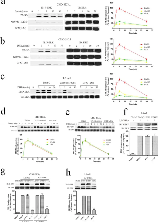
Involvement of Ca2+and PKC in HCA
1-mediated ERK1/2 Activation
The pertussis toxin-sensitive Ga subunit can directly activate
PKC, resulting in ERK1/2 phosphorylation in CHO and COS cells [13]. And our previous studies have demonstrated that PKC played an important role in HCA2and HCA3-induced ERK1/2
activation [12,14]. Therefore, two inhibitors of PKC were used to determine whether PKC was involved in the pathway leading to HCA1-mediated ERK1/2 phosphorylation. The CHO-HCA1
cells were pretreated with 1mM of GF109203X (GFX) or 10mM of Go6983 for 1 h, followed by the agonists L-lactate and 3,5-DHBA in a time course. As shown in Figs. 4a and 4b, both treatment with GF109203X and Go6983 resulted in dramatic decreases (.60%) in ERK1/2 activation. A similar result was observed during 3,5-DHBA-mediated ERK1/2 activation in L6 cells (Fig. 4c). Collectively, these data demonstrated that PKC played a determinant role in HCA1-mediated ERK1/2 activation.
We also evaluated the effect of PLC and PLD, the upstream signaling molecules of PKC, in the HCA1-mediated ERK1/2
signaling pathway. The results showed that PLC inhibitor U73122 (10mM) and PLD inhibitor FIPI (1mM) could not block the activation of ERK1/2 induced by HCA1in both CHO-HCA1and
L6 cells (Figs. 4d, 4e and 4f). Intriguingly, activated HCA1
receptors signal to ERK1/2 via PKC-dependent but PLC- and PLD-independent pathways, leading us to believe that calcium might play an important role in this process. Previous studies have shown that L-lactate causes a rapid increase of intracellular Ca2+
in CHO-K1 cells expressing HCA1receptors [15]. Accordingly,
we investigated whether or not intracellular and extracellular Ca2+
was involved in HCA1-stimulated ERK1/2 phosphorylation.
Pretreatment with the extracellular Ca2+chelator EGTA (5 mM)
significantly inhibited ERK1/2 phosphorylation in both CHO-HCA1and L6 cells (Figs. 4 g and 4 h). However, the intracellular
Ca2+ chelator BAPTA-AM (50mM) did not impair ERK1/2
activation by HCA1receptors in both CHO-HCA1and L6 cells.
Taken together, the results of the present study indicated that stimulation of HCA1 receptors by agonists lead to ERK1/2
activation via PLC and PLD-independent and extracellular Ca2+
and PKC -dependent pathway.
Involvement of PI3K and Src in HCA1- mediated ERK1/2 Activation
Activation of several GPCR has been shown to increase the activity of Src-family tyrosine kinases and Src has been demon-strated to be a critical regulator of GPCR activity, modulating receptor internalization, desensitization and coupling to ERK1/2 and RTK [16]. Previous studies have reported that PI3K and Src are involved in ERK1/2 activation in response to Gi-coupled
receptors [17,18]. Our previous work also demonstrated that PI3K and Src played an important role in both HCA2 and HCA3
mediated ERK1/2 phosphorylation [12,14]. Using CHO-HCA1
cells treated with the PI3K inhibitor wortmannin and the Src inhibitor PP2, we found that both wortmannin and PP2 abolished HCA1-stimulated ERK1/2 phosphorylation (Figs. 5a and 5b),
suggesting that both PI3K and Src kinases played important roles in HCA1-mediated ERK1/2 activation in CHO-HCA1cell lines.
In L6 cells, pretreatment with the PI3K inhibitor wortmannin showed a similar result as seen in CHO-HCA1 cells (Fig. 5c).
However, inhibition of Src by the selective Src kinase inhibitor PP2, did not attenuate HCA1-induced ERK1/2 activation in L6
cells (Fig. 5d). Taken together, these results indicated that PI3K plays an important role in HCA1-mediated ERK1/2 activation.
GbcPlays a Central Role in HCA1-induced ERK1/2 Activation
Gbc subunits bind to and activate PI3K, which is a known
mediator of Gbc-stimulated ERK1/2 activation [17]. To test the
involvement of Gbc-subunits in HCA1-mediated ERK1/2
activa-tion, CHO-HCA1 and L6 cells were preincubated with Gbc
specific inhibitor M119K (20mM) for 4 hrs [19], followed by stimulation with lactate or 395-DHBA for different lengths of time. As shown in Fig. 6, pretreatment with M119K resulted in dramatic decreases (.60%) in HCA1-induced ERK1/2 activation
in both CHO-HCA1and L6 cells, which suggested that the Gbc
subunit might play a central role in HCA1-induced ERK1/2
activation.
5 min. b, Serum-starved L6 cells were pretreated with DMSO or tyrphostin A9 (1mM) or AG1478 (1mM) for 1 h, and then stimulated with 3 mM 3,5-DHBA for for indicated time periods. Serum-starved CHO-HCA1cells (c and d) or L6 cells (e) were pretreated with DMSO or 10mM AG1024 for 2 hrs, and the cells were then stimulated with 10 mM L-Lactate (c) or 300mM 3,5-DHBA (d) for CHO-HCA1cells and 3 mM 3,5-DHBA for L6 cells (e) for indicated time periods. f, Serum-starved L6 cells were pretreated with DMSO or PP2(10mM) or Go6983(10mM) or wortmannin (200 nM) for 1 h, or pretreated with M119K (20mM) or both M119K (20mM) and wortmannin (200 nM) for 4 hrs, and the cells were then stimulated with 5 mM 3,5-DHBA for 5 min. The data shown are representative of at least three independent experiments. Error bars, S.E. for three replicates. Data were analyzed by using the Student’s t test (*p,0.05, **p,0.01, ***p,0.001). IB, immunoblot; P-ERK, phospho-ERK.
doi:10.1371/journal.pone.0093041.g007
Effect of Growth Factor Receptor- Transactivation in HCA1-mediated ERK1/2 Activation
Many GPCRs can activate RTKs (receptor tyrosine kinases) in the absence of RTK ligands, a phenomenon called transactivation [20,21]. Our previous studies have reported that both HCA2and
HCA3mediated ERK1/2 activation is PDGFR
transactivation-dependent in CHO cells and EGFR transactivation in A431 cells [12,14]. Accordingly, we investigated whether PDGFR transacti-vation and EGFR transactitransacti-vation played a role in agonist-stimulated ERK1/2 phosphorylation via HCA1. CHO-HCA1
and L6 cells were preincubated with the PDGF receptor-selective receptor tyrosine kinase inhibitor tyrphostin A9 (1mM) for 1 h, followed by stimulation with 300mM 395-DHBA for CHO-HCA1
cells and 3 mM 395-DHBA for L6 cells for different lengths of time. As shown in Figs. 7a and 7b, in CHO-HCA1cells, there was
only a moderate inhibition (about 25%) of HCA1-mediated
ERK1/2 activation. In contrast, in L6 cells, there was no inhibition of ERK1/2 phosphorylation compared with cells treated with agonist alone. Lactate stimulation also exhibited a similar result in the CHO-HCA1cells (Fig. 7a). As the inhibition of
HCA1-mediated ERK1/2 activation by tyrphostin A9 was
relatively small and most of the tyrphostin tyrosine kinase inhibitors were not really specific, the reduction of HCA1
-mediated ERK1/2 phosphorylation by tyrphostin A9 was likely to be unspecific effects.
To assess the role of EGFR transactivation in HCA1-induced
ERK1/2 activation in cells that endogenously express HCA1, L6
cells were utilized for further investigation. Serum-starved L6 cells were treated with AG1478 (100 nM), an EGFR specific tyrosine kinase inhibitor, for 1 h before exposing them to 3 mM 3,5-DHBA. As shown in Fig. 7b, AG1478 pretreatment have no inhibition of ERK1/2 phosphorylation compared with cells treated with agonist alone.
Previous studies have demonstrated that Src can regulate IGF-I receptor [22], and Src kinase can substitute for the receptor kinase in phosphorylating and activating IGF-I receptor [23]. Next, we investigate whether IGF-1R transactivation was involved in HCA1-mediated ERK1/2 activation. CHO-HCA1 and L6 cells
were preincubated with a selective insulin like growth factor-I (IGF-I) receptor tyrosine kinase inhibitor tyrphostin AG 1024 (10mM) for 2 hrs, followed by stimulation with 300mM 39 5-DHBA for CHO-HCA1 cells and 3 mM 395-DHBA for L6 for
different lengths of time. As shown in Figs. 7d and 7e, in both AG1024 pretreated CHO-HCA1 and L6 cells, ERK1/2
phos-phorylation was decreased over 50% compared with cells treated with agonist alone. Lactate stimulation also exhibited similar results in CHO-HCA1 cells (Fig. 7c), showing that IGF-1R
transactivation is involved in HCA1-induced ERK1/2 activation
in both CHO-HCA1and L6 cells.
To further determine whether HCA1can activate IGF-1R, L6
cells were treated with 3,5-DHBA for 5 min, as shown in Fig7f, 3,5-DHBA treatment induced about two fold IGF-1R phosphor-ylation. Pretreatment with PP2 inhibitor resulted in moderate decreases (about 15%) in HCA1-induced IGF-1R activation.
However, M119K or wortmannin pretreatment resulted in more notable decreases (40 and 55% respectively) in HCA1-mediated
IGF-1R phosphorylation, simultaneous inhibition of Gbc and
PI3K resulted in a nearly complete inhibition of IGF-1R phosphorylation (fig. 7f), suggesting the involvement of Gbcand
PI3K in HCA1-mediated IGF-1R phosphorylation.
Arrestin3 is Involved in HCA1Internalization, but Arrestins are not Involved in HCA1-mediated ERK1/2 Activation
To evaluate the role of arrestins in the regulation of HCA1
internalization and ERK1/2 activation, we used specific siRNAs to reduce the expression of arrestin2 and arrestin3 in HEK-293 cells stably expressing HCA1receptors. The endogenous
expres-sion of arrestins was effectively and specifically knocked-down by specific siRNA treatment but was unaffected in cells treated with non-specific or control siRNAs (Fig. 8a). Silencing arrestin3 effectively inhibited HCA1internalization, whereas knock-down of
arrestin2 had no effect on the internalization of HCA1receptors
(Fig. 8b). We further investigated the effect of knock-down of arrestins on ERK1/2 activation, and no difference was observed between control and knock-down cells (Fig. 8c). Taken together, arrestin3 might be involved in HCA1receptor internalization, but
both arrestins were not required for HCA1-mediated ERK1/2
activation.
Discussion
Lactate is an important metabolic intermediate released by skeletal muscle and other organs including the adipose tissue, which converts glucose into lactate under the influence of insulin [5]. Two recent studies showed that lactate was the endogenous ligand of hydroxy-carboxylic acids (HCAs) receptor 1 [1,15]. And lactate was a specific agonist of HCA1as it did not activate the
closely related receptors HCA2and HCA3. Activation of HCA1in
adipocytes by lactate results in the inhibition of lipolysis at physiologically relevant lactate concentrations (1 to 20 mM) [1], suggesting that HCA1 could be a new target for dyslipidemia
treatment without the unwanted side effect of cutaneuous flushing. As a metabolite of glucose, lactate concentrations rise in vivo following a glucose load [1], and thus HCA1 may also serve a
regulatory role for glucose metabolism. It has been suggested that lactate plays a role in insulin signaling, particularly in insulin mediated anti-lipolytic effects [5]. It has also been suggested that HCA1may play a role in muscle glucose and fatty acid metabolism
[6]. However, the underlying molecular mechanisms for HCA1
signaling remain largely unknown. In the current study, we focused on a detailed characterization of HCA1-mediated MAPK
signalling pathways.
In the present study, the CHO-K1 cell line was selected as a cellular model system for characterizing HCA1receptor signaling
pathways as it was a commonly used cell line for investigating GPCR coupling to various signaling pathways. For better delineation of HCA1-mediated phosphorylation of ERK1/2, we
also used L6 cell line, a rat skeletal muscle cell line, which endogenously expressed rat HCA1receptors, in our current study.
The HCA1 receptor was a Gi protein-coupled receptor, upon
stimulation by agonists, HCA1 receptors triggered an inhibitory
stably expressing HCA1-EGFP were transfected with specific arrestin siRNA or a non-specific control siRNA,72 hrs after transfection, cells were stimulated with 20 mM lactate for 60 min and examined with confocal microscopy as described under ‘Experimental Procedures.’ c, 72 hrs after transfection with specific arrestin siRNA or non-specific control siRNA, cells were stimulated with 20 mM lactate for the indicated time periods and immunoblotted using monoclonal anti-phospho-MAPK E10 (Thr202/Tyr204), and then the blots were stripped and reprobed for total ERK1/2 to control for loading. The data and pictures shown are representative of at least three independent experiments. Error bars, S.E. for three replicates. Data were analysed by Student’sttest (***P,0.001).
doi:10.1371/journal.pone.0093041.g008
effect on adenylate cyclase that led to a decrease of intracellular cAMP levels in a PTX-sensitive manner (Figs. 1a and 1b). Additionally, both CHO-K1 stably expressing HCA1and L6 cell
lines showed a time-dependent activation of ERK1/2 in response to L-lactate or 395-DHBA, peaking at approximately 5 min and returning to basal levels at 30 min, however, the activation of ERK1/2 was significantly attenuated in the presence of PTX (Figs. 3a and 3b). These results indicated that the essential involvement of a heterotrimeric Giprotein in ERK1/2
phosphor-ylation at an early stage was common to both CHO and L6 cells. Previous studies have shown that L-lactate causes a rapid increase of intracellular Ca2+in CHO-K1 cells expressing HCA
1
receptors [16]. We next evaluated the role of PKC in the regulation of HCA1-induced ERK1/2 phosphorylation using
specific inhibitors. Our present data demonstrated that the HCA1-induced ERK1/2 activation was blocked by Go6983 and
GF109203x, PKC inhibitors, suggesting that the PKC pathway participates in ERK1/2 activation (Figs. 4a, 4b and 4c). The involvement of PLC and PLD as a contributor to HCA1-mediated
ERK1/2 activation was assessed by incubating cells with a PLC inhibitor, U73122 or a PLD inhibitor FIPI. Our results shown that both U73122 and FIPI exhibited no significant inhibition of ERK1/2 phosphorylation by activated HCA1(Figs. 4d, 4e and 4f).
Furthermore, we found that HCA1-induced ERK1/2 activation
was abolished by the depletion of extracellular Ca2+ by the
chelator EGTA but not by BAPTA-AM, an intracellular Ca2+
chelator in both CHO-HCA1and L6 cells, suggesting that Ca2+
channel may play an important part in HCA1-mediated ERK1/2
activation (Figs. 4 g and 4 h). Taken together, these data suggested the involvement of extracellular Ca2+ and PKC in HCA
1
-mediated ERK1/2 activation.
Moreover, phosphatidylinositol-39 kinases (PI3K) and Src family non-receptor tyrosine kinases have each been proposed as early intermediates in the pathway to induce EGF receptor transactivation [24,25]. In the present study, we observed that PI3K involved in IGF-1R transactivated phosphorylation of ERK1/2, whereas the Src kinase was not required for HCA1
-induced IGF-1R transactivation in L6 cells.
There is a growing body of evidence to suggest that the transactivation of growth factor receptors is another mechanism by which GPCRs mediate ERK1/2 phosphorylation [20]. Our previous study demonstrated that, in CHO-K1 cells, both HCA2
and HCA3-mediated ERK1/2 activation was potently inhibited
by the PDGF receptor-selective inhibitor tyrphostin A9, and in A431 cells, the EGF receptor-selective inhibitor AG1478 was found to significantly impair ERK1/2 activation. Our present research showed that PDGFR and EGFR were well possible playing no role in HCA1-induced ERK1/2 phosphorylation in
CHO-K1 and L6 cells. In contrast, HCA1-mediated ERK1/2
phosphorylation was found to be significantly impaired by AG1024, an insulin-like growth factor-1 receptor specific tyrosine kinase, in both two cell lines. These results suggested that a transactivation of insulin-like growth factor-1 receptor participa-teed in HCA1-mediated ERK1/2 phosphorylation. Previous study
also have shown that the insulin-like growth factor-1 receptor can be transactivated in response to GPCR ligands such as thrombin [26] and angiotensin II [27].
In addition, we observed that pretreated the cells with M119K, an inhibitor of Gbcsubunit-dependent signaling [19], effectively
attenuated the IGF-1R receptor phosphorylation and ERK1/2 activation triggered by HCA1(Figs. 6a, 6b and 6c). Simultaneous
inhibition of Gbc and PI3K resulted in a nearly complete
inhibition of IGF-1R phosphorylation. These results indicated that Gbcsubunit might act as an early signal mediating HCA1
-induced IGF-1R receptor transactivation. The major effects of Gi
activation on ERK1/2 cascade appear to be mediated via its Gbc
subunits [28,29]. Previous studies have shown that Gi-type GPCRs
stimulate Ca2+mobilization through the binding of G
bcsubunits
to PLC [30,31]. It has also been reported that the best understood mechanism whereby the Gbc subunits stimulate ERK1/2 is
through the ‘transactivation’ of classical receptor tyrosine kinases, e.g., the EGF and platelet-derived growth factor (PDGF) receptors [21]. Thus, we postulated that upon stimulation of HCA1 by
agonists, activated Gi protein impaired cAMP production and
released Gbc subunits, the free Gbc subunits caused IGF-1R
transactivation.
Arrestins are traditionally recognized as playing a well-established role in the termination of receptor-G-protein coupling and the initiation of clathrin-dependent internalization [32]. However, there is a growing body of evidence indicates that arrestins function as signal transducers for many GPCRs to mediate ERK1/2 activation [33]. Arrestins are required for later phase activation of the ERK1/2 pathway mediated by angiotensin II type 1A (AT1A) [34],b2-adrenergic [35], vasopressin 2 [36], and parathyroid hormone (PTH) [37] receptors, whereas, in the dopamine D2 and D3 receptor [38] and the formyl peptide receptor (FPR) [39], arrestins have been found to play no role or only a minor role in the activation of the ERK1/2 pathway. Our results using siRNA showed that arrestin3 was required for agonist-mediated HCA1 internalization, whereas knockdown of
arrestin2 or arrestin3 using siRNA had no effect on ERK1/2 activation. These results were in good agreement with our previous observation for the HCA2-mediated activation of the
ERK1/2 pathway [40].
In conclusion, we have characterized the molecular mechanisms of HCA1-mediated activation of the ERK1/2 pathway and
demonstrated that the Gbcsubunit dissociated from the activated
Gi protein played a central role in the regulation of HCA1
-activated ERK1/2 phosphorylation via PKC pathway activation and IGF-1R transactivation. Furthermore, we found arrestin-2 and arrestin-3 had no effect on HCA1-mediated ERK1/2
activation by using arrestin-2/3 specific siRNA, whereas HCA1
internalization was arrestin3-dependent. However, additional investigations will be necessary to further clarify the role of the ERK1/2 pathway in HCA1-mediated insulin-dependent
inhibi-tion of lipolysis.
Supporting Information
Figure S1 a, L6 cells were transfected with specific HCA1
siRNA or a nonspecific control siRNA (The HCA1 siRNA sequence was 59- ACCUGGAAGUCGAGCACUA -39, whereas 59-AAACUCUAUCUGCACGCUGAC-39 was used for nonspe-cific control). A total of 96 hrs after transfection, cells were harvested, mRNA levels of GAPDH and HCA1 were measured by quantitative real-time–PCR. b, A total of 96 hrs after transfection with specific HCA1 siRNA or nonspecific control siRNA, L6 cells were stimulated with DMSO or 3 mM 3,5-DHBA for 5 min and immunoblotted using monoclonal anti- phospho-MAPK E10 (Thr202/Tyr204), and then the blots were stripped and reprobed for total ERK to control for loading. The data shown are representative of at least three independent experiments. Error bars, S.E. for three replicates. Data were analyzed by using the Student’s t test (***p,0.001). IB, immunoblot; P-ERK, phospho-ERK.
Author Contributions
Conceived and designed the experiments: GL JL. Performed the experiments: GL HW LW RC. Analyzed the data: GL HW LW.
Contributed reagents/materials/analysis tools: GL. Wrote the paper: GL LW.
References
1. Liu C, Wu J, Zhu J, Kuei C, Yu J, et al. (2009) Lactate inhibits lipolysis in fat cells through activation of an orphan G-protein-coupled receptor, GPR81. J Biol Chem 284: 2811–2822.
2. Taggart AK, Kero J, Gan X, Cai TQ, Cheng K, et al. (2005) (D)-beta-Hydroxybutyrate inhibits adipocyte lipolysis via the nicotinic acid receptor PUMA-G. J Biol Chem 280: 26649–26652.
3. Ahmed K, Tunaru S, Langhans CD, Hanson J, Michalski CW, et al. (2009) Deorphanization of GPR109B as a receptor for the beta-oxidation intermediate 3-OH-octanoic acid and its role in the regulation of lipolysis. J Biol Chem 284: 21928–21933.
4. Ahmed K, Tunaru S, Offermanns S (2009) GPR109A, GPR109B and GPR81, a family of hydroxy-carboxylic acid receptors. Trends Pharmacol Sci 30: 557– 562.
5. Ahmed K, Tunaru S, Tang C, Muller M, Gille A, et al. (2010) An autocrine lactate loop mediates insulin-dependent inhibition of lipolysis through GPR81. Cell Metab 11: 311–319.
6. Ge H, Weiszmann J, Reagan JD, Gupte J, Baribault H, et al. (2008) Elucidation of signaling and functional activities of an orphan GPCR, GPR81. J Lipid Res 49: 797–803.
7. Lorenz K, Schmitt JP, Schmitteckert EM, Lohse MJ (2009) A new type of ERK1/2 autophosphorylation causes cardiac hypertrophy. Nat Med 15: 75–83. 8. Sun Y, Cheng Z, Ma L, Pei G (2002) Beta-arrestin2 is critically involved in CXCR4-mediated chemotaxis, and this is mediated by its enhancement of p38 MAPK activation. J Biol Chem 277: 49212–49219.
9. Schwindinger WF, Robishaw JD (2001) Heterotrimeric G-protein betagamma-dimers in growth and differentiation. Oncogene 20: 1653–1660.
10. Ahn S, Kim J, Hara MR, Ren XR, Lefkowitz RJ (2009) {beta}-Arrestin-2 Mediates Anti-apoptotic Signaling through Regulation of BAD Phosphorylation. J Biol Chem 284: 8855–8865.
11. Pu J, Peng G, Li L, Na H, Liu Y, et al. (2011) Palmitic acid acutely stimulates glucose uptake via activation of Akt and ERK1/2 in skeletal muscle cells. J Lipid Res 52: 1319–1327.
12. Li G, Deng X, Wu C, Zhou Q, Chen L, et al. (2011) Distinct kinetic and spatial patterns of protein kinase C (PKC)- and epidermal growth factor receptor (EGFR)-dependent activation of extracellular signal-regulated kinases 1 and 2 by human nicotinic acid receptor GPR109A. J Biol Chem 286: 31199–31212. 13. van Biesen T, Hawes BE, Raymond JR, Luttrell LM, Koch WJ, et al. (1996)
G(o)-protein alpha-subunits activate mitogen-activated protein kinase via a novel protein kinase C-dependent mechanism. J Biol Chem 271: 1266–1269. 14. Zhou Q, Li G, Deng X, H X, Chen L, et al. (2012) Activated Human
Hydroxy-Carboxylic Acid Receptor-3 Signals to MAP Kinase Cascades via the PLC-Dependent PKC and MMP-Mediated EGFR Pathways. Br J Pharmacol. 15. Cai TQ, Ren N, Jin L, Cheng K, Kash S, et al. (2008) Role of GPR81 in
lactate-mediated reduction of adipose lipolysis. Biochem Biophys Res Commun 377: 987–991.
16. Luttrell DK, Luttrell LM (2004) Not so strange bedfellows: G-protein-coupled receptors and Src family kinases. Oncogene 23: 7969–7978.
17. Lopez-Ilasaca M, Crespo P, Pellici PG, Gutkind JS, Wetzker R (1997) Linkage of G protein-coupled receptors to the MAPK signaling pathway through PI 3-kinase gamma. Science 275: 394–397.
18. Kranenburg O, Verlaan I, Hordijk PL, Moolenaar WH (1997) Gi-mediated activation of the Ras/MAP kinase pathway involves a 100 kDa tyrosine-phosphorylated Grb2 SH3 binding protein, but not Src nor Shc. EMBO J 16: 3097–3105.
19. Kirui JK, Xie Y, Wolff DW, Jiang H, Abel PW, et al. (2010) Gbetagamma signaling promotes breast cancer cell migration and invasion. J Pharmacol Exp Ther 333: 393–403.
20. Pierce KL, Luttrell LM, Lefkowitz RJ (2001) New mechanisms in heptahelical receptor signaling to mitogen activated protein kinase cascades. Oncogene 20: 1532–1539.
21. Gschwind A, Zwick E, Prenzel N, Leserer M, Ullrich A (2001) Cell communication networks: epidermal growth factor receptor transactivation as the paradigm for interreceptor signal transmission. Oncogene 20: 1594–1600. 22. Bromann PA, Korkaya H, Courtneidge SA (2004) The interplay between Src
family kinases and receptor tyrosine kinases. Oncogene 23: 7957–7968. 23. Peterson JE, Kulik G, Jelinek T, Reuter CW, Shannon JA, et al. (1996) Src
phosphorylates the insulin-like growth factor type I receptor on the autopho-sphorylation sites. Requirement for transformation by src. J Biol Chem 271: 31562–31571.
24. Lin AL, Zhu B, Zhang W, Dang H, Zhang BX, et al. (2008) Distinct pathways of ERK activation by the muscarinic agonists pilocarpine and carbachol in a human salivary cell line. Am J Physiol Cell Physiol 294: C1454–1464. 25. Lopez-Ilasaca M, Gutkind JS, Wetzker R (1998) Phosphoinositide 3-kinase
gamma is a mediator of Gbetagamma-dependent Jun kinase activation. J Biol Chem 273: 2505–2508.
26. Du J, Brink M, Peng T, Mottironi B, Delafontaine P (2001) Thrombin regulates insulin-like growth factor-1 receptor transcription in vascular smooth muscle: characterization of the signaling pathway. Circ Res 88: 1044–1052. 27. Zahradka P, Litchie B, Storie B, Helwer G (2004) Transactivation of the
insulin-like growth factor-I receptor by angiotensin II mediates downstream signaling from the angiotensin II type 1 receptor to phosphatidylinositol 3-kinase. Endocrinology 145: 2978–2987.
28. Hawes BE, van Biesen T, Koch WJ, Luttrell LM, Lefkowitz RJ (1995) Distinct pathways of Gi- and Gq-mediated mitogen-activated protein kinase activation. J Biol Chem 270: 17148–17153.
29. Crespo P, Xu N, Simonds WF, Gutkind JS (1994) Ras-dependent activation of MAP kinase pathway mediated by G-protein beta gamma subunits. Nature 369: 418–420.
30. Dickenson JM, Hill SJ (1998) Involvement of G-protein betagamma subunits in coupling the adenosine A1 receptor to phospholipase C in transfected CHO cells. Eur J Pharmacol 355: 85–93.
31. Dorn GW, 2nd, Oswald KJ, McCluskey TS, Kuhel DG, Liggett SB (1997) Alpha 2A-adrenergic receptor stimulated calcium release is transduced by Gi-associated G(beta gamma)-mediated activation of phospholipase C. Biochem-istry 36: 6415–6423.
32. Luttrell LM, Lefkowitz RJ (2002) The role of beta-arrestins in the termination and transduction of G-protein-coupled receptor signals. J Cell Sci 115: 455–465. 33. Lefkowitz RJ, Shenoy SK (2005) Transduction of receptor signals by
beta-arrestins. Science 308: 512–517.
34. Ahn S, Wei H, Garrison TR, Lefkowitz RJ (2004) Reciprocal regulation of angiotensin receptor-activated extracellular signal-regulated kinases by beta-arrestins 1 and 2. J Biol Chem 279: 7807–7811.
35. Shenoy SK, Drake MT, Nelson CD, Houtz DA, Xiao K, et al. (2006) beta-arrestin-dependent, G protein-independent ERK1/2 activation by the beta2 adrenergic receptor. J Biol Chem 281: 1261–1273.
36. Ren XR, Reiter E, Ahn S, Kim J, Chen W, et al. (2005) Different G protein-coupled receptor kinases govern G protein and beta-arrestin-mediated signaling of V2 vasopressin receptor. Proc Natl Acad Sci U S A 102: 1448–1453. 37. Gesty-Palmer D, Chen M, Reiter E, Ahn S, Nelson CD, et al. (2006) Distinct
beta-arrestin- and G protein-dependent pathways for parathyroid hormone receptor-stimulated ERK1/2 activation. J Biol Chem 281: 10856–10864. 38. Beom S, Cheong D, Torres G, Caron MG, Kim KM (2004) Comparative
studies of molecular mechanisms of dopamine D2 and D3 receptors for the activation of extracellular signal-regulated kinase. J Biol Chem 279: 28304– 28314.
39. Gripentrog JM, Miettinen HM (2008) Formyl peptide receptor-mediated ERK1/2 activation occurs through G(i) and is not dependent on beta-arrestin1/2. Cell Signal 20: 424–431.
40. Li G, Shi Y, Huang H, Zhang Y, Wu K, et al. (2010) Internalization of the human nicotinic acid receptor GPR109A is regulated by G(i), GRK2, and arrestin3. J Biol Chem 285: 22605–22618.