Interleukin-2/15 Receptor
b
Chain Prevents Disease
Progression in Experimental Arthritis
Tiantian Zhang1., Xuehua Bai1., Xiaohua Mao1,2
*
1Key Laboratory of Ministry of Education for Developmental Genes and Human Diseases, School of Life Sciences, Southeast University, Nanjing, China,2Department of Biochemistry, School of Medicine, Southeast University, Nanjing, China
Abstract
The role of interleukin (IL)-15 in the pathogenesis of rheumatoid arthritis (RA) is well established; however, systemic knockdown of IL-15 receptor (IL-15R) for reduction in inflammation at local sites has not been demonstrated. In this study, the therapeutic effect of intravenously administered siRNA targeting thebchain of IL-15R which is shared by the receptor for IL-2 was examined in rats with adjuvant-induced arthritis (AA). Polyethylenimine (PEI)-complexed siRNA nanoparticles could easily accumulate in arthritic paws of AA rats. In the paws, the nanoparticles were avidly taken up by macrophages and to a lesser extent by T cells. Weekly administered IL-2/15Rb siRNA polyplexes were capable of decreasing disease progression in AA rats, with striking inhibition of clinical, radiologic, and histologic features of RA. The observed therapeutic effect was associated with reduced expression of proinflammatory mediators in the inflamed joints. Thus, this study provides evidence that IL-2/15Rbcould be targeted for the treatment of RA.
Citation:Zhang T, Bai X, Mao X (2013) Systemic Delivery of Small Interfering RNA Targeting the Interleukin-2/15 ReceptorbChain Prevents Disease Progression in Experimental Arthritis. PLoS ONE 8(11): e78619. doi:10.1371/journal.pone.0078619
Editor:Pierre Bobe´, INSERM-Universite´ Paris-Sud, France
ReceivedApril 30, 2013;AcceptedSeptember 16, 2013;PublishedNovember 5, 2013
Copyright:ß2013 Zhang et al. This is an open-access article distributed under the terms of the Creative Commons Attribution License, which permits unrestricted use, distribution, and reproduction in any medium, provided the original author and source are credited.
Funding:This study was supported by National Natural Science Foundation of China (81071445). The URL of the funder’s website is www.nsfc.gov.cn. The funder had no role in study design, data collection and analysis, decision to publish, or preparation of the manuscript.
Competing Interests:The authors have declared that no competing interests exist.
* E-mail: [email protected]
.These authors contributed equally to this work.
Introduction
Rheumatoid arthritis (RA) is characterized by synovial hyper-plasia and persistent inflammation that is now recognized to result from the interaction among macrophages, T cells, B cells, and nonhematopoietic cells such as fibroblasts. These interactions are facilitated by the actions of cytokines released from the activated cells that then, through both autocrine and paracrine mechanisms, induce the production of other proinflammatory cytokines, which together contribute to the pathogenesis of this disease and ultimately lead to joint damage [1,2]. Accordingly, biologic agents that block the actions of specific cytokines or immunue regulators have emerged as major therapies in RA. However, no final victory has been achieved over this joint damaging and potentially life-threatening systemic autoimmune disease, as the effects of current biologics are only partial and nonresponses are common. A plausible explanation for variable response to the biologic agents is that distinct cytokines/immune regulators may mediate discrete effects at different disease stages, and thus, there may be optimal targets defined by the relative disease stage of intervention [1].
Based on the work pioneered by McInnes et al. [3,4], the
importance of IL-15 in the development and amplification of the inflammatory process in RA has been demonstrated. In RA, IL-15 is expressed primarily by macrophages as well as by fibroblast-like synoviocytes and endothelial cells [5]. It exhibits pleiotropic proinflammatory effects on numerous target cell types relevant to a variety of inflammatory conditions [6] and was thought to be at
the apex of the cytokine cascade created in the inflamed joints [7]. This notion was strengthened by the fact that TNFablockade does not affect synovial expression of IL-15 whereas neutralizing IL-15 significantly inhibits the production of proinflammatory cytokines including TNFa in RA synovial cell cultures [8,9]. Most importantly, IL-15 plays a key role in sustaining the fundamentally important cognate interactions between T cells and macrophages which are involved in the production and release of most proinflammatory cytokines, chemokines and metalloproteinase enzymes [1,6]. In clinical studies, the link between serum IL-15 levels and disease severity in patients with early arthritis has been demonstrated [10]; in addition, genetic variants in IL-15 were shown to associate with joint destruction in RA in a multicohort study, suggesting a direct role for this cytokine in articular bone erosion [11,12]. As IL-15 is implicated in initiation and perpetuation of the inflammation and mediates osteoclastogenesis in RA, IL-15 signaling pathway could possibly be targeted therapeutically. In support of this view, several IL-15- or IL-15 receptor (IL-15R)-directed monoclonal antibodies and fusion proteins have been effective in ameliorating RA in animal models [13–15].
delivered siRNA targeting the IL-15Rbchain which is also shared by IL-2 receptor was assessed in experimental arthritis. We speculate that IL-2/15Rb siRNA formulated with polyethyleni-mine (PEI) would be avidly taken up by inflamed macrophages, inhibit the production and release of most proinflammatory cytokines and thus prevent disease progression in rats with adjuvant-induced arthritis (AA).
Results
Design of siRNA against rat IL-2/15Rb
IL-15 is a cytokine that binds a heterotrimeric receptor containing a unique a chain (IL-15Ra), a b chain that is also found in the receptor for IL-2, andcc[17]. IL-15Rabinds IL-15 with high affinity, but transduces signals only in the presence of the IL-2/15Rbandcc. Sinceccis shared by receptors for IL-4, IL-7, IL-9, IL-15, and IL-21 and a loss of cc function causes immunodeficiencies in both humans and mice [18], it is anticipated that knockdown of cc may lead to severe systemic side effects. In addition, it is unknown whether certain cytokines such as IL-4 and IL-9 play an inflammatory role in RA. In view of the well-established role of IL-15 and possible involvement of IL-2 in RA pathogenesis [19,20], we attempted to test the feasibility of
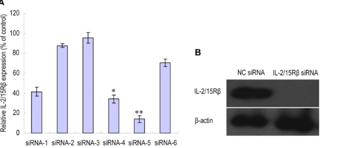
in vivosilencing of IL-2/15Rbfor the treatment of this disease. For this purpose, six siRNA sequences targeting rat IL-2/15Rbwere designed. Peritoneal macrophages were transfected with each of the six siRNA duplexes and the silencing of IL-2/15Rb mRNA was measured by quantitative RT-PCR (qPCR). As shown in Figureoˆ 1A, the most potent effect was observed with siRNA-5, which successfully resulted in an 80% mRNA reduction relative to nonspecific negative control siRNA (NC siRNA). The silencing effect of siRNA-5 was further confirmed by Western blotting (Figureoˆ 1B). In the following experiments, siRNA-5 was used as IL-2/15Rb-specific siRNA.
Preparation and properties of PEI/siRNA complexes
In vivo-jetPEI, a linear, endotoxin-free, cationic polymer
currently used for the delivery of therapeutic nucleic acids in clinical trials was chosen as siRNA carrier. PEI/siRNA complexes were prepared by mixingin vivo-jetPEI with siRNA at N:P ratio of
6 at which the PEI could tightly bind siRNA as analyzed by gel retardation assay (not shown). The resulting PEI/siRNA com-plexes displayed an average hydrodynamic diameter of about 246 nm and an average zeta potential of+28 mV as determined by DLS (Figuresoˆ 2A, B). To examine the uptake of PEI/siRNA polyplexes by macrophages, PEI-formulated Cy3-siRNA (N:P = 6) was incubated with rat peritoneal macrophages in the extracellular medium for 12 h. Next, intracellular uptake was analyzed by flow cytometry. As shown in Figureoˆ 2C, about 53% of the particles were internalized. Western blot analysis revealed that IL-2/15Rb
siRNA polyplexes were capable of knocking down IL-2/15Rb
expression in rat peritoneal macrophages by 60% compared to negative control siRNA (Figureoˆ 2D). The silencing capability of the PEI/IL-2/15RbsiRNA complexes also indicated that IL-2/ 15RbsiRNA could be released from the endocytosis vesicles. Tissue distribution of systemically administered PEI/ siRNA complexes
Nanoparticles are usually taken up by liver, spleen and other parts of the reticuloendothelial system (RES). On the other hand, small particles can easily penetrate kidney’s filtering systems. To determine tissue uptake and kinetics of PEI/siRNA complexes in the context of the inflammatory disorder, Cy5 was used to monitor the in vivo distribution of systemically applied PEI/siRNA
nanoparticles. The long-wavelength emission spectrum of this fluorophore renders it distinguishable from background auto-fluorescence and thus very suitable forin vivoimaging [21]. AA
rats were injected through the tail vein with a single dose of Cy5-siRNA formulated with the in vivo-jetPEI and tissue
distribution was visualized at 6 and 12 h using Caliper IVIS imaging system. As shown in Figureoˆ 3A, Cy5-siRNA could accumulate in arthritic paws, and the accumulation pattern was similar at 6 h and 12 h after intravenous injection. To examine the distribution in detail, rats were sacrificed at 12 h after whole animal assay, and major organs including joints (without skin) were isolated. As shown in Figureoˆ 3B, the kidneys were found to possess the strongest signals among all tissues examined, whereas the liver, spleen, and joint displayed background to moderate signals. Note that the signal intensity in the excised
Figure 1.In vitrovalidation of siRNA sequences designed to target rat IL-2/15Rb.Six different IL-2/15RbsiRNA sequences were designed and rat peritoneal macrophages were transfected with each of the six siRNA duplexes or with a nontargeting negative control siRNA (NC siRNA) using X-tremeGENE siRNA transfection reagent (Roche). RNA and proteins were prepared at 24 and 48 h post-transfection, respectively. (A) IL-2/15RbmRNA levels measured by quantitative real time-PCR (qPCR). Results were normalized to GAPDH and are presented as the percentage of NC siRNA. *P,0.05; **P,0.01 versus NC siRNA. (B) Western blot analysis of IL-2/15Rbprotein levels in macrophages transfected with siRNA-5. The detection of actin expression was performed to monitor protein loading.
doi:10.1371/journal.pone.0078619.g001
joints was weaker than that in whole-animal assay, which was probably due to inadvertent loss of inflamed soft tissue when the joint was isolated. Fluorescence signals were hardly detected in lungs and hearts. Therefore although our PEI/siRNA complex-es, like most siRNA nanoparticlcomplex-es, are subjected to clearance from the blood through RES and renal filtration, they also efficiently accumulate in the inflamed joints.
In vivocellular uptake of systemically administered PEI/
siRNA complexes
Two predominant cell types in the synovial infiltrate of RA, the phagocytic macrophages and the non-phagocytic T lymphocytes, were chosen to assess the preferential immune cell type targeted by PEI/siRNA particles. To this end, we collected blood, spleen, liver, kidney, and inflamed joints at three time points (2, 8, and 24 h post intravenous injection) from AA rats injected with PEI/ Cy3-siRNA complexes and performed flow cytometry to examine the in vivocellular uptake of the nanocomplexes. Representative
flow cytometry staining for CD11b+cells and CD3+cells at 24 h post injection is presented in Figuresoˆ 4A and B. Over the 24 h time course the proportion of CD11b- and CD3-positive cells positive for the fluorescence tended to gradually increase (Figuresoˆ 4C, D). Although the PEI-encapsulated siRNA was detected in all of the tissues examined, it was preferentially taken up by macrophages and T cells in the paw. At 24 h, approximately 37% of CD11b+ cells and approximately 13% of CD3+ cells isolated from the paw had engulfed the Cy3-siRNA polyplexes. Note that at any time points in any tissues examined, CD11b+
cells took up siRNA polyplexes more efficiently than CD3+cells. Lastly, it is worth mentioning that while kidney accumulates most PEI/siRNA particles as demonstrated by in vivo imaging,
macrophages and T cells in this tissue seem less efficient in taking up the particles.
Systemic delivery of PEI/IL-2/15RbsiRNA nanoparticles inhibits inflammation in experimental arthritis
To evaluate thein vivo efficacy of silencing IL-2/15Rbfor RA
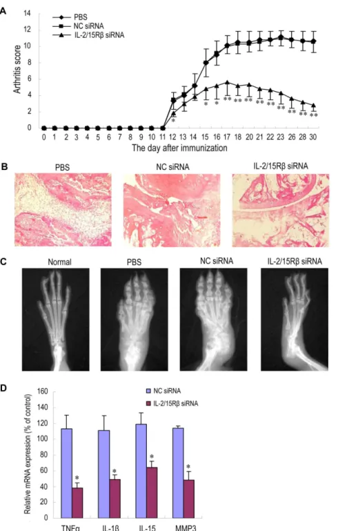
treatment, AA rats were intravenously injected with PEI/IL-2/ 15Rb siRNA once per week for three weeks. Compared with phosphate-buffered saline (PBS) and NC siRNA control groups, administration of IL-2/15Rb siRNA nanoparticles significantly reduced clinical signs of arthritis, as assessed by paw swelling and arthritis scores (Figureoˆ 5A). The maximum effect was observed at the end of the observation period. To objectively measure the extent of joint destruction, rats were subjected to radiography on day 31, then killed, and paws were isolated for histological evaluation of the ankle joints. Histological and radiographic examination confirmed these findings, showing that the reductions in synovial tissue inflammation and in cartilage/bone destruction were striking in the group treated with IL-2/15RbsiRNA (Figuresoˆ 5B, C).
To assess the effect of silencing IL-2/15Rbon the expression of proinflammatory mediators in the arthritic joints, RNA was prepared from ankle joints of AA rats receiving different treatment, and cytokine expression was analyzed by qPCR. As shown in Figureoˆ 5D, expression of two well-known inflammatory cytokines TNFa and IL-1b in the group receiving IL-2/15Rb
Figure 2. Physical and biological characterization of PEI/siRNA complexes.siRNA was complexed within vivo-jetPEI at N:P ratio of 6. (A) Hydrodynamic size distribution, and (B) zeta potential of PEI/NC siRNA complexes analyzed by DLS. (C) Flow cytometric analysis of the uptake of siRNA polyplexes in rat peritoneal macrophages. Cells were incubated with PEI-formulated Cy3-siRNA (40 nM) for 12 h. As negative control, cells were incubated with PEI/NC siRNA. (D) Western blot analysis of IL-2/15Rbsilencing in rat peritoneal macrophages. Macrophages were transfected with PEI/ IL-2/15RbsiRNA complexes for 48 h.
siRNA decreased sharply as compared with NC siRNA-treated controls. Reduction in the transcript for MMP-3, which partici-pates in cartilage/bone digestion, was also observed. Interestingly, reduction in IL-15 mRNA production upon Il-2/15Rb knock-down was statistically significant but not so drastic compared with the reduced expression of TNFa, IL-1b, and MMP-3.
Discussion
Previously we showed that immunotoxins targeting IL-15R-bearing cells (e.g. activated macrophages and T cells) can attenuate disease severity in rats with AA [15]. In view of the toxic moiety of immunotoxins and a wide distribution of IL-15R in multiple tissues, frequent administration of the immunotoxins may cause systemic side effects. It has been reported that siRNA can ensure a therapeutic effect for several weeks in non-dividing cells such as tissue macrophages [16], thus we believe inhibiting activation of IL-15R+ cells via siRNA-mediated gene silencing would be safer than eliminating IL-15R+cells by cytotoxic protein agents. In this study we assessed the therapeutic efficiency of IL-2/ 15Rb siRNA for systemic delivery in experimental arthritis. As expected, administration of IL-2/15RbsiRNA decreased disease progression in AA rats, with striking inhibition of the clinical, radiologic, and histologic features of RA.
Because naked siRNA is subject to degradation by endogenous enzymes, and is too large and too negatively charged to freely cross cellular membranes, the issue of effective and non-toxic delivery is a key challenge and serves as the most significant barrier between siRNA technology and its therapeutic application [22]. It is widely recognized that viral vectors are highly efficient delivery systems for nucleic acids, but their clinical application is hindered by their induction of toxic immune responses and inadvertent gene expression changes following random integration into the host genome [23]. Cationic polymers, such as PEI, poly-L-lysine, and chitosan, can bind and condense large nucleic acids into stabilized nanoparticles and are used to be polymeric gene transfection materials as promising alternatives of viral vectors. Among them, PEI, which has strong escape capacity from the endosome due to the so-called ‘‘proton sponge’’ effect, is usually a gold standard of polymeric transfection agent [24]. We therefore chose to usein vivo-jetPEI, a linear PEI-based transfection reagent for in vivo
experiments, as IL-2/15Rb siRNA delivery vehicle. It has been reported thatin vivo-jetPEI-formulated siRNA at N/P ratio of 8
does not induce inflammation and toxicity after systemic delivery [25]. In the future we plan to evaluate potential toxicity of the PEI/siRNA complexes at N/P ratio of 6 used in the present study. There are few studies of systemic application of anticytokine siRNA by the intravenous route as a therapeutic tool in RA [26– 28]. IL-15 contributes to the pathogenesis of arthritis by maintaining the activation of synovial macrophages which in turn are responsible for the production and release of most proin-flammatory factors, including IL-15 itself. The observed thera-peutic effect of IL-2/15RbsiRNA on AA rats was associated with decreased expression of proinflammatory mediators in the ankle joints, including TNFa, IL-1b, MMP-3, and IL-15. Compared with marked influence on the expression of TNFa, IL-1b, and MMP-3, reduction in IL-15 mRNA production upon IL-2/15Rb
knockdown was not so drastic, consistent with the role of IL-15 signaling in the amplification of the inflammatory network in RA. Because TNFais a major proflammatory cytokine in RA, and it is IL-15, not IL-2, that induces the production of abundant TNFaby synovial macrophages and T cells [4], we speculate while IL-2/ 15Rbis a common signaling subunit shared by IL-2 and IL-15, the antiarthritic effect of IL-2/15Rb siRNA may be primarily ascribed to the inhibition of IL-15 signaling. This view is strengthened by the fact that IL-15, not IL-2, plays a role in the development of osteoclasts which were reported to be involved in the pathological destruction of bone [29]. On the other hand however, IL-2, like IL-15, may also stimulate the proliferation of T cells, B cells, and NK cells [30] that all participate in RA Figure 3. Tissue distribution of PEI/Cy5-siRNA polyplexes.The
particles were administered as a single dose (0.6 mg siRNA/kg) to AA rats via tail vein. Images were taken with IVIS imaging system. 0 h represents the non-injected control. (A)In vivofluorescence images of the arthritic paws at 6 and 12 h post-injection. Shown are represen-tative of 3 rats per group. (B) Fluorescence images of major organs. Rats were killed 12 h after injection. Hind ankle joints and various organs (heart, liver, spleen, lung, and right kidney) from three rats were isolated and imaged.
doi:10.1371/journal.pone.0078619.g003
pathogenesis, the antiarthritic effect of IL-2/15RbsiRNA can thus be secondarily attributed to the inhibition of IL-2 signaling.
In tissue biodistribution experiments, PEI/siRNA complexes showed greater selective accumulation in kidney and mild or intermediate incorporation by liver and spleen. This is consistent with classic clearance of a majority of intravenously administered foreign particles by renal filtration and RES uptake. It has been reported that siRNA is barely detectable in normal joints and is increased in the inflamed joints of arthritic mice [26]. Similarly, in this study significant amounts of PEI/siRNA particles were found to accumulate in the inflamed joints, which was most likely resulted from the enhanced permeability and retention (EPR) effect due to the abnormal and leaky vasculature in the joints of RA. Thus, although further studies will be necessary, based on imaging data, the PEI/siRNA complexes are preferentially retained in kidney 12 h after intravenous injection, followed by liver, joints, and spleen, whereas accumulation is barely detectable in heart and lung. Carriers such as liposomes, drug-polymer conjugates, and PLGA nanoparticles have been utilized for EPR effect-based passive targeting to the inflamed synovium [26,27,31]. High uptake of the PEI/siRNA complexes by arthritic paws confirms that PEI-mediated siRNA delivery has the potential for the treatment of RA.
Nanoparticles experience rapid clearance by the kidney if they are smaller than 10 nm in diameter [32,33]. In addition, the physicochemical characteristics of nanoparticles such as particle size and surface charge can dramatically affect clearance mechanism via glomerular basement membrane (GBM)-mediated disruption of siRNA nanoparticles. It has been recently suggested that cationic polymer-based siRNA nanoparticles being positive in zeta potential and being <100 nm or smaller in hydrodynamic
diameters can deposit and disassemble at the kidney GBM [34,35]. Considering the size of our PEI/siRNA particles (average diameter 246 nm), it is unlikely that they could access the GBM and disassemble there to generate components that are small enough to cross into the urinary space. Therefore, although PEI/ siRNA nanoparticles are primarily accumulated in kidney over a period of 12 h after administration, renal filtration does not seem to be the main elimination pathway for the clearance of these particles from circulation. To gain a more complete picture of tissue uptake and elimination pathway of jetPEI/siRNA particles, additional studies over a longer time course as well as assessment of tissues (including urine) no studied here are needed.
A critical requirement for achieving in vivo RNA interference
using a systemic approach is to ensure the delivery of siRNA to the cytoplasm of the cell. In the present study, a significant proportion of macrophages in the inflamed paws ingested Cy3-siRNA polyplexes following intravenous injection, consistent with marked alleviation of disease severity in AA rats treated with IL2/15Rb
siRNA polyplexes. In addition, considering that RA is a systemic inflammatory disease, manifested locally as erosion of the joints, and that the pathogenesis of RA involves systemic-derived cellular infiltration and cytokine production [36,37], it is possible that CD11b+ cells transfected by PEI/IL2/15Rb siRNA particles in other places might migrate to inflamed joints to counterbalance local inflammation. Besides avid uptake of the particles by macrophages, a notable proportion of T cells isolated from the
paws also entrapped the siRNA, which could be attributed to the surface charge of the PEI/ siRNA complexes. Perhaps T cells, being nonphagocytic cells, prefer ingesting cationic particles [38]. Since both macrophages and T cells are involved in the pathogenesis of RA, systemic inhibition of macrophage/T cell activation via downregulating IL-15 and IL-2 signaling may result in an enhanced anti-arthritic effect. It has been recently reported that a combined therapy with anti-CD3 and anti-TNF leads to a long-term amelioration of established arthritis in an animal model [39].
In conclusion, PEI-complexed IL-2/15RbsiRNA particles are capable of downregulating IL-2/15Rbin vitroin difficult-to-silence
peritoneal macrophages from rats.In vivoexperiments demonstrate
that the siRNA polyplexes can easily accumulate in arthritic paws and a significant proportion of macrophages and T cells in the target tissue can take up the particles. As expected, weekly administered PEI-complexed IL-2/15Rb siRNA is effective in reducing disease severity in AA rats, confirming that IL-2/15Rb
could be therapeutically targeted for the treatment of RA.
Materials and Methods
Ethics statement
All animal experiments were performed in accordance with the guideline of and approved by the Committee on Laboratory Animals of Southeast University, China. All surgery and in vivo imaging were performed under sodium pentobarbital anesthesia, and all efforts were made to minimize suffering.
siRNA duplexes
Six siRNA duplexes directed against rat IL-2/15Rb and a negative control (NC) siRNA were synthesized by GenePharma (Shanghai, China). The sense sequences for each of the IL-2/ 15Rb siRNA were as follows: siRNA-1, 59 CGGAGAUGUAA-CAUAAGCUTT; siRNA-2, 59 GAGGAUGCAUCCGUAUUCA-TT; siRNA-3, 59GGAAGUGCUUGACAGAGAUTT; siRNA-4, 59GCCUAUGGGAACAGCAUAATT; siRNA-5, 59 GAAGGGA-UGUCUACCAAUATT; siRNA-6, 59 GGGAUGGGAAGGAU-CAUAATT. To assess silencing efficiency, siRNA duplexes (40 nM) were separately transfected into rat peritoneal macro-phages using X-tremeGENE siRNA transfection reagent (Roche). After 24 h, total RNA was extracted using TRIzol extraction (Takara, Japan) according to the manufacturer’s instructions. Then RNA was reverse-transcribed into cDNA with a mix of oligo dT and random primers. SYBR green (Takara, Japan) based quantitative real-time PCR (qPCR) was performed to measure the silencing of IL-2/15Rb mRNA using GAPDH as an endogenous control. The primer pairs for IL-2/ 15Rb and GAPDH were: IL-2/15Rb forward, 59 CTTCTTG-TCCTGCGTCTG; reverse, 59GGATGTGGCACTTGAGAA; GAPDH forward, 59GCAAGAGAGAGGCCCTCAG; reverse 59TGTGAGGGAGATGCTCAGTG. The PCR reaction was performed at 95uC for 30 s, followed by 40 cycles of 95uC for 5 s and 60uC for 34 s. IL-2/15Rb expression was normalized to GAPDH and calculated as the percentage of NC siRNA. The most potent siRNA was further confirmed by Western blotting. Figure 4.In vivocellular uptake of siRNA polyplexes after single systemic administration.Arthritis rats were injected intravenously with 0.3 mg/kg Cy3-siRNA formulated with PEI. The rat receiving phosphate buffered saline (PBS) was used as control. Cellular uptake of the Cy3-siRNA was evaluated by flow cytometry 2, 8, and 24 hours after injection using anti-CD3 and anti-CD11b mAbs. (A,B) Representative flow cytometry dot plots at 24 h. (C,D) Histograms showing percentages of Cy3-siRNA uptake within the gated CD3 or CD11b positive cells. Each value is an average of two determinations from two independent experiments. Data are representative of two independent experiments.
doi:10.1371/journal.pone.0078619.g004
NC siRNA labeled with Cy3 or Cy5 (Cy3-siRNA, Cy5-siRNA) was purchased from RiboBio (Guangzhou, China).
Preparation of PEI/siRNA complexes
In vivo-jetPEI was ordered from PolyPlus Transfection. PEI/
siRNA complexes were formed at an N/P ratio of 6 following the recommendations of the manufacturer.
Properties of PEI/siRNA complexes
The particle size and surface charge of PEI/siRNA complexes (N/P ratio of 6) were measured by dynamic light scattering (DLS).
Peritoneal macrophage isolation and cell culture Rat peritoneal macrophages were isolated by adhesion to plastic culture dishes 3 days after intraperitoneal injection of 4% thioglycollate media (Sigma). They were cultured at 37uC in high glucose DMEM medium (Gibco) containing 10% fetal bovine serum (FBS), 100 U/mL penicillin, and 100mg/mL streptomycin.
Western blot analysis
Rat peritoneal macrophages were incubated with 40 nM siRNA within PEI/siRNA nanoparticles or transfected with siRNA using X-tremeGENE siRNA transfection reagent. 48 h after transfec-tion, cells were harvested and lysed on ice in lysis buffer containing 50 mM Hepes (pH 7.5), 100 mM NaCl, 1 mM EDTA, 10% glycerol, 1% NP-40, and 0.5 mM PMSF. Cellular debris was removed and protein was collected by centrifugation. Protein concentrations in the extracts were measured with Pierce BCA protein assay kit. Equal amounts (30mg) of protein were resolved by 10% SDS-PAGE and electrotransferred to PVDF membranes. Then the membranes were blocked in 2.5% bovine serum albumin (BSA)-TBST (1.0 M Tris-Cl, pH 8.0, 150 mM NaCl, 0.05% Tween-20), incubated with rabbit anti-IL-2Rb (M-20) or anti-bactin (R-22) antibodies (Santa Cruz Biotechnology, USA) at a dilution of 1:2000, and followed by probing with horseradish peroxidase-labeled goat anti-rabbit antibody (Wuhan Boster Biological Technology, China).
Arthritis induction and treatment
All animal experiments were done following the guidelines approved by the Committee on Animals of Southeast University, China. Wistar male rats (specific pathogen-free) were purchased from Shanghai Laboratory Animal Center of Chinese Academy of Sciences. Rats were housed in a clean pathogen-free environment. To produce adjuvant-induced arthritis (AA), the footpad of the left hind paw was injected intradermally with 100mL Freund’s
complete adjuvant (CFA) containing 10 mg/mL heat-killed Bacille Calmette-Guerin (BCG) freeze-dried powder (National Institutes for Food and Drug Control, China) in sterile liquid paraffin on day 0. About 11 days after immunization arthritis was developed in CFA-uninjected paws of some rats, and by day 13 AA was induced in all immunized rats.
For therapeutic treatment studies, from day 10 on, rats with ongoing arthritis in the treatment group (n = 6) were injected intravenously with PEI/IL-2/15RbsiRNA complexes (0.3 mg/kg siRNA) once a week for three weeks (on days 10, 17, and 24). The other 2 groups (n = 6) were injected with phosphate buffered saline (PBS) or PEI/NC siRNA, respectively.
Arthritis in each paw was scored as previously described [15]. Three paws except the CFA-injected left hind paw were scored, so the highest possible score per rat was 12.
In vitrocellular uptake of PEI/siRNA complexes
Forin vitrocellular uptake studies, rat peritoneal macrophages
were seeded in 24-well plates at 26105cells/1.88 cm2.
Cy3-siRNA-loaded PEI with an N:P ratio of 6 were incubated with macrophages for 12 h. Then the cells were trypsinized into single cell suspension with 0.25% trypsin-EDTA, collected by centrifu-gation at 1000 rpm for 10 min, washed three times with PBS, and resuspended in 500mL of PBS containing 1% FBS. Intracellular
uptake was analyzed by flow cytometry. The transfection efficiency was calculated as the percentage of the fluorescence- emitting cells in the total number of cells.
In vivoimaging
AA rats that had developed overt arthritis with a mean clinical score of at least 9 (three paws except the CFA-injected paw) were used forin vivo imaging. Rats were randomly assigned to three
groups (n = 3 per group). PEI/Cy5-siRNA nanoparticles were injected as a single dose (0.6 mg siRNA/kg) via tail vein. AA rats that did not receive PEI/Cy5-siRNA served as control. The rats were anaesthetized by isoflurane inhalation. At different time points after injection, thein vivoimages were observed with Caliper
IVIS Spectrum imaging system (excitation 640 nm, emission 680 nm) and recorded by a built-in CCD camera. After 12 h, the rats were killed, and the excised paws and major organs were also imaged.
In vivocellular uptake of PEI/siRNA complexes
Rats that had developed overt arthritis with a mean clinical score of at least 9 were used. The rats were injected intravenously with a single dose of Cy3-siRNA (0.3 mg/kg) formulated with PEI. As controls, other rats were injected with PBS. Blood, liver, spleen, kidney, and paws were harvested at 3 time points (2, 8, and 24 h post injection). Mononuclear cells from blood and single cell suspensions from liver, spleen, and kidney were prepared using standard procedures. For cell isolation from arthritic paws, right hind ankle joints were isolated and digested with 1 mg/mL of collagenase D (Roche) for 60 min at 37uC. Isolated cells were stained with anti-rat CD11b PerCP-eFluorH710 or anti-rat CD3 FITC (eBioscience, San Diego, CA) for 20 min at 4uC and analyzed by flow cytometry. The percentage of CD11b+or CD3+ cells positive for Cy3 fluorescence was given as the percentage of cells in the upper right and lower right quadrants.
Histologic analysis
Right hind paws were resected above the ankle joint, fixed in 10% phosphate buffered formaldehyde solution, and decalcified in 10% EDTA. The joints were then dehydrated, embedded in paraffin and sectioned longitudinally. Serial 4-mm sections of ankle joints were stained with hematoxylin and eosin (H&E).
Radiographic assessment
On day 31, three rats from each group were selected at random and anesthetized by intraperitoneal injection of sodium pentobar-bital. Normal rats were used as control. Radiographs of the right hind paws were obtained with a lumina XR system (Caliper IVIS Spectrum).
Quantitation of mRNA for proinflammatory cytokines Right hind ankle joint were isolated by removing the skin and then stored at280uC until use. Total RNA was obtained using TRIzol extraction (Takara, Japan) according to the manufactur-er’s instructions (n = 3 in each group). The RNA was reverse transcribed into complementary DNA (cDNA) with a mix of oligo
dT and random primers. The mRNA amounts for TNFa, IL-1b, IL-15, and MMP3 were quantified by qPCR as described previously [15]. The PCR reaction was performed at 95uC for 30 s, followed by 40 cycles of 95uC for 5 s and 60uC for 34 s. Cytokine expression was normalized to GAPDH and presented as percentage of the PBS controls.
Statistics
Statistical analyses were performed using the non-parametric one-way ANOVA. The results were shown as mean6SD.
Acknowledgments
We thank Fengqin Miao for her technical assistance with histologic analysis and Zhenyi Su for his assistance with flow cytometry.
Author Contributions
Conceived and designed the experiments: XM TZ XB. Performed the experiments: TZ XB. Analyzed the data: TZ XB XM. Wrote the paper: XM TZ.
References
1. Brennan FM, McInnes IB (2008) Evidence that cytokines play a role in rheumatoid arthritis. Joˆ Clin Invest 118: 3537–3545.
2. McInnes IB, O’Dell JR (2010) State-of-the-art: rheumatoid arthritis. Ann Rheum Dis 69: 1898–1906.
3. McInnes IB, al-Mughales J, Field M, Leung BP, Huang FP, et al. (1996) The role of interleukin-15 in T-cell migration and activation in rheumatoid arthritis. Nat Med 2: 175–182.
4. McInnes IB, Leung BP, Sturrock RD, Field M, Liew FY (1997) Interleukin-15 mediates T cell-dependent regulation of tumor necrosis factor-alpha production in rheumatoid arthritis. Nat Med 3: 189–195.
5. Asquith DL, McInnes IB (2007) Emerging cytokine targets in rheumatoid arthritis. Curr Opin Rheumatol 19: 246–251.
6. McInnes IB, Gracie JA (2004) Interleukin-15: a new cytokine target for the treatment of inflammatory diseases. Curr Opin Pharmacol 4: 392–397. 7. Waldmann TA, Tagaya Y (1999) The multifaceted regulation of interleukin-15
expression and the role of this cytokine in NK cell differentiation and host response to intracellular pathogens. Annu Rev Immunol 17: 19–49. 8. Ernestam S, af Klint E, Catrina AI, Sundberq E, Enqstrom M, et al. (2006)
Synovial expression of IL-15 in rheumatoid arthritis is not influenced by blockade of tumour necrosis factor. Arthritis Res Ther 8: R18.
9. Andersson AK, Feldmann M, Brennan FM (2008) Neutralizing IL-21 and IL-15 inhibits pro-inflammatory cytokine production in rheumatoid arthritis. Scandoˆ Joˆ Immunol 68: 103–111.
10. Gonza´lez-A´ lvaro I, Ortiz AM, Alvaro-Gracia JM, Castan˜eda S, Dı´az-Sa´nchez B, et al. (2011) Interleukin 15 levels in serum may predict a severe disease course in patients with early arthritis. PLoS One 6(12): e29492.
11. Knevel R, Krabben A, Brouwer E, Posthumus MD, Wilson AG, et al. (2012) Genetic variants in IL15 associate with progression of joint destruction in rheumatoid arthritis: a multicohort study. Ann Rheum Dis 71: 1651–1657. 12. Schett G, Gravallese E (2012). Bone erosion in rheumatoid arthritis:
mechanisms, diagnosis and treatment. Nat Rev Rheumatol 8: 656–664. 13. Ruchatz H, Leung BP, Wei XQ, McInnes IB, Liew FY (1998) Soluble IL-15
receptor alpha-chain administration prevents murine collagen-induced arthritis: a role for IL-15 in development of antigen-induced immunopathology. Joˆ Immunol 160: 5654–5660.
14. Ferrari-Lacraz S, Zanelli E, Neuberg M, Donskoy E, Kim YS, et al. (2004) Targeting IL-15 receptor-bearing cells with an antagonist mutant IL-15/Fc protein prevents disease development and progression in murine collagen-induced arthritis. Joˆ Immunol 173: 5818–5826.
15. Wang D, Deng X, Leng X, Mao X (2010) Interleukin-15 receptor-directed immunotoxins atteunuate disease severity in rat adjuvant arthritis. Mol Immunol 47:1535–1543.
16. Bartlett DW, Davis ME (2006) Insights into the kinetics of siRNA-mediated gene silencing from live-cell and live-animal bioluminescent imaging. Nucleic Acids Res 34: 322–333.
17. Fehniger TA, Caligiuri MA (2001) Interleukin 15: biology and relevance to human disease. Blood 97: 14–32.
18. Schindler CW (2002) Series introduction. JAK-STAT signaling in human disease. Joˆ Clin Invest 109: 1133–1137.
19. Kurreeman FA, Daha NA, Chang M, Catanese JJ, Begovich AB, et al. (2009) Association of IL2RA and IL2RB with rheumatoid arthritis: a replication study in a Dutch population. Ann Rheum Dis 68: 1789–1790.
20. Kuuliala A, Nissinen R, Kautiainen H, Repo H, Leirisalo-Repo M (2006) Low circulating soluble interleukin 2 receptor level predicts rapid response in patients with refractory rheumatoid arthritis treated with infliximab. Ann Rheum Dis 65: 26–29.
21. Gao J, Liu W, Xia Y, Li W, Sun J, et al. (2011) The promotion of siRNA delivery to breast cancer overexpressing epidermal growth factor receptor through anti-EGFR antibody conjugation by immunoliposomes. Biomaterials 32: 3459–3470.
22. Whitehead KA, Langer R, Anderson DG (2009) Knocking down barriers: advances in siRNA delivery. Nat Rev Drug Discov 8: 129–138.
23. Akhtar S, Benter IF (2007) Nonviral delivery of synthetic siRNAs in vivo. Joˆ Clin Invest 117: 3623–3632.
24. Guo S, Huang Y, Jiang Q, Sun Y, Deng L, et al. (2010) Enhanced gene delivery and siRNA silencing by gold nanoparticles coated with charge-reversal polyelectrolyte. ACS Nano, 4: 5505–5511.
25. Bonnet ME, Erbacher P, Bolcato-Bellemin AL (2008) Systemic delivery of DNA or siRNA mediated by linear polyethylenimine (L-PEI) does not induce an inflammatory response. Pharm Res 25: 2972–2982.
26. Khoury M, Louis-Plence P, Escriou V, Noel D, Largeau C, et al. (2006) Efficient new cationic liposome formulation for systemic delivery of small interfering RNA silencing tumor necrosis factor alpha in experimental arthritis. Arthritis Rheum 54: 1867–1877.
27. Khoury M, Escriou V, Courties G, Galy A, Yao R, et al. (2008) Efficient suppression of murine arthritis by combined anticytokine small interfering RNA lipoplexes. Arthritis Rheum 58: 2356–2367.
28. Ye C, Bhan A, Deshpande V, Shankar P, Manjunath N (2013) Silencing TNF-a in macrophages and dendritic cells for arthritis treatment. Scandoˆ Joˆ Rheumatol 42: 266–269.
29. Ogata Y, Kukita A, Kukita T, Komine M, Miyahara A, et al. (1999) A novel role of 15 in the development of osteoclasts: inability to replace its activity with IL-2. Joˆ Immunol 162: 2754–2760.
30. Bachmann MF, Oxenius A (2007) Interleukin 2: from immunostimulation to immunoregulation and back again. EMBO Rep 8: 1142–1148.
31. Mitragotri S, Yoo JW. (2011) Designing micro- and nano-particles for treating rheumatoid arthritis. Arch Pharm Res 34: 1887–1897.
32. Choi HS, Liu W, Misra P, Tanaka E, Zimmer JP, et al. (2007) Renal clearance of quantum dots. Nat Biotechnol 25: 1165–1170.
33. Choi HS, Ipe BI, Misra P, Lee JH, Bawendi MG, et al. (2009) Tissue- and organ-selective biodistribution of NIR fluorescent quantum dots. Nano Lett 9: 2354–2359.
34. Zuckerman JE, Choi CH, Han H, Davis ME (2012) Polycation-siRNA nanoparticles can disassemble at the kidney glomerular basement membrane. Proc Natl Acad Scioˆ Uoˆ Soˆ A 109: 3137–3142.
35. Naeye B, Deschout H, Caveliers V, Descamps B, Braeckmans K, et al. (2013) In vivo disassembly of IV administered siRNA matrix nanoparticles at the renal filtration barrier. Biomaterials 34: 2350–2358.
36. Howard KA, Paludan SR, Behlke MA, Besenbacher F, Deleuran B, et al. (2009) Chitosan/siRNA nanoparticle-mediated TNF-alpha knockdown in peritoneal macrophages for anti-inflammatory treatment in a murine arthritis model. Mol Ther 17: 162–168.
37. Arend WP (2001) Physiology of cytokine pathways in rheumatoid arthritis. Arthritis Rheum 45: 101–106.
38. Fro¨hlich E (2012) The role of surface charge in cellular uptake and cytotoxicity of medical nanoparticles. Intoˆ Joˆ Nanomedicine 7: 5577–5591.