Ultrastructural transneuronal degeneration study
of axonal elements within the paratrigeminal nucleus
in sinoaortic deafferented rats
Estudo ultraestrutural de degeneração transneuronal dos elementos axonais
do núcleo paratrigeminal de ratos com desnervação sino-aórtica
Cristofer André Caous1, Ricardo Luís Smith2, Edna Freymuller Haapalainen3, Charles Julian Lindsey2
Study carried out at Universidade Federal de São Paulo – UNIFESP, São Paulo (SP), Brazil.
1 Instituto do Cérebro – InCe, Hospital Israelita Albert Einstein – HIAE, São Paulo (SP), Brazil; Universidade Federal de São Paulo – UNIFESP, São Paulo (SP), Brazil. 2 Universidade Federal de São Paulo – UNIFESP, São Paulo (SP), Brazil.
3 Centro de Microscopia Eletrônica, Universidade Federal de São Paulo – UNIFESP, São Paulo (SP), Brazil.
Corresponding author: Cristofer André Caous – Avenida Albert Einstein, 627/701 – Morumbi – Zip code: 05651-901 – São Paulo (SP), Brazil – Phone: (55 11) 2151-1366 – E-mail: cacaous@gmail.com
Received on: Mar 19, 2012 – Accepted on: Apr 12, 2012
Conflict of interest: none. ABSTRACT
Objective: Morphological study that searched to authenticate the presence of sinoaortic baroreceptor inputs within the dorsolateral medullary nucleus under electron microscopy analysis. Methods:
After a 5-day survival period, 9 baroreceptor-denervated rats deeply anaesthetized with equithesin were transcardially perfused and their brains were histologically processed. Results: The neuronal cytoarchitecture of the paratrigeminal nucleus comprehends afferent projections from other nuclei that have a distributive character regarding visceral and nociceptive functions in the cardiovascular reflex integration response. Conclusion: The medial portion of the nucleus receives afferent projections of the rostral ventrolateral medulla, as shown by retrograde neurotracing studies. The present results show that the medial extent of the paratrigeminal nucleus contains degenerated axoplasmic cellular components in sinoaortic deafferented rats. The number of degenerated axonal fibers was also larger in this area of the nucleus.
Keywords: Nerve degeneration; Trigeminal nucleus, spinal; Baroreflex; Denervation; Microscopy, electron; Medulla oblongata
RESUMO
Objetivo: Estudo morfológico que buscou verificar, por meio de microscopia eletrônica, a presença de aferências de receptores sino-aórticos em núcleo localizado na região dorso-lateral bulbar.
Métodos: Após 5 dias de sobrevida, 9 ratos com desnervação sino-aórtica anestesiados com equitesina foram submetidos à perfusão transcardíaca, e o encéfalo de cada um deles foi processado histologicamente. Resultados: A citoarquitetura neuronal do núcleo paratrigeminal compreende projeções aferentes de outros núcleos
que apresentam uma característica distributiva em relação às funções viscerais e nociceptivas na integração do reflexo cardiovascular.
Conclusão: A porção medial do núcleo recebe projeções aferentes da região rostro-ventrolateral do troncoencefálico, confirmadas por meio de estudos com rastreadores neuronais. Os resultados indicam que a região medial do núcleo paratrigeminal contém o maior número de fibras axonais degeneradas.
Descritores: Degeneração neural; Núcleo espinal do trigêmeo; Barorreflexo; Desnervação; Microscopia eletrônica; Bulbo
INTRODUCTION
The paratrigeminal nucleus (Pa5) cytoarchitecture is typified by a fusiform neuronal collection with distributive characteristics regarding visceral and nociceptive functions
in the cardiovascular reflex integration response(1-3).
The Pa5, a dorsal lateral medullary structure within the spinal trigeminal complex, has been studied with
emphasis to morphological(4,5) and functional aspects. The
latest participates in cardiovascular(6-8), respiratory(9,10) and
thermoregulatory reflex mechanisms(11).
The Pa5 was primarily physiologically identified in rats by means of bradykinin microinjected into the dorsolateral medulla, which significantly increased
the arterial blood pressure response(12). The B
2
carefully identified in the spinal trigeminal tract, Pa5
and nucleus tractus solitarius (NTS)(13,14). As known,
arterial blood pressure modulation is physiologically regulated by negative feedback of the cardiovascular reflex through baroreceptors located in the carotid sinus and aortic arc. Neural mechanisms underlie the arterial blood pressure (AP) to the sympathetic nervous system, and those referred to peripheral
including the sympathetic nervous system to AP(15).
These biological sensors are activated by each cardiac systole, producing action potentials transmitted by the vagal (aortic baroreceptors) and glossopharyngeal
(carotid baroreceptors) nerves to the NTS(16,17). Vagal
and glossopharyngeal nerve fibers have excitatory synapses in the NTS, considered the primary medullary
or central terminal site for baroreceptor afferents(18-20).
The excitatory fibers of the NTS project to the caudal ventrolateral nucleus (CVL) and activate inhibitory preganglionic cardiovascular neurons within the
rostroventrolateral reticular nucleus (RVL)(21). This
regulatory mechanism responds to a hypertensive situation and is always activated in order to provide biological homeostasis, by causing reduction of
mean arterial pressure(22,23). On the other hand, if
a hypotensive condition occurs, the sympathetic cardiovascular tone activity increases the arterial pressure since the baroreceptors are not triggered. Consequently, NTS and CVL neuronal activities are diminished and RVL cardiac cycle-locked neurons
are not inhibited(24), up regulating the cardiovascular
physiological system.
Retrograde tract-tracing studies have evidenced
distinct projection fields in the Pa5(2,5). Microinjections
of latex microspheres containing fluorescent substances have revealed sets of labeled Pa5 neurons organized along the extent of the rostral-caudal tract. Ultrastructure investigation based on a degenerated nerve fiber technique has great value in tracing interneuronal connections. The aim of the present investigation was to verify the existence of sinoaortic baroreceptor inputs within the paratrigeminal nucleus in deafferented rats. Electron microscopy was performed to confirm the identification of degenerated axon terminals characterized by increased electron opacity of the axoplasm. The morphology and distribution of degenerating synapses in the target nucleus were analyzed from distinct locations, although a quantification of terminals seen in these samples taken from different parts of the nucleus was not performed in the present study. Our results validate the presence of degenerated synaptic elements to describe the distribution of the terminals with respect to the
somata, and proximal and distal dendrites of the target cells.
OBJECTIVE
The aim of this morphological study was to authenticate the presence of sinoaortic baroreceptor inputs within this dorsolateral medullary nucleus under electron microscopy analysis.
METHODS
Experimental animal handling
The experimental design was approved by the Research Ethics Committee of the Universidade Federal de São Paulo (UNIFESP) and stands in accordance with the guide for the care and use of laboratory animals, as adopted and promulgated by the US National Institutes of Health. Care was taken to ensure that animals did not suffer at any stage in the experimental procedure. The animals were housed individually, with a 12-hour dark-light cycle. Food and water were available ad libitum.
Sinoaortic baroreceptor denervation
Three-month-old male normotensive Wistar EPM-1 strain(25)
rats weighing 290 to 320g were anaesthetized with
thiopental 20mg.kg-1 and chloral hydrate 300mg.kg-1,
i.p.(26) Animals that did not attain an adequate
anesthetic level within 10 minutes after anesthetic administration were not used. The level of anesthesia was verified recurrently by assessing limb withdrawal reflexes to noxious pinching. Sinoaortic baroreceptor
denervation(27) was performed in nine rats. The carotid
bifurcation area was dissected on both sides of the trachea. The aortic nerve, superior laryngeal nerve and superior cervical chain were isolated and bilaterally transected under a binocular surgical microscope (Carl Zeiss, Germany). The connective tissue and fibers of the carotid artery wall were excised and swabbed with 10% phenol in ethanol solution. Baroreflex response was experienced after sinoaortic denervation by means
of a phenylephrine hydrochloride injection (10µg.kg-1,
deafferented rats. A wide spectrum of veterinary antibiotics was administered by intramuscular route after the surgery.
Histological procedure
Within 2 to 4 days survival period, rats were deeply anaesthetized with equithesin (4.5ml/kg, i.p.) and placed on a respirator for intracardiac perfusion (via the apex of the left ventricle) by means of 2% paraformaldehyde and 2% glutaraldehyde in a 0.1M cacodylate buffer at pH 7.35 to 7.45. The perfusion pump was stopped when the upper and lower limbs became rigid. The brain stems were removed from the skull and samples were taken from caudal to rostral areas of the Pa5, secondarily fixed in buffered 1% osmium tetroxide, block-stained with uranyl acetate, dehydrated in a crescent ethanol series followed by propylene oxide transition, and embedded
in an Araldite® resin blend. The Pa5 was identified in
0.5µm toluidine-stained sections under light microscopy. Ultrathin sections of silver interference color were cut and samples were collected according to distinct
projection fields of the Pa5(2) and finally mounted on
300-mesh grids. Electron micrographs were taken of Pa5 cross-sections and quantitative observations were based on counts of 20 fields on random grid squares selected from each projection segment. All examinations were performed under a Jeol 1200 EXII transmission electron microscope (Peabody, MA).
Patterns of degeneration
Several degeneration patterns at the electron microscopy level have been described by morphological studies. Different chemical or temporal factors can interfere in ultrastructural element analysis, therefore, it is very important to describe the main variables applied herein. The Wallerian axonal degeneration, also known as electron dense degeneration, was the type considered for this study. This kind of degeneration is characterized foremost by augmented electron opacity of the axoplasm. It is worth remembering that electron opacity may vary depending on fixation and animal handling. Other features like cellular element changes were observed accompanying the increase in axoplasmic electron density identification. Early changes were seen particularly in mitochondria, axoplasmic reticulum and synaptic vesicles. The mitochondria became globoid, sometimes crenated, with stacked mitochondrial cristae. The axoplasmic reticulum often swelled and the synaptic vesicles became larger. Also, a floccose appearance of the axoplasm leads to the perceptible darkening observed at later phases. The degenerated
fibers commonly lose their continuity and arrangement chains of fragments. Dark synaptic buttons remain allocated to the postsynaptic elements in early stages of degeneration.
RESULTS
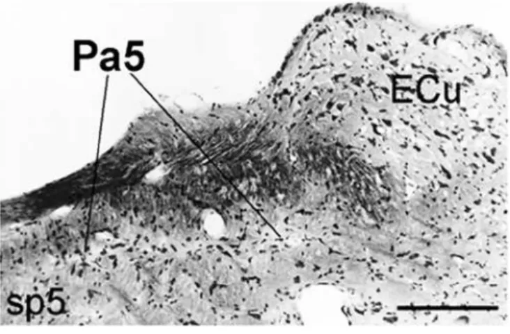
The fine structure of the entire Pa5, which had ultrathin slices randomly collected, was examined under a transmission electron microscope. Within 2 to 4 days of postoperative period, electron microscopy analysis showed degenerated axon terminal fibers and identified synaptic elements in the medial extent of the paratrigeminal nucleus (Pa5, Figure 1), which projects to other nuclei functionally involved in central neuronal cardiovascular control. The bilateral cross-sections of the Pa5 revealed that the number of degenerated axonal fibers in the vicinity of synaptic elements were strictly distributed within the areas that receive afferent fibers from NTS and RVL as described by Caous et
al.(2). Again, the Pa5 region considered the medial
extent of the nucleus was properly identified according to its morphological cytoarchitecture under light microscopy analysis of the nissil-stained tissue sections as established by previous studies.
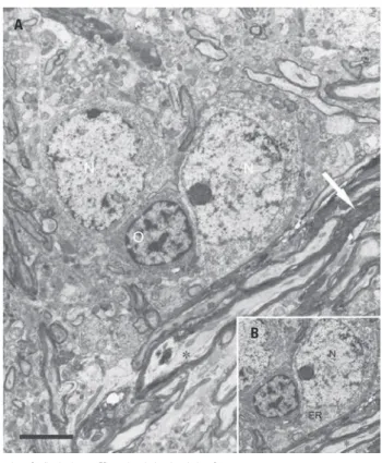
Despite the main finding of the present investigation that comprehended degenerated axonal elements in the same innervation site that receives projections from RVL or NTS, other detailed observations should be described as follows. Transverse sections of the paratrigeminal nucleus (Figure 2) contain a compact area of a nerve cell that constitutes the nucleus and surrounding cytoplasm depicted by a conspicuous and spherical nucleolus and arising from the top of the cell body is a thick myelinated degenerating axon (A, B and
Pa5: paratrigeminal nucleus; ECu: external cuneate; sp5: spinal trigeminal tract. Bar=100µm
A
C
E
B
D
F
N: nucleus; At: axon terminal; s: synapse; sv – synaptic vesicles; Ax: axon; Den: dendrite; white arrows show degenerated axonal fibers (except for picture F). Bars=0.5µm
Figure 2. Electron micrographs from cross-sections of rat paratrigeminal nucleus (Pa5) after the sinoaortic baroreceptor denervation. In A and B, note the myelinated degenerated axons in the contralateral Pa5. In C, a myelinated degenerated axon was observed in the ipsilateral Pa5. In D and E, the asymmetric synaptic pattern and a degenerated axon terminal (*) were shown. In F, typical elements as gap junctions were depicted in the Pa5
C). The presence of gap junctions could be also observed between dendrites (F) and an oligodendrocyte is shown between neuronal perykaria where two proximal cell bodies (type 2 perykaria) with a prominent nucleolus encompassed by degenerated and non-degenerated fibers were observed (Figure 3). Axon terminals (At) containing pleomorphic clear vesicles form asymmetrical synapses with dendrites, some intact (D) and other presenting a dark degenerated characteristic (E). These buttons contacting larger dendrites were generally small-to-medium in size, made asymmetric-type synaptic contacts, and contained pleomorphic vesicles of medium-to-large size (Figure 4A).
On the basis of synaptic polarity, vesicle size, and the nature of the pre- and postsynaptic elements, fundamentally axo-dendritic and axo-somatic types could be distinguished. Small occurrences of axo-somatic synapses were perceived and axo-dendritic synapses were predominantly identified. All synapses with a non electron-dense core were round and asymmetric with a
A
B
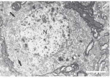
N: nucleus; O: oligodendrocyte; ER: rough endoplasmic reticulum. Bar=4µm
Figure 3. Typical electron microscopic findings after sinoaortic baroreceptor denervation in the paratrigeminal nucleus. The electron micrograph (A) demonstrates a neuron cell body surrounded by normal myelinated axonal fibers (*, B) and degenerated fibers (white arrow)
A
B
sv: synaptic vesicles; Den: dendrite; s: synapse; At: axon terminal. Bars=0.5µm
thick postsynaptic membrane element. Mitochondria of variable shapes were identified, as well as dispersed rough endoplasmic reticulum throughout the cytoplasm. Synaptic contact on the soma of Pa5 neurons was small and covered, on average, only 4% of the soma surface. The most striking feature of the synaptic structure of the Pa5 was that more than 90% of all synapses were axo-dendritic (Figure 4B). Dense degenerated axon fibers with the same shape and type patterns (Figure 5) were observed in the entire examined target nucleus. Dendritic spines were rare and no synapses between two vesicle-containing structures, axo-axonic or dendro-dendritic synapses, were identified.
responses to smaller blood pressure increments, while the NTS would be activated in surges greater than
40mmHg(30).
Ultrastructure and electrophysiological inferences
Degenerated cellular components identified herein represent barosensitive neurons recorded in the Pa5
in earlier studies(9). This functional pathway between
the Pa5 and RVL was evidenced through cardiocycle-locked neurons recorded in the RVL that respond to
bradykinin microinjections in the Pa5(6,12). Our findings
hypothesize that the medial extent of the Pa5 containing degenerated axoplasmatic cellular components after sinoaortic denervation, may be the same one that receives afferent projections of the rostroventrolateral medulla as shown in previous retrograde neurotracing studies, in which a distributive characteristic of the Pa5 was based on the topographical organization of
neuroanatomical primarily afferent connections(2,5,31),
although this relevant information should be confirmed by neurotracing and electron microscope conjugated experiments.
General discussion
After sinoatrial node denervation and ultrastructural inspection of the paratrigeminal nucleus, the described degenerating fibers and terminals allow us to imply that this nucleus has the necessary connections to contribute to cardiovascular reflexes. Also, it can be claimed that the terminals in question are confined to a specific portion of the paratrigeminal nucleus that corresponds mainly to its medial portion. The vagus nerve has sensitive and motor fibers in the cervical region and in the nodose ganglia; both ascend to a medullar level through the dorsolateral medullary region and the
islands of neuropil of the Pa5(32), and the aortic and
carotid baroreceptors were first identified in vagal
and glossopharyngeal nerves, respectively(33); both
nerves have somatic inputs to the Pa5. The afferent projection areas from NTS and RVL inserted in the medial extent of the nucleus contained electron-dense degenerated axonal fibers. This finding may confirm the baroreflex component characteristic of the nucleus. The present neuroanatomical data suggest that the Pa5 can be considered a sensitive information gateway to visceral reflexes.
CONCLUSION
This neuroanatomical substrate or pathway that involves baroreceptor elements can be associated with N: nucleus. Bar=4µm
Figure 5. Electron micrograph of a type-2 neuron in the paratrigeminal nucleus without axosomatic synaptic contacts. Observe a round oval structure, stacks of rough endoplasmic reticulum, Golgi apparatus (black arrows) and a myelinated degenerated axon (*)
DISCUSSION
Cardiovascular and baroreceptor functions of the Pa5
The Pa5 is already confirmed to be the only extra-solitary structure of the central nervous system which receives primary afferent fibers from vagus, trigeminal,
and glossopharyngeal nerves(28). Despite the fact that
the nucleus does not present interneuron cells(4), it
integrates cardiorespiratory and thermoregulatory or
thermosensitive reflexes(29), since the Pa5 lesion with
sympathetic activation and its cardiovascular effects. In conclusion, after the sinoatrial node denervation the paratrigeminal nucleus was inspected at the electron microscopic level, degenerated axonal fibers or terminals were identified in the medial extent of the nucleus. The present result, notwithstanding the morphological work, complements other research on the Pa5 regarding its function as an important relay for pressure-sensitive information to the cardiovascular reflex arc.
ACKNOWLEDGMENTS
The present study was supported by Fundação de Amparo à Pesquisa do Estado de São Paulo (FAPESP). Drs. Charles Lindsey, Ricardo Smith and Edna Freymuller are recipients of “Bolsa de Produtividade em Pesquisa 1” from the Conselho Nacional de Pesquisa e Desenvolvimento Tecnológico (CNPq). Dr. Cristofer Caous is a neuroscientist researcher at Hospital Israelita Albert Einstein.
REFERENCES
1. Alioto OE, Lindsey CJ, Koepp J, Caous CA. Sensory sciatic nerve afferent inputs to the dorsal lateral medulla in the rat. Auton Neurosci. 2008; 140(1-2):80-7.
2. Caous CA, de Sousa Buck H, Lindsey CJ. Neuronal connections of the paratrigeminal nucleus: a topographic analysis of neurons projecting to bulbar, pontine and thalamic nuclei related to cardiovascular, respiratory and sensory functions. Auton Neurosci. 2001;94(1-2):14-24.
3. Caous CA, Koepp J, Couture R, Balan AC, Lindsey CJ. The role of the paratrigeminal nucleus in the pressor response to sciatic nerve stimulation in the rat. Auton Neurosci. 2008;140(1-2):72-9.
4. Chan-Palay V. The paratrigeminal nucleus. I. Neurons and synaptic organization. J Neurocytol. 1978;7(4):405-18.
5. Saxon DW, Hopkins DA. Efferent and collateral organization of paratrigeminal nucleus projections: an anterograde and retrograde fluorescent tracer study in the rat. J Comp Neurol. 1998;402(1):93-110.
6. Caous CA, Balan A, Lindsey CJ. Bradykinin microinjection in the paratrigeminal nucleus triggers neuronal discharge in the rat rostroventrolateral reticular nucleus. Can J Physiol Pharmacol. 2004;82(7): 485-92.
7. Spyer KM. Neural organisation and control of the baroreceptor reflex. Rev Physiol Biochem Pharmacol. 1981;88:24-124.
8. Yu YG, Caous CA, Balan AC, Rae GA, Lindsey CJ. Cardiovascular responses to sciatic nerve stimulation are blocked by paratrigeminal nucleus lesion. Auton Neurosci. 2002;98(1-2):70-4.
9. Balan Júnior A, Caous CA, Yu YG, Lindsey CJ. Barosensitive neurons in the rat tractus solitarius and paratrigeminal nucleus: a new model for medullary, cardiovascular reflex regulation. Can J Physiol Pharmacol. 2004;82(7):474-84. 10. Dampney RA, Goodchild AK, Tan E. Identification of cardiovascular cell groups
in the brain stem. Clin Exp Hypertens A. 1984;6(1-2):205-20.
11. Amini-Sereshki L, Zarrindast MR. Brain stem tonic inhibition of thermoregulation in the rat. Am J Physiol. 1984;247(1Pt2):R154-9.
12. Lindsey CJ, Buck HS, Fior-Chadi DR, Lapa RC. Pressor effect mediated by bradykinin in the paratrigeminal nucleus of the rat. J Physiol. 1997;502 (Pt1):119-29.
13. Campos MM, Ongali B, Thibault G, Neugebauer W, Couture R. Autoradiographic distribution and alterations of kinin B(2) receptors in the brain and spinal cord of streptozotocin-diabetic rats. Synapse. 2005;58(3):184-92.
14. Couture R, Toma N, Barbot L. SR142801 behaves as a tachykinin NK-3 receptor agonist on a spinal nociceptive reflex in the rat. Life Sci. 2000;66(1): 51-65.
15. Guild SJ, Austin PC, Navakatikyan M, Ringwood JV. Malpas SC. Dynamic relationship between sympathetic nerve activity and renal blood flow: a frequency domain approach. Am J Physiol Regul Integr Comp Physiol. 2001; 281(1):R206-12.
16. Panneton WM, Loewy AD. Projections of the carotid sinus nerve to the nucleus of the solitary tract in the cat. Brain Res. 1980;191(1):239-44. 17. Ciriello J. Brainstem projections of aortic baroreceptor afferent fibers in the
rat. Neurosci Lett. 1983;36(1):37-42.
18. Seller H, Illert M. The localization of the first synapse in the carotid sinus baroreceptor reflex pathway and its alteration of the afferent input. Pflugers Arch. 1969;306(1):1-19.
19. Schreihofer AM, Guyenet PG. The baroreflex and beyond: control of sympathetic vasomotor tone by GABAergic neurons in the ventrolateral medulla. Clin ExpPharmacol Physiol. 2002;29(5-6):514-21.
20. Schreihofer AM, Sved AF. Nucleus tractus solitarius and control of blood pressure in chronic sinoaortic denervated rats. Am J Physiol. 1992;263(2Pt 2): R258-66.
21. Granata AR, Numao Y, Kumada M, Reis DJ. A1 noradrenergic neurons tonically inhibit sympathoexcitatory neurons of C1 area in rat brainstem. Brain Res. 1986;377(1):127-46.
22. Guertzenstein PG, Silver A. Fall in blood pressure produced from discrete regions of the ventral surface of the medulla by glycine and lesions. J Physiol. 1974;242(2):489-503.
23. Urbanski RW, Sapru HN. Evidence for a sympathoexcitatory pathway from the nucleus tractus solitarii to the ventrolateral medullary pressor area. J Auton Nerv Syst. 1988;23(2):161-74.
24. Yu YG, Lindsey CJ. Baroreceptor-sensitive neurons in the rat paratrigeminal nucleus. Auton Neurosci. 2003;105(1):25-34.
25. Festing MF. The choice of animals in toxicological screening: inbred strains and the factorial design of experiment. Acta Zool Pathol Antverp. 1980;(75):117-31. 26. Valenstein ES. A note on anaesthetizing rats and guinea pigs. J Exp Anal
Behav. 1961;4:6.
27. Krieger EM. Neurogenic hypertension in the rat. Circ Res. 1964;15:511-21. 28. Ciriello J, Hrycyshyn AW, Calaresu FR. Glossopharyngeal and vagal afferent
projections to the brain stem of the cat: a horseradish peroxidase study. J Auton Nerv Syst. 1981;4(1):63-79.
29. Kilduff TS, Sharp FR, Heller HC. Progressive activation of paratrigeminal nucleus during entrance to hibernation. Am J Physiol. 1988;255(1Pt2):R178-81. 30. Sousa LO, Lindsey CJ. Cardiovascular and baroreceptor functions of the
paratrigeminal nucleus for pressor effects in non-anaesthetized rats. Auton Neurosci. 2009;147(1-2):27-32.
31. de Sousa Buck H, Caous CA, Lindsey CJ. Projections of the paratrigeminal nucleus to the ambiguous, rostroventrolateral and lateral reticular nuclei, and the solitary tract. Auton Neurosci. 2001;87(2-3):187-200.
32. Kalia M, Sullivan JM. Brainstem projections of sensory and motor components of the vagus nerve in the rat. J Comp Neurol. 1982;211(3):248-65. 33. Panneton WM, Burton H. Projections from the paratrigeminal nucleus and