Identification and Characterization of Lipase
Activity and Immunogenicity of LipL from
Mycobacterium tuberculosis
Jun Cao1, Guanghui Dang1, Huafang Li1, Tiantian Li1, Zhiguo Yue2, Na Li2, Yajun Liu2, Siguo Liu1*, Liping Chen1*
1Division of Bacterial Diseases, State Key Laboratory of Veterinary Biotechnology, Harbin Veterinary Research Institute, Chinese Academy of Agricultural Sciences, Harbin, PR China,2Heilongjiang Provincial Hospital for Prevention and Treatment of Tuberculosis, Harbin, PR China
*[email protected](SL);[email protected](LC)
Abstract
Lipids and lipid-metabolizing esterases/lipases are highly important for the mycobacterial life cycle and, possibly, for mycobacterial virulence. In this study, we expressed 10 mem-bers of the Lip family ofMycobacterium tuberculosis. Among the 10 proteins, LipL dis-played a significantly high enzymatic activity for the hydrolysis of long-chain lipids. The optimal temperature for the lipase activity of LipL was demonstrated to be 37°C, and the optimal pH was 8.0. The lipase active center was not the conserved motif G-x-S-x-G, but rather the S-x-x-K and GGG motifs, and the key catalytic amino acid residues were identi-fied as G50, S88, and K91, as demonstrated through site-directed mutagenesis experi-ments. A three-dimensional modeling structure of LipL was constructed, which showed that the GGG motif was located in the surface of a pocket structure. Furthermore, the subcellular localization of LipL was demonstrated to be on the mycobacterial surface by Western blot analysis. Our results revealed that the LipL protein could induce a strong humoral immune response in humans and activate a CD8+T cell-mediated response in mice. Overall, our
study identified and characterized a novel lipase denoted LipL fromM.tuberculosis, and demonstrated that LipL functions as an immunogen that activates both humoral and cell-mediated responses.
Introduction
Tuberculosis (TB) is an infectious disease caused by bacteria in theMycobacterium tuberculosis
complex, which includesM.tuberculosis,M.bovis,M.africanum,M.microti, andM.canettii
[1,2]. TB remains one of the world’s deadliest communicable diseases. In 2013, an estimated 9.0 million people developed TB, and 1.5 million died from the disease [3]. The emergence of multi-drug resistant (MDR) and extensively drug resistant (XDR) mycobacterial strains further complicates the prevention and control of TB. Approximately 480,000 people worldwide devel-oped MDR-TB in 2013. It is estimated that approximately 9.0% of the MDR-TB cases had
OPEN ACCESS
Citation:Cao J, Dang G, Li H, Li T, Yue Z, Li N, et al. (2015) Identification and Characterization of Lipase Activity and Immunogenicity of LipL from
Mycobacterium tuberculosis. PLoS ONE 10(9): e0138151. doi:10.1371/journal.pone.0138151
Editor:Joyoti Basu, Bose Institute, INDIA
Received:April 12, 2015
Accepted:August 25, 2015
Published:September 23, 2015
Copyright:© 2015 Cao et al. This is an open access article distributed under the terms of theCreative Commons Attribution License, which permits unrestricted use, distribution, and reproduction in any medium, provided the original author and source are credited.
Data Availability Statement:All relevant data are within the paper and its Supporting Information files.
XDR-TB; which underscores the challenges of preventing or treating TB [3]. Therefore, the development of effective vaccines and screening of new drugs against TB is of utmost impor-tance. The elaboration of the pathogenic molecular mechanisms of TB infection may provide valuable insights for vaccine development and new drug screening. Although much has been learned about the pathogenM.tuberculosis, many aspects of the molecular pathogenicity ofM.
tuberculosisremain to be elucidated.
Lipids and esterases/lipases associated with lipid metabolism are very important in the mycobacterial life cycle. Eight percent of theM.tuberculosisgenome encodes products that participate in lipid metabolism, and approximately 30–40% of the dry weight ofM.tuberculosis
corresponds to lipids [4,5].M.tuberculosisis a slow-growing intracellular pathogen, and the special pathological characteristic of its infection is granuloma. During the formation phase of granuloma,M.tuberculosisgoes into a dormant state, in which the bacteria accumulate lipids in the form of lipid inclusion bodies (LIBs) [6,7]. Most of these lipids consists of tri-acylglycer-ols (TAG) and may originate from host lipid degradation and/or fatty acid absorption [7,8]. It has been reported thatM.tuberculosisin the center of granulomas can also accumulate lipids from the degradation of immune cells [6,9]. Furthermore,M.tuberculosiscan convert macro-phages, which are colonized byM.tuberculosis, to exhibit a foamy phenotype and accumulate LIBs through the foamy macrophages [10]. On the one hand, the lipids provide energy toM.
tuberculosis, and on the other hand, these lipids may also provide materials for the synthesis of
M.tuberculosiscell membrane and cell well lipids [11].
Because lipids are important for the survival and pathogenicity ofM.tuberculosis, the ester-ases/lipases associated with lipid metabolism are considered critical virulence factors forM.
tuberculosis[12]. Lipases catalyze the hydrolysis of ester bonds in long-chain acylglycerols to release fatty acids and glycerol. During infection,M.tuberculosisalso relies on its lipases to hydrolyze host cell lipids to release fatty acids, which serve as its energy source [13,14].
Lipases differ from esterases due to their ability to hydrolyze substrates with long-chain acylglycerols at the oil-water interface, whereas esterases can only hydrolyze substrates with short-chain acylglycerols [15,16]. TheM.tuberculosisgenome contains a markedly high num-ber of lipolytic enzymes, of which the 21 memnum-bers of a family called Lip (A to W, except K and S) have been annotated as putative esterases or lipases based on the presence of the G-x-S-x-G motif, which is characteristic of theα/βhydrolase-fold family [17,18]. In this study, therv1497
gene fromM.tuberculosisH37Rv encoding LipL, which was previously annotated as a putative lipase, was overexpressed inM.smegmatis. We identified and characterized the lipase activity by using the purified LipL protein. Moreover, purified LipL was inoculated in BALB/c mice to evaluate its immunogenicity.
Materials and Methods
Ethics statement
Blood samples were obtained from TB patients who were admitted to the Heilongjiang Provin-cial Hospital for Prevention and Treatment of Tuberculosis, Harbin, China. The sera from healthy adult donors were from Harbin, China. Informed written consent was obtained from all participants. This project was approved by the Ethics Committee of Heilongjiang Provincial Hospital for Prevention and Treatment of Tuberculosis.
The mice used in this study were purchased from Vital River, Beijing, China. The animal experiments were performed in accordance with animal ethics guidelines and approved proto-cols. The animal experiment was approved by the Animal Ethics Committee of Harbin Veteri-nary Research Institute of Chinese Academy of Agricultural Sciences. The study approval
number was SQ20142049. All the experiments were designed to minimize the numbers of ani-mals used, and every effort was made to minimize pain and distress to the aniani-mals.
Bacterial strains and culture condition
Escherichia coliDH5αand BL21 (DE3) (Novagen, Darmstadt, Germany) strains were used as host strains for cloning and expression experiments.E.coliwere grown on Luria-Bertani (LB) broth or agar containing appropriate antibiotics, ampicillin or kanamycin, at 50μg/ml; Liquid
M.smegmatiscultures were grown in Middlebrook 7H9 medium (BD Biosciences) supple-mented with 10% oleic acid/albumin/dextrose/catalase enrichment (10% OADC, BD Biosci-ences), 0.05% Tween 80 (Amresco), and 0.2% glycerol, containing kanamycin, at 25μg/ml. Transformants were selected on Middlebrook 7H10 solid media supplemented with 25μg/ml kanamycin when necessary. Plates were incubated at 37°C for 3 to 4 days forM.smegmatis.
Gene amplification and plasmids construction
The parent vector pMV261 (kind gift from Lu Yu) was a promoterlessEscherichia coli -myco-bacteria shuttle plasmid. A pair of primers named“AI-PF”and“AI-PR”was designed
(Table 1) according to the sequence from NCBI and used to amplify the acetamidase promoter fromM.smegmatismc2155 genome DNA. The pMV261 inserted with the acetamidase pro-moter is an acetamide-inducible vector named pAI. Then“Linker-N”(Table 1) containing His-tag was added to vector pAI to generate pAIN vector whose His-tag was at the N-terminal.
The primers (Table 2) were designed to amplify the 10 Lip family genes ofM.tuberculosis. PrimeSTAR Max DNA polymerase was used for PCR amplification. PCR involved pre-dena-turation at 95°C for 5 min, 30 cycles of denapre-dena-turation at 98°C for 10 sec, annealing at 60°C for 5 sec, elongation at 72°C for 8 sec, and a post-elongation at 72°C for 1 min. The 10 Lip family genes were then cloned into the pAIN vector.
Table 1. Primers used in this study.
Primer Sequence (5’!3’)
Primers for vector
AI-PF CTAGGTACCAGTGACGCGGTCTCAAGCG
AI-PR CTAGGATCCAAAACTACCTCGGGCATGTGG
Linker-NF GATCCTCACCACCACCACCACCACACTAGTCAGCTGCAGAATTCATATGCATCGATGGTT
Linker-NR AACCATCGATGCATATGAATTCTGCAGCTGACTAGTGTGGTGGTGGTGGTGGTGAG
Primers for mutations
G49-F GGTTCGCTGGCGGAGCGCTGGCGGTGT
G49-R TCCGCCAGCGAACCGGCGCCCCGGAAA
G50-F GGTTCGGTGCCGGAGCGCTGGCGGTGT
G50-R TCCGGCACCGAACCGGCGCCCCGGAAA
G51-F GGTTCGGTGGCGCAGCGCTGGCGGTGTA
G51-R TGCGCCACCGAACCGGCGCCCCGGAAA
S88-F GATGGTGTTCGCGGCGACCAAGGGCA
S88-R TCGCCGCGAACACCATCGGCGCGGAAT
K91-F GCGGGCATGACGGCCACGGTCATCCA
K91-R GCCGTCATGCCCGCGGTCGCCGAGAA
S361-F GCTTGGGCGGCGCGATCGGCTGGACA
S361-R TCCAGCCGATCGCGCCGCCCAAG
The template plasmid pAIN-lipL and the complementary mutagenic oligonucleotide pairs (Table 1) were used for PCR amplification to introduce the G49A, G50A, G51A, S88A, K91A, and S361A substitutions. The PCR products were digested withDpnIto damage the methyl-ated template plasmid. The resultant mutant plasmids were transformed intoE.coliDH5α cells, and all of the substitutions were confirmed by Sanger sequencing.
Expression and purification of Lip family proteins from
M
.
tuberculosis
TheM.smegmatismc2155 cells were transformed with the positive recombinant plasmids by electroporation and incubated on 7H10 agar plates containing 25μg/ml kanamycin. After being incubated for 3 days at 37°C, single colonies were selected and grown in 5 ml of 7H9 broth with 0.05% Tween 80 and 25μg/ml kanamycin for 3 days. Expression of His-tagged recombinant proteins inM.smegmatiswas performed in 7H9 medium supplemented with 10% OADC, 0.05% Tween 80 and 0.2% glycerol, containing 25μg/ml kanamycin. The cultur-ing condition was 37°C at a shakcultur-ing speed of 160 rpm. Acetamide (Sigma-Aldrich) was added to final concentration of 0.2% (w/v) whenM.smegmatiswere grown to an optical density at 600 nm (OD600) of ~ 2.0 for the expression of His-tagged recombinant proteins and grown for another 20 hours. Cells were harvested by centrifugation at 5,000 rpm for 10 min at 4°C. The cell pellets were washed twice and then re-suspended in ice-cold washing buffer (20 mM Tris-HCl, 150 mM NaCl, pH 8.0) and then passed through a high-pressure cell disruptor at 4°C (model Tso.75, Constant Systems, UK). The cell debris was removed by centrifugation at 15,000 rpm for 30 min at 4°C. The supernatant was loaded onto a Ni2+Sepharose 6 Fast Flow (GE Healthcare, USA) equilibrated with buffer A (20 mM Tris-HCl, 150 mM NaCl, 10 mM
Table 2. Primers used for PCR of 10 Lip family genes fromM.tuberculosis.
Gene Primer direction Primer sequence (5’!3’) Amplicon Size(bp)
lipI Sense CCGGAATTCGCCCAGTTTGGACAACACCGCC 958
(rv1400c) Antisense CCGGTTAACTCCGTGTAGCACAACCCGTAGCG
lipL Sense CCGGAATTCGATGGTTGACACCGGAGTCGATCA 1285
(rv1497) Antisense CCGGTTAACGCCCGCCGCGGCTCC
lipM Sense CCGGAATTCGGGCGCTCCTCGACTCATCCA 1291
(rv2284) Antisense CCGGTTAACTGGGATCGCAACCGCGGGG
lipN Sense CCGGAATTCGACCAAGAGTCTGCCAGGTGTG 1126
(rv2970c) Antisense CCGGTTAACAACCCGGCTAAGGTGCGCG
lipQ Sense CCGGAATTCGCACATCGCCAGCGTGACTTCG 1261
(rv2485c) Antisense CCGGTTAACGCTGGCCGGAGGTGACGACA
lipT Sense CCGGAATTCGGCCCTGGAGTCGGCTAC 1531
(rv2045c) Antisense CCGGTTAACATTCGCCAGCGAAAACCCGTC
lipU Sense CCGGAATTCGGCGGTCCGGCCGGTGCTA 889
(rv1076) Antisense CCGGTTAACCCCGGTGGCCTCGCGGATGTA
lipV Sense CCGGAATTCGCCCGAAATCCCCATCGCC 670
(rv3203) Antisense CCGGTTAACGCGCGGTCCCAGTCGACT
lipY Sense CGCCATATGTGTCTTATGTTGTTGCGTTGCC 1309
(rv3097c) Antisense CCGGTTAACGGCGGCGATACCGAGTTG
lipZ Sense CCGGAATTCGACATCACCGAGTGTCCGGGAG 862
(rv1834) Antisense CCGGTTAACGTCGACGAGCAGCGATAGCGC
The restriction sites integrated into the sequences are underlined.
imidazole, pH 8.0) after being passed through a filter membrane (Millipore, 0.22μm, USA). The column was washed with 10 column volumes of buffer A and an imidazole concentration gradient (20, 40, 60, 80, 100, and 200 mM) buffer. Proteins were eluted with elution buffer (buffer A containing 500 mM imidazole and 20% glycerol). The purified protein was then fur-ther purified using anion exchange chromatography RESOURCETMQ column (1 ml) (GE Healthcare, USA), and chromatography Superdex 75 (GE Healthcare, USA). The collected protein samples were analyzed by 12% sodium dodecyl sulfate polyacrylamide gel electropho-resis (SDS-PAGE) and concentration was determined by using a Pierce BCA protein assay kit (Pierce, USA).
Lipase assay
Lipase activity was assayed by measuring the amount ofp-nitrophenol (p-NP) released from
p-NP ester substrate with varying lengths of fatty acids [16]. These substrates includedp-NP acetate (C2),p-NP butyrate (C4),p-NP caproate (C6),p-NP caprylate (C8),p-NP laurate (C12),p-NP myristate (C14),p-NP palmitate (C16), andp-NP stearate (C18). Thep-NP esters were dissolved in isopropanol at a concentration of 10 mM. The total lipase activity was assayed using the cell lysate supernatants of 10 recombinantM.smegmatis. The standard lipase activity assays were performed in 100μl reaction system consisting of a final concen-tration of 0.5 mMp-NP esters substrate and the buffer (pH 8.0) of 80 mM H3BO3, 80 mM H3PO4, 300 mM NaCl, 0.3% Triton X-100 and 20% glycerol. The reaction mixture with increasing concentrations gradient (1, 2, 3, and 4μg) of purified protein was incubated at 37°C for 40 min and the release ofp-NP was determined by measuring spectrophotometri-cally at 405 nm. One unit of enzyme activity was defined as the amount of enzyme releasing 1μmol ofp-NP per min at 37°C under standard reaction conditions.
Substrate specificity was determined by usingp-NP esters with different aliphatic side chains (C2-C18), as mentioned above. The optimum temperature of the enzyme reaction was determined in the same substrate solution at various temperatures ranging from 35°C to 60°C. The optimal pH for lipase activity was determined by assaying the hydrolytic activity at pH ranging from 6.0 to 9.0, using an established spectrophotometric method [16]. The effects of nonionic detergent Tween 20 and Tween 80, anionic detergent SDS and serine proteinase inhibitor phenylmethylsulfonyl fluoride (PMSF, Sigma-Aldrich) on lipase activities were exam-ined by adding increasing concentrations gradient of each into the reaction system. All reac-tions were conducted in triplicate.
Molecular modeling
The 3D model of LipL was generated using SWISS-MODEL (http://swissmodel.expasy.org/). Briefly, a new modeling project was started, and the target sequence file in the FASTA format was uploaded to search for templates. The top-ranked structure based on similarity with the target sequence was selected as the template structure. A 3D model of LipL was constructed with template by the Modeler module. The structural figures were prepared using the program PyMOL (http://www.pymol.org/).
Subcellular fractionation of mycobacteria and immunoblotting
through a high-pressure cell disruptor, and the resulting lysates were centrifuged at 3,000 g to remove any unbroken cells. The lysate supernatant was centrifuged at 27,000 g at 4°C for 1 hour, and the resulting supernatant was transferred into conical tubes (50 ml, Nest). The pellet was resuspended in lysis buffer containing 1 mM PMSF and 2 mM lysozyme (Sigma-Aldrich), and centrifuged at 27,000 g at 4°C for 1 hour to obtain the pellet, which mostly comprised of the cell wall fraction (CW). The supernatants obtained from the above-described two centrifu-gations were pooled together and centrifuged at 100,000 g and 4°C for 4 hours. The cyto-plasmic (Cy) fraction was the supernatant obtained from this centrifugation. A final centrifugation at 100,000 g and 4°C for 4 hours was performed to remove the any cell mem-brane contaminant. Both pellets were pooled and considered cell memmem-brane (CM) fraction. All of the fractions were resuspended in 10 mM NH4HCO3buffer.
The protein concentrations of each fraction were determined using the BCA protein assay kit (Pierce, USA). Equal amounts of each fraction (20μg) were separated by 12% SDS-PAGE and transferred onto a nitrocellulose membrane (Pall Corporation, NY, USA). The membranes were then blocked with 5% skimmed milk in PBS + 0.05% Tween 20 (PBST) at 37°C for 2 hours. The membranes were exposed to mouse anti-His-tag mAb at 1/2,000 dilution or rabbit anti-KatG antiserum at 1/1,000 dilution and incubated for 1 hour at 37°C. After washing three times for 10 min each, the membranes were incubated with Dylight 680-labeled goat anti-mouse IgG antibody or Dylight 800-labeled goat anti-rabbit IgG antibody (KPL, Gaithersburg, MD, USA) at 1/10,000 dilution, respectively. Antiserum againstM.tuberculosisKatG was used as a quality control to ensure that there was no cytoplasmic contamination in the other frac-tions. The bands were detected using the Odyssey infrared imaging system (LI-COR Biosci-ences) in both the 700 nm and 800 nm channels.
Study population and serological characterization
Sera samples were obtained from 51 TB patients who were admitted to the Heilongjiang Pro-vincial Hospital for Prevention and Treatment of Tuberculosis, Harbin, China, and 45 clini-cally healthy donors. The diagnoses of TB patients were confirmed by examination of sputum-positive smears for acid-fast bacillus. The patients with pulmonary TB were categorized into two groups as follows. Group 1 (n = 29) comprised patients who had been diagnosed with TB for the first time and had no history of TB treatment. Group 2 (n = 22) comprised relapsed TB patients who were previously treated for TB and disease relapsed after the initial completion treatment. Clinically healthy donors were those who showed no symptoms of TB and had been vaccinated withM.bovisBCG during infancy. All of the sera samples were negative for immu-nodeficiency virus.
Immunization of mice, flow cytometry analysis for spleen lymphocytes
and statistical analysis
The BALB/c mice used in this study were housed in 4 groups and given one week to acclimate to the housing facility. Mice were maintained in cages fitted with micro-isolators connected to air circulator. The cages had bedding, feed and water. The environmental temperature, humid-ity, and light were in the standard conditions conducive to mice activity. During housing, mice were monitored every day for health status and no adverse events were observed. Breeder was cleaned in the morning every day. A completed ARRIVE guidelines checklist is included inS1 Checklist.
In this study, 5–6 weeks old female BALB/c mice (Vital River, Beijing) maintained under specific pathogen-free conditions were used. Mice were randomly divided into 2 groups. Groups of mice (n = 6 per group) were immunized with the LipLMsrecombinant protein and PBS (mock-immunized). LipLMswas purified from recombinantM.smegmatisstrains. The mice were immunized at the age of 6–7 weeks by the subcutaneous injection of 100μg of recombinant protein in a 200μl volume formulated in protein plus incomplete Freund’s adju-vant (IFA) or PBS plus IFA. The mice were immunized two times at an interval of 14 days. The spleens of the mice were harvested 14 days after the second immunization.
The mice were allowed to rest for two weeks after the secondary immunization. Mice were euthanized by cervical dislocation in the laboratory and the spleens were then removed asepti-cally. Single-cell spleen suspensions were prepared aseptically, and spleen lymphocytes were obtained using a mice spleen lymphocyte separation reagent kit (TBD). The spleen lympho-cytes were washed three times in PBS and resuspended in RPMI-1640 supplemented with 20% fetal bovine serum (FBS, Gibco) and 1% penicillin-streptomycin. The cells were then counted, and adjusted to a final concentration of 1 × 10⁶cells/ml and grown in 24-well plates. The cells were stimulated with the corresponding purified protein at a concentration of 10μg/ml and incubated at 37°C for 12 hours. The cells were then washed twice and incubated with specific surface antibodies, including anti-CD3-APC, anti-CD4-FITC, and anti-CD8-PE (Invitrogen, USA), for 1 hour at room temperature. The cells were washed twice and then processed for flow cytometry analysis (FACSAria flow cytometer, BD Biosciences), and the data were ana-lyzed using FlowJo software (http://www.flowjo.com/).
Each experiment was independently repeated three times. Two-tailed Student’sttest was used for the analysis of statistical significance (Pvalue) in this study, and Prism software (ver-sion 5.0; GraphPad, San Diego, CA, USA) was used for these analyses. APvalue of less than 0.05 was considered significant (P<0.05;P<0.01;P<0.001; ns, not significant).
Results
Cloning, expression, and purification of LipL
biochemistry activity from the inclusion bodies using several renaturation conditions. However, active proteins were not obtained. Thus, we constructed the acetamide-inducible vector pAIN forM.smegmatis, in which mycobacterial proteins could be expressed in soluble form. We cloned thelipLgene into the pAIN vector to obtain the recombinant plasmid plipL. The LipL protein was expressed as a His-tagged fusion protein inM.smegmatisin both soluble and insol-uble forms. The solinsol-uble component was purified by affinity chromatography on a Ni-NTA col-umn, followed by an anion-exchange chromatography RESOURCETMQ column, and finally by Superdex 75. The samples were analyzed by SDS-PAGE, and the results showed that the purified protein had an expected molecular mass of approximately 48.5 kDa (S1A Fig).
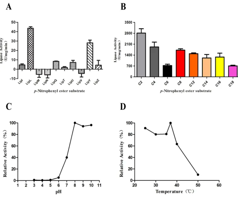
Fig 1. Analysis of lipase activity.(A) The total lipase activities were assayed using cell lysate supernatants from the 10 recombinantM.smegmatisstrains. Thep-NP palmitate (C16) was used as the substrate for the lipase activities analysis. The values represent the means±standard deviations (SD) of three
independent experiments. (B) Lipase activity of LipL towardsp-NP esters with various chain lengths (C2, acetate; C4, butyrate; C6, caproate; C8, caprylate; C12, laurate; C14, myristate; C16, palmitate; and C18, stearate). The values represent the means±SD of three independent experiments. (C) The effect of pH on lipase activity of LipL. (D) The effect of temperature on lipase activity of LipL.
To confirm that the purified proteins were LipL, Western blot analysis and high-perfor-mance liquid chromatography mass spectrometry (HPLC-MS) were performed. The Western blot analysis results showed that the purified product band that reacted with His-tag anti-bodies was approximately 50 kDa, in agreement with the expected molecular weight of LipL (S1B Fig). The purified LipL protein was further validated by HPLC-MS analysis. The result obtained from searching NCBI databases using the HPLC-MC data matched LipL (GenBank accession no. NP216013) fromM.tuberculosisH37Rv with a score of 292. The results of the Western blot and HPLC-MS assays confirmed that the purified protein was in fact LipL.
Substrate specificity of LipL
Lipases, which hydrolyze long-chain esters, are considered much more crucial for the pathoge-nicity ofM.tuberculosisthan esterases, which can only hydrolyze short-chain esters. To investi-gate the substrate specificity of the purified LipL protein, we applied a wide range of esters, particularly the long-chain esters ofp-NP (C12to C18), as substrates in the enzymatic activity detection experiment. The substrates includedp-NP acetate (C2),p-NP butyrate (C4),p-NP caproate (C6),p-NP caprylate (C8),p-NP laurate (C12),p-NP myristate (C14),p-NP palmitate (C16), andp-NP stearate (C18). The results showed that LipL displayed rather high activity against both long-chain and short-chainp-NP esters (Fig 1B). Therefore, the relatively high activity displayed by LipL with long-chain esters (C12to C18) suggested that it could be classi-fied as a lipase.
Effect of pH and temperature on lipase activity of LipL
The lipase activities of LipL were analyzed within the pH range from 3.0 to 10.0 and the tem-perature range from 25°C to 50°C usingp-NP laurate (C12) as the substrate. The optimal activ-ity was detected at pH 8.0 and 37°C (100% relative activactiv-ity). LipL possessed activities over a range of pH values from 7.0 to 10.0, but had reduced activity in acidic conditions with pH val-ues from 3.0 to 7.0 (Fig 1C). LipL also displayed activities over a broad range of mild tempera-tures from 25°C to 50°C (Fig 1D). LipL retained 91%, 63%, and 10% relative activity at 25°C, 40°C, and 50°C, respectively. This finding indicated that lower temperatures favored LipL lipase activities while temperatures greater than 40°C severely damaged LipL lipase activities.
Effects of detergents on LipL activity
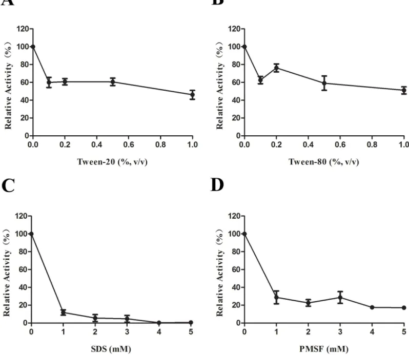
We next assessed the effect of various detergents on LipL lipase activity. The addition of the nonionic detergent Tween 20 or Tween 80 resulted in reductions in the relative lipase activity by approximately 40% (Fig 2A and 2B). The most prominent reductions in the relative lipase activities of approximately 85% and 75% were observed upon the addition of the anionic deter-gent SDS and the serine proteinase inhibitor PMSF, respectively (Fig 2C and 2D).
Site-directed mutagenesis
LipL contains the G-x-S-x-G motif, which represents the characteristic feature of theα/β hydrolase-fold family. In addition to the most common motif G-x-S-x-G, the GGG and S-x-x-K motifs, which were recently discovered to have a direct linkage to enzymatic activity, were also present in the LipL amino acid sequence. To identify the residues in the LipL primary structure that are necessary for catalytic activity, site-directed mutagenesis of the important residues within the above-mentioned three motifs was performed.
G50A, G51A, S88A, K91A, and S361A (Fig 3A), were generated by PCR using the template plasmid pAIN-lipL and the mutagenic oligonucleotide pairs. The alanine residue was chosen as a substitute residue because it lacked a bulky side chain and therefore would likely not have any steric and electrostatic effects. Moreover, it would not destroy the main-chain conforma-tion even in the presence of other residues. The analysis of the activities of the mutants com-pared with that of the wild-type LipL showed that each of the G50A, S88A, and K91A substitutions resulted in an approximately 90% loss of activity, whereas the G49A and G51A
Fig 2. The effect of detergents on lipase activity of LipL.(A) Addition of Tween 20 at concentration ranging from 0%–1% in the LipL lipase activity assays. (B) Addition of Tween 80 at concentration ranging from 0%–1%. (C) Addition of SDS at concentration ranging from 0 mM–5 mM. (D) Addition of PMSF at concentration ranging from 0 mM–5 mM. Data represent the means±SD of three independent experiments.
substitutions resulted in losses in activities of 70% and 60%, respectively (Fig 3B). The S361A substitution resulted in an approximately 20% loss of lipase activity. In general, the G50 residue in the GGG motif and the S88 and K91 residues in the S-x-x-K motif showed a significant impact on the lipase activity. Therefore, we considered the GGG and S-x-x-K motifs as the cat-alytically central part of LipL and that, the G50, S88, and K91 residues in these motifs were essential for the lipase activity.
3D Model of LipL
Because the GGG and S-x-x-K motifs were identified as crucial LipL for lipase activity and spe-cifically residues G50, S88, and K91 were determined to be catalytically critical, to further obtain insights into the possible location of the catalytic residues within the overall topological organization of LipL, a 3D model was built using the SWISS MODEL server with EstA carbox-ylesterase, which possesses the highest sequence homologies with LipL, as the template (PDB code: 3ZYT).
As expected, the model (Fig 4A) showed that LipL consisted of severalαhelices andβ sheets, and this result was in agreement with theα/βhydrolase-fold family. The six predicted critical residues are clearly shown in the ribbon diagram of LipL. The secondary structures of the predicted motifs that form the catalytic center of lipase activity are displayed in the next three ribbon diagrams. The GGG motif is located in the junction of anαhelix and aβsheet, and the G50 residue is located in the center of this motif (Fig 4B). The partially transparent sur-face representation of the GGG motif enabled us to better visualize the location of this motif (Fig 4F). A substrate binding pocket was observed in the surface of the GGG motif (blue and red colors). These features of the GGG motif are consistent with our biochemical results. The
Fig 3. The analysis of the lipase activities of the mutants.The six residue substitutions of site-directed mutagenesis were G49A, G50A, G51A, S88A, K91A, and S361A. (A) A schematic of the LipL protein sequence with predicted catalytic motif and the mutations. (B) The experiment was performed in the standard lipase activity assays. The values represent the means±SD of three independent experiments.
Fig 4. Homology model of LipL (template: EstA carboxylesterase, PDB code: 3ZYT).(A) Ribbon view of LipL model which consisted of severalαhelices andβsheets. The predicted active residues are shown in different colors. (B) Enlarged view of GGG motif (residues G49 and G51 are shown in blue; residue G50 is shown in red). (C) The close-up view of the S-x-x-K motif (residue S88 is shown in red; residue K91 is shown in blue). (D) Enlarged view of the G-x-S-x-G motif (residue S361 is shown in purple). (E) The partially transparent surface representation of LipL. The GGG motif is highlighted shown in blue and red colors. It is noteworthy that the GGG motif is formed a catalytic pocket surface structure. (F) The close-up view of the surface representation of the GGG motif.
S-x-x-K motif is located in a helix structure (Fig 4C); the S88 residue is shown in red color, and the K91 residue is depicted in blue.Fig 4Dshows the conserved G-x-S-x-G motif, which is located in aβsheet, and the S361 residue is shown in purple. In fact, the whole protein with a partially transparent surface was constructed (Fig 4E). It is noteworthy that the GGG motif does in fact form a catalytic pocket surface structure. However, the surface structures of the S-x-x-K and G-x-S-x-G motifs are hidden from sight because they are inside the protein. The close-up view of the surface structure of the GGG motif is highlighted inFig 4F.
Subcellular location of LipL in mycobacteria
To investigate the subcellular location of LipL in mycobacteria, subcellular fractions prepared fromM.smegmatisoverexpressing LipL were probed with anti-His monoclonal antibody to detect the presence of this protein. The possible contamination of the cell wall components by cytoplasmic components was excluded by probing the fractions with antiserum against KatG ofM.tuberculosis. The presence of KatG in the cytoplasmic fraction and its absence in the cell wall fraction validated the purity of the cell wall preparations. The Western blot results showed that the protein bands that exhibited strong intensity were recognized in the cell wall and the cell membrane fractions, but was also detected in cytoplasmic fraction albeit at a weaker inten-sity (Fig 5AandS2 Fig). This result suggested that LipL was present in the cell wall and the cell membrane, and to a lesser extent in the cytoplasm.
Immunogenicity of LipL
The results of the subcellular localization experiment suggested that LipL was mainly located in the cell membrane and cell wall. We therefore hypothesized that LipL may be accessible to the host immune system. To investigate whether the host humoral immune system would react to LipL, an indirect ELISA was performed to evaluate the reaction of the sera of TB patients using the recombinant protein LipLMs, which was expressed and purified fromM.smegmatis. Because we primarily obtained purified LipLEfrom theE.coliexpression system, which showed no biochemical activity, the difference in the immuno-reactivities of LipLMsand LipLEwas also determined through ELISA assays.
The data revealed that the sera of all infected patients mounted significantly higher antibody responses against LipLMscompared with those obtained from the healthy controls (p= 0.0006; Fig 5B). However, in contrast to LipLMs, the use of LipLEas the detection antigen resulted in no significant differences in the sera reactions between TB patients and healthy controls (p>0.05; Fig 5C). The overall reactions of all of the sera samples to LipLEwere stronger than those to LipLMs(Fig 5C). The results clearly indicated that a LipL-specific humoral immune response was induced in TB patients. The results also inferred that LipLEcould barely react with the LipL-specific antibody, indicating that it lost its immuno-reactivity as well as its biochemical activity. Because the patient samples included sera from two groups of newly infected patients and relapsed patients, we also assayed the difference in the B cell responses elicited by these two groups against LipLMs. The results demonstrated that the sera from the newly infected group responded better than those from the relapsed group (p= 0.0173;Fig 5D).
Relative proportion of spleen CD4
+and CD8
+T cell subsets
Fig 5. Subcellular localization, the humoral and cell-mediated immune responses of LipL.(A) Subcellular localization of LipL inM.smegmatis. Bacteria were lysed and fractionated to separate the cytoplasm (Cy) from the cell wall (CW). Equal amounts of protein (20μg) from each fraction were subjected to SDS-PAGE, transferred onto a nitrocellulose membrane, and probed with either monoclonal anti-His antibodies (top) or rabbit anti-KatG antiserum (bottom). (B and C) The humoral responses induced by the recombinant LipLMs(B) and LipLE(C) proteins in TB patients compared with healthy
weeks after the last immunization and stimulated with LipLMsprotein in a 24-well plate for 12 hours. The splenocytes were harvested, stained with fluorescein labeled antibodies, and sub-jected to flow cytometry to determine the percentages of CD3+CD8+and CD3+CD4+T cells.
Two weeks after protein immunization, the number of CD3+CD8+T cells in the LipLMs -immunized mice increased, and the percentage was significantly higher than that of the control group (p<0.0001;Fig 5EandS3A Fig), whereas the number of CD3+CD4 T cells in the
LipLMsgroup was significantly decreased compared with the control group (p= 0.006;Fig 5F andS3B Fig). Three weeks after immunization, the percentage of CD3+CD8+T cells in the LipLMs-immunized group was also significantly higher than that in the control group (p= 0.0066;Fig 5EandS3A Fig), while the difference in the number of CD3+CD4+T cells in the LipLMsgroup compared with the control group was not significant (p>0.05;Fig 5Fand S3B Fig).
Our data demonstrated that the LipL protein can effectively activate CD8+T cell-mediated response, which was stronger than the CD4+T cell response. These results suggested that LipL could elicit significantly strong cell immune responses and particularly a cytotoxic effect in mice.
Discussion
Although it is well known that esterases/lipases are crucial for the pathogenicity of mycobacte-ria [18], the esterases/lipases from mycobacteria have not been sufficiently characterized. This could, at least partly, be due to the difficulty in obtaining soluble and biochemically active ester-ase/lipase products in theE.coliexpression system [6,16,20]. Esterases/lipases usually form inclusion bodies without biochemical activity in theE.coliexpression system, and their enzy-matic activities cannot be observed unless they are successfully renatured [15,17,21–23]. Pri-marily, we attempted to express the members of the Lip family ofM.tuberculosisin theE.coli
system. All of the recombinant proteins formed inclusion bodies and failed to renaturate. To obtain natural and biochemically active products, a robust and acetamide-inducible expression vector for the host bacteriumM.smegmatis, denoted pAI, was constructed as described previ-ously [24,25].
We overexpressed 10 hypothetical esterases/lipases belonging to the Lip family ofM. tuber-culosisin theM.smegmatisexpression system. The total lipase activities were assayed using the cell lysate supernatants from the 10 recombinantM.smegmatisstrains using a previously described method [16]. Interestingly, we found that the highest lipase activity was observed with the strain overexpressing thelipLgene (Fig 1A), which was markedly higher than that obtained for the positive control LipY, the most intensively studied lipase inM.tuberculosis
[15,17,26–31]. Therefore, we focused on the LipL protein in the subsequent studies. The sub-strate specificity of LipL was investigated through lipase activity assays using purified LipLMs fromM.smegmatis. Meanwhile, experiments with LipY were performed as the positive control (S4 Fig). The results showed that LipL could not only hydrolyze short-chain esterases but also had a rather high activity against long-chain lipids (Fig 1B). The optimal temperature for enzyme activity was found to be 37°C, and the optimal pH was identified as 8.0. Both of these characteristics reflect the adaptation of mycobacteria to the host environment.
were assayed (n = 51 for patients; n = 45 for healthy controls). (D) ELISA reaction of LipLMsto the sera of either TB group 1 or group 2 patients were assayed
(n = 29 for group 1; n = 22 for group 2). Student’sttest was used for analysis of statistical significance (Pvalue). OnlyPvalues of<0.05 were considered
significant (*P<0.05;**P<0.01;***P<0.001; ns, not significant). (E) Graphical representation of spleen CD3+CD8+T cell percentages from LipL Msand
PBS immunized mice. (F) Graphical representation of spleen CD3+CD4+T cell percentages from LipL
Msand PBS immunized mice. The data represent the
means±standard errors of the means (SEM) of three independent experiments, and the statistically significant differences were revealed using unpaired
Student’sttest (*P<0.05;**P<0.01;***P<0.001; ns, not significant).
Of these 10 recombinant proteins, six had the G-x-S-x-G consensus sequence, which is con-sidered a characteristic feature of theα/βhydrolase-fold family [21]. In addition, five of these proteins had the S-x-x-K motif, which is conserved in the carboxylesterase VIII family [32], and four had the GGG motif, which had been reported in certain esterases [33]. Among the 10 proteins, while some had one or two of the three motifs, LipL had all three motifs. To deter-mine which motif was responsible for the lipase activity of LipL, a site-directed mutagenesis experiment was performed. The lipase activity detection of the mutants showed that G50A, S88A, and K91A substitutions resulted in almost 90% loss of lipase activity compared with the wild-type protein (Fig 3B). This result indicated that the lipase activity was related to the S-x-x-K and GGG motifs. In addition, the key amino acid residues of the active center of the lipase were determined through the site-directed mutagenesis. Through homologous modeling, we found that the GGG motif formed the pocket structure on the three dimensional surface (Fig 4E and 4F). A previous study revealed that the GGG motif was considered an oxyanion hole motif, which is consistent with the findings of our modeling study [33]. Furthermore, the rib-bon diagram showed that LipL was composed of multipleαhelices andβsheets structure (Fig 4A), further suggesting that LipL belongs to theα/βhydrolase-fold family. We revealed the overall topological organization of LipL through homology modeling, and the predicted active center was consistent with the experimental results of the site-directed mutagenesis experi-ment. This finding provides information that has important reference value for further detailed analysis of the structure and function of LipL.
Although the LipL protein had no predicted signal peptides according to the SignalP 3.0 tool, which indicated that LipL is not secretedviathe classical pathway, this finding cannot exclude the possibility of its secretion through non-classical mechanisms. Regarding non-clas-sical secretion mechanisms, important secretory proteins, such as ESAT-6 and CFP-10, also contain no signal sequence in their N-termini. However, LipL was predicted to be an exported protein by the Gpos-PLoc (http://www.csbio.sjtu.edu.cn/bioinf/Gpos-multi/), Phobius (http:// www.ebi.ac.uk/Tools/pfa/phobius/), and PSORTb 3.0.2 (http://www.psort.org/psortb/index. html) tools [19,34]. The result from the Western blot analysis of the mycobacterial subcellular fractions showed that LipL was located on the cell wall and cell membrane, suggesting the like-lihood of the secretion of LipL.
Proteins expressed on the bacterial surface are accessible to the host’s humoral or cellular immune system and are therefore important humoral or/and cellular immunogens [35–37]. Given its subcellular location in mycobacteria, we thus hypothesized that LipL could induce host humoral or cell-mediated immune responses. The strong humoral response against LipL elicited in TB patients indicated that LipL was expressed during the infection period in TB patients and LipL has good immunogenicity. According to the antigen-antibody reaction results, LipLElost its immuno-reactivity compared with LipLMs. The reason might be due to the LipLEcould not correctly foldviaresolution from an inclusion body, which suggested that the immunogenicity of LipL was mostly derived from spatial epitopes and that a probable lin-ear conformation of LipLEcould not appropriately react with LipL-specific antibodies. This finding also suggested that the use of theM.smegmatisexpression system was a good strategy for the expression and purification of mycobacterial antigens for the development of both immuno-diagnostic methods and vaccine components. Similarly, theM.smegmatisexpression system will be more practical for obtaining soluble and natural mycobacterial proteins during the functional characterization of a gene fromM.tuberculosis[25,38].
our study, the newly infected patients (group 1) responded better against LipL than the relapsed patients (group 2). This difference may be attributed to the difference in immunoge-nicity amongM.tuberculosisantigens. Alternatively, it is also likely that the group 2 patients could not induce sufficient immune responses against TB, resulting in relapse.
CD4+T cells and CD8+T cells are crucial components of acquired immune responses and are important for protective immunity againstM.tuberculosis. CD4+T cells produce cytokines to activate macrophages, while CD8+T cells recognize antigenic peptides loaded in the cytosolic compartment and capable of effecting anti-microbial mechanisms, which are absent or less prominent in CD4+T cells [40]. Although the role of CD8+T cells in TB is less understood than that of CD4+T cells, CD8+T cells were recently hypothesized to markedly contribute to optimal immunity and protection againstM.tuberculosis[41–43]. In our study, the analysis of the cellu-lar immune response elicited by LipL in mice showed that the number of CD8+T cells was signif-icantly increased, whereas the number of CD4+T cells was not changed. This finding suggested that the LipL protein could effectively activate the CD8+T cell-mediated response, which may contribute to protective immunity against TB. Taken together, the results showed that theM.
tuberculosislipase LipL could induce both B and T cell immune response, thus LipL could poten-tially be used as a target for the development of clinical diagnostic reagents and as a vaccine can-didate antigen alone or in combination with other immunogenic antigens for TB vaccine design. In addition, LipL was up-regulated inM.tuberculosiscultured under hypoxic conditions for 12 days [17]. We speculated that LipL provides a potential target for drug screening.
Conclusions
The LipL protein was successfully expressed in and purified fromM.smegmatis, and its charac-terizaiton demonstrated that it was a lipase with high enzymatic activity to hydrolyze long-chain lipids. The key amino acid residues of the active center of LipL were determined through site-directed mutagenesis and homologous modeling experiments. Given its potential surface location in mycobacteria, LipL was demonstrated to induce a strong humoral immune response and activate a CD8+T cell-mediated response.
Supporting Information
S1 Checklist. Completed“The ARRIVE Guidelines Checklist”for reporting animal data in this manuscript.
(PDF)
S1 Fig. The purification and Western blot analysis of LipLMsfromM.smegmatis.(A)
Anal-ysis of purified LipLMsprotein by SDS-PAGE. Proteins were eluted with elution buffer at a gra-dient concentration of imidazole. Lanes: 1, eluate eluted with 100 mM imidazole; 2, eluate eluted with 200 mM imidazole; 3, eluate eluted with 500 mM imidazole; M, molecular mass markers. (B) LipL protein was confirmed by Western blot. The protein band reacted with His-tag antibodies. Lanes: 1–2, eluate eluted with 200 mM imidazole; 3–4: eluate eluted with 500 mM imidazole; M, molecular mass markers.
(TIF)
S2 Fig. Subcellular localization of LipL inM.smegmatis.Bacteria were lysed and fractionated to separate the cytoplasm (Cy) from the cell wall (CW). Equal amounts of protein (20μg) from each fraction were subjected to SDS-PAGE, transferred onto a nitrocellulose membrane, and probed with either monoclonal anti-His antibodies (top) or rabbit anti-KatG antiserum (bot-tom).
S3 Fig. Cell-mediated immune response of LipL.Histograms showed the CD3+CD8+T cell subsets and the CD3+CD4+T cell subsets from LipLMsand PBS immunized mice. (A) The per-centage of CD3+CD8+T cells in the LipLMsgroup was significantly higher than that of the control group. (B) The percentage of CD3+CD4+T cells in the LipLMsgroup was significantly decreased compared with the control group (day 21 not significant).
(TIF)
S4 Fig. The lipase activity of LipY.Lipase activity of LipY towardsp-NP esters with various chain lengths (C2, acetate; C4, butyrate; C6, caproate; C8, caprylate; C12, laurate; C14, myris-tate; C16, palmimyris-tate; and C18, stearate). The values represent the means ± SD of three indepen-dent experiments.
(TIF)
Acknowledgments
We thank P. Hairong Huang for the generous gift ofM.smegmatismc2155.
Author Contributions
Conceived and designed the experiments: SL LC. Performed the experiments: JC GD HL TL ZY NL YL. Analyzed the data: JC SL LC. Contributed reagents/materials/analysis tools: SL LC. Wrote the paper: JC LC.
References
1. Brosch R, Gordon SV, Marmiesse M, Brodin P, Buchrieser C, Eiglmeier K, et al. A new evolutionary scenario for theMycobacterium tuberculosiscomplex. Proc Natl Acad Sci U S A. 2002; 99(6):3684–9. Epub 2002/03/14. doi:10.1073/pnas.052548299PMID:11891304; PubMed Central PMCID:
PMC122584.
2. Supply P, Marceau M, Mangenot S, Roche D, Rouanet C, Khanna V, et al. Genomic analysis of smooth tubercle bacilli provides insights into ancestry and pathoadaptation ofMycobacterium tuberculosis. Nat Genet. 2013; 45(2):172–9. Epub 2013/01/08. doi:10.1038/ng.2517PMID:23291586; PubMed Central PMCID: PMC3856870.
3. World Health Organization. Global Tuberculosis Report. Geneva: World Health Organization; 2014.
4. Camus JC, Pryor MJ, Medigue C, Cole ST. Re-annotation of the genome sequence of Mycobacterium tuberculosis H37Rv. Microbiology. 2002; 148(Pt 10):2967–73. Epub 2002/10/09. PMID:12368430.
5. Cole ST, Brosch R, Parkhill J, Garnier T, Churcher C, Harris D, et al. Deciphering the biology of Myco-bacterium tuberculosisfrom the complete genome sequence. Nature. 1998; 393(6685):537–44. Epub 1998/06/20. doi:10.1038/31159PMID:9634230.
6. Dhouib R, Laval F, Carriere F, Daffe M, Canaan S. A monoacylglycerol lipase fromMycobacterium smegmatisInvolved in bacterial cell interaction. J Bacteriol. 2010; 192(18):4776–85. Epub 2010/07/06. doi:10.1128/JB.00261-10PMID:20601476; PubMed Central PMCID: PMC2937407.
7. Garton NJ, Christensen H, Minnikin DE, Adegbola RA, Barer MR. Intracellular lipophilic inclusions of mycobacteria in vitro and in sputum. Microbiology. 2002; 148(Pt 10):2951–8. Epub 2002/10/09. PMID: 12368428.
8. Daniel J, Deb C, Dubey VS, Sirakova TD, Abomoelak B, Morbidoni HR, et al. Induction of a novel class of diacylglycerol acyltransferases and triacylglycerol accumulation inMycobacterium tuberculosisas it goes into a dormancy-like state in culture. J Bacteriol. 2004; 186(15):5017–30. Epub 2004/07/21. doi: 10.1128/JB.186.15.5017-5030.2004PMID:15262939; PubMed Central PMCID: PMC451596.
9. Kaufmann SHE. How can immunology contribute to the control of tuberculosis? Nat Rev Immunol. 2001; 1(1):20–30. doi:10.1038/35095558PMID:11905811.
11. Kremer L, de Chastellier C, Dobson G, Gibson KJ, Bifani P, Balor S, et al. Identification and structural characterization of an unusual mycobacterial monomeromycolyl-diacylglycerol. Mol Microbiol. 2005; 57(4):1113–26. Epub 2005/08/11. doi:10.1111/j.1365-2958.2005.04717.xPMID:16091048.
12. Vromman F, Subtil A. Exploitation of host lipids by bacteria. Curr Opin Microbiol. 2014; 17:38–45. Epub 2014/03/04. doi:10.1016/j.mib.2013.11.003PMID:24581691.
13. Rodriguez JG, Hernandez AC, Helguera-Repetto C, Aguilar Ayala D, Guadarrama-Medina R, Anzola JM, et al. Global adaptation to a lipid environment triggers the dormancy-related phenotype of Myco-bacterium tuberculosis. mBio. 2014; 5(3):e01125–14. Epub 2014/05/23. doi:10.1128/mBio.01125-14 PMID:24846381; PubMed Central PMCID: PMC4030484.
14. Ehrt S, Schnappinger D.Mycobacterium tuberculosisvirulence: lipids inside and out. Nat Med. 2007; 13(3):284–5. Epub 2007/03/08. doi:10.1038/nm0307-284PMID:17342139.
15. Mishra KC, de Chastellier C, Narayana Y, Bifani P, Brown AK, Besra GS, et al. Functional role of the PE domain and immunogenicity of theMycobacterium tuberculosistriacylglycerol hydrolase LipY. Infect Immun. 2008; 76(1):127–40. Epub 2007/10/17. doi:10.1128/IAI.00410-07PMID:17938218; PubMed Central PMCID: PMC2223678.
16. Zhang M, Wang JD, Li ZF, Xie J, Yang YP, Zhong Y, et al. Expression and characterization of the car-boxyl esterase Rv3487c fromMycobacterium tuberculosis. Protein Expr Purif. 2005; 42(1):59–66. Epub 2005/06/09. doi:10.1016/j.pep.2005.03.022PMID:15939293.
17. Deb C, Daniel J, Sirakova TD, Abomoelak B, Dubey VS, Kolattukudy PE. A novel lipase belonging to the hormone-sensitive lipase family induced under starvation to utilize stored triacylglycerol in Myco-bacterium tuberculosis. J Biol Chem. 2006; 281(7):3866–75. Epub 2005/12/16. doi:10.1074/jbc. M505556200PMID:16354661; PubMed Central PMCID: PMC1523426.
18. Singh G, Jadeja D, Kaur J. Lipid hydrolizing enzymes in virulence:Mycobacterium tuberculosisas a model system. Crit Rev Microbiol. 2010; 36(3):259–69. Epub 2010/05/27. doi:10.3109/1040841X. 2010.482923PMID:20500016.
19. Vizcaino C, Restrepo-Montoya D, Rodriguez D, Nino LF, Ocampo M, Vanegas M, et al. Computational prediction and experimental assessment of secreted/surface proteins fromMycobacterium tuberculo-sisH37Rv. PLoS Comput Biol. 2010; 6(6):e1000824. Epub 2010/06/30. doi:10.1371/journal.pcbi. 1000824PMID:20585611; PubMed Central PMCID: PMC2891697.
20. Cotes K, Dhouib R, Douchet I, Chahinian H, de Caro A, Carriere F, et al. Characterization of an exported monoglyceride lipase fromMycobacterium tuberculosispossibly involved in the metabolism of host cell membrane lipids. Biochem J. 2007; 408(3):417–27. Epub 2007/09/06. doi:10.1042/ BJ20070745PMID:17784850; PubMed Central PMCID: PMC2267359.
21. Canaan S, Maurin D, Chahinian H, Pouilly B, Durousseau C, Frassinetti F, et al. Expression and char-acterization of the protein Rv1399c fromMycobacterium tuberculosis. A novel carboxyl esterase struc-turally related to the HSL family. Eur J Biochem. 2004; 271(19):3953–61. Epub 2004/09/18. doi:10. 1111/j.1432-1033.2004.04335.xPMID:15373841.
22. Shen G, Singh K, Chandra D, Serveau-Avesque C, Maurin D, Canaan S, et al. LipC (Rv0220) is an immunogenic cell surface esterase ofMycobacterium tuberculosis. Infect Immun. 2012; 80(1):243–53. Epub 2011/11/01. doi:10.1128/IAI.05541-11PMID:22038913; PubMed Central PMCID:
PMC3255680.
23. Lun S, Bishai WR. Characterization of a novel cell wall-anchored protein with carboxylesterase activity required for virulence inMycobacterium tuberculosis. J Biol Chem. 2007; 282(25):18348–56. Epub 2007/04/13. doi:10.1074/jbc.M700035200PMID:17428787.
24. Daugelat S, Kowall J, Mattow J, Bumann D, Winter R, Hurwitz R, et al. The RD1 proteins of Mycobacte-rium tuberculosis: expression inMycobacterium smegmatisand biochemical characterization. Microb Infect. 2003; 5(12):1082–95. doi:10.1016/s1286-4579(03)00205-3
25. Noens EE, Williams C, Anandhakrishnan M, Poulsen C, Ehebauer MT, Wilmanns M. Improved myco-bacterial protein production using aMycobacterium smegmatisgroEL1DeltaC expression strain. BMC Biotechnol. 2011; 11:27. Epub 2011/03/29. doi:10.1186/1472-6750-11-27PMID:21439037; PubMed Central PMCID: PMC3076238.
26. Singh VK, Srivastava V, Singh V, Rastogi N, Roy R, Shaw AK, et al. Overexpression of Rv3097c in
Mycobacterium bovisBCG abolished the efficacy of BCG vaccine to protect againstMycobacterium tuberculosisinfection in mice. Vaccine. 2011; 29(29–30):4754–60. Epub 2011/05/14. doi:10.1016/j. vaccine.2011.04.086PMID:21565242.
28. Saxena AK, Roy KK, Singh S, Vishnoi SP, Kumar A, Kashyap VK, et al. Identification and characterisa-tion of small-molecule inhibitors of Rv3097c-encoded lipase (LipY) ofMycobacterium tuberculosisthat selectively inhibit growth of bacilli in hypoxia. Int J Antimicrob Agents. 2013; 42(1):27–35. Epub 2013/ 05/21. doi:10.1016/j.ijantimicag.2013.03.007PMID:23684389.
29. Singh VK, Srivastava M, Dasgupta A, Singh MP, Srivastava R, Srivastava BS. Increased virulence of
Mycobacterium tuberculosisH37Rv overexpressing LipY in a murine model. Tuberculosis (Edinb). 2014; 94(3):252–61. Epub 2014/03/19. doi:10.1016/j.tube.2014.02.001PMID:24631199.
30. Brust B, Lecoufle M, Tuaillon E, Dedieu L, Canaan S, Valverde V, et al.Mycobacterium tuberculosis
lipolytic enzymes as potential biomarkers for the diagnosis of active tuberculosis. PLos One. 2011; 6 (9):e25078. Epub 2011/09/22. doi:10.1371/journal.pone.0025078PMID:21966416; PubMed Central PMCID: PMC3178603.
31. Delorme V, Diomande SV, Dedieu L, Cavalier JF, Carriere F, Kremer L, et al. MmPPOX inhibits Myco-bacterium tuberculosislipolytic enzymes belonging to the hormone-sensitive lipase family and alters mycobacterial growth. PLos One. 2012; 7(9):e46493. Epub 2012/09/28. doi:10.1371/journal.pone. 0046493PMID:23029536; PubMed Central PMCID: PMC3460867.
32. Arpigny JL, Jaeger KE. Bacterial lipolytic enzymes: classification and properties. Biochem J. 1999; 343 Pt 1:177–83. Epub 1999/09/24. PMID:10493927; PubMed Central PMCID: PMC1220539.
33. Infanzon B, Valenzuela SV, Fillat A, Pastor FI, Diaz P. Unusual carboxylesterase bearing a GGG(A)X-type oxyanion hole discovered inPaenibacillus barcinonensisBP-23. Biochimie. 2014; 104:108–16. Epub 2014/06/15. doi:10.1016/j.biochi.2014.06.003PMID:24929101.
34. Gardy JL, Brinkman FS. Methods for predicting bacterial protein subcellular localization. Nat Rev Micro-biol. 2006; 4(10):741–51. Epub 2006/09/12. doi:10.1038/nrmicro1494PMID:16964270.
35. Delogu G, Pusceddu C, Bua A, Fadda G, Brennan MJ, Zanetti S. Rv1818c-encoded PE_PGRS protein ofMycobacterium tuberculosisis surface exposed and influences bacterial cell structure. Mol Micro-biol. 2004; 52(3):725–33. Epub 2004/04/23. doi:10.1111/j.1365-2958.2004.04007.xPMID:15101979.
36. Delogu G, Brennan MJ. Comparative immune response to PE and PE_PGRS antigens of Mycobacte-rium tuberculosis. Infect Immun. 2001; 69(9):5606–11. Epub 2001/08/14. PMID:11500435; PubMed Central PMCID: PMC98675.
37. Brennan MJ, Delogu G, Chen Y, Bardarov S, Kriakov J, Alavi M, et al. Evidence that mycobacterial PE_PGRS proteins are cell surface constituents that influence interactions with other cells. Infect Immun. 2001; 69(12):7326–33. Epub 2001/11/14. doi:10.1128/IAI.69.12.7326-7333.2001PMID: 11705904; PubMed Central PMCID: PMC98818.
38. Poulsen C, Holton S, Geerlof A, Wilmanns M, Song YH. Stoichiometric protein complex formation and over-expression using the prokaryotic native operon structure. FEBS Lett. 2010; 584(4):669–74. Epub 2010/01/21. doi:10.1016/j.febslet.2009.12.057PMID:20085764.
39. Choudhary RK, Mukhopadhyay S, Chakhaiyar P, Sharma N, Murthy KJ, Katoch VM, et al. PPE antigen Rv2430c ofMycobacterium tuberculosisinduces a strong B-cell response. Infect Immun. 2003; 71 (11):6338–43. Epub 2003/10/24. PMID:14573653; PubMed Central PMCID: PMC219563.
40. Dorhoi A, Reece ST, Kaufmann SHE. 2012. Immunity to Intracellular Bacteria, p. 973–1000. In Paul W. E., (ed.), Fundamental immunology, 7th ed. Lippincott Williams & Wilkins, Philadelphia, PA.
41. Kaufmann SH. Tuberculosis vaccines: time to think about the next generation. Semin Immunol. 2013; 25(2):172–81. Epub 2013/05/28. doi:10.1016/j.smim.2013.04.006. PMID:23706597
42. Cooper AM. Cell-mediated immune responses in tuberculosis. Annu Rev Immunol. 2009; 27:393–422. Epub 2009/03/24. doi:10.1146/annurev.immunol.021908.132703PMID:19302046.