ReD. Inst. Med. trop. Stjo pau¿o 29(51: 284.288, s etembr o-oÌLtubro, 1987
l:.' :
,:,. 'ttt'
i:, :,
AMERICAN TRYPANOSOMIASIS (CHAGAS' DISEASE} IN CONVENTIONAL AND GERMFREE
RATS AND MICE (1}
''''
Marcelo E. SILVA (2). Elizio A. EVANGELTSTA (3)1acgì¡êslR. NICOLT (3), Eduardo A. BAMBTRRA (4) & Dnio
C.
Germf.ree (cF) and conventional (CV) CFW (LOB) mice and Wistar and Sprâgue Dawley rats were infected with Trypanosoma cruzi. The disease was more severe
in
thecF
thanin
the CV animals as revealed by: (1) an ea¡lier ând mo¡e intense parasitemiâ; (2) a more precocious mortality; (3) â twice enlarged spleen: (4) a more intense cell and tissue pa¡asitism; (5) visceral signs of cardiac failure.KEY WORDS: Chagas'disease; Trypanosomiasis; Germfree rats; Germfi:ee mice.
The difference between germfree (GF) and
conventional (CV) animals of the same species
is not restricted
to
the absence of the normal florai¡
the former. To this châracteristics, the lack ofreaction of the host to germs and a seriesofdirect effects ofthese germs on the host should
be added (cOR,DON & pESTr, 19?1).
GF animals, v/hen infected with pathogens
may exhibit higher or loq¡er resistance
to
theinfecting organism (GORDON
&
PESTI, 19?1; PLEASANTS, 1974).cF
mice are five tÍmes mo_ re resistant and show more intense mitogenie ând immunogenlc rqsponses to bâcterialendoto-xins than their CV counterpa¡ts
(KlyONO
etal., 1980). FINERTY et al. (19?2)infected
cF
and CV mice with Plasmodium berghei; the rise inparasitemia was more Þrecocious in CV animals even âlthough antibodies could be detected eâr
SUMMARY
INTRODUCTION
Uer in cF mice which also had higher plasma immu, noglobulin titers. On infection with
Eperythro-zoon coccoides, CV reacted more efficien y than
GF mice(HYDE et al., 19?2). VIEIRA et aI., (1985) presented preliminary evidences thât
schistoso-miasis mansoni was less severe
in G!'
than inCV mice.
In the present work, the infection with Try.
panosoma cruzi s/as studied in GF and CV mice
and rats.
MATERIAL AND METHODS
{1) This wolk was Ðrtlacted Îloln the M. S. thesis ofMES.
12) Brâsil-Plesent adalress: DeÞârtâmento de Nutrição, Escolâ cle Fârmácia, unive¡sidacte Fealerât de ouro preto, ouro preto, Mc.,
13) Depârtamento de Bioqu¡mica lmunologia, Instituto de ciêncras Biolóeicâs, Unjversictâ.te F.ederât de Minâs ceraÍs (UI¡MG),
Belo Honzonte, MA., B¡asit.
(4) Departâmenio de Anatomia pâtotógica, Faculdade de Mealicina, UFMG, Bero Ho¡izonre. Mo.. B¡âß
Colombian and Y st¡ains of T.
cluzi
weremaintained in CV mice and in irradiated CV rats.
To obtain the inocula, the animâls were killed under ether anesthesia. Blood was collected
SILVA. M. E : EVANGELISTA, E. A.: NICOLI, J. R.i BAMBIR,R,A. E- A, & VIEIR,A, E. C _ ArnE¡ICAN TIYPANOSOMiASß fChâSAS, diseâse) in conventionâl and germftee rats ând mice. Rev. I¡sú. Med. t¡op. Sáo paulo, 29:284 2BB. r98?.
heaú of rats
with
syringes containing hepârin. The evâluation of the number oftrypomastigo-tes'was done according to BRENER (1962). The
adjustment of the number of parasites
to
thedesired inocula was done by dilution with blood obtâined from uninfected animals. The manipu lâtions were performed
in
a laminar flow hood.The blood v¡as transferred to sterile amfroules.
A sâmple was seeded in thioglycollate medium
and brain-heart broth for control of asepsis. The
ampoules were then seâled in a flâme and
intro-duced into the isolâtor. Contamination wâs ne-ver detected in the control samples after
incuba-tion at 28.C and 3?"C.
Breeding nuclei of
cF
(LOB) mice ândcF
Wistar râts were supplied by Dr. Morris pollard,University of Notre Dâme, Notre Dame, Indiana,
USA. A breeding nucleus of GF Sprague-Dawley rats was obtained from Dr. Edward Balish,
Uni-versity of lryisconsin, Mâdison, Wisco¡si¡, USA.
Those ânimals are free of áll demonstrable
mi-crobes. Mice, however, bear a leukemogenic
vi-rus acquired prenâtally which remains latent un-less activated by radiâtion (POLLARD, 1965).
The CV animâls were derived from the GF
colony. Rats andmice support very well the
con-ventionalization.
malswere killed under ether anesthesia. The âni-mals were opened ventrally from the neck to the
end of the abdomen and immersed in 4q.
fc.rrnal-dehyde solution. Fragments meâsuring 2-4mm
were removed from the thymus, liver, spleen,
lymphonodes, brain, and skeletal, cardiac, and
smooth muscles, The fragments were fixed in
4% formaldehyde solution and processed for pa
râffin
embedding. The sect.ions were stainedwith hematoxylin-eosin. The slides v¡ere
exami-ned by only one person who did not have access
to the codification of the slides, whose
identifi-cation was done only after each reporthâd alrea-dy been written.
The control of isolator asepsis was done ac cording to WAGNER (1959).
RESULTS Mice
GF rats and mice were maintained in
flexi-ble f¡lâstic isolators (TREXLER, 1959) an¿t mani
pulated according
to
established procedures(PLEASANTS, 1974). Unless otherwise stated,
tåe CV counterpârts were also maintained ih iso
lâtors and handled accordingly.
GF ând CV 21 and 56 days otd Wistâr rats and 14 months old Sprague-Dâv¡ley rats we¡e inoculated inhaperitoneâlly (i. p.)with t0s trypo: mâstigotqs per g¡âm of body weight of
Colom-biân strain ofT. cruzi. GF ând CV three months
old CFür (LOB) mice were inoculated i. p. with
I
x
104 trypomastigotes of Y strâin of T. cruzi.GF and CV 21 days old CFW (LOB) mice were
inoculated similarly with 1.0-1.3 x 10a trypomâs tigotes of Y strain of T. cruzi.
Blood trypomastigotes were counted daily,
as described by BRENER (1962).
The expetiment was run
until
the death ofthe animâls. In experiments with rats, some
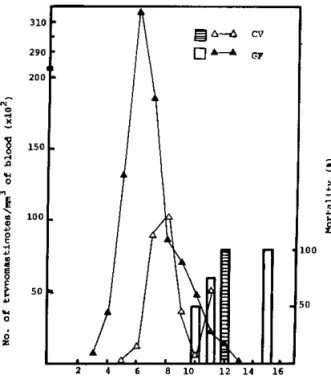
ani-Figure 1 shows the evolutior of the pa¡asitêmiâ and the cumulative mortality ofsix GF and four
CV three months old male C¡'W mice infected
with
I
x
104 blood forms of trypomastigotes ofT. cruzi, Y strain. An earlier ând more intense
5
8
r50a
È50
SILVA M E ; EvANGELrsrA. E. A.. Nrcolr, J. R.;BAMBTRRA, E. A. & vIEr'.A. E. o. Ame¡ican ,.ypanosomiåsis {châgas,
dFease) in conventionâì alrd gelmftee râts and mice Rev. Ins¿. Me.t. t¡op. Sáo pãulo, z9:2g4 288. l98z. parasitemia q'âs observed in the GF group. The
mortality also was more precocious
in
¿hecF
than in CV group, even though two
cF
ânimalssurvived up
to
the 15ù day âftet infection.At
the 12t¡ day, aU CV and 66Ea of tinecF
ânimalswere dead.
The experimentwas repeated with fourmale
and ñve fema-le GF and six male and tqrelve fe_ male CV, 21 dâys old mice infected
with
1.0-1.3x
10" tryÞomastigotes ofy
strain of T.cruzi-Again, both pa.rasitemia and mortality were mo reÞrecociousin the GF group, aìthough thepeâk
of parasitemia was highet
in
CV mice.In
this experiment, the CV animâls were keptin
the CV animal room. After the beginningofthe expe_ riment, therewas â drop in the temperaturethat
affeCted mostly the CV mice.
Rats
A preliminary experiment
with
one female ând one male GF a¡d two female CV 14 monthsold Sprague-Dâv¡ley rats was caFied out. A hi_ gher pârasitemia and a more precocious death
ì¡/ere observed in GF group.
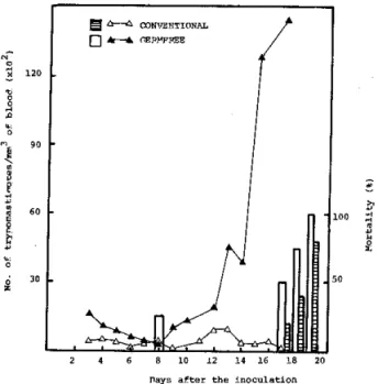
Figure 2 sho\Ã's the pÐasitemia and the
morta-ìity
of five GF and six CV Wistar mâle2l
davsold rats v,¡eighing approximately 60g and inocu_
lâted with 6 x 10⁶ trypomâstigotes of Colombian
shain
of T. c¡.uzi. The patasitemia was muchhigher in the
cF
group reaching 150.000 trypo_mastigotes/mm3 of blood. Themortâlity was also
more precocious
in
the GF eroup.At
the 21sr day, allcF
had died whereas one CV lived ufrto the 47h dây, when it was sacrifrced.
The histopâthological findings were similar
in
rats
and mice fromthe
above mentionedgroups. Macroscopicâlty: (1) the spleens from
eF
animals were twice the size of CV animals: (2) pronounced signs of congestive cardiac insuffi,
ciency (such âs, ascites ând generalized visceral congestion)
i¡
GF animals. Microscopically: G)more intense cell ând tissue pa¡asitism
in
GFthan in CV animals; (2) the parasitism was much
more intense in,the organs rich
in
celts of themononuclea¡ phagocytic system ând
in
the pa_renchyma of liver, âdrenâls and muscles (car_ diâc, skeletal ând smooth) of both
eF
and CV ânimâls.In
the central nervous system. therewere sparse and discrete inflammatory lesions
a¡d ceuular parasitism.
Figute 3 shov/s â sample of the histopatho_ logic firdings in hearts of GF' and CV mice. The
more Ìntense agtessiveness of the disease in GF
ânimals is evident.
The experimentráas repeated with four male and four female GF and three male and three
female CV, 56 days old Wistal rats weighing ap_
proximately 1109, The animals were Ínoculated
with
12x
106 trypomastigotes of Colombianshain ofT. ctuzi, The f,arasitemia was nore pre-cocious and higher in the
cF
groups. Atl animalssurvived to the ?1"t day, when they were killed.
On histopathologicâl examination, no diffe_
rence between the GF and the CV animals could
be detected.
A
discrcte focal parasitismin
the musculâr and macrophagic phagocytic systemcould be visuâlized. g
I 3so
E
ñ
El H t¡t¡eqnns fl l.{, er}lF¡€!
Fis. 2 Parasitemia and cumulâtive mortãliùy of semftees and conventionalt days old WistâÌ male mts weighing 60g, inoculated with 6x10' blood forms ofU strâin of T¡ypåDosoEå cruri.
3
I'
Mice a-re the ânimals most widely used as hosts in experimental Chagas' disease. They
de-velop an acute and a chronic phase
followi¡g
infection with T. cruzi. Figure 1 shows
that
GFSILVA. M E.: EVANGELISTA. E. A.: NICOLL J. R.: BAMBIRRA. E. A. & VIEIRA. E. C. - AùrericÂn t¡\'påììosonì ia sis , Châgâs disease) ¡n conventjonal and gemìfree rats and mjce Rer. Inst. ìled. t¡op. Siào P¡ulÒ. 29:281.288. t98?
mice had an ea¡lie¡ and higher parasitemia than
thet
CV counterparts These ¡esults may be explained by the fact
that
GF mice have a ìessdeveloped lymphoid system when compared
with CV mice, impâiring the cell mediated im-mune lesponse (SZERI et â1., 19?6). ROGERS
&
BALISH (1978) ând GOODMAN et al. (19?8)reported evidences fo¡ the "immunological
im-maturity'of
GF animals. The rate ofgamma-cell and tissue parasitism
in
GF mice may be explained by the smaller number of cells invol ved in immunological defense such as:'tymphocytes (OLSON& WOSTMAN, 1966a ) and neutro
phils, monocytes. and eosinophils (OLSON & wosTMANN. 1966b).
Moreover peritoneal macrophâges from GF
mice a¡e possibìy less activâted th,an those ob,
tained from CV animals. Macrophages ftom GF
animals have smaller and more spherical nuclei, fewer mitochondria and a higher number
oflyso-some-like granules per unit voìume ofcytoplasm
(WOODWARD, 19?8). These differences may be possibly related to the lack of stimuli from
intes-tinål flora {WOODWARD, 19?8).
The highe¡ parasitemia found in CV mice in
the second experiment might be explained by
the lower tempera¿ure of the ¡oom where the
animals were housed.
lt
is well known that thereis a raise in parasitemiâ at lower temperatures rKOLODNY, 1940). Nevertheless. the death ¡âte
and the histopathological findings confirmed
the ¡esults of the hrst experimenl.
Figue 2 shows tÌìat, in rats, the leveìs
ofparasi-temia were higher in the GF than in CV group.
ln 21 days oìd animals, the mo¡tality was earlie¡ and totâl in the GF group.
All
six GF and fourout
five CVrâts
died. The highmortality
ofyoung Iats infected-
with
T. cruzi conhÌms theresults ofKOLODNY (1940) and CULBERTSON & KESSLER (1942), who showed that the disea se is more seve¡e in yolrnger animals. The mor-phological findings a¡e comÞâtible with the
pa-rasitological data, i. e., the disease was more se-vere in GF than in CV rats.
In older rats there was no mortality
i¡
either group of animals. The pârasitemia, again. wasmore precocious and higher
in
the GF than int}le CV rats. The histopathological findings were
similar for both groups.
The ¡esults ¡eported herein show that Cha
gas'disease is more severe
in
GF thanin
CVrats and mice. Further q,ork
will
be ca¡ried outto
elucidâte the reason fo¡ the observed diffe-rences.globulin synthesis is 50 times higher in CV
tha[
i¡
GF mice (SELL & FAHEY, 1964). These dataale also suggestive of a slower humoral immune response in GF than in CV mice. The
histopatho-logicâl dâtâ conh¡m
that
the disease is muchiì.1
i'.,:
1
SILVA, M. E.; EVANGELISTA, E. A.: NICOLI, J- R.: BAMBIRR'A, E. A. & VIEIRA. E. C- Amencan hypanosomiâsis (Chagâs'
clisease) ìn conventionâl and germftee rats ând mice. Rev. Inst. Med. ¿¡op. Siio Paulo, 29:284 288.
198?-Tdpanosom¡ase americana (doença de Chagas) em rÀtos e camundongos convencionais e
isen-tos de g€rmes
Camundongos CFIJÍ¡ (LOB) e ratos Wistar e Sprague Dawley isentos de germes (GF) e
con-vencionais (CV) foram infectados com TrypÀno.
soma cruzi. A doençâ foi mais grave nos animais
cF
do que nos Cv, o que foi demonstrado por: (1) uma parasitemia mais precoce e mais intensa; (2) uma mortalidade mâis precoce; (3) baço duas vezes maior; (4) um parasitismo celular e tissularmais intenso: (5) si¡âis viscerais de insuficiência
cardÍaca.
ACKNOWLEDGEMENTS RESUMO
This work vt'as supported by Financiadora
de Estudos e Projetos (FINEP) and by conselho Nacionâl de Desenvolvimento Cientlflco e
Tec-nológico (CNPq). The technical help of Ronilda
M- de Paulâ and Carlos Gomes Silva is
acknow-ledged.
ponse to LPS in germfree, Eschêrichiâ coli
monoâssôc¡â-ted ând conventionâl mice. J. Immunol.,124:36 41, 1980. KOLODNY, M. H. - Studies on age resistance âgâinst tlypanosome infecbion. L Theresistance of¡ats ofdifferent ages to infection with T. cruzi. Am€¡. J. Hyci,,29: 13-24, 1939.
KOIODNY, M. H. - The efrect ofenvironmental üempe
!âtule upon expedrnentâl llypânosomiasis (T. r¡uzi) of
râts. Am€¡. J. Hys.,32: 2l-23,
1940-OLSON, G. B. & WOSTMANN, B. S. - Lymphocytopuie
sis, plâsmocytopoiesis âîd ceuul prclife¡ation in non-antÍgenical¡y stimulåted germfree mice. J. Immunol., 9?: 287 274, lgß8à.
ol-soN, G. B. & wOSTMANN, B. s. - cellulù andhumo-ral immune response of germftee mice stimulated 1l'lth
?s IIGGor Salmoneua ¿ypbi¡r¡rrium. J. Immunol., 97: 2?5-286, 1966b_
BRENER, Z.-Thempeutic activity ând cliteûon ofcure onmiceexpe meDtauyinfectedwithTrypânosor¡acruzi. ney.Ihst. Meal. trop. S. Påulo,4:389 398,1962.
CULBEF,TSON, J. t. & KESSLER, W. R. - Age re8is-tance of mice to TrypanosoDå cruri. J, Paråsi¿., 28: 155,158, 1942.
FINERTY, J. F.; TOBIE,J. E. d¿ EVANS, C. B.-Aniibody
and lÌÌlmunoglobuliì synthesis 1¡ gelmflee ând conven-tional mice infected with Plasmo¿lium bc¡sh€i- Âne¡. J. trop. Meit. Hyg., Z1:499 505, 19?2.
GOODMAN, M- G.i FIDLER, J. M. & {¡EIGLE, W. O.
-Nonspeciñc aclivation of EuÌinelys¡Þhocytes. ûr. Cells respondÍng riitogenicålly to 2 mercâptoetl¡ânol are typi cal unstimulated Iymphocytes. J. Immunol., t2l:
1899-1904,1978:
GORDON, H. A. & PESTI, L. - the gnotobiotic anÍma¡
as a toolin the study ofhosi miclobial relâtionship. Bâct. Rev..35:390 429. 19?1.
10_
REFERENCBS
PLEASANTS, J. R. - Onotobiotlcs. ID: MELBY JR., E.
C. & ALTMAN, N. H. ed. - Hândbook oflaboratory åDi. hat scie¡ce. Clevelând, CRC Press, 1974. p. rrg 1?4. POLLARD, M. vlIâl status of germi:ee mice. Natl. cân-c€r Inst. MoDogl¡Þh.,20: 16?-1?2, 1965.
ROGERS, T. J. & BALISI¡, E. Efrect oI the systemlc candidiasis on blastosenesis of lympbocytes tom gelm' Aee and conventional rabs. Infect. Immu¡.. 20: 142-150. 19,r8.
SELL, S- & F'AHEY, J. L. * Relâtionship- between
" glo
bulin merâbohsm ând low serum ? slobulir in gerrnfree mice. J.ImD¡¡¡oI.. 93: 8l-8?.
1964-SZER,I, I.; ÀNDERLi{, P-: BANOS, Z. & R,ADNAI, B,
- Decæâsed cellulâl lespohse lÌ1 gelm-fiee ânimâls. Actâ
Microbiol. Acâal. Sci. h\ns.,Zst 23r 234, 1978.
TRE)iLER, P. C. - The use of plastics in the desigz¡ of
ùolator systems. Ann. N. Y, Acad. Sci., ?8:29-36, 1959.
VIEIE¡., L- Q.; MORAES E SAN"OS, T. & VIEIRA, E.
C. - AlteIabions in cell numbe¡, size and couâgercontent iÌ livers ofconventionâl ând germfree mice beùlng Schis
tosoma nansoni.In: WOSTMANN. B. S.. ed. - C€¡mfr€è res€årcb: Microflora. Cobtrol ând lts âppllcatron to the biomedlcal sclences, New York, Aìan R. Liss, 1985. p. 235 238.
14.
I]YDE, C- L.i FINERTY, J. F. & EVANS, C. B. -å.ntibody and immunogtobulin synthesis in germtee and conven tional mice i¡fected with EDerythroroor coccoides. Âmer.
J. troD. Med. HyA.,2l: 506'511, I9?2.
I<IYONO, H.: McGHEE, J. R. & MICHALEK, s. M. -LPs Ëeulâtion of the immune response: compâ¡iron
ofrcs-1.
1'1.
WAGNER, M. - Determi4âtion of germftee status. AnD,
N. Y. Acad. Sci., ?E:89'101, 1950.
wooDwARD, B. -A st€rdological ultrast¡ucturâI Âtudy oipelitonea] macrophages Aom gelmtree and conventio nally'reaÌed mice. CeU Tiis. Rês.,192: l5?'166. r978.