52 Case Report
Brazilian Journal of Urology
Official Journal of the Brazilian Society of Urology
Vol. 27 (1): 52-54, January - February, 2001
SUBCUTANEOUS AND TESTICULAR METASTASIS FROM PROSTATIC
ADENOCARCINOMA WITH NEUROENDOCRINE DIFFERENTIATION
FRANCISCO T. DÉNES, ARTUR H. BRITO, ANDRÉ M. DOS SANTOS
Division of Urology, School of Medicine, State University of São Paulo, SP, Brazil
ABSTRACT
The pathological finding of testicular metastasis in cases of disseminated prostatic adenocarcinoma is rare, but was more frequently reported in the past, when bilateral castration was performed more often. The existence of skin and subcutaneous metastasis adds a worse prognosis, because generally it is sign of advanced disease with an average survival time of less than one year. The synchronous occurrence of such metastasis has not been described previously, neither their association to neuroendocrine differentiation. The presence of such differentiation of prostatic adenocarcinoma represents a very unfavorable prognostic factor, as suggested in recent literature. Herein, we discuss the case of a 53 year old man, who presented with macroscopic hematuria and frequency associated to several painless subcutaneous nodules in left axilla and shoulder, as well as in the lower abdominal wall. The right testis was painful, endured and on rectal examination, the prostate was dif-fusely enlarged. Serum PSA was elevated, reaching 1760 ng/ml and prostatic biopsy disclosed a Gleason 10 prostatic adenocarcinoma with neuroendocrine differentiation. The same pathological pattern was detected in the right testis and in all subcutaneous nodules, documented by positive staining of chromogranin, a marker of neuroendocrine cells. He was submitted to a prostate tunnelization and maximal androgen blockade plus adju-vant chemotherapy, nevertheless, he died 5 months latter.
Key words: prostate; prostatic neoplasms; neoplasm metastasis; testis; subcutaneous; neuroendocrine differ-entiation
Braz J Urol, 27: 52-54, 2001
INTRODUCTION
Advanced prostatic adenocarcinoma with symptomatic testicular metastasis is a rare condition, with only 70 cases presented until 1990 (1). Subcuta-neous metastasis is even less frequent, with only 14 cases presented in the literature (2). To our knowl-edge, this is the first case in which both types of me-tastasis are synchronously present and neuroendocrine differentiation of the prostatic adenocarcinoma is his-tologically confirmed, both in the prostate and in the metastatic tissues.
CASE REPORT
A 53 years old patient of African origin presented an isolated episode of painless
53
UNUSUAL METASTASES FROM PROSTATIC CARCINOMA
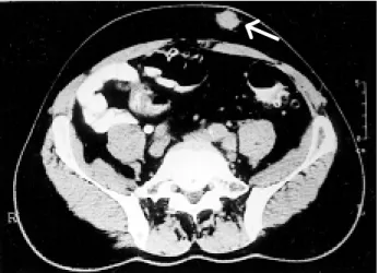
ng/ml and prostatic biopsy disclosed a Gleason 10 prostatic adenocarcinoma in all fragments. A bone scan revealed diffuse metastatic involvement of the vertebral spine and costal arches, while abdominal CT disclosed left bladder wall and lower ureteral infiltration by a large prostatic neoplasm. This exam also demonstrated the subcutaneous abdominal le-sion (Figure-1). As urinary symptoms and
hema-turia worsened, antiandrogen therapy was initiated, with decrease of PSA. However, due to persistence of urinary symptoms and hematuria, he was sub-mitted to prostatic tunnelization, associated to
bi-lateral orchiectomy and excisional biopsy of 4 ab-dominal and thoracic subcutaneous lesions. Patho-logic examination of the prostatic tissue confirmed the presence of Gleason 10 prostatic adenocarci-noma with areas of neuroendocrine differentiation. Examination of the right testis and all subcutane-ous nodules also confirmed the presence of meta-static neuroendocrine tumor, positively stained for chromogranin, a specific marker of neuroendocrine cells (Figure-2). Two months after surgery, he was clinically improved, with significant weight gain and without urinary symptoms or hematuria. The PSA levels dropped to 53 ng/ml, and maximal an-drogen blockade with cyproterone citrate was ini-tiated, nevertheless he still had generalized pain and adjuvant chemotherapy was initiated, even so, he died 5 months latter.
DISCUSSION
In the past, when antiandrogenic drugs were yet unavailable, orchiectomy was the treatment of choice in advanced adenocarcinoma of the prostate. In many patients, silent microscopic metastatic in-volvement of the testes was discovered on pathologi-cal examination, being described in 2.4 to 4% of the cases reviewed with this specific interest (1). Symp-tomatic testicular metastases are very rare, signaling to an advanced disease, with undetermined, but
gen-←
←
←
←
←
Figure 1 - Abdominal CT shows subcutaneous nodule in the left anterior wall.
Figure 2 - A)- Neuroendocrine tumor with chromogranin positive in cellular cytoplasm (immunolabeling, X400). B)- Solid groups of cuboidae cells with scanty cytoplasm, nuclei hypercromatic, grouped in organoid arrangement in microscopic section of the right testis, showing prostatic adenocarcinoma metastasis (HE, X 100).
54
UNUSUAL METASTASES FROM PROSTATIC CARCINOMA
erally poor prognosis (1). Subcutaneous metastases were previously presented in 14 patients, most of them in terminal stages of the disease, with survival of less than 12 months, despite therapy (2). The synchro-nous occurrence of such metastases has not been de-scribed previously. Moreover, the presence of neu-roendocrine differentiation may explain the aggres-sive behavior of the prostatic adenocarcinoma in this patient, as it is usually associated to a very unfavor-able prognosis. Cohen et al., analyzing 17 patients who died of prostatic cancer, disclosed that 15 exhib-ited neuroendocrine differentiation (3). However, additional studies are needed in order to establish neuroendocrine differentiation as an independent prognostic factor and to evaluate new therapeutic options against these tumors, particularly unrespon-sive to current modes of therapy.
REFERENCES
1. Kirkali Z, Reid R, Deane RF, Kyle KF: Silent testicular metastasis from carcinoma of prostate. Brit J Urol, 66: 205-207, 1990.
2. Delima A, Mohamed A, Yalla SV, Burros HM: Prostatic carcinoma metastasizing to skin and subcutaneous tissues. Urology, 2: 663-665, 1973. 3. Cohen RJ, Glezerson G, Haffejee Z, Afrika D: Prostatic carcinoma: histological and immuno-histological factors affecting prognosis. Brit J Urol, 66: 405-410, 1990.
________________________ Received: November 16, 2000 Accepted after revision: February 9, 2001
______________________ Correspondence address: Dr. André Meirelles dos Santos Rua Bartira, 59 / 41