online | memorias.ioc.fiocruz.br
Dengue 4 in Ceará, Brazil: characterisation of epidemiological
and laboratorial aspects and causes of death during the first
epidemic in the state
Izabel Letícia Cavalcante Ramalho1,2, Fernanda Montenegro de Carvalho Araújo1,3/+,
Luciano Pamplona de Góes Cavalcanti4, Deborah Nunes Melo Braga5,
Anne Carolinne Bezerra Perdigão3, Flavia Barreto dos Santos6, Fernanda de Bruycker Nogueira6,
Kiliana Nogueira Farias da Escóssia7, Maria Izabel Florindo Guedes2
1Laboratório Central de Saúde Pública, Setor de Virologia, Fortaleza, CE, Brasil 2Universidade Estadual do Ceará, Rede Nordeste de Biotecnologia, Fortaleza, CE, Brasil 3Centro Universitário Christus, Fortaleza, CE, Brasil
4Universidade Federal do Ceará, Departamento de Saúde Comunitária, Fortaleza, CE, Brasil 5Instituto de Prevenção de Câncer, Fortaleza, CE, Brasil
6Instituto Oswaldo Cruz-Fiocruz, Laboratório de Imunologia Viral, Rio de Janeiro, RJ, Brasil 7Secretaria de Saúde do Estado do Ceará, Núcleo de Vigilância Epidemiológica, Fortaleza, CE, Brasil
BACKGROUND The first dengue cases in Brazil with laboratory confirmation occurred in the northern region of the country,
with the isolation of two serotypes, dengue virus 1 (DENV-1) and DENV-4. In Ceará, the introduction of DENV-4 was reported during a DENV-1 epidemic in 2011, with only two isolations.
OBJECTIVES The aim of this study was to characterise the first DENV-4 epidemic in the state of Ceará, Brazil.
METHODS The study population was composed of patients with suspected dengue that were reported to health care units
from January to December 2012. The laboratory confirmation of infection was made by viral isolation, reverse transcription polymerase chain reaction (RT-PCR), AgNS1, immunohistochemistry and IgM enzyme-linked immunosorbent assay (ELISA).
MAIN CONCLUSIONS In the study year, 72,211 suspected dengue cases were reported and 51,865 of these cases (71.8%) were
confirmed to be positive. Co-circulation of three serotypes, DENV-1, DENV-3 and DENV-4, was detected with a predominance of DENV-4 (95.3%). Most cases were not severe, but there were 44 fatal outcomes. DENV-4 Genotype II was identified for the first time in Ceará.
Key words: dengue 4 - epidemiology - deaths - genotyping - Ceará/Brazil
Dengue is one of the most serious public health prob-lems in Brazil and in the world, especially in tropical and subtropical regions, and it affects approximately 390 million people every year, of which 96 million are clini-cally manifest.(1) The disease is caused by dengue virus (DENV), an arbovirus belonging to the Flaviviridae family and to the Flavivirus genus, with four antigeni-cally distinct serotypes, DENV-1 to 4.(2) The four DENV serotypes exhibit high genotype variability;(3) genomic sequencing studies have characterised five genotypes for DENV-1, six genotypes for DENV-2, five genotypes for DENV-3 and four genotypes for DENV-4.(4)
In Brazil, the first cases of dengue with laboratory confirmation occurred in Boa Vista, Roraima, in 1982, with the isolation of DENV-1 and DENV-4 serotypes. (5) This outbreak was restricted to the northern region
of the country, with no major consequences. After its introduction in Brazil, DENV-4 was isolated again in 2008 in Manaus-AM and in 2010 it was isolated in Boa Vista-RR; by the beginning of July 2011, this serotype
doi: 10.1590/0074-02760180320
+ Corresponding author: [email protected] Received 5 July 2018
Accepted 11 September 2018
had been isolated in 10 Brazilian states, including Ceará. (6,7,8) In the years 2011 and 2012, DENV-4 was
respon-sible for epidemics in most states and coincided with the simultaneous circulation of the four serotypes in the Brazilian territory. DENV-4 was first isolated in Ceará in 2011 during a DENV-1 epidemic.(9)
The aim of this study was to characterise the clini-cal, epidemiological and laboratorial aspects of the first DENV-4 epidemic in Ceará, Brazil.
SUBJECTS AND METHODS
Ethical aspects - The study was approved by the Research Ethics Committee of the State University of Ceará, under opinion No. 913.484 and CAAE No. 38014414.6.0000.5534, and followed the Guidelines and Norms Regulating Research on Human Subjects estab-lished in Council Resolution 466/12 National Health/MS.
Case definitions
Case of suspected dengue - Patients with acute febrile illness, with a maximum duration of seven days, with at least two of the following symptoms: headache, retro-or-bital pain, myalgia, arthralgia, prostration and rash.
Dengue case confirmed by laboratory criteria - A suspected case with a positive laboratory test was identi-fied as a case of dengue confirmed by laboratory criteria.
Dengue case confirmed by clinical-epidemiological criteria - After the autochthonous dengue virus circu-lation was confirmed, acute cases of dengue were con-firmed by clinical-epidemiological criteria.
Deaths from dengue - The criteria for suspected dengue death were based on recent reports of fever (up to seven days) without apparent bacterial infection, the presence of rash, and the presence of cavity effusion and/or bleeding.(9) The bodies were autopsied with the consent of the family, and samples of blood, liquor and viscera were collected and sent to the laboratory to iden-tify the cause of death.
Laboratory diagnostic methods - The specific labo-ratory diagnosis of dengue was performed in LACEN-CE through at least one of the following laboratory tech-niques described below:
Viral isolation - Viral isolation was performed by inoculation into the Aedes albopictus C6/36 cell line(10) and DENV serotypes were identified by an indirect im-munofluorescence (IFI) test using monoclonal antibod-ies specific for the four serotypes,(11) and/or reverse tran-scription polymerase chain reaction (RT-PCR).(12)
NS1 antigen capture [NS1 enzyme-linked immuno-sorbent assay (ELISA)] - To capture NS1 antigen, the Platelia™ Dengue NS1 Ag-ELISA kit (Bio-Rad Labo-ratories, Marnes-La-Coquette, France) was used accord-ing to the manufacturer’s instructions. All samples were tested between 1-7 days after the onset of symptoms.
IgM antibodies capture - Anti-DENV IgM antibody capture was performed on serum samples from patients > 6 days after symptom onset using the Dengue IgM Capture ELISA kit (Panbio, Brisbane, Australia) accord-ing to the manufacturer’s instructions.
Detection of NS1 and IgM in LCR were performed as described by Araújo et al. and Soares et al.(13,14)
Molecular methods
Viral RNA extraction - Viral RNA was extracted us-ing the QIAamp Viral Mini Kit (Qiagen, Inc., Valencia, USA), following the manufacturer’s instructions, and it was stored at -80ºC until RT-PCR was performed.
RT-PCR - Conventional RT-PCR, described by Lanciotti et al.,(12) was used for detection and typing of DENV from acute phase samples (up to seven days of disease). The method consists of a two-step reaction, where the first step was performed by RT followed by a PCR. The protocol detects the four serotypes simultane-ously in a semi-nested procedure, generating amplified products with specific sizes for each DENV serotype.
Genotyping - The RT-PCR technique for sequencing was performed according to the protocol described by Miagostovich et al.(15) Oligonucleotides were used to am-plify overlapping fragments of approximately 900 base pairs (bp) along the DENV-4 E gene. Amplified and pu-rified cDNA fragments were sequenced in both direc-tions using the BigDye Terminator Cycle Sequencing Ready Reaction kit version 3.1 (Applied Biosystems®, California, USA). After the sequencing reaction, the products were purified using Centri-Sep Spin Columns (Princeton Separation, New Jersey, USA) or a DyeEx 2.0 Spin Kit (QIAGEN, Inc., California, USA). The DNA was resuspended in 10 μL of formamide and transferred to a 96-well plate (MicroAmp Optical 96 Well Reac-tion Plate - Applied Biosystems, California, USA). The plate was sent to the PDTIS/Fiocruz DNA Sequencing Platform and the products were analysed by capillary electrophoresis on an ABI 3730 DNA Analyser (Applied Biosystems®, California, USA).
For analysis of the sequenced products, the program Chromas® 1.45 (http://www.technelysium.com.au/chro-mas14x.html) was used. Sequence identity was deter-mined by BLAST (http://blast.ncbi.nlm.nih.gov/Blast. cgi). Sequence alignment was performed using the soft-ware ClustalW2 (http://www.ebi.ac.uk/Tools/msa/clust-alw2/), and the phylogenetic tree was constructed with a substitution model based on maximum likelihood, with support of the bootstrap test (1000 pseudo-replicas) using the MEGA 5 Program (http://www.megasoftware.net/).
Immunohistochemistry - The immunohistochemical technique was performed on 10% formalin-fixed viscera fragments with the streptavidin-biotin-alkaline phos-phatase (SAAP) technique at the Evandro Chagas Insti-tute (IEC, Pará, Brazil). Sections of approximately 3 to 4 μm were stained with anti-DENV-2 polyclonal primary antibodies produced in mice by the IEC Arboviruses Sector. Negative and positive controls were performed for each group of samples tested.(16)
RESULTS
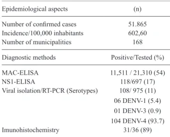
In 2012, 72,211 suspected cases of dengue were reported in 184 municipalities of Ceará. Among these cases, 51,865 (71.8%) were confirmed in 168 munici-palities (91.3%), with a cumulative incidence of 602.6 cases/100,000 inhabitants, Table I.
The main criterion for confirmation of cases was clinical-epidemiological (78%). These cases were con-centrated in the months of April and May, reaching a peak transmission in the capital city of Fortaleza, with an incidence of 1,558.84 cases/100,000 inhabitants.
A total of 21,310 patients had blood samples collected and sent to the laboratory. The IgM ELISA test was the most widely used method in laboratory diagnosis, con-firming 54% (11,511/21,310) of the cases. NS1 ELISA confirmed 17% (118/697) of the cases tested. The tech-nique of immunohistochemistry applied to the investiga-tion of the deaths confirmed dengue in 86.11% (31/36) of the cases analysed, Table I.
TABLE I
Epidemiological and laboratory aspects of dengue cases occurred in Ceará, Brazil, 2012
Epidemiological aspects (n)
Number of confirmed cases 51.865
Incidence/100,000 inhabitants 602,60 Number of municipalities 168
Diagnostic methods Positive/Tested (%)
MAC-ELISA 11,511 / 21,310 (54)
NS1-ELISA 118/697 (17)
Viral isolation/RT-PCR (Serotypes) 108/ 975 (11)
06 DENV-1 (5.4) 01 DENV-3 (0.9) 104 DENV-4 (93.7) Imunohistochemistry 31/36 (89)
MAC-ELISA: IgM antibody capture-enzyme-linked immu-nosorbent assay; RT-PCR: reverse transcription polymerase chain reaction.
TABLE II
Distribution of confirmed cases of dengue, by gender and age group, Ceará, Brazil, 2012
Variables (n) (%)
Gender
Female 30,050 57.92
Male 21,828 42.07
Age range (years)
< 1 613 1.18
1 a 4 1,609 3.10
5 a 9 3,005 5.79
10 a 14 5,028 9.69
15 a 19 5,943 11.46
20 a 29 12,621 24.33
30 a 39 9,318 17.96
40 a 49 6,648 12.81
50 a 59 4,110 7.92
60 a 69 1,944 3.75
70 a 79 763 1.47
≥ 80 276 0.53
Total 51,878 100.00
analysed by viral isolation and subsequent sequencing of the E gene, allowing the identification of the circulating genotype II in the state of Ceará (Fig. 1).
Among the cases with positive viral isolation, four cases of DENV-1 and DENV-4 co-infection were identi-fied, and all four resulted in a cure (data not shown).
Among all the cases, predominance was observed in females (57.9%) and in the age group 20 to 29 years (24.3%), Table II.
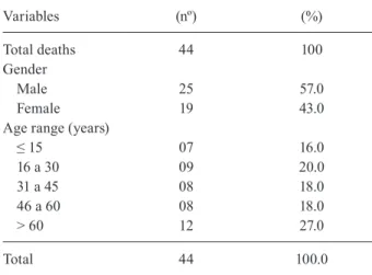
In 2012, a total of 44 deaths from dengue were con-firmed in 16 different municipalities, with a mean age of 36 years, ranging from five months to 91 years. Deaths were concentrated between March and June (Fig. 2), and 36 (82%) cadavers were autopsied. A higher percentage of deaths occurred among males (57%, 25/44) and those older than 60 years of age (Table III).
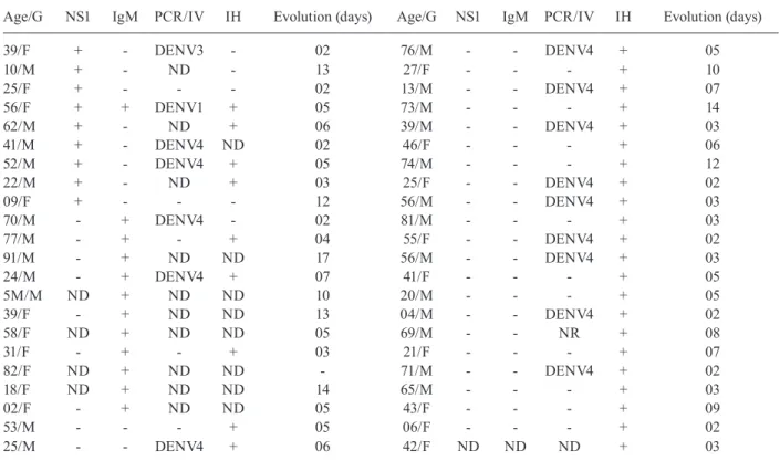
Viral isolation and/or RT-PCR identified the infec-tious serotype in 16 (36.4%) of the deaths, with DENV-4 being the serotype identified in 1DENV-4 (87.5%) of these. Immunohistochemistry confirmed 86.2% (31/36) of the cases, Table IV.
The most frequent symptoms were fever (77.8%), vomiting (72.2%), abdominal pain (69.4%), cough (69.4%), dyspnoea (61.1%) and respiratory distress (61.1%). Co-morbidities were reported in 58.3% of the deaths,
pri-marily hypertension (30.5%), heart disease (30.5%) and diabetes mellitus (11.1%). The main associated conditions were smoking (30.5%), alcoholism (25.0%) and obesity (22.2%), Table V.
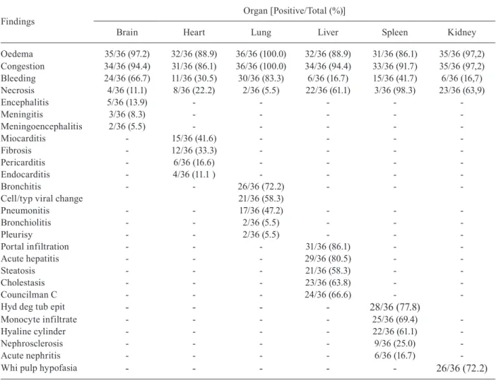
General histopathological findings such as oedema, congestion and haemorrhage predominated in the lung, brain, liver and heart. These findings also highlighted the presence of hepatitis in 80.5%, pneumonitis in 47%, myocarditis in 42% and encephalitis in 14% of the deaths that occurred in 2012, Table VI. Among the deaths, 22.7% (10/44) were positive for dengue in CSF.
DISCUSSION
Among the confirmed cases of dengue in Ceará, re-gardless of the criteria of confirmation adopted, 99.6% did not progress to severe forms. In the city of Aracaju, during the epidemic in 2012, this percentage was 96.1%. (17) Both cities exhibited percentages very close to those
reported during epidemics in other Brazilian cities, where most of the symptomatic cases detected by surveillance services in Brazil progressed to less severe forms.(18)
The percentage of reported cases that were confirmed by laboratory criteria was 22%, which is considered su-perior to percentage of confirmations recommended by the Brazilian Ministry of Health for epidemics, which is 10% of reported cases.(19) It should be emphasised that, for serious cases, the recommendation is always to look for laboratory techniques that confirm the diagnosis, es-pecially for cases that progress to severe forms of dis-ease and/or death.
The majority of the confirmed cases occurred in the population of 20 to 59 year old patients and in the female gender. This age range is clearly explained by the age distribution of the Brazilian population, but in relation to gender, it is worth noting that other analytical studies conducted in Brazil do not indicate a significant differ-ence in the risk of illness for the female sex. There are several hypotheses to justify the higher number of fe-male cases, especially the fact that dengue is a disease predominantly transmitted in the home and that Brazil-ian women are more likely than men to seek care.(17)
The confirmed cases were concentrated in the cap-ital of Ceará, and beginning in the month of July, there was a significant reduction in the incidence, confirming the seasonality of dengue in Ceará, as well as in part of the northeast.(20)
During the 2012 epidemic, the co-circulation of three serotypes was detected, but with a marked predomi-nance of DENV-4 (93.7%), which was similar to what occurred in other Brazilian states that same year.(21) The predominance and dispersion of DENV-4 appeared to be predictable, since it had recently been reintroduced in Brazil, with a large part of the population still sus-ceptible to the disease.
From the phylogenetic analysis based on the E gene, one of the samples positive for serotype 4 was charac-terised as belonging to genotype II, the same one that circulated in the north, northeast and southeast regions of the country in the year 2011.(22,23,24) Genotype II, iden-tified in our study, has been the most prevalent in the Brazilian states to date.
TABLE III
Classification, gender and age group of deaths confirmed by dengue, Ceará, Brazil, 2012
Variables (nº) (%)
Total deaths 44 100
Gender
Male 25 57.0
Female 19 43.0
Age range (years)
≤ 15 07 16.0
16 a 30 09 20.0
31 a 45 08 18.0
46 a 60 08 18.0
> 60 12 27.0
Total 44 100.0
TABLE IV
Laboratory confirmation in dengue death cases (n = 44), in 2012, Ceará, Brazil
Age/G NS1 IgM PCR/IV IH Evolution (days) Age/G NS1 IgM PCR/IV IH Evolution (days)
39/F + - DENV3 - 02 76/M - - DENV4 + 05
10/M + - ND - 13 27/F - - - + 10
25/F + - - - 02 13/M - - DENV4 + 07
56/F + + DENV1 + 05 73/M - - - + 14
62/M + - ND + 06 39/M - - DENV4 + 03
41/M + - DENV4 ND 02 46/F - - - + 06
52/M + - DENV4 + 05 74/M - - - + 12
22/M + - ND + 03 25/F - - DENV4 + 02
09/F + - - - 12 56/M - - DENV4 + 03
70/M - + DENV4 - 02 81/M - - - + 03
77/M - + - + 04 55/F - - DENV4 + 02
91/M - + ND ND 17 56/M - - DENV4 + 03
24/M - + DENV4 + 07 41/F - - - + 05
5M/M ND + ND ND 10 20/M - - - + 05
39/F - + ND ND 13 04/M - - DENV4 + 02
58/F ND + ND ND 05 69/M - - NR + 08
31/F - + - + 03 21/F - - - + 07
82/F ND + ND ND - 71/M - - DENV4 + 02
18/F ND + ND ND 14 65/M - - - + 03
02/F - + ND ND 05 43/F - - - + 09
53/M - - - + 05 06/F - - - + 02
25/M - - DENV4 + 06 42/F ND ND ND + 03
Age/G: age/gender; F: female; M: male; NS1: NS1 ELISA; PCR/IV: polymerase chain reaction and/or viral isolation; IH: imuno-histochemistry; ND: not done; (+): positive; (-) negative.
A larger number of samples should be sequenced for further investigation of the genotypic variants possibly present in the state of Ceará. Although no differences have been reported in the progression of the disease in patients with two different circulating genotypes, gen-otypic surveillance is necessary to identify not only the genotypes already described but also variants within the genotypes. These variants may present nucleotide alterations that may lead to changes in the viral rep-lication process, such as changes in important amino acids in the viral structure. The genotype identifica-tion in 2012 played an important role in the molecular and genotypic surveillance of DENV-4 in the state of Ceará, since until then, there was no knowledge of the circulating genotype.
The higher incidence of male deaths can be explained by cultural influences, among other factors. There is evidence that men tend to seek health services less of-ten than women, which can lead to a late diagnosis and thereby decrease the chances of early detection of warn-ing signs, which increases the risk of death.(24) One limi-tation of this study was that the new classification of cas-es proposed by the World Health Organization (WHO) was only considered in Brazil in 2014, when these data had already been recorded. Thus, we cannot consider the aspects related to the classification of cases, since there was no record of warning signs in the information sys-tem of the Ministry of Health before 2014.
TABLE V
Frequency of clinical manifestation, comorbidity and risk factor reported among dengue deaths (n = 36)
occurred in Ceará, Brazil
Variables
Total
n (%) Variables
Total (n)
Clinical manifestation Comorbidity
Fever 28 (78) Cardiopathy 12
Vomiting 26 (72) Hypertension 11
Cough 25 (69) Diabetes 4
Abdominal pain 25 (69) Hematological disease 2
Dyspnea 22 (61) Asthma 2
Resp. distress 22 (61) Kidney disease 2 Headache 17 (47) Atherosclerosis 2 Irritability 16 (44) Dyslipidemia 1
Diarrhea 15 (42) Anemia 1
Myalgia 15 (42) Osteoporosis 1
Prostration 14 (39) Lung emphysem 1 Somnolence 14 (39) Risk factor
Hepatomegaly 13 (36) Smoking 12
Convulsion 11 (42) Alcoholism 9
Alt sensorium 10 (28) Obesity 8
Exanthema 9 (25) Sequel to stroke 2 Hematemesis 8 (22) Transplanted 1
Arthalgia 7 (19) Pregnancy 1
TABLE VI
Findings of necropsies of dengue cases that evolved to death, by organ affected, Ceará, Brazil, 2012
Findings
Organ [Positive/Total (%)]
Brain Heart Lung Liver Spleen Kidney
Oedema 35/36 (97.2) 32/36 (88.9) 36/36 (100.0) 32/36 (88.9) 31/36 (86.1) 35/36 (97,2) Congestion 34/36 (94.4) 31/36 (86.1) 36/36 (100.0) 34/36 (94.4) 33/36 (91.7) 35/36 (97,2) Bleeding 24/36 (66.7) 11/36 (30.5) 30/36 (83.3) 6/36 (16.7) 15/36 (41.7) 6/36 (16,7) Necrosis 4/36 (11.1) 8/36 (22.2) 2/36 (5.5) 22/36 (61.1) 3/36 (98.3) 23/36 (63,9)
Encephalitis 5/36 (13.9) - - - -
-Meningitis 3/36 (8.3) - - - -
-Meningoencephalitis 2/36 (5.5) - - - -
-Miocarditis - 15/36 (41.6) - - -
-Fibrosis - 12/36 (33.3) - - -
-Pericarditis - 6/36 (16.6) - - -
-Endocarditis - 4/36 (11.1 ) - - -
-Bronchitis - - 26/36 (72.2) - -
-Cell/typ viral change 21/36 (58.3)
Pneumonitis - - 17/36 (47.2) - -
-Bronchiolitis - - 2/36 (5.5) - -
-Pleurisy - - 2/36 (5.5) - -
-Portal infiltration - - - 31/36 (86.1) -
-Acute hepatitis - - - 29/36 (80.5) -
-Steatosis - - - 21/36 (58.3) -
-Cholestasis - - - 23/36 (63.8) -
-Councilman C - - - 24/36 (66.6) -
-Hyd deg tub epit - - - - 28/36 (77.8)
Monocyte infiltrate - - - - 25/36 (69.4)
-Hyaline cylinder - - - - 22/36 (61.1)
-Nephrosclerosis - - - - 9/36 (25.0)
-Acute nephritis - - - - 6/36 (16.7)
-Whi pulp hypofasia - - - 26/36 (72.2)
It is also worth mentioning the high number of deaths during the 2012 dengue epidemic ranked second in Brazil in relation to the number of confirmed deaths.(18)
The CSF positivity was 20% lower than that found during the DENV-2 and DENV-3 epidemics, where the positivity reached 48,8%, suggesting less involvement of the CNS during the circulation of the DENV-4 serotype.(25)
Most autopsies revealed some type of comorbidity (61.36%), as identified in Singapore.(26) The presence of these comorbidities, such as heart disease and hyperten-sion, are indicated as risk factors for death.(27)
In the study period, acute pulmonary oedema was the primary immediate cause of death, reflecting the severe involvement of the respiratory system, clinically evidenced by the occurrence of respiratory distress and/ or dyspnoea in 61% of the cases.
The autopsies had great relevance, considering that these studies made possible the diagnosis of dengue in 81.81% (36/44) of the deaths. The histological findings of oedema, congestion and haemorrhage in a large per-centage of deaths have been described in the literature, due to haemodynamic changes associated with dengue. (28) Histopathological changes in specific organs, such as
necrosis, inflammatory processes, steatosis, degenera-tive changes, mainly in the liver, lung, heart and brain, have been reported in other studies.(28)
The lethality was higher than considered acceptable by the Brazilian Ministry of Health. Ceará maintains a high rate of lethality when compared to other states.(23) This may be explained in part by the historical circula-tion of dengue for almost 30 years, and the existence of a local surveillance system together with a laboratory and a death verification service that identifies most of the suspected deaths.(9)
In conclusion - The epidemic showed the expected pattern, with most cases resulting in a cure. The labo-ratory confirmation of diagnosis, in detecting the new circulating serotype and confirming the emerging epi-demic, was shown to be a reliable tool for disease sur-veillance. DENV-4 Genotype II was identified in Ceará for the first time.
AUTHORS’ CONTRIBUTION
DNMB, ACBP, FBS, FBN, KNFE and MIZG reviewed the final version of the article. All authors read and approved the final manuscript.
REFERENCES
1. Bhatt S, Gething PW, Brady OJ, Messina JP, Farlow AW, Moyes CL, et al. The global distribution and burden of dengue. Nature. 2013; 496(7446): 504-7.
2. Gubler DJ. Dengue and dengue hemorrhagic fever. Clin Micro Rev. 1998; 11(3): 480-96.
3. Holmes EC, Twiddy SS. The origin, emergence and evolutionary genetics of dengue virus. Infect Genet Evol. 2003; 3(1): 19-28.
4. Chen R, Vasilakis N. Dengue - quo tu et quo vadis? Viruses. 2011; 3(9): 1562-608.
5. Osanai CH, Travassos da Rosa AP, Tang AT, do Amaral RS, Passos AD, Tauli PL. Outbreak of dengue in Boa Vista, Roraima. Pre-liminary report. Rev Inst Med Trop São Paulo. 1983; 25(1): 53-4.
6. de Figueiredo RMP, Naveca FG, Bastos MS, Melo MN, Viana SS, Mourão MPG, et al. Dengue virus type 4, Manaus, Brazil. Emerg Infect Dis. 2008; 14(4): 667-9.
7. Temporão JG, Penna GO, Carmo EH, Coelho GE, Azevedo SS, Nunes MRT, et al. Dengue virus serotype 4, Roraima state, Brazil. Emerg Infect Dis. 2011: 17(5): 938-40.
8. Nogueira RMR, Eppinhghaus ALF. Dengue virus type 4 arrives in the state of Rio de Janeiro: a challenge for epidemiological surveil-lance and control. Mem Inst Oswaldo Cruz. 2011; 106(3): 255-6.
9. Cavalcanti LPG, Braga DN, da Silva LM, Aguiar MG, Castiglioni M, Silva Jr JU, et al. Postmortem diagnosis of dengue as an epidemio-logical surveillance tool. Am J Trop Med Hyg. 2016; 94(1): 187-92.
10. Igarashi A. Isolation of a singh’s Aedes albopictus cell clone sen-sitive to Dengue and Chikungunya viruses. J Gen Virol. 1978; 40(3): 531-44.
11. Gubler DJ, Kuno G, Sather GE, Velez M, Oliver A. Mosquito cell cultures and specific monoclonal antibodies in surveillance for dengue viruses. Am J Trop Med Hyg. 1984. 33(1): 158-65.
12. Lanciotti RS, Calisher CH, Gubler DJ, Chang GJ, Vorndam AV. Rapid detection and typing of dengue viruses from clinical sam-ples by using reverse transcriptase-polymerase chain reaction. J Clin Microbiol. 1992; 30(3): 545-51.
13. Araújo FMC, Brilhante RS, Cavalcanti LP, Rocha MF, Cordeiro RA, Perdigão ACB, et al. Detection of the dengue non-structural 1 antigen in cerebral spinal fluid samples using a commercially available enzyme-linked immunosorbent assay. J Virol Methods. 2011; 177(1): 128-31.
14. Soares CN, Faria LC, Peralta JM, de Freitas MR, Pccuioni-Sohler M. Dengue infection: neurological manifestations and cerebrospi-nal fluid (CSF) acerebrospi-nalysis. J Neurol Sci. 2006; 249(1): 19-24.
15. Miagostovich MP, dos Santos FB, Fumian TM, Guimarães FR, da Costa EV, Tavares FN, et al. Complete genetic characterization of a Brazilian dengue virus type 3 strain isolated from a fatal out-come. Mem Inst Oswaldo Cruz. 2006; 101(3): 307-13.
16. Hall WC, Crowell TP, Watts DM, Barros VL, Kruger H, Pinheiro F, et al. Demonstration of yellow fever and dengue antigens in formalin-fixed paraffin-embedded human liver by immunohisto-chemical analysis. Am J Trop Med Hyg. 1991; 45(4): 408-17.
17. Cunha PEL, Bohland AK. Dengue: descrevendo a epidemia em Aracaju, Sergipe, Brasil, 2008. Rev Bras Med Fam Comunidade. 2012; 7(25): 247-54.
18. Silva EM, Jesus SRR, Fonseca ISS. Epidemiologia da Dengue no Brasil no ano de 2012. Ciências Biológicas e da Saúde. 2014; 2(2): 69-78.
19. MS/SAS - Ministério da Saúde/Secretaria de Atenção a Saúde. Diretrizes para a organização dos serviços de atenção à saúde em situação de aumento de casos ou de epidemia de dengue. Brasília: Ministério da Saúde; 2013.
20. Viana DV, Ignotti E. A ocorrência da dengue e variações me-teorológicas no Brasil: revisão sistemática. Rev Bras Epidemiol. 2013; 16: 2401-256.
21. Villabona-Arenas CJ, Oliveira JL, Capra CS, Balarini K, Loureiro M, Theoto PF, et al. Detection of four dengue serotypes suggests rise in hyperendemicity in urban centers of Brazil. PLoS Negl Trop Dis. 2014; 8(2): e2620.
22. Nunes M, Faria N, Vasconcelos H, Medeiros D, de Lima CS, Car-valho V, et al. Phylogeography of dengue virus serotype 4, Brazil, 2010-2011. Emerg Infect Dis. 2012; 18(11): 1858-64.
23. Vicente CR, Pannuti CS, Urbano PR, Felix AC, Cerutti Jr C, Herbinger KH, et al. First phylogenetic analysis of dengue virus serotype 4 circulating in Espírito Santo state, Brazil, in 2013 and 2014. Epidemiol Infect. 2018; 146(1): 100-6.
24. Travassos C, Viacava F, Pinheiro R, Brito A. Utilização dos serviços de saúde no Brasil: gênero, características familiares e condição social. Rev Panam Salud Publica/Pan Am J Public Health. 2002; 11(5): 365-73.
25. Araújo FM, Araújo MS, Nogueira RM, Brilhante RS, Oliveira DN, Rocha MFG, et al. Central nervous system involvement in dengue: a study in fatal cases from a dengue endemic area. Neu-rology. 2012; 78(10): 736-42.
26. Leo YS, Thein TL, Fisher DA, Low JG, Oh HM, Narayanan RL, et al. Confirmed adult dengue deaths in Singapore: 5-year multi-center retrospective study. BMC Infect Dis. 2011; 11(1): 123.
27. Nascimento LB, Oliveira PS, Magalhães DP, França DDS, Mag-alhães LA, Silva JB, et al. Caracterização dos casos suspeitos de dengue internados na capital do estado de Goiás em 2013: período de grande epidemia. Epidemiol Serv Saude. 2015; 24: 475-84.