Availability on the Coupling of Photosynthetic
Electron Transport and CO
2
-Assimilation in
Marine Phytoplankton
Nina Schuback1*, Christina Schallenberg2, Carolyn Duckham1, Maria T. Maldonado1, Philippe D. Tortell1,3
1Departement of Earth, Ocean and Atmospheric Sciences, University of British Columbia, Vancouver, BC, Canada,2School of Earth and Ocean Sciences, University of Victoria, Victoria, BC, Canada,3Department of Botany, University of British Columbia, Vancouver, BC, Canada
Abstract
Iron availability directly affects photosynthesis and limits phytoplankton growth over vast oceanic regions. For this reason, the availability of iron is a crucial variable to consider in the development of active chlorophyll a fluorescence based estimates of phytoplankton primary productivity. These bio-optical approaches require a conversion factor to derive ecologically-relevant rates of CO2-assimilation from estimates of electron transport in
pho-tosystem II. The required conversion factor varies significantly across phytoplankton taxa and environmental conditions, but little information is available on its response to iron limita-tion. In this study, we examine the role of iron limitation, and the interacting effects of iron and light availability, on the coupling of photosynthetic electron transport and CO2
-assimila-tion in marine phytoplankton. Our results show that excess irradiance causes increased decoupling of carbon fixation and electron transport, particularly under iron limiting condi-tions. We observed that reaction center II specific rates of electron transport (ETRRCII, mol
e- mol RCII-1s-1) increased under iron limitation, and we propose a simple conceptual
model for this observation. We also observed a strong correlation between the derived con-version factor and the expression of non-photochemical quenching. Utilizing a dataset from in situ phytoplankton assemblages across a coastal–oceanic transect in the Northeast
subarctic Pacific, this relationship was used to predict ETRRCII: CO2-assimilation
conver-sion factors and carbon-based primary productivity from FRRF data, without the need for any additional measurements.
Introduction
The photosynthetic assimilation of inorganic CO2into organic carbon by marine
phytoplank-ton accounts for almost half of total global primary productivity [1], and variations in OPEN ACCESS
Citation:Schuback N, Schallenberg C, Duckham C, Maldonado MT, Tortell PD (2015) Interacting Effects of Light and Iron Availability on the Coupling of Photosynthetic Electron Transport and CO2-Assimilation in Marine Phytoplankton. PLoS
ONE 10(7): e0133235. doi:10.1371/journal. pone.0133235
Editor:Amanda M Cockshutt, Mount Allison University, CANADA
Received:February 6, 2015
Accepted:June 25, 2015
Published:July 14, 2015
Copyright:© 2015 Schuback et al. This is an open access article distributed under the terms of the Creative Commons Attribution License, which permits unrestricted use, distribution, and reproduction in any medium, provided the original author and source are credited.
Data Availability Statement:All relevant data are within the paper and its Supporting Information files.
phytoplankton primary productivity can profoundly affect ecosystem dynamics and global cli-mate (e.g. [2–5]). However, despite its recognized importance, it remains challenging to accu-rately quantify marine primary production at the temporal and spatial resolution needed to relate its variability back to external environmental conditions. In vast oceanic regions, the availability of iron (Fe) limits marine phytoplankton primary productivity [6–8]. This element plays a fundamental role in the photosynthetic electron transport chain (ETC) and therefore the conversion of light energy to organic carbon products [9–11].
Approaches currently used to measure phytoplankton primary production quantify rates at different points of the photosynthetic process (evolution of O2, assimilation of CO2, electron
transport in photosystem II). These various rates can be decoupled in response to changes in environmental conditions or phytoplankton taxonomy [12]. For this reason, it is likely that iron limitation will affect the conversion factors between these various productivity metrics. Phytoplankton CO2-assimilation can be measured directly using the radioisotope tracer14C
[13,14]. This technique has been widely applied in biological oceanography over the past 60 years, despite a number of well-known limitations (e.g. low spatial and temporal resolution, high cost and labour intensity, bottle artifacts due to exclusion of grazers and contamination, requirement for radio-isotopes, ambiguity of whether net or gross production is measured [14–18]). In recent years, bio-optical approaches have emerged as an attractive alternative to overcome these limitations. Chlorophyllafluorescence (ChlF) yields, measured by Pump and Probe, FRR, or PAM fluorometry, can be used to estimate rates of linear electron transport (i.e. rates of charge separation) in photosystem II (ETRRCII) [19–23], thus providing a measure of
gross photosynthesis. Being non-intrusive, instantaneous and relatively inexpensive, these approaches can be used to examine phytoplankton photophysiology at unmatched spatial and temporal resolution, and improve the coverage of productivity estimates over vast oceanic domains.
Despite significant potential, active ChlF approaches are currently not widely applied to monitor rates of phytoplankton primary productivity. This is due, in part, to uncertainty in the conversion of ETRRCIIto ecologically relevant rates of CO2-assimilation [12,24]. Numerous
studies conducted over the past decades have collectively shown that the conversion factor linking ETRRCIIto CO2-assimilation in phytoplankton is not constant, but changes in response
to taxonomy and environmental conditions [12,20,24–53]. On the physiological level, ETR R-CIIand CO2-assimilation can be uncoupled by a number of energy-allocation processes that
evolved to maximize photosynthetic efficiency while preventing photodamage. Marine phyto-plankton evolved an exceptional photosynthetic plasticity to achieve this balance under low nutrient and fluctuating light conditions. A number of recent studies have examined this fine-tuning of electron transport and energy allocation within the phytoplankton photosynthetic apparatus, providing mechanistic insight into the processes decoupling CO2-assimilation and
photosynthetic electron transport (e.g. [54–63]).
In this study, we examine the interacting effects of iron levels and instantaneous light avail-ability on the coupling of ETRRCIIand CO2-assimilation in marine phytoplankton. We derived
rates of ETRRCIInormalized to PSII reaction center content (mol e-mol RCII-1s-1), resulting in
a conversion factor consisting of two parameters: the amount of chlorophylla(chla) function-ally connected to each RCII (1/nPSII, mol chla-1mol RCII-1), and the electron requirement for
carbon fixation (Fe:C, mol e-mol C). Working with natural phytoplankton assemblages in the
Northeast subarctic Pacific, and mono-specific phytoplankton cultures in the laboratory, we conducted simultaneous measurements of FRRF-derived ETRRCIIand14C-based CO2
-assimi-lation over a range of irradiances (PvsE curves) under high and low iron conditions. Our results demonstrate significant and interactive effects of irradiance and iron availability on the cou-pling of ETRRCIIand CO2-assimilation, with an increase in the conversion factorFe:C/nPSII
decoupling appeared to be caused by the effects of increased excitation pressure on the photo-synthetic ETC, resulting in a strong correlation between the derived conversion factor and the expression of non-photochemical quenching (NPQ) in the antennae of PSII. This correlation can, in turn, be used to derive rates of carbon-based productivity from FRRF data, without the need for any additional measurements.
Methods
In this study, we utilized three separate datasets. First, we examined the coupling of ETRRCII
and CO2-assimilation in a mixed phytoplankton assemblage during a 6 day ship-board iron
addition experiment in iron-limited waters of the subarctic Pacific (Fig 1). Secondly, we con-ducted experiments with two mono-specific phytoplankton cultures grown under controlled light and iron conditions in the laboratory. These experiments were conducted to examine the physiological effects of iron and light on the conversion factorFe:C/nPSII, in the absence of
potentially confounding taxonomic shifts. Finally, we applied the results obtained from the iron addition experiment to derive a conversion factor predicting rates of CO2-assimilation
along a coastal to open ocean transect in the NE subarctic Pacific (Line-P,https://www. waterproperties.ca/linep/) (Fig 1). All fieldwork for this project was conducted under the authorization and permits of Fisheries and Oceans Canada.
Iron addition experiment
All fieldwork was conducted on board theCCGS John P.Tullyin August—September 2013. A 6 day iron addition experiment was initiated at P20 (49°34 N, 138°40 W) (Fig 1), located in iron-limited HNLC waters. Water was collected before dawn from 7 m depth using a trace metal clean pumping system and an on-deck class 100 laminar flow hood (cf. [64]). In order to eliminate macro-zooplankton, the water was pre-filtered through acid washed 200μm Nitex mesh. Six trace metal-cleaned 10 L cubitainers were rinsed and filled in random order. Tripli-cate iron-addition treatments were amended with 1 nM Fe (ammonium iron (II) sulfate hexa-hydrate ((NH4)2Fe(SO4)26H2O), dissolved in 0.05M HCl), while triplicate controls were left
unamended. Cubitainers were kept in on-deck incubators continuously supplied with seawater pumped from 5 m depth. Light intensity was adjusted to ~ 50% of full sunlight with neutral density screening and irradiance was continuously logged using a LI-1000 radiation sensor (LI-COR, USA), located 2 m above the incubator. This level of light reduction was chosen to avoid exposing the phytoplankton to irradiances higher than in situ values. On days 1, 3 and 5 at exactly 2 hours after local sunrise, 500 mL of water were sub-sampled from each cubitainer using trace metal clean techniques. Sub-samples were analyzed for total chlorophylla concen-tration ([chla]), photophysiological parameters and rate measurements as outlined below. On the last day of the experiment, additional samples were collected for pigment analysis by high pressure liquid chromatography (HPLC), and the determination of absorption spectra using the quantitative filter technique (QFT) [65].
Laboratory culturing
artificial seawater medium AQUIL [68], prepared as described by Maldonado et al. [69]. All cultures were kept at 19°C in continuous, sub-saturating light (ca. 40μmol quanta m-2s-1). Growth was monitored by daily measurements of in vivo chlafluorescence using a Turner 10-AU Fluorometer, and cultures were kept in exponential growth phase using semi-continu-ous batch culturing [70]. Cultures were considered acclimated when growth rates during ca. 40 cell divisions (five successive transfers), varied by<15% [70]. Acclimated, exponentially grow-ing cells were used to inoculate triplicate 200 mL cultures. These 200 mL cultures were sub-sampled several times for FRRF measurements (see below), which demonstrated that cells maintained steady-state photophysiology throughout the sampling phase. During early to mid-exponential phase, each replicate culture was sampled for duplicate ETRRCII-PvsE curves,
duplicate14C-PvsE curves and triplicate [chla] samples. Sterile, trace metal clean techniques
were used at all times.
Station sampling
In addition to the iron addition experiment, seawater samples were collected at five hydro-graphic stations (P4, P12, P16, P20, and P26) spanning a coastal to open ocean transect in the NE subarctic Pacific (Fig 1). Collection of water column hydrographic profiles was performed with a CTD (SeaBird Electronics, model 911 plus) equipped with a dissolved oxygen sensor (SBE 43), fluorometer (Seapoint), and an underwater photosynthetically active radiation (PAR) sensor (Biospherical QSP-400). At each of the stations, water was collected from Niskin bottles at three depths exactly two hours after local sunrise and processed immediately for rate measurements, photophysiological parameters, and [chla] as described below.
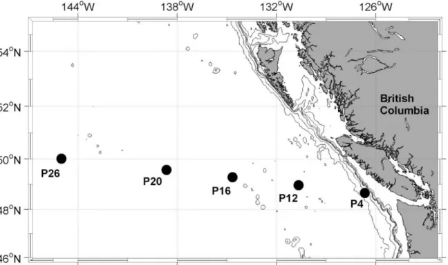
Fig 1. Map of sampling stations along the Line-P transect in the NE subarctic Pacific.The iron addition experiment was initiated at station P20, located in iron-limited high nutrient low chlorophyll (HNLC) waters. Sampling depths at other stations along the transect were: 30 m at P4; 5 m, 25 m and 40 m at P12, P16, P20 and P26.
For the 3 sets of experiments outlined above, samples for [chla] were filtered onto pre-com-busted 25 mm glass fiber filters (GF/F) using low vacuum pressure (<5 mm Hg) and analyzed following the method of Welschmeyer [71]. In the field, triplicate 100–300 mL samples were filtered and stored at -20°C until analysis within three weeks of collection. In the laboratory, triplicate culture samples (10 mL, 20 mL and 30 mL) were collected and analyzed immediately. Each sample was analyzed in duplicate.
Carbon assimilation
For both laboratory and field work, rates of carbon assimilation were measured as small vol-ume PvsE curves in a custom built photosynthetron [72]. In the field, 300 mL of water were spiked with 150μCi NaH14CO3(final concentration 0.5μCi mL-1, 52.5 mCi mL-1specific activ-ity) (Perkin-Elmer) immediately after sampling. Spiked samples were mixed gently but thor-oughly, aliquoted into 20 ml glass scintillation vials and placed into the photosynthetron. Temperature was kept within 1°C of in situ temperature by circulating water from a water-bath through an aluminum cooling jacket (the offset from in situ temperature was larger for station samples because samples from different depth had to be incubated simultaneously). Light was provided by high power light emitting diodes (LEDs) located under each scintillation vial. Each PvsE curve consisted of 11 light levels spanning intensities from 3 to 600μmol quanta m-2s-1. Actual light intensities were measured before and after each experiment using a 4πquantum sensor (QSL-2100, Biospherical Instruments) immersed in water inside a scintillation vial. Incubations lasted for 3–4 hours and were ended by gentle filtration onto 25 mm GF/F filters. Filters were pre-combusted to reduce nominal pore size to approx. 0.4μm. For each curve, three time-zero samples were taken by filtering 20 mL immediately after spiking. The total14C activity added was determined from three 1 mL aliquots of the spiked sample added to 1 mL 1 M NaOH. All work was done under low light and filters were stored in scintillation vials at -20°C until processing within 1 month of the experiment. During laboratory processing, 500μL of 3 M HCl was added to each filter and vials were left to degas for>24 hours to elimi-nate any inorganic14C remaining in the samples. Ten mL of scintillation cocktail (Scintisafe
plus, Fisher) were added to each vial, and vials were then vortexed and left to stand in the dark for>12 hours before analysis on a liquid scintillation counter (Beckman). Disintegrations per minute (DPM) were derived from scintillation counts using a quench curve prepared from commercial14C standards (Perkin-Elmer). DPM were converted to units of carbon biomass
following Knap et al. [73].
The14C protocol used for laboratory cultures was the same as outlined above with the
fol-lowing exceptions. We spiked 80 mL of exponentially growing culture with 40μCi NaH14CO3
and 3 mL aliquots were incubated in the photosynthetron for 30 minutes. Duplicate curves were measured for each sample. The incubation was terminated by adding 1 mL of 1 M HCl to each vial and samples were dried completely, omitting the filtration step. After drying, salts were re-suspended in 1 mL MilliQ water. For both laboratory and field measurements
14C-PvsE curves were fit following Webb et al. [74], as described below.
Chl
a
fluorescence parameters and ETR
RCII(ST) flash protocols consisted of 100 flashlets with 1.0μs length and 2.5μs interval
(46200μmol quanta m-2s-1peak power intensity, resulting in a ST flash length of 250μs, pro-viding ~5–10 quanta per RCII). The excitation power was selected at the beginning of the cruise to saturate the observed fluorescence transients within the first half of the ST excitation protocol. Our experience indicates that this approach offers the best signal-to-noise ratio in the recovered parameters, while accommodating significant variations in the photosynthetic prop-erties of the local phytoplankton populations along the cruise track, without re-adjusting of the excitation protocol. Excitation power was provided by an array of eight LEDs at four wave-lengths centered on 445 nm, 470 nm, 505 nm, and 530 nm (equal intensity at each wavelength; seeS1 Figfor more information on the spectral distribution). We measured steady state light curves (SSLC), where each sample was exposed to 10 actinic‘background’irradiances ranging from 0 to 1000μmol quanta m-2s-1, also provided at four wavelengths (S1 Fig). The relatively long duration of the SSLCs in this study could create some potential for the settling of cells which could influence the ChlF yield. However, our sampling region is known to be dominated by small cells [66], which should have a slow settling rate. Equally, the laboratory isolates used during this study stay in suspension for many hours, and it is thus unlikely that rapid settling of cells drastically altered our results.
All ChlF yields and parameters described below were derived by an iterative non-linear fit-ting procedure, applying the four parameter biophysical model of Kolber et al. [21] to a mean of 20 consecutive ST flashes using custom software (Z. Kolber). This software accounts for a formation of fluorescence quenching, most likely due to formation of a P680 triplet, which reduces the maximum fluorescence yield attainable during the ST flash by 3–6%. Throughout the SSLC, ST flashes were applied continuously (at 1 s interval), while the length of each light step was optimized to allow all derived parameters to reach steady state (ca. 5 min). ChlF yields and parameters corresponding to each light level were obtained from the mean of the last three acquisitions at each light level. In this way, we derived the fluorescence yields Foand Fm(in
dark regulated state) as well as F0and F
m0(in the light regulated state for each light level of the
SSLC). Fo0was calculated as Fo0= Fo/(Fv/Fm+ Fo/Fm0) [75]. Even though this derivation has
become widely accepted in the literature, we caution here that it might not hold for values derived under high background irradiance (see [76]) and varying stress levels experienced by natural phytoplankton assemblages.
Five fluorescence signals, Fo, Fm, F0, Fm0and Fo0were used to calculate ChlF parameters,
fol-lowing Roháček [77]. In the dark-regulated state, we derived the commonly used Fv/Fmratio as
Fv/Fm= (Fm-Fo)/Fm[78]. For each light level of the SSLC protocol we have calculated the
fol-lowing ChlF parameters: (1) The photochemical quenching of variable fluorescence, Fq0/Fv0=
(Fm0-F0)/(Fm0-Fo0), which quantifies the fraction of functional RCII in the open state (i.e.
pri-mary quinone acceptor QAin the oxidized state) [79]. (2) The maximum quantum yield of
PSII photochemistry, Fv0/Fm0= (Fm0-Fo0)/Fm0, which can be used to quantify the extent to
which photochemistry in PSII is limited by competition with thermal decay of excitation energy [75]. (3) The overall quantum efficiency of photochemical energy conversion in PSII at a given light intensity (note that numerous definitions for this parameter exist in the literature), Fq0/Fm0= (Fm0-F0)/Fm0=ФPSII0(the product of Fq0/Fv0and Fv0/Fm0[19]). Furthermore, the
func-tional absorption cross section of PSII,σPSII(Å2RCII-1), was derived from the rate of closure of
RCII in the dark-regulated and at each light-regulated state [20,21]. The connectivity parame-ter,ρ, was also calculated, but not used in our analysis.
Rates of charge separation (i.e. ETRRCII) in functional RCII (mol e-mol RCII-1s-1) were
ETRRCII ¼E sPSII0 Fq0
Fv0
6:022 10 3
ð1Þ
where E (μmol quanta m-2s-1) is the actinic irradiance at each light level,σPSII0(Å2RCII-1) is
the functional absorption cross section at E and Fq0/Fv0is the photochemical capacity of PSII at
E. The number 6.022 x 10⁻³converts
μmol quanta to quanta and Å2to m2. Because of potential systematic errors in the calculation of Fo0, we also calculated ETRRCIIas
ETRRCII¼E sPSII
ðFq0=Fm0Þ ðFv=FmÞ
6:022 10 3
ð2Þ
which does not require the knowledge of Fo0. Both calculations are equivalent, assuming that
non-photochemical quenching processes affecting ChlF can be adequately accounted for in either the absorption term (Eq 1) and the efficiency term (Eq 2). WhileEq 2does not require Fo0(which was not measured directly) orσPSII0(which is difficult to derive at high irradiances),
it does rely on parameters measured in the fully dark-regulated state, which can be difficult to achieve infield assemblages. For all ETRRCIIcalculated during our iron addition experiment
(n = 345) the difference between values calculated in both ways ranged from 0.5 to 21% with a mean coefficient of variance of 5.5%. Both approaches thus provided similar results in the anal-ysis of our data, and the differences observed were not systematically related to the treatment (highvslow Fe).
Non-photochemical quenching (NPQ) at each light level was estimated as the normalized Stern-Volmer quenching coefficient, defined as NPQNSV= (Fm0/Fv0)-1 = Fo0/Fv0[65].
Quantifi-cation of NPQ using NPQNSVinstead of the more commonly used Stern-Volmer coefficient of
quenching, defined as NPQSV= (Fm-Fm0)/Fm[80], is appropriate for our data-set, as it resolves
differences between NPQ present in the dark-regulated state.
P
vs
E curves
Measurements of CO2-assimilation and ETRRCIIwere plotted against irradiance, and the
expo-nential model of Webb et al. [74] was fit to the data using a non-linear least squares regression procedure in Matlab. For the CO2-assimilation data, an intercept parameter was added to force
the regression through the origin and provide a good fit in the linear part of the PvsE curve [28,81]. For both rates of productivity, we derived the light saturated maximum rate Pmaxand
the light utilization efficiencyα. When photoinhibition was observed at high irradiances, the data-points were excluded from the fitting procedure.
Derivation of conversion factor
Because we derived ETRRCIIin units of mol e-mol RCII-1s-1and CO2-assimilation in units of
mol C mol chla-1s-1, the conversion factor between the two rates accounts for changes in chla
functionally associated with each RCII (1/nPSII, mol chlamol RCII-1) and the number of
charge separations in RCII needed per CO2-assimilated into organic carbon products (Fe:C,
mol e-mol C-1).
ETRRCIIðmol e mol RCII 1s 1
Þ
CO2assimilationðmol C mol chla 1s 1Þ¼Fe:C
mol e mol C
1
nPSII
mol chla mol RCII
ð3Þ
In this approach, we attribute the observed decoupling between ETRRCIIand CO2
one conversion factor obscures the mechanistic underlying of the observed decoupling. Never-theless, as we will show, our approach has the potential to provide FRRF-derived estimates of phytoplankton primary productivity in carbon units without the need for many of the auxiliary measurements and inherent assumptions used in previous studies.
The value of 1/nPSIIis known to change significantly as a function of taxonomy [22], light
[22,82], macro-nutrients [83], and iron availability [84–90]. Therefore we could not assume a constant value for 1/nPSII, as has been done in most previous studies [24]. Although 1/nPSIIcan
be directly measured from oxygen flash yield experiments (e.g. [91–93]), the approach is labour-intensive and not practical for routine field sampling. A new approach to derive [RCII] directly from FRRF measurements has been developed [94,95], but not implemented in our study because the inherent assumption that the ratio of rate constants of photochemistry and fluorescence (kp/kf)
is confined to a narrow range, does not hold under varying levels of iron limitation [62,87,94]. Having established a relationship between light intensity and rates of CO2-assimilation and
ETRRCIIfor each sample, we were able to model the light dependency of the conversion factor
Fe:C/nPSII. This approach allowed us to observe how the coupling of ETRRCIIand CO2
-assimi-lation is modulated by incident irradiance, and how, in turn, iron limitation influences the light-dependent response. Additionally, we usedαand Pmaxof each rate to derive the conver-sion factor under sub-saturating and saturating light conditions, respectively.
Results
Effect of iron addition on phytoplankton community composition,
photophysiology, ETR
RCIIand CO
2-assimilation in the NE subarctic
Pacific
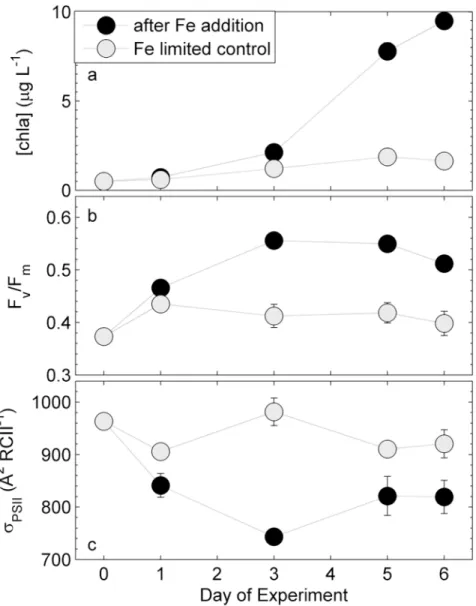
Phytoplankton assemblages at station P20 in the NE subarctic Pacific (Fig 1) responded strongly to iron addition in a ship-board incubation experiment (Fig 2). Six days after iron addition, [chla] increased by an order of magnitude, whereas the control (i.e. no iron addition) showed only a small increase in [chla]. This result confirms that the initial phytoplankton assemblage was iron-limited (Fig 2A), and that we were able to carry out the manipulation experiment without significant contamination of the control bottles. The slight increase in [chl a] in the control treatments is likely attributable to a decrease in grazing pressure and to changes in the light environment (i.e. lower and less fluctuating light). Iron addition also signif-icantly affected phytoplankton photophysiology, as demonstrated by rapid changes in the parametersσPSIIand Fv/Fmderived in the dark-regulated state (Fig 2B and 2C). Fv/Fminitially
increased in both treatments, but then remained low in the control while continuing to increase in the iron addition treatment (Fig 2B). While the functional absorption cross-section of PSII, σPSII(Å2RCII-1), remained high and relatively constant in the iron-limited control, it declined
rapidly after iron addition, and remained ~25% lower than that of the initial phytoplankton assemblage (Fig 2C). The observed changes in Fv/FmandσPSIImay have resulted from both,
photophysiological responses and from changes in species composition. CHEMTAX analysis of pigments sampled on day 6 of the experiment showed that the addition of iron changed the taxonomic composition of the phytoplankton assemblage (S2 Fig). Most prominently, the abundance of chlorophytes decreased from 7% to 1%, prymnesiophytes decreased from 55% to 22%, pelagophytes increased from 17% to 39%, and diatoms increased from 1% to 16% in iron amended bottles. A similar response has been observed in previous iron addition experiments conducted in this region [96].
We measured PvsE curves of short-term CO2-assimilation and ETRRCIIfive times during
and ETRRCII. Chlorophylla-normalized CO2-assimilation showed a small, though not
statisti-cally significant, increase after iron addition (Fig 3A–3E). The observed increase in the chla -normalized rate was small, because cellular chlacontent increased in parallel with CO2
-assimi-lation (under all nutrient limitations, cellular chlain phytoplankton is drastically reduced, a condition referred to as chlorosis, e.g. [97]). The strong effect of iron addition on CO2
-assimila-tion can be seen more clearly when rates are normalized to volume. Indeed, volume-normal-ized CO2-assimilation rates increased more than 8-fold after iron addition in this experiment
(S3 Fig). In contrast to rates of CO2-assimilation, ETRRCIIdecreased significantly after iron
addition, when compared to the iron-limited control treatment (Fig 3F–3J).
The response of CO2-assimilation and ETRRCIIto iron addition is further visualized inFig 4,
which shows changes in light-limited slopes (α) and light saturated rates (Pmax), as well as the derived conversion factorFe:C/nPSIIforαand Pmax, throughout the experiment. Values for
αand Pmaxwere derived from the14C-based and FRRF-based PvsE curves shown inFig 3. No
Fig 2. Response of chlabiomass and photophysiology during the on-board iron addition experiment.
Shown are changes in (a) [chla], (b) Fv/Fm, and (c)σPSII. Error bars represent standard errors from three biological replicates and are sometimes smaller than the symbol.
statistically significant change in values ofαcould be determined for either chla-normalized CO2-assimilation, ETRRCIIorFe:C/nPSII(p-value>0.05). Similarly, the Pmaxfor chla
-normal-ized CO2-assimilation remained relatively constant in the control, and did not show a
statisti-cally significant increase after iron addition (p-value>0.05) (Fig 4D). In contrast, there was a significant (p-value<0.05) decrease in Pmaxfor ETRRCIIfollowing iron-addition, as compared
to the control treatments, which exhibited a small increase in this variable over the course of
Fig 3. Response of rates of CO2-assimilation (mol C mol chla-1s-1) and ETRRCII(mol e-mol RCII-1s-1) during the iron addition experiment.Both rates were measured as a function of irradiance, and PvsE curves were fit with the exponential model of Webb et al. [74]. Shown are mean values from three biological replicates where error bars represent standard error of mean and are sometimes smaller than symbols.
the experiment (Fig 4E). The observed changes in the Pmaxfor CO2-assimilation and ETRRCII,
resulted in a decrease inFe:C/nPSIIin the iron addition treatment compared to the relatively
constant value observed in the iron-limited control (Fig 4F). This difference was statistically significant for the last 2 days of the experiment (p-value<0.05). When compared to the initial value on day 0 of the incubation, the conversion factorFe:C/nPSIIfor Pmaxdecreased by 66%
after iron addition, and by 16% in the iron-limited control (Fig 4F). These results indicate that
Fig 4. Time-course ofα(a-c) and Pmax(d-f) of CO2-assimilation, ETRRCIIand the derived conversion factorΦe:C/nPSIIduring the iron addition
experiment.The conversion factorΦe:C/nPSIIunder light limiting conditions is derived from values in (a) and (b). Similarly, the conversion factorΦe:C/nPSIIat
light saturation is derived from the values in (d) and (e). The error in (a), (b), (c), and (d) is the 95% confidence interval of the parameter derived from the fit to data from three biological replicates, and the error in (c) and (f) is the propagated error from (a)/(b) and (d)/(e), respectively.
the iron-dependent changes inFe:C/nPSIIare most readily apparent under high irradiance
con-ditions where photosynthesis is light-saturated.
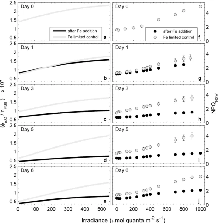
To better explain the iron-dependent decrease in ETRRCIIandFe:C/nPSIIobserved in our
data, we examined changes in additional FRRF-derived ChlF parameters, measured on day 3 after iron addition. We choose day 3 for the in-depth analysis of our data, but trends observed on this day were representative of those observed throughout the experiment. The parameter Fq0/Fv0represents the efficiency of charge separation in functional RCII (Fig 5A). It is an
esti-mate of the fraction of open RCII (i.e. QAoxidized) at any given light level, and therefore
always equals one at zero irradiance. On day 3 after iron addition, we observed higher Fq0/Fv0
for the iron-limited control at all irradiance levels (Fig 5A), indicating a greater fraction of open reaction centers. The parameter Fv0/Fm0, the efficiency of excitation energy capture by the
Fig 5. Light dependency of ChlF-derived parameters from FRRF measurements on day three after iron addition and in the iron-limited control treatment.The parameter Fq0/Fv0(a) represents the efficiency
of charge separation in functional RCII and is an estimate of the fraction of open RCII (i.e. QAoxidized) at any
given light level. The parameter Fv0/Fm0(b) represents the efficiency of excitation energy capture by the
fraction of open RCII and can be used to quantify the extent to which non-photochemical quenching in the PSII antenna competes with photochemistry for excitation energy. The parameter Fq0/Fm0(c) represents the
overall quantum efficiency of photochemical energy conversion in PSII (Φ0
PSII). See text for a full description
of these parameters and their interpretation. Error bars represent standard errors from three biological replicates and are often smaller than symbols.
RCII is limited by thermal energy dissipation in the antenna [75]. This parameter was signifi-cantly reduced in the iron-limited control relative to the iron addition treatment (Fig 5B), indi-cating that the efficiency of excitation energy transfer in the light-harvesting antenna was comprised. The overall efficiency of charge separation per quantum absorbed in PSII (Fq0/Fm0)
is the product of Fq0/Fv0and Fv0/Fm0[19,77]. On day 3, at all light levels, Fq0/Fm0was higher in
the iron addition treatment than in the iron-limited control (Fig 5C).
We used our PvsE measurements of CO2-assimilation and ETRRCIIto examine the
light-dependent response of the conversion factorFe:C/nPSII. Our results (Fig 6) show thatFe:C/nPSII
increased with increasing irradiance, regardless of iron treatment and day of the experiment (Fig 6A–6E). However, this light-dependent increase was much more pronounced in the iron-limited control treatment. It is important to note that the magnitude and light-dependency of Fe:C/nPSIIin the iron-limited control treatment changed over the course of the experiment
rela-tive to the initial sample (Fig 6A). This shift inFe:C/nPSIIin the absence of iron addition likely
reflects changes in light quality and quantity in the incubation bottles relative to the ambient water column.
Also shown inFig 6is the light and iron dependency of NPQNSV, estimated as Fo0/Fv0. This
parameter showed a light and iron-dependent response that was remarkably similar toFe:C/
nPSII, with values increasing with increasing light, regardless of treatment and day of the
experi-ment, and decreasing in response to iron addition (Fig 6F–6J). The NPQNSVvalues measured
in our initial sample (Fig 6F) were higher than those measured in either control or iron addi-tion treatments during the following days. We attribute this effect to a more stable light envi-ronment in the incubation bottles, relative to in situ irradiance levels.
Given the similar light and iron-dependent responses ofFe:C/nPSIIand NPQNSV, we sought
to examine the relationship between these two variables. In order to do so, however, it was nec-essary to derive NPQNSVandFe:C/nPSIIvalues at a standard set of light levels, matching those
of the FRRF derived ETRRCII-PvsE curves. For each sample, ETRRCII-PvsE curves consisted of
14 light levels spanning from 0 to 1000 µmol quanta m-2s-1. These light levels did not exactly match those used for the CO2-assimilation experiments. We thus used the PvsE curve fits of
our14C data to derive the CO2-assimilation values at light levels matching those of the ETR R-CII-PvsE curves. In this way, we were able to compile a dataset of 298 paired values for NPQNSV
andFe:C/nPSII, derived from 27 sets of ETRRCIIand14C PvsE curves during the iron addition
experiment. Plotting theseFe:C/nPSIIvalues against the corresponding NPQNSVreveals a strong
and statistically significant correlation (R2= 0.70, p-value<0.0001, for quadratic fit) (Fig 7).
Effects of iron limitation on photophysiology and rates of ETR
RCIIand
CO
2-assimilation in mono-specific phytoplankton cultures
Using methods analogous to those applied to mixed phytoplankton assemblages in the NE subarctic Pacific; we measured PvsE curves of CO2-assimilation and ETRRCIIin mono-specific
laboratory cultures of two open ocean phytoplankton species. The results, summarized in
Table 1, show similar trends as observed in our field data. Steady-state growth rates (μ, d-1) in the low iron cultures were 68% and 49% of iron-replete growth rates inT.oceanicaand C.polylepis, respectively (Table 1). For both species, Fv/Fmin iron-limited cultures was reduced
(by 32% and 20% inT.oceanicaandC.polylepis, respectively). In iron-limitedT.oceanica, σPSIIincreased by 15%, while it increased by 5% inC.polylepis. The iron dependent changes
inμ, Fv/FmandσPSIIwas statistically significant in both species (one tailed p-value<0.0001
and<0.01 forT.oceanicaandC.polylepis, respectively). Chlorophylla-normalized CO2
we observed a 90% increase in ETRRCIIat PmaxinT.oceanicaunder iron-limited growth
condi-tions.C.polylepisalso exhibited an increase in ETRRCIIat Pmaxunder iron-limited conditions,
but this increase was not statistically significant (p-value>0.05). Regardless of species-specific differences, both species showed the same trend of increasedFe:C/nPSIIand NPQNSVunder
iron limitation (Table 1), which is consistent with our field observations. Furthermore, the
Fig 6. Changes in the light dependency of the conversion factorΦe:C/nPSII(a-e) and NPQNSV(f-j) over the course of the iron addition experiment. Units of inΦe:C/nPSIIare (mol e- mol C) / (mol chlamol RCII-1). The curves were derived by dividing corresponding values of ETRRCIIand CO2-assimilation from the PvsE curves presented inFig 3. NPQ was estimated as the normalized Stern-Volmer quenching coefficient NPQNSV= Fo0/Fv0and is unitless [65]. Error bars are the standard error from three biological replicates and often smaller than symbols.
Fig 7. Relationship between the conversion factorΦe:C/nPSIIand NPQNSVvalues during the iron addition experiment.Values ofΦe:C/nPSIIwere
derived from PvsE curves of CO2-assimilation and ETRRCIIat irradiances corresponding to each ETRRCII-PvsE curve light level. Units ofΦe:C/nPSIIare (mol e-mol C-1) / (mol chlamol RCII-1). NPQ
NSVvalues were derived as Fo0/Fv0for each light level of the SSLC. Data points represent means and standard
errors for parameters derived from three biological replicates. A quadratic fit through all data points (Φe:C/nPSII= -733.21 NPQ2+8792.4 NPQ–1477.1) is
statistically significant (R2= 0.70, p-value<0.0001).
doi:10.1371/journal.pone.0133235.g007
Table 1. Effect of iron limitation on photophysiology in two mono-specific phytoplankton cultures grown in the laboratory.
T.oceanica C.polylepis
[Fe] 42nM 0.13nM 42nM 1.28nM
μ(d-1) 1.27±0.14 (n = 6)*** 0.41±0.09 (n = 5) 0.53±0.12 (n = 5)** 0.27±0.05 (n = 4)
Fv/Fm 0.63±0.01*** 0.43±0.01 0.51±0.02** 0.41±0.03
σPSII 643±3 742±16 591±7 621±3
PmaxCO2-assimilation 0.030±0.004 0.035±0.005 0.032±0.009 0.028±0.009
PmaxETRPSII 174±9* 330±21 370±26* 506±65
PmaxФe:C/ nPSII 5874±648* 9225±1502 11691±3730 18145±6091
NPQNSV 0.37–0.47*** 0.58–0.75 0.5–0.59*** 0.72–0.79
*p-value<0.05 **p-value<0.01 ***p-value<0.0001
species-specific differences observed in our laboratory experiments are consistent with changes in phytoplankton assemblage composition observed in our iron addition experiment, where the abundance of diatoms (lowerFe:C/nPSII) was increased in the iron addition treatment and
the abundance of prymnesiophytes (higherFe:C/nPSII) was decreased (S2 Fig).
Thalassiosira oceanicaandChrysochromulina polylepiswere grown in steady state iron-replete and iron-limited conditions. The mean growth rateμ, derived from successive mea-surements in semi-continuous batch cultures, is given in d-1. The error is the SD of 3 biological
replicates, and number of consecutive batch transfers (ca. 4 cell divisions per transfer) used to calculate growth rates are given in brackets. Fv/FmandσPSIIare values from cultures in the
dark regulated state (10 min of 5μmol quanta m-2s-1at 730 nm), measured on the day of CO2-assimilation experiments. The error is SD of 3 biological replicates. Changes in these
parameters are statistically significant forT.oceanica(p-value<0.0001) andC.polylepis (p-value<0.01). Pmaxfor CO2-assimilation (mol C mol chla-1s-1) and ETRRCII(mol e- mol
RCII-1s-1) were derived from PvsE curves as described in the methods section. The error is the 95% confidence interval of the Pmaxderived from the fit to data from 6 whole curve
measure-ments (duplicate curves each from 3 biological replicates). The conversion factorFe:C/nPSIIfor
Pmaxwas derived as the quotient of Pmaxfor ETRRCIIand Pmaxfor CO2-assimilation. The error
is the propagated error from numerator and denominator. NPQNSVwas estimated as Fo0/Fv0
from the last ST acquisition during each light level of the PvsE curves. The values shown are from the first and last step of the PvsE curves (4 and 800μmol quanta m-2s-1). Each NPQNSV
value is the mean of 2 values measured on 3 biological replicates. Changes in response to iron limitation are statistically significant for both species (p-value<0.0001).
Discussion
Our results provide new insight into the effects of iron and light availability on the coupling between CO2-assimilation and photosynthetic electron transport in natural phytoplankton
assemblages and mono-specific laboratory cultures. We show that both of these environmental variables significantly influenceFe:C/nPSII, which has important implications for the use of
FRRF measurements to infer rates of CO2-assimilation in oceanic waters. Below, we first
dis-cuss the observed increase inFe:C/nPSIIunder excess light and low iron conditions in the
con-text of previously reported values. We then discuss the effects of iron and light on
phytoplankton photophysiology, and suggest a simple conceptual explanation for the observed increase in ETRRCIIunder iron limitation. We hypothesize, that iron and light-dependent
changes inFe:C/nPSIIare driven by the need to dissipate excess excitation energy, caused by
either excess light, or the effects of iron limitation on the ETC. In this context, we discuss the correlation betweenFe:C/nPSIIand NPQNSV, and examine the potential significance of this
finding in the context of marine primary productivity studies.
Magnitude of the observed conversion factor
The conversion factorFe:C/nPSII, derived from our measurements of ETRRCIIand CO2
-assimi-lation, varied significantly in response to light and iron availability. In our field experiment, the addition of iron caused the value ofFe:C/nPSIIat light saturation (Pmax) to decrease by 66%
within 6 days (Fig 4F). Furthermore, short-term changes in light availability had a major effect on the value ofFe:C/nPSII, and this effect was enhanced under iron limitation. A recent
meta-analysis of variability in experimentally determinedFe:Cfrom 14 field studies found values
has been done in most previous studies [24],Fe:Cvalues on day 3 of the iron-addition
experi-ment range from 13 to 39 mol e-mol C-1. Using a constant value of 1/n
PSIIfor both treatments
is unlikely to be realistic. Even though iron-limited phytoplankton possess less chlaper cell, 1/nPSII, the ratio of chlato RCII, has frequently been observed to increase under low iron
con-ditions [84,85,88,90,98]. If we thus assume 700 mol chlamol RCII-1for the iron-limited con-trol treatment and 500 mol chlamol RCII-1for the iron addition treatment [85],
Fe:Cranges
from 13 to 28 mol e-mol C-1. TheseFe:Cvalues represent the range observed across different
irradiance levels in our PvsE experiments. At the time of sampling, cells in the on board incuba-tor were exposed to ~ 40 µmol quanta m-2s-1. Assuming 700 and 500 mol chlamol RCII-1for the iron-limited and iron-replete treatments, respectively, we deriveFe:Cvalues of ~18 and
~15 mol e-mol C-1. Values ofFe:Cestimated from our data are thus within the range reported
in previous field studies [24], with no estimate falling below the theoretical minimum of 4 mol e-mol C-1.
Ideally, measurements of ETRRCIIand CO2-assimilation should be performed
simulta-neously on the same sample, eliminating differences in incubation time and spectral quality of the light sources used. As discussed in detail in the supplementary material, the differences in spectral distribution of the light sources used for FRRF and14C measurements could have led to an underestimation of absolute values ofFe:C/nPSII(S1 Fig). However, these differences
can-not explain the large iron dependent changes we observed inFe:C/nPSII, since the absorption
spectra of iron-limited and iron-enriched treatments did not differ drastically (S1 Fig). Further-more, differences in incubation times could have influence the absolute magnitude of the derived conversion factor. Incubation times used for the PvsE curves were ca. 5 min for FRRF measurements (applied incrementally to the same sample), vs. 3–4 hours in the field and 30 min in the laboratory for14C-assimilation experiments (light levels applied simultaneously to
different samples). As has been shown by Halsey et al. [16,17,99] and Pei and Laws [18], the use of fixed incubation times for cells growing at different growth rates could lead to an overes-timation of our conversion factorFe:C/nPSIIin the iron-limited relative to iron-replete samples.
Additionally, the longer incubation time in CO2-assimilation experiments might have
exacer-bated cumulative processes such as photodamage under excess irradiance. To address this issue, we did not utilize the part of the PvsE curves showing photo-inhibition. However, we cannot rule out any differential cumulative effects of photoinhibition on ETRRCIIand14
C-assimilation at Pmax. This could potentially decrease CO2-assimlation at Pmaxrelative to ETR R-CIIat Pmaxand lead to overestimation of ourFe:C/nPSIIvalues at Pmax. Notwithstanding these
potential sources of uncertainty in the absolute value ofFe:C/nPSII, the good agreement between
our estimatedFe:C(assuming ~ 500–700 mol chlamol RCII-1) and those of previous studies
suggests that our observations are robust. More importantly, potential offsets in the absolute values ofFe:C/nPSIIdo not diminish the significance of the relative, iron and light-dependent
changes we observed in this parameter (discussed below).
Interacting effects of iron and light on the conversion factor
Φ
e:C/n
PSII Our data show strong and interacting effects of iron and light availability on the conversion factorFe:C/nPSIIin phytoplankton field assemblages and mono-specific laboratory cultures(Fig 4C, 4fand6,Table 1). It has been shown that the magnitude of both 1/nPSIIandFe:Cvary
significantly between phytoplankton taxa (e.g. [22,93]). Changes inFe:C/nPSIIin field
any particular set of environmental conditions (i.e. iron limitation). In the following, we discuss the observed changes inFe:C/nPSIIfrom a predominantly photophysiological point of view,
since our laboratory results specifically demonstrate such physiological effects.
Numerous metabolic processes, acting between ETRRCIIand CO2-assimilation can act to
increaseFe:C, and therefore the conversion factorFe:C/nPSII(e.g. [61,59,100]). In addition to its
role in reducing CO2to organic carbon products, reductant (NADPH) formed at the end of the
ETC can also be used for nitrate and sulphate reduction [101], photorespiration [102], or respi-ration via the malate shunt [103]. These alternative pathways decouple ETRRCIIfrom CO2
-assimilation, increasing the value ofFe:C. Similarly, before the formation of NADPH,
pseudo-cyclic electron flow can reduce O2and create a water-water cycle of electron transport, also
increasingFe:C(e.g. [104]). Pseudo-cyclic electron transport pathways can divert electrons
from the ETC before (short water-water cycling, e.g. [105]) or after PSI (Mehler-reaction, e.g. [106]). Cyclic electron transport (CET) around PSII [107,108] and charge recombination in PSII [109,110], act more closely to the initial charge separation in RCII, and can also cause an increase inFe:C.
We suggest that the higherFe:C/nPSIIobserved in response to iron limitation and
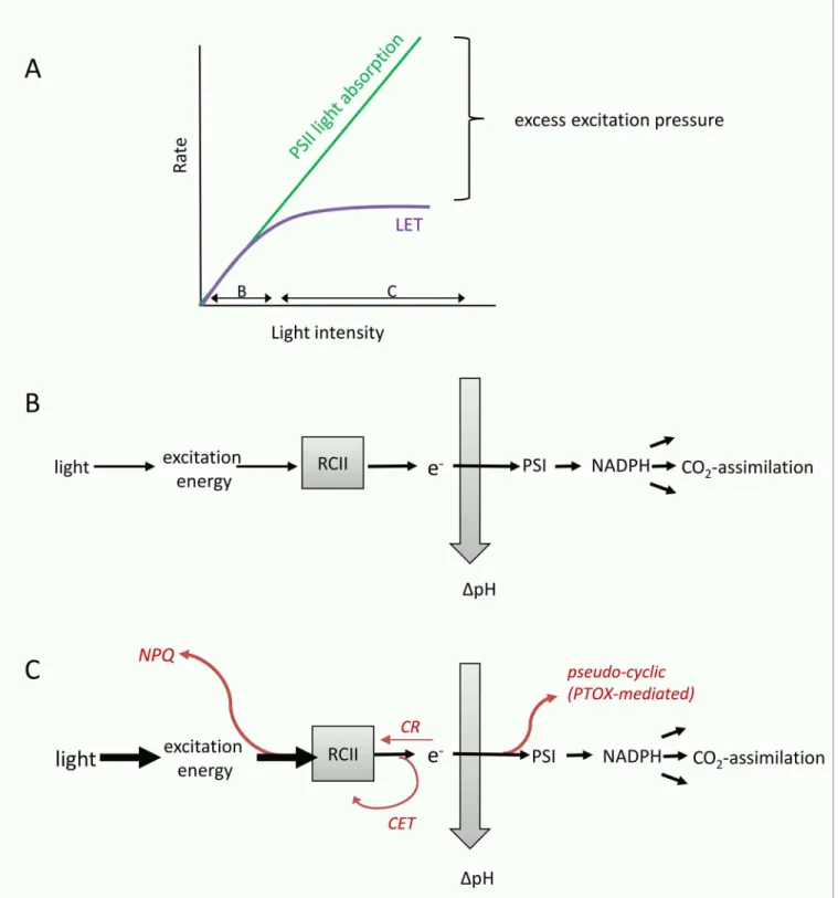
short-term increases in incident irradiance during the PvsE experiments (Fig 6A–6E) results predom-inantly from increases in the alternative electron flow pathways prior to reductant formation. These pathways, which are diagramed conceptually inFig 8, can act as‘safety valves’to keep the primary quinone acceptor QAoxidized when excitation pressure on the ETC is high,
thereby decreasing the potential of damage to RCII [111,54,55,61,56,104,112,60].
Iron limitation directly affects the photosynthetic ETC and thereby modulates the light-dependent changes in the conversion factorFe:C/nPSII(Fig 6A–6E). Importantly, iron
limita-tion has been shown to alter the stoichiometry of ETC components (i.e. expression of iron-rich PSI and cytochromeb6fcomplexes is down-regulated to a higher extent than PSII) (e.g.
[62,84,116–118]). Low levels of electron acceptors downstream of PSII ultimately restrict the flow of electrons away from PSII during light exposure. This exacerbates the need for short (i.e. acting before PSI) alternative electron flow pathways to dissipate excess excitation energy and prevent over-reduction of RCII (Fig 8). A number of recent studies have suggested that re-rout-ing electrons to a midstream plastoquinol oxidase (PTOX) to bypass the electron flow bottle-neck of PSI is a common strategy in open ocean phytoplankton [113,55,54,56–58,62]. Importantly, up-regulation of pseudo-cyclic electron flow under iron limitation not only pro-tects RCIIs from photodamage, but also helps to maintain a highΔpH across the thylakoid membrane, providing energy for cell maintenance and growth [62,119]. Cyclic electron flow around PSII [107,108,114,61,120] and increases in charge recombination at PSII [109,110,115] are two additional mechanisms that can act to prevent over-reduction and damage of RCII when excitation pressure is high and the electron flow bottleneck is prior to PSI. Unlike PTOX-mediated water-water cycling, these processes do not contribute to an increase inΔpH across the thylakoid membrane. They would, however, contribute to a high ETRRCIIand therefore
Fe:C/nPSII(Fig 8).
While ambient light intensity has a well-documented effect on values of 1/nPSII, these
changes act on timescales longer than those of short-term PvsE experiments, and are thus unlikely to have caused the light-dependent changes we observed inFe:C/nPSII(Fig 6). On
lon-ger time-scales, however, iron limitation causes a reduction of chlaper cell (chlorosis), and an increase in chlaper functional RCII (1/nPSII) [87,90]. This well documented response, which
Fig 8. Conceptual diagram visualizing the concept of excess excitation pressure and its dissipation before and after charge separation in RCII.(A) Absorption of light energy by pigments in the light harvesting antenna of PSII cannot be controlled biologically, and rises linearly with incident light intensity. However, rates of linear electron transport (LET) and CO2-assimilation saturate at a light intensity determined by the physiological state of the phytoplankton,
resulting in a typical PvsE curve. Under optimal growth conditions, it is the resupply of NADP-(predominantly from CO2-assimilation) which limits LET, while under short-term exposure to excess light and under iron limitation, the‘bottleneck’of LET will be located before PSI. Whenever exitonic influx exceeds the chemical outflux at the level of RCII, excess excitation pressure needs to be safely dissipated to prevent photodamage. (B) Under optimal growth conditions and sub-saturating light, all absorbed photons are used for charge separation in RCII, and the majority of electrons will be used for LET and CO2-assimilation,
In summary, we suggest that it is the effect of high excitation pressure, which causes a de-coupling of ETRRCIIand CO2-assimilation. This high excitation pressure may be a result of
short-term exposure to excess irradiance as well as the effect of iron limitation on the ETC. This purely photophysiological interpretation can be extended to observations made in mixed phytoplankton communities. Here, fluctuating light and low iron conditions will select for spe-cies with the best ability to control high excitation pressure by adjusting the flow of excitation energy into, and the flow of electrons out of PSII.
Iron limitation increases ETR
RCIITo our knowledge, this is the first study which shows that ETRRCIIincreases under iron
limita-tion. This observation may seem counter-intuitive, and it is important to emphasize that our results do not imply an overall increase in photosynthetic electron transport under low iron conditions. Rather, our observations point to an increase in the rate of charge separation at each individual RCII, independent of the reduced total cellular concentration of these RCII. We show that the overall efficiency of PSII photochemistry in the light-regulated state, Fq0/Fm0
(=ФPSII0), is reduced under iron limitation (Fig 5C), as expected. However, deconvolution of
this parameter into its constituents Fq0/Fv0(Fig 5A) and Fv0/Fm0(Fig 5B) shows that Fq0/Fv0,
rep-resenting the fraction of open RCII (QAoxidized) at each given light level, increased under
iron limitation. We hypothesize that this is likely achieved by increased alternative electron transport pathways acting to keep RCIIs open (QAoxidized) and bypassing the electron flow
bottleneck at PSI, when excitation pressure is high (Fig 8). In contrast to Fq0/Fv0, the parameter
Fv0/Fm0is much lower when iron is limiting (Fig 5B), indicating that the excitation energy
transfer in the antennae is compromised.
Based on our experimental observations, we suggest a simple mechanistic explanation for the observed increase in ETRRCIIunder iron limitation. Cellular iron demand can be
signifi-cantly reduced by economizing on iron-rich components of the photosynthetic apparatus and
‘funneling’more electrons down fewer RCIIs (i.e., increasing ETRRCII). In line with this
expla-nation is the observation that values ofσPSIIare high under iron limitation, and rapidly
decrease after iron addition (Fig 2) [121,122,84,85,123–126,87]. Strzepek et al. [127] suggested that increasedσPSIIcompensates for fewer iron-rich photosynthetic reaction centers in
South-ern Ocean phytoplankton species. Similarly, Ryan-Keogh et al. [128] noted that increasing the absorption cross section of RCs by the expression of isiLHCs allows cells to reduce the cellular iron requirement while maintaining the same light absorption capacity.
In conclusion, our results and interpretation support a scenario where photosynthetic elec-tron flow has been fine-tuned to maximize energy conversion as well as photo-protection under conditions where ETC component abundance and stoichiometry are compromised by the availability of iron.
Link to NPQ
NSVAbove, we discussed how mechanisms acting down-stream of the initial charge separation in RCII are likely to be enhanced under conditions of high excitation pressure, resulting in high comprises the functioning of the ETC and has been shown to create a‘bottle neck’for LET before PSI. Under these conditions, PTOX-mediated pseudo-cyclic electron flow (e.g. [54–58,62,105,113]), cyclic electron transport around PSII (e.g. [107,108,114]), and charge recombination in RCII (e.g.
[109,110,115]), have been suggested to safely dissipate excess excitation energy after RCII (but before PSI). Up-regulation of these alternative electron flow pathways could explain the high ETRRCII(andΦe:C/nPSII) observed in our iron-limited samples. Excess excitation energy can also be dissipated in the light
harvesting antenna, before charge separation in RCII. Collectively, a number of different molecular processes dissipating excess excitation energy in the PSII antenna can be quantified as NPQNSV.
before reaching RCII [104].Fig 8shows schematically the‘safety mechanisms’used for the dis-sipation of excess energy at both sides of RCII. Because processes dissipating excess excitation pressure in the antenna also quench ChlF yields measured by FRRF, they have collectively been called non-photochemical quenching (NPQ). NPQ, which is present in all oxygenic photosyn-thetic organisms, encompasses a wide variety of mechanisms acting to dissipate absorbed light energy as heat before it reaches RCII [129–134]. Following the approach of McKew et al.[80], we estimated NPQ from FRRF measurements as so-called normalized Stern-Volmer quench-ing (NPQNSV). We observed a strong correlation between the conversion factorFe:C/nPSIIand
the expression of NPQNSV(Fig 7). We note thatFe:C/nPSIIand NPQNSVare not entirely
inde-pendent parameters, and therefore the strong correlation observed inFig 7is in part a result of their co-dependence on the ChlF parameter Fv0(which we used in the derivation of both
NPQNSVandFe:C/nPSII).
At this point, the relationship betweenФe:C/nPSIIand NPQNSVshown inFig 7is empirical
rather than mechanistic. However, while there are a number of processes which will influence
Фe:C/nPSIIand NPQNSVdifferentially, there are many processes related to the amount of
excita-tion pressure experienced by the ETC that would influence both in a consistent manner. Numerous studies have shown thatФe:Cincreases if light is saturating, i.e. when excitation
pressure is high (e.g. [33,36,40]). Clearly, excess light would also increase the expression of NPQNSV. Indeed, very recent work has pointed to a mechanistic link between alternative
elec-tron sinks involving PTOX and the expression of NPQNSV[105].
A possible approach towards improved prediction of CO
2-assimilation
from FRRF data
While it remains to be seen how strong the correlation betweenFe:C/nPSIIand NPQNSV(Fig 7)
may be for other datasets, our results provide a potential basis for improved estimates of CO2
-assimilation from FRRF measurements alone. A number of factors make this approach more desirable than the use of static, regional conversion factors. First, the magnitude ofFe:C/nPSIIin
phytoplankton assemblages will be determined by a multitude of interacting environmental variables. The use of NPQNSVas an integrated physiological measure of environmental effects
on electron transport processes will therefore help to constrain the relationship betweenFe:C/
nPSIIand various environmental stressors. Secondly, as our data show, the magnitude ofFe:C/
nPSIIcan vary significantly within the same sample in response to short-term variations in
inci-dent light. Such small scale changes would be lost using a static (regional) conversion factor, but are captured with our NPQNSV-based approach, as every single ETRRCIIestimate is paired
with a corresponding NPQNSVestimate. Finally, a non-static conversion factor is crucial if the
goal is to monitor the effects of environmental change on marine primary productivity, since physiological responses to environmental change will likely affect the conversion factor itself before productivity changes are observed.
As a test of the validity of our approach, we used theFe:C/nPSIIvs. NPQNSVcorrelation
determined from our iron addition experiment (Fig 7) to predict the CO2-assimilation rates
from FRRF-derived ETRRCIIand NPQNSVmeasured along the Line-P transect. In this case, in
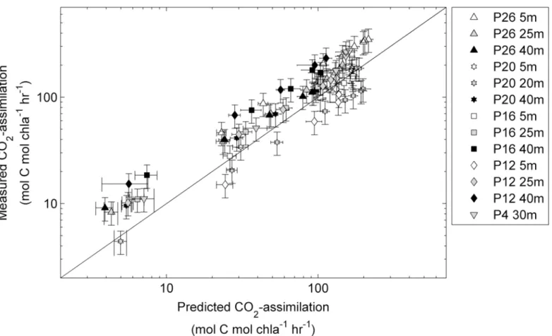
situ phytoplankton assemblages were collected from within and below the mixed layer, and rate measurements were conducted immediately after collection, without any experimental manipulation (seemethods). As shown inFig 9, we obtained a strong correlation between the predicted and measured CO2-assimilation rates (Spearman’sr= 0.90, n = 95 and two-tailed
correction of our data. The RMSE for the values predicted using our approach and measured values is 48.4 mol C mol chla-1hr-1. This error represents ~ 10% of the total range of values observed along the transect during this study, suggesting that rates of productivity can be pre-dicted with reasonable accuracy. In comparison with our approach, computation of CO2
-assimilationfrom FRRF data assuming a constant 1/nPSIIvalue of 500 mol chlamol RCII-1and
4 mol e-mol C-1, significantly under-predicts observed CO2-assimilation rates (RMSE = 837.3
mol C mol chla-1hr-1). Even if we use a constant conversion factor derived from the average of
theFe:C/nPSIImeasured during our iron addition experiment, the model error remains larger
compared to that derived using our variable, NPQNSV-based conversion factor (Fig 7). Our
data therefore show significant potential in the application of a variable, NPQNSV-derived
con-version factor and associated quantification of carbon uptake rates from FRRF data.
Conclusion
Deriving rates of phytoplankton CO2-assimilation from bio-optical approaches like FRRF has
the potential to provide estimates of primary production at unprecedented spatial and tempo-ral resolution. High resolution measurements, covering large oceanic regions, are essential for
Fig 9. Rates of CO2-assimilation (mol C mol chla-1hr-1) derived from FRRF measurements plotted against rates measured by14C-assimilation
experiments.Samples were taken at one to three depths at five stations along Line-P in the NE subarctic Pacific (seeFig 1). FRRF based PvsE curves were used to derive ETRRCIIand NPQNSVat 8 light levels for each sample, andΦe:C/nPSIIvalues were then derived from the relationship presented inFig 7.Φe:C/
nPSIIand ETRRCIIfor each light level were used to calculate CO2-assimilation rates. Error bars for predicted CO2-assimilation rates represent the propagated
error from the ChlF yields of the last three ST acquisitions of each light level during the FRRF PvsE curve used to derive NPQNSVand ETRRCII. Error bars for
measured CO2-assimilation rates represent the mean coefficient of variance derived from all duplicate measurements (n = 46). The correlation between all
predicted and measured data points (n = 95) was statistically significant (Spearman’sr= 0.90, two-tailed p-value<0.0001). All statistics are for non log-transformed data.
such measurements are indispensable for the development and validation of algorithms esti-mating global marine primary productivity from remote sensing.
Crucial to this approach is a sound characterization of the conversion factor between FRRF-derived ETRRCIIand primary productivity in carbon units. Our data demonstrate that the
con-version factor varies significantly in response to iron and light availability in phytoplankton field assemblages and mono-specific laboratory cultures. We interpret the observed variability in the conversion factorFe:C/nPSIIas a manifestation of the extreme photophysiological
flexi-bility which evolved in phytoplankton to maximize growth under dynamic light and nutrient regimes [135,136]. We hypothesize that, to a large extent, changes inFe:C/nPSIIrepresent a
suite of coordinated photophysiological adjustments acting to balance light absorption with CO2-assimilation under given environmental conditions. These will be manifested on the
phys-iological as well as on the taxonomic level. On the taxonomic level, a low nutrient and / or fluc-tuating light environment will select for species with the best ability to control high excitation pressure by adjusting the flow of excitation energy into, and the flow of electrons out of PSII (manifested in changes of NPQNSV, 1/nPSIIandFe:C). Future studies will be needed to evaluate
the relationship between NPQNSVandFe:C/nPSIIin a number of oceanic regions in order to
evaluate the potential for improved CO2-assimilation estimates from FRRF data.
Supporting Information
S1 Fig. Spectral distribution of light sources used for FRRF and Photosynthetron assays, and absorption spectra of phytoplankton assemblages on day 6 of the iron-addition experi-ment.(a) The FRRF instrument used during this study contains LEDs with peak output at four wavelengths (445 nm, 470 nm, 503 nm, 530 nm). In our FRRF instrument, excitation as well as actinic background irradiance is applied from the same LEDs. (b) Spectral distribution of the LEDs used in the photosynthetron used for14C-uptake experiments. (c) Spectral overlap of the two light sources. The overlap is good in the region of maximal light absorption by photosyn-thetic pigment (ca. 450 nm). However, in direct comparison with the photosynthetron, the FRRF instrument provides a higher proportion of photons in the region>480 nm. This could have led to an underestimation of ETRRCIIvalues relative to CO2-assimilation values measured
in the photosynthetron, resulting in an under-estimate ofFe:C/nPSII. In addition to knowledge
of spectral differences in the light sources used (a-c), spectral correction of our data would require light absorption spectra of the phytoplankton assemblages examined. Relative absorp-tion spectra of the phytoplankton communities on day 6 after iron-addiabsorp-tion (measured using the quantitative filter technique [65]) are shown in (d-f). Spectra from 3 biological replicates of the control (d) and two biological replicates of the iron addition treatment (e) were averaged, and these spectra are shown together in panel (f). The results show relatively small changes in the relative light absorption between the two treatments, and it is unlikely that these changes would have significantly influenced the large iron and light-dependent effects inFe:C/nPSII.
Because we did not measure absorption spectra for all sampling points of the iron addition experiment and stations along the transect, we were unable to spectrally correct our data. Fur-thermore, because we are not deriving absolute values forFe:C/nPSII, we did not apply a
con-stant correction factor (estimated from e.g. the data shown in a-f). (TIFF)
S2 Fig. Phytoplankton assemblage composition on day 6 of the iron addition experiment.
experiment. One to1.5 L of water were filtered on 25 mm GF/F and stored at -80°C until analy-sis. Pigments were extracted and quantified as described by Taylor et al. [137]. Pigment ratios were then used to estimate phytoplankton assemblage composition using CHEMTAX as described by Taylor et al. [137]. The initial pigment ratio matrix used for our data was taken from Lee et al. [138], table 5, which is specific to North Pacific phytoplankton isolates. (TIFF)
S3 Fig. Response of volume normalized rates of CO2-assimilation (mol C m-3hr-1) during
the iron addition experiment.The rates were measured as a function of irradiance, and PvsE curves were fit with the exponential model of Webb et al. [74]. Shown are mean values from three biological replicates where error bars represent standard error of mean and are some-times smaller than symbols. Results shown in this figure confirm a strong stimulatory effect of iron additions on primary productivity in the experimental bottles.
(TIFF)
Acknowledgments
We wish to thank Marie Robert of the Institute of Ocean Sciences (IOS) for providing berths on board the John P. Tully Line-P cruises and logistical and technical support at sea. We also thank Ania Posacka, Andreas Müller and Walter Green for assistance during field sampling and Yannick Huot, Dave Semeniuk, Anna Hippmann and Clara Hoppe for critical reading of the manuscript. We thank Zbigniew Kolber for support with the FRRF instruments and David Suggett and two anonymous reviewers for their comments on earlier versions of the
manuscript.
Author Contributions
Conceived and designed the experiments: NS CS PDT MTM. Performed the experiments: NS CS CD. Analyzed the data: NS PDT MTM. Wrote the paper: NS PDT MTM.
References
1. Field CB, Behrenfeld MJ, Randerson JT, Falkowski P. Primary production of the biosphere: integrat-ing terrestrial and oceanic components. Science. 1998; 281: 237–240. PMID:9657713
2. Falkowski PG, Barber RT, Smetacek V. Biogeochemical Controls and Feedbacks on Ocean Primary Production. Science. 1998; 281: 200–206. doi:10.1126/science.281.5374.200PMID:9660741
3. Beardall J, Raven JA. The potential effects of global climate change on microalgal photosynthesis, growth and ecology. Phycologia. 2004; 43: 26–40. doi:10.2216/i0031-8884-43-1-26.1
4. Hays GC, Richardson AJ, Robinson C. Climate change and marine plankton. Trends Ecol Evol. 2005;
20: 337–344. doi:10.1016/j.tree.2005.03.004PMID:16701390
5. Chavez FP, Messié M, Pennington JT. Marine Primary Production in Relation to Climate Variability and Change. Annu Rev Mar Sci. 2011; 3: 227–260. doi:10.1146/annurev.marine.010908.163917
6. Moore JK, Doney SC, Glover DM, Fung IY. Iron cycling and nutrient-limitation patterns in surface waters of the World Ocean. Deep Sea Res Part II Top Stud Oceanogr. 2001; 49: 463–507.
7. Boyd PW, Jickells T, Law CS, Blain S, Boyle EA, Buesseler KO, et al. Mesoscale iron enrichment experiments 1993–2005: Synthesis and future directions. science. 2007; 315: 612–617. PMID: 17272712
8. Behrenfeld M, Westberry T, Boss E, O’Malley R, Siegel D, Wiggert J, et al. Satellite-Detected Fluores-cence Reveals Global Physiology of Ocean Phytoplankton. Biogeosciences. 2009; 779–794.
9. Raven JA, Evans MCW, Korb RE. The role of trace metals in photosynthetic electron transport in
O2-evolving organisms. Photosynth Res. 1999; 60: 111–150. doi:10.1023/A:1006282714942