Active Site Loop Conformation Regulates
Promiscuous Activity in a Lactonase from
Geobacillus kaustophilus
HTA426
Yu Zhang1,2,3‡, Jiao An1,2‡, Guang-Yu Yang1‡, Aixi Bai2, Baisong Zheng2, Zhiyong Lou4, Geng Wu1, Wei Ye1, Hai-Feng Chen1, Yan Feng1,2*, Giuseppe Manco5*
1State Key Laboratory of Microbial Metabolism, School of Life Sciences and Biotechnology, Shanghai Jiao Tong University, Shanghai, People’s Republic of China,2Key Laboratory for Molecular Enzymology and Engineering of Ministry of Education, Jilin University, Changchun, People’s Republic of China,3 Environmental Science Research and Design Institute of Zhejiang Province, Hangzhou, People’s Republic of China,4Laboratory of Structural Biology, School of Medicine, Tsinghua University, Beijing, People’s Republic of China,5Institute of Protein Biochemistry, National Research Council, Naples, Italy
‡These authors contributed equally to this work.
*[email protected](YF); [email protected](GM)
Abstract
Enzyme promiscuity is a prerequisite for fast divergent evolution of biocatalysts. A phospho-triesterase-like lactonase (PLL) fromGeobacillus kaustophilusHTA426 (GkaP) exhibits main lactonase and promiscuous phosphotriesterase activities. To understand its catalytic and evolutionary mechanisms, we investigated a“hot spot”in the active site by saturation mutagenesis as well as X-ray crystallographic analyses. We found that position 99 in the ac-tive site was involved in substrate discrimination. One mutant, Y99L, exhibited 11-fold im-provement over wild-type in reactivity (kcat/Km) toward the phosphotriesterase substrate
ethyl-paraoxon, but showed 15-fold decrease toward the lactonase substrateδ -decanolac-tone, resulting in a 157-fold inversion of the substrate specificity. Structural analysis of Y99L revealed that the mutation causes a*6.6Åoutward shift of adjacent loop 7, which may cause increased flexibility of the active site and facilitate accommodation and/or catalysis of organophosphate substrate. This study provides for the PLL family an example of how the evolutionary route from promiscuity to specificity can derive from very few mutations, which promotes alteration in the conformational adjustment of the active site loops, in turn draws the capacity of substrate binding and activity.
Introduction
Enzyme promiscuity can function as a starting point in divergent evolution for generating a specific enzyme in the presence of selective stress. A better understanding of catalytic promis-cuity can improve our knowledge of protein evolution and ancestry as well as providing new tools for protein engineering and biotechnological applications [1–3]. One of the most impor-tant models for studying enzyme promiscuity is the enzyme that degrades synthetic
OPEN ACCESS
Citation:Zhang Y, An J, Yang G-Y, Bai A, Zheng B,
Lou Z, et al. (2015) Active Site Loop Conformation Regulates Promiscuous Activity in a Lactonase from
Geobacillus kaustophilusHTA426. PLoS ONE 10(2): e0115130. doi:10.1371/journal.pone.0115130
Academic Editor:Israel Silman, Weizmann Institute
of Science, ISRAEL
Received:August 13, 2014
Accepted:November 19, 2014
Published:February 23, 2015
Copyright:© 2015 Zhang et al. This is an open
access article distributed under the terms of the
Creative Commons Attribution License, which permits unrestricted use, distribution, and reproduction in any medium, provided the original author and source are credited.
Data Availability Statement:All PDB files are
available from the Research Collaboration for Structural Bioinformatics (RCSB) Protein Data Bank database (accession numbers:3TN3 and 3TN5).
Funding:This study was supported by National
organophosphate (OP) compounds, including most agricultural pesticides and chemical war-fare agents, which first appeared on this planet during the 20th century [4]. Currently, enzy-matic detoxification of OP compounds has become an important subject worldwide, because continuous and excessive use of OP compounds has led to the contamination of many terrestri-al and aquatic ecosystems [5]. Indeed, most OP degrading enzymes are known promiscuous enzymes [6–9]. One typical example is phosphotriesterase (PTE, EC 3.1.8.1) from the soil bac-teriaBrevundimonas(formerlyPseudomonas)diminuta(bdPTE), which has high catalytic effi-ciency for hydrolyzing a variety of neurotoxic OP compounds [10,11]. The same protein and a closely related protein with 90% sequence identify were identified inFlavobacterium sp. (OPH) [12] andAgrobacterium radiobacter(OpdA) [13], respectively. In addition to a wide substrate spectrum of OP substrates,bdPTE was recently reported to possess promiscuous lactonase and esterase activities [6]. Furthermore, a number of PTE remote homologs have been character-ized from several microorganisms and shown to proficiently hydrolyze various lactones with weak PTE activity [14–19]. These new enzymes that emerged were designated as PTE-like lac-tonases (PLL) [14] and were ascribed to a new family in the amidohydrolase superfamily [14,
18]. It has been hypothesized that PTEs evolved from an unknown member of the PLL family in response to changing environmental conditions [14]. Promiscuous activities in PLL mem-bers have been successfully enhanced by molecular evolution in the laboratory. The obtained mutants, which possess several mutational sites, showed a significant increase in PTE activities [20–23]. However, to date, the detailed evolutionary relationships between PLL enzymes and PTE enzymes has not been fully understood.
The three-dimensional structures of several PLL enzymes have been solved previously [16–
20]. Structural comparison of the PLLs withbdPTE [24] showed that the overall structures su-perpose very well. All of these enzymes belong to the amidohydrolase superfamily and possess an essentially identical binuclear metal center embedded within the (β/α)8-TIM barrel fold
[25]. Remarkably, PLLs differ frombdPTE in the length and the topology of surface loops 7 and 8. Both loops 7 and 8 in the amidohydrolase superfamily are most often found in contact with the bound substrates and playing important role in determination of substrate specificity [26]. A chimeric PLL enzyme fromDeinococcus radiodurans(DrOPH) containing these addi-tional loops frombdPTE did not exhibit enhanced PTE activity as expected, most likely due to the difficulty to replicate in a different environment the features provided by a specific loop [17,20]. Amino acid Trp131 in the active site of PTEs is conserved, while at the same position in PLLs a Tyr has been found. The aromatic side chain of Trp131 inbdPTE plays an important role in the interaction with the leaving aromatic ring of the OP substrate throughπ-stacking
in-teractions, which facilitates substrate binding to the active site [27]. However, the tyrosine resi-due in PLLs appears to stabilize the lactone ring during catalysis, and a mutant Y97W in the PLL enzyme fromSulfolobus solfataricus(SsoPox) showed a 3-fold increase in PTE activity [18]. Indeed, the crystal structure ofGeobacillus kaustophilusHTA426 (GkaP) shows that the binding site for the putative leaving group is lined with a series of very hydrophobic residues, including Tyr99, Tyr100, Trp271, and Pro275. These findings suggested that Tyr99 ofGkaP may be involved in substrate discrimination. However, so far no study has explored this con-served tyrosine residue in PLL family members.
In this study, a PLL enzyme from the thermophilic bacteriumGeobacillus kaustophilus HTA426 (GkaP) was characterized. To explore its promiscuous catalytic mechanism, a satura-tion mutagenesis library at posisatura-tion 99 was constructed and screened for enhanced PTE activi-ty. The structural factors controlling both the catalytic efficiency and substrate specificity were further identified by crystal structure analysis and molecular docking. These results showed that position 99 in the active site of PLLs could be evolved to develop greater PTE’s substrate specificity. Importantly, the structural analysis indicated that the dramatic movement of the
Competing Interests:The authors have declared
active loop 7 ofGkaP provides a catalytically competent active site architecture to account for the catalytic enhancements observed in the evolved mutant. This work shows that active site loop conformation regulates promiscuous activity and alters the capacity for both substrate binding and efficient catalysis.
Results and Discussion
Sequence and structure similarity
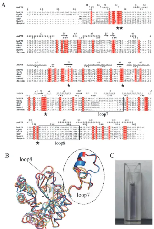
The structural-based sequence alignment of theGkaP gene with the reported PLLs and PTEs is shown inFig. 1A.GkaP is most closely related to the PLL from thermophilicGeobacillus stear-othermophilus(GsP) with an identity of 98%. It also showed sequence identity to the PLL from the extremely radioresistant bacteriumDeinococcus radiodurans(DrOPH) and PLL from hy-perthermophilic archaeonSulfolobus solfataricus(SsoPox) with an identity of 58% and 33%, re-spectively. In comparison to PTEs,GkaP was distantly related to mesophilicbdPTE and OpdA, with sequence identities of 24% and 25%, respectively. The sequence alignment indicated that GkaP contains entirely conserved residues (His23, His25, Lys145, His178, His206 and Asp266) within the binuclear metal center. The structure of theGkaP (PDB ID 3TN3) was also com-pared to the PLL and PTE members (Fig. 1B). All structures possess a (β/α)8-barrel fold and a
binuclear metal center, which is typical of the amidohydrolase superfamily. Both loop 7 and loop 8, which are hypothesized to bind substrates of the amidohydrolase superfamily [26], overhang the binuclear metal center. Superposition of the structure ofGkaP with those of PLL and PTE members produced the RMSD of all Cαatoms ranging from 0.272 to 1.084 Å, indicat-ing that these proteins are spatially homologous. Similar to other PLL enzymes, the length of loop 7 inGkaP is 12 residues shorter thanbdPTE and OpdA, and loop8 inGkaP has almost the same length as inbdPTE and OpdA, but its topology structure is more detached from the protein core than that ofbdPTE and OpdA. Previous studies have presumed that the elonga-tion of loop 7 inbdPTE forms a shortα-helix, which may provide an active“cap”that narrows
the active site mouth and may thereby increase PTE preference toward the substrate paraoxon [14].
TheGkaP gene was cloned into pET-28a (+) with an N-terminal His6-tag and subsequently
overexpressed inE. coliBL21 (DE3). The recombinant protein was expressed at a high yield (*50 mg/L) in the 2YT medium containing 1 mM CoCl2. The purified Co2+-containingGkaP
appeared purple colored (Fig. 1C), as also observed in another PLL enzyme,DrOPH, that has 58% homology toGkaP. It is noteworthy that this purple color was different than the brown color that was observed with Co2+-containing GsP, though both proteins only differed by four
residues in the N-terminal tail. Since the coloring was attributed to a charge transfer between an active site tyrosine residue andβ-metal complex [17,28], these proteins may have
substan-tial variations in the microenvironment of the active site due to the long distance interactions, despite a homology as high as 98%.
Catalytic promiscuity of
GkaP
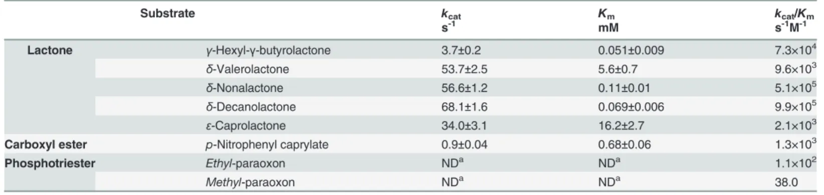
We next testedGkaP activities against lactonase, phosphotriesterase, and esterase substrates, respectively. The hydrolytic activities of the recombinantGkaP against an array of lactones were detected and the values of the kinetic parameters are summarized inTable 1. We found that the enzyme efficiently hydrolyzes six-membered ring lactones containing a hydrophobic side chain. The best substrate was found to beδ-decanolactone, withkcat,Km, andkcat/Km
val-ues of 68.08 s-1, 0.069 mM, and 9.9 × 10⁵s-1M-1, respectively. The kinetic curve showed that the enzyme reached a saturated state quickly at low substrate concentration*0.5 mM (S1A
Fig 1. Sequence, structure and color ofGkaP.(A) Structure-based sequence alignment betweenGkaP (gi|56420041) and other phosphotriesterase (PTE) homologous proteins. A structure-based sequence alignment was performed using ClustalW and ESPript (http://espript.ibcp.fr). The relationships of these sequences to those found in PTE fromBrevundimonas diminuta(bdPTE; gi|2098312), PTE fromAgrobacterium radiobacter(OpdA; gi|15212234), PLL fromGeobacillus stearothermophilus(GsP; gi|15899258), PLL from
Deinococcus radiodurans(DrOPH; gi|15805954), and PLL fromSulfolobus solfataricus(SsoPox; gi|15899258) are shown for comparison. Conserved residues that constitute the binuclear metal center are labeled in asterisks. The loop 7 and loop 8 regions are indicated in the black square. (B) Superposition ofGkaP (yellow) with other related PTE homologous protein structures.bdPTE (red) (PDB ID 1DPM), OpdA (blue) (PDB ID 2D2G), GsP (pink) (PDB ID 3F4D),DrOPH (orange) (PDB ID 3FDK), andSsoPox (cyan) (PDB ID 2VC7). (C) Visible purple color for purifiedGkaP. The protein concentration is approximately 8 mg/mL.
which is a typical PTE substrate. This was exemplified by the fact that the saturation kinetic curve was not obtainable up to a 5 mM concentration ofethyl-paraoxon (S1B Fig.). The catalytic efficiency (kcat/Km) was estimated to be 100 s-1M-1under the pseudo-first-order condition
([s]<<Km), which is over 100-fold higher than that measured for the PLLDrOPH (kcat/Km 0.83 s-1M-1) [17]. In addition, PLL members have been shown to catalyze the ester hydrolysis
[15]. We also detected a significant esterase activity forGkaP withp-nitrophenyl caprylate, which had akcat/Kmof 1.3× 10³s-1M-1(Table 1;S1C Fig.). The activity analysis demonstrated thatGkaP
was catalytically promiscuous and had proficient lactonase activity similar to currently reported PLLs (kcat/Km), which was in the range of 105–106s-1M-1[14,17]. Similar to other PLLs,GkaP
exhibited weak PTE activity, which was approximately 105-fold lower thanbdPTE.
Enhancement of PTE activity in
GkaP
To enhance the promiscuous PTE activity ofGkaP, the residue contributing to substrate bind-ing was selected and mutated. The conserved Trp131 ofbdPTE, which is a residue located at the entrance of the binuclear metal center, has been reported to participate in substrate binding through hydrophobic interactions between its indole ring and the aromatic ring of the para-oxon [27,29]. In PLL members, a conserved tyrosine is present at the position that corresponds to Trp131 inbdPTE/OpdA. This residue is important for lactonase activity, because the hy-droxyl group forms hydrogen bonds with the carbonyl group of the lactone substrate and stabilizes the lactone ring in a favorable state during catalysis [18]. Therefore, we mutated the Tyr99 residue ofGkaP by saturated mutagenesis to incorporate all 20 possible amino acids in order to assess if a specific residue could increase the PTE activity. We constructed a small mutation library with approximately 200 clones. All clones with obvious activity changes were sequenced, which provided specific mutants of interest for further purification and enzymatic characterization.
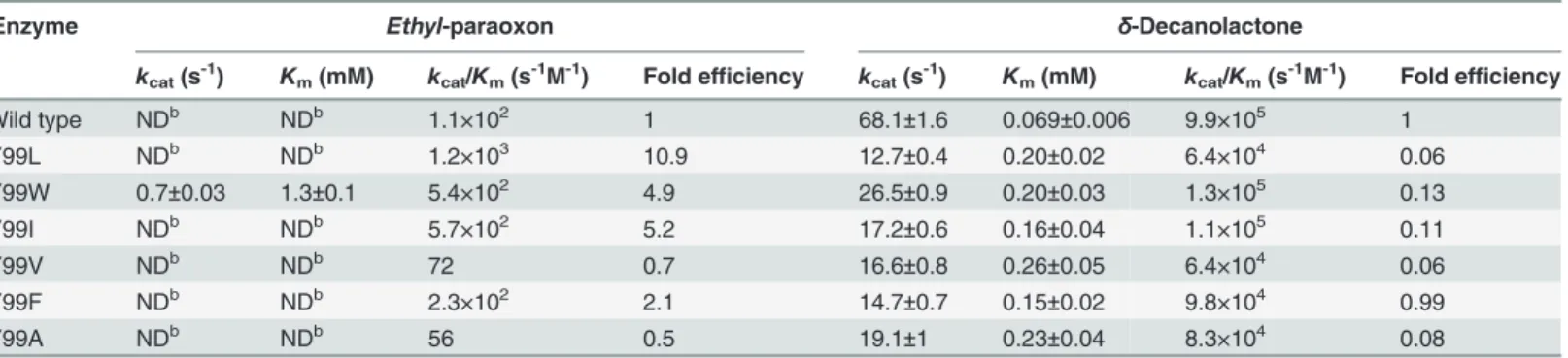
The hydrophobic Leu, Trp, and Phe residues were chosen for further characterization be-cause they induced a substantial increase in PTE activity (up to 11-fold). In addition, we choose Ala, which has the smallest side chain and was used as a minimum reference to study the func-tion of the other side chains. The saturafunc-tion kinetic curves of the mutants forethyl-paraoxon were not obtainable, with the exception of the Y99W mutant (S2 Fig.). The catalytic efficiency (kcat/Km) were estimated under the pseudo-first-order condition ([s]<<Km) and are detailed inTable 2. The mutant Y99A had a 2-fold decrease inkcat/Km, which suggested that the volume Table 1. Kinetic parameters forGkaP at 75°C.
Substrate kcat Km kcat/Km
s-1 mM s-1M-1
Lactone γ-Hexyl-γ-butyrolactone 3.7±0.2 0.051±0.009 7.3×10⁴
δ-Valerolactone 53.7±2.5 5.6±0.7 9.6×10³
δ-Nonalactone 56.6±1.2 0.11±0.01 5.1×10⁵
δ-Decanolactone 68.1±1.6 0.069±0.006 9.9×10⁵
ε-Caprolactone 34.0±3.1 16.2±2.7 2.1×10³
Carboxyl ester p-Nitrophenyl caprylate 0.9±0.04 0.68±0.06 1.3×10³
Phosphotriester Ethyl-paraoxon NDa NDa 1.1×10²
Methyl-paraoxon NDa NDa 38.0
aNot determined, saturation kinetics could not be attained up to a 5mM concentration. Only the catalytic ef
ficiency (kcat/Km) can be estimate under pseudo
and polarity of the side chain at position 99 is important for the catalysis. The Y99W mutant exhibited the smallestKmvalue compared to WT and other mutants, which is beneficial for
substrate binding. However, its overall 5-fold increase inkcat/Kmsuggests that this substitution
may affectethyl-paraoxon hydrolysis. The Y99F mutant also exhibited a 2-fold improvement inkcat/Km. The kinetic analysis revealed that Leu at position 99 provided the largest increase in
PTE activity, and mutant Y99L displayed an 11-fold enhancement inkcat/Kmtowardethyl
-paraoxon. These results indicated that the hydrophobic side chain substitutions, such as leu-cine, phenylalanine, or tryptophan, were favored at position 99 in order to sustain the activity against a phosphotriester substrate.
All of the Y99X mutants had reduced lactonase activity as measured byδ-decanolactone hy-drolysis, which showed significantly elevatedKmvalues (2–3 fold) towardδ-decanolactone and
a 3–5-fold decrease inkcatvalues relative to wild-type (Table 2). The finding that all of these
mutants had an increase inKmsuggests that the hydroxyl group of Tyr99 plays a crucial role in binding of the substrate to the wild-type enzyme. The Y99X mutants showed increased PTE ac-tivity and decreased lactonase acac-tivity. A more substantial effect occurred with mutant Y99L, which caused a specificity switch of 157-fold. These results unambiguously confirm the impor-tance of position 99 in substrate discrimination byGkaP.
BecausebdPTE mainly hydrolyzes OP pesticides, a variety of pesticides (Fig. 2, g-l) were used to test the activities of the active mutants described above (Table 3). Both wild-type and mutants favored pesticide substrates with diethyl side chains. The specific activity of the Y99W mutant against all of the pesticides was 2–4-fold higher than the wild-type enzyme. Strikingly, the mutant Y99L dramatically enhanced catalytic activity toward all pesticides from 4 to 90-fold. It is noteworthy that the activity improvement for thiophosphoryl pesticides is much higher than that for the phosphoryl pesticides. While the activity of Y99L increased 4- to 6-fold for themethyl-paraoxon andethyl-paraoxon, the enhancement was 70- to 90-fold for their thiophosphoryl counterpart (methyl- orethyl-parathion). Therefore, Tyr99 inGkaP can evolve enzymatic function and markedly enhance the weak PTE activity.
X-ray structures and analysis of the wild-type
GkaP and mutant Y99L
To gain a better understanding of the structural basis for the changes in both catalytic spectra and activity caused by the mutations, X-ray crystal structures of the wild-type protein and mu-tant Y99L were solved at a resolution between 1.50 and 1.75 Å. The details of the data collection and structure refinement are summarized inTable 4. The overall structure ofGkaP contained
Table 2. Kinetic parameters ofGkaP wild type and mutants with phosphotriesterase or lactonase activitya.
Enzyme Ethyl-paraoxon δ-Decanolactone
kcat(s-1) Km(mM) kcat/Km(s-1M-1) Fold efficiency kcat(s-1) Km(mM) kcat/Km(s-1M-1) Fold efficiency
Wild type NDb NDb 1.1×10² 1 68.1±1.6 0.069±0.006 9.9×10⁵
1
Y99L NDb NDb 1.2×10³ 10.9 12.7±0.4 0.20±0.02 6.4×10⁴ 0.06
Y99W 0.7±0.03 1.3±0.1 5.4×10² 4.9 26.5±0.9 0.20±0.03 1.3×10⁵ 0.13
Y99I NDb NDb 5.7×10² 5.2 17.2±0.6 0.16±0.04 1.1×10⁵ 0.11
Y99V NDb NDb 72 0.7 16.6±0.8 0.26±0.05 6.4×10⁴
0.06
Y99F NDb NDb 2.3×10² 2.1 14.7±0.7 0.15±0.02 9.8×10⁴ 0.99
Y99A NDb NDb 56 0.5 19.1±1 0.23±0.04 8.3×10⁴ 0.08
aAll assays at 75°C.
bNot determined, saturation kinetics could not be attained.
an (β/α)8barrel fold, which is in agreement with the structures of other PLL enzymes.
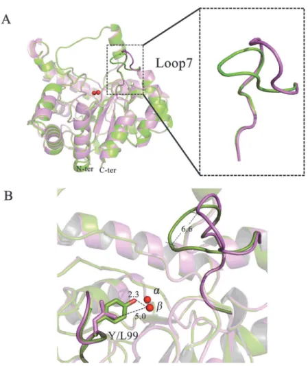
Com-pared to the structure of wild-typeGkaP, there were no major structural changes induced by these mutations. The root mean square deviation (RMSD) for all backbone atoms between the structures were all under 0.3 Å. The main structural feature that was noticeably different be-tween the mutants was in the conformational variability of loop 7 (Fig. 3A). The overlaid struc-tures of Y99L and wild-typeGkaP showed a conspicuous outward shift of the active site in the region of loop 7 by a distance of approximately 6.6 Å (Fig. 3B). This loop contains several
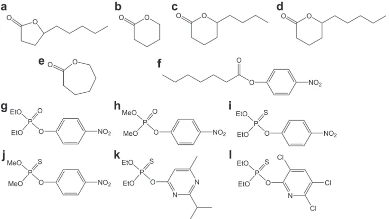
Fig 2. Structures of the substrates used in this study.(a)γ-hexyl-γ-butyrolactone; (b)δ-valerolactone; (c)δ-nonalactone; (d)δ-decanolactone; (e)ε -caprolactone; (f)p-nitrophenyl caprylate; (g)ethyl-paraoxon; (h)methyl-paraoxon; (i)ethyl-parathion; (j)methyl-parathion; (k) diazinon; (l) chlorpyrifos.
doi:10.1371/journal.pone.0115130.g002
Table 3. Specific activities of the wild-typeGkaP and mutants with several organophosphate pesticides.
OP pesticide Specific activity (mU/mg)a
WT Y99W Y99L
Ethyl- paraoxon 242.0±4.5 499.5±4.2 1035.1±67.8
Methyl-paraoxon 61.9±5.9 164.7±18.2 392.2±55.4
Ethyl- parathion 15.5±0.5 37.7±2.2 1072.5±34.0
Methyl-parathion 5.6±0.4 21.2±1.7 519.7±40.5
Diazinon 36.4±0.3 38.6±4.5 1882.2±56.6
Chlorpyrifos 1.5±0.2 3.6±0.7 7.3±1.4
aOne unit (U) of enzymatic activity was de
fined as the amount of enzyme that catalyzes the hydrolysis of 1μmol substrate per minute at 75°C.
residues (Arg230, Ile233, Met236, and Val237) that are found within a large binding pocket and may function in substrate specificity. A recent study has proposed that remote mutations in OpdA result in significant changes in the conformational distribution of loop 7. The domi-nant open conformation of loop 7 may provide the proper steric space for binding OP sub-strates. The closed state is optimally pre-organized for paraoxon hydrolysis, but seems to block access to the active site [30]. We identified an open conformation of loop 7 in Y99L mutant. From the structural analysis, replacement of Tyr99 with Leu created an extra 2.7 Å space in the distance between the Cδof Leu99 and theβ-metal (Fig. 3B). An explanation could be the
vanish-ing of the interaction between Arg230 and Tyr99. In the wild type enzyme, a svanish-ingle water mole-cule at position 713 was found to form two hydrogen bonds with the hydroxyl group of Tyr99 (2.5 Å) and the guanidine group of Arg230 (3.2 Å), respectively (S3 Fig.). This hydrogen-bonded bridge may stabilize the conformation of loop 7 and kept it at close state. However the Y99L mu-tation disrupts the hydrogen-bonded network, which in turn pushes the flexible Arg230 against residues of loop 7 favoring the open conformation. The loop movement causes expansion of the
Table 4. Data collection and refinement statistics forGkaP wild type and mutant.
Structure w.t.GkaP Y99L
Data collection statistics
Cell parameter: a, b, c (Å) 51.31, 88.15, 89.99 51.36, 80.30, 92.20
Space group P21 P21
Wavelength used (Å) 0.97916 0.97916
Resolution (Å)a 50.0–1.6(1.66–1.6) 50.0–1.75(1.81–1.75)
No. of molecule/asymmetric unit 2 2
No. of unique reflections 99136 74050
No. of all reflections 706232 552363
Completeness (%)a 95.8 (93.2) 99.8 (100)
Redundancy 7.1(6.8) 7.5(7.4)
I/σ(I) 34.6 (3.7) 13.1 (3.6)
Rmergeb(%) 5 (43.1) 13.2 (47.5)
Refinement statistics
Rworkc(%) 18.12 22.19
Rfree(%) 19.82 24.93
RMSD bond lengths (Å) 0.0067 0.0057
RMSD bond angle (°) 1.0636 0.8796
Overall B-factor (Å2) 24.42 27.376
Final model (Number of protein atoms) 5093 5088
Final model (H2O molecules) 738 505
Ramachandran plot
Res. in most favored regions 625 627
Res. in additionally allowed regions 10 11
Res. in generously allowed regions 0 0
aValues in parentheses correspond to the highest resolution shell. bR
merge=ΣhklΣi|I(hkl)i-<I(hkl)>|/ΣhklΣiI(hkl)i, where<I(hkl)>is the mean intensity of the observationsI(hkl)iof
reflection hkl.
cR
work=Σ||Fobs||Fcalc||/Σ|Fobs|, whereFobsandFcalcare the observed and calculated structure-factor
amplitudes, respectively.Rfreewas calculated asRworkusing a randomly selected subset of*10% of
unique reflections not used for structure refinement.
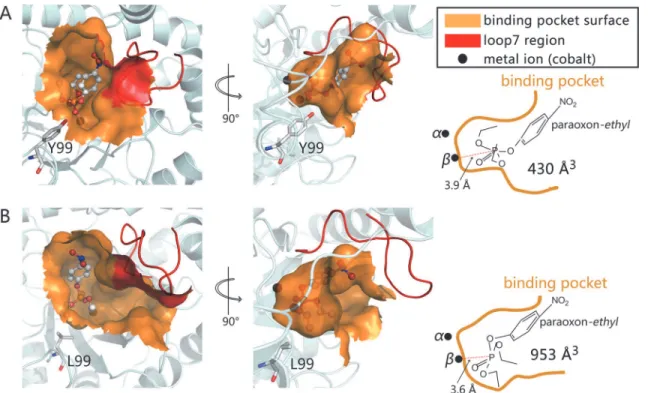
substrate binding pocket volume to 953 Å3as calculated by CASTp online, which is approximate-ly double the volume of the wild-typeGkaP space (430 Å3). In addition, the hydrophobic Leu
side chain may increase the affinity for phosphotriester substrates in the binding site. To qualitatively estimate the significant enhancement in the activity of theGkaP mutant Y99L, a structure analysis andin silicodocking experiments were conducted. The results indi-cated that the distance between the phosphorus atom ofethyl-paraoxon and theβ-cobalt ion is
3.85 Å in the wild-type (Fig. 4A), but 3.6 Å in the Y99L mutant, respectively (Fig. 4B). In con-trast to the wild-type binding model, the phosphorus center of the substrate in the Y99L mu-tant models are closer to theβ-cobalt ion, which would result in strong polarization of the P-O bond and benefit catalysis.
To gain a better understanding for the preference of Y99L mutant for the thiophosphoryl pesticides substrates (Table 3), we performed substrate docking and 10 ns’MD simulations on Y99L complex withethyl-paraoxon (P = O) and diazinon (P = S) bound, respectively. The binding free energies are -10.83±3.19 kcal/mol for Y99L-paraoxon and -22.36±4.37 kcal/mol for Y99L-diazinon, which confirmed that the mutant Y99L did favors P = S over the phospho-ryl pesticides substrates. However, the structural basis of this change is not clear and need fur-ther research to reveal.
Fig 3. Crystal structure analysis ofGkaP wild-type and mutant Y99L.(A) Overlay of the structures of wild-typeGkaP (green), and Y99L (purple). (B) Overlay of loop 7 regions of wild-typeGkaP (green) and Y99L (purple). The metal ions are shown in red spheres. Distances are shown inÅ.
Concluding remarks
In a previous paper [21] some of us reported the effect of mutations Phe and Leu at position Trp263 and Trp at Tyr97 as able to increase the PTE activity and specificity inSsoPox, a finding suggested to be related to the widening of the leaving group subsite. This has been confirmed by recent structural analyses showing that W263X induces very subtle changes in the loop 8 positioning whereas have no effect on the very short loop 7 [31]. Here we demonstrated that mutagenesis of the Tyr99 in the active site ofGkaP can markedly enhance its promiscuous PTE activity. This residue presumably contributes to the process of the natural divergent evolu-tion in the PTE family. The structural analyses revealed that the enhancement of enzyme activ-ity could be caused by alterations in the dynamics and flexibilactiv-ity of substrate-binding loop 7, which creates a wide and open state of the active site. The transition between the close (wild type) and open (mutant) conformational states governed by loop 7 movement is directly asso-ciated with the regulation of the substrate specificity. Our study provides an example that a lim-ited mutation in a promiscuous enzyme may lead to structural changes and benefit for an alternative binding substrate and efficient catalysis. It also suggests that the conformation of ac-tive site loop plays an essential role for regulating promiscuous activity of the enzyme, reinforc-ing the need to consider the interplay between active site architecture and catalysis in future protein engineering.
Materials and Methods
Reagents, bacterial strains, and plasmids
All substrates were purchased from Sigma-Aldrich (Shanghai, China), and the chemical struc-tures are shown inFig. 2. Restriction enzymes, T4 DNA ligase, LA TaqDNA polymerase,
Fig 4. Binding model ofethyl-paraoxon into the active site of wild-typeGkaP (A), and Y99L (B).Dockings were performed using AutoDock 4.0 on a rigid protein structure. Binding pockets are shown at the surface. Loop 7 is shown in red. The cobalt ions are labeled asαandβ. Substrateethyl-paraoxon is shown as ball-and-stick. Y99 and L99 are shown as stick models (gray carbons).
kanamycin, and isopropyl 1-thio-β-D-galactopyranoside (IPTG) were obtained from
Fermen-tas (Vilnius, Lithuania) and TaKaRa (Kyoto, Japan). The molecular mass marker for SDS-PAGE was obtained from Fermentas. Ni-NTA agarose was purchase from Qiagen (Chats-worth, CA). All other chemicals were reagent grade.Geobacillus kaustophilusHTA426 was purchased from the Japan Collection of Microorganisms (JCM No.12893). The expression vec-tor pET-28a (+) as well asE. colistrainsXL1-blue and BL21-CodonPlus (DE3)-RIL, which were used for cloning and protein expression, respectively, were purchased from Invitrogen (Carls-bad, CA).
Cloning of the GK1506 gene and DNA manipulation
Standard molecular cloning and transformation techniques were employed as described by Sambrooket al[32]. The gene encoding a PLL (GenBank ID: 3183579) was amplified by PCR fromGeobacillus kaustophilusHTA426 genomic DNA using the primers listed inS1 Table. PCR conditions were 95°C for 5 min, followed by 30 cycles at 95°C for 40 s, 56°C for 40 s, 72°C for 90 s, and a final extension at 72°C for 8 min. The amplified gene was cloned into the pET-28a (+) vector with an N-terminal 6×His tag. The recombinant plasmid was then transformed intoE. coliXL1-blue electrocompetent cells and plated onto 2YT agar plate containing 50μg/mL
kanamycin overnight. The insert sequence was confirmed by DNA sequencing.
Expression and purification of
GkaP
The recombinant plasmid was transformed intoE. coliBL21-CodonPlus (DE3)-RIL electro-competent cells. Cells were grown at 37°C in 2YT medium supplemented with 50μg/mL
kanamycin and 1.0 mM CoCl2until the OD600reached 0.6–0.8. Protein expression was then
induced by the addition of 1.0 mM IPTG and the culture was grown for 12 h at 26°C. Cells were harvested by centrifugation and resuspended in 50 mM Tris-HCl (pH8.0) containing 0.2 mM Co2+and 5 mM imidazole. After ultrasonic cell disintegration, the cytosolic fraction
was heated at 60°C for 30 min and centrifuged at 12000 rpm for 20 min to remove heat-in-duced protein aggregates. The supernatant was added to a Ni2+-chelating affinity column and
eluted with 100 mM imidazole in 20mM Tris-HCl (pH 7.9) and 10 mM NaCl for an approxi-mate total of 10 mL. The purity of the fusion enzymes (wild type and mutant) was assessed on 15% SDS-PAGE and enzymes were diafiltrated twice with 50 mM Tris-HCl (pH 8.0).
Kinetics measurements of
GkaP
The lactonase, phosphotriesterase, and esterase hydrolyses ofGkaP were monitored by absor-bance changes in a UV-2550 spectrophotometer (Shimadzu, Kyoto, Japan) at a constant tem-perature of 75°C with 1 mL reaction volumes (path length = 1cm). Analysis of reaction samples for each substrate was performed at a constant enzyme concentration. Theδ
-decano-lactone substrate was dissolved in dimethyl sulfoxide (DMSO), whereas thep-nitrophenyl caprylate and OP substrates in acetonitrile as stock solutions. For enzymatic kinetics assay, ali-quots of the stock were added to the reaction buffer for defined concentrations. The hydrolysis of lactone was monitored using a pH-sensitive colorimetric assay [33]. Briefly, the reactions were performed in 2.5 mM Bicine (pH 8.3) containing 0.2 M NaCl, 0.2 mM cresol purple, and 0.02–20 mM lactone substrate. Upon mixture of the substrate with the enzyme, the decrease in absorbance was monitored at 577 nm (ε577= 47300 M-1cm-1, 1%DMSO). The enzyme was
corrected for the background rate of spontaneous hydrolysis in the absence of enzymes, which were subtracted from the enzymatic rates. The kinetic parameters (kcat,Km) were obtained by
fitting the data to the Michaelis-Menten equation [V = S×E×kcat/(S+Km)] or to the
pseudo-first-order form of it at S<<Km(V = S×E×kcat/Km) with GraphPad Prism software (Graphpad, San Diego, CA), where V is the initial velocity, E is the enzyme concentration, S is the substrate concentration. The final concentrations used in the assay to determine specific activities toward the tested OP pesticides were 1mM, 1mM, 0.25 mM, and 0.25 mM for paraoxon, parathion, di-azinon, and chlorpyrifos, respectively. The rate of paraoxon and parathion hydrolysis was mea-sured at a wavelength of 405nm as described above. The rate of diazinon hydrolysis was measured at a wavelength of 228nm (ε228= 3300 M-1cm-1), and chlorpyrifos was measured at
276 nm (ε276= 2790 M-1cm-1). Control reactions were performed in the absence of enzyme.
One unit of enzymatic activity was defined as the amount of enzyme that catalyzed the hydro-lysis of 1μmol substrate per minute. All assays were performed at least in triplicate. Enzyme
concentrations were determined using the Bio-Rad protein assay kit II (Bio-Rad, Richmond, CA).
Site-directed mutagenesis
The mutantsY99W, Y99I, and Y99V were constructed using the QuikChange site-directed mu-tagenesis protocol [34]. The pET-28a (+) plasmid carrying the wild-type or mutantGkaP gene was used as a template for PCR with LA Taq polymerase (TaKaRa). All mutagenic primers are listed inS1 Table. The PCR reactions were as follows: initial denaturation for 5 min at 94°C, followed by 16 cycles of 94°C for 30 s, 57°C for 30 s, and 72°C for 8 min. A final elongation step at 72°C was performed for 30 min. The final PCR reaction was then incubated with 2 U DpnI (Fermentas) at 37°C for 1 h to remove the methylated template, and 2μL of the reaction was
transformed into competent E. coli BL21-CodonPlus (DE3)-RIL cells. The expression and pu-rification procedures of these mutants were the same as described for the wild-type construct.
Site-saturation mutagenesis and screening procedure
Saturation mutagenesis at Tyr99 was performed and the primers used are listed inS1 Table. The target amino acid position was coded by NNK (forward) and MNN (reverse), where N = A, G, C, or T, K = G or T, and M = A or C. Whole plasmid PCR reactions and transformations were performed as described in the method for site-directed mutagenesis. A library of clones was screened for paraoxon hydrolysis activity compared to the activity of wild type using a 96-well plate assay. Approximately 200 randomly selected colonies were picked with sterilized toothpicks and placed into individual wells of 96-well plates, which were filled with 200μL of
2YT containing 50μg/mL kanamycin and 1.0 mM CoCl2. The plates were shaken overnight at
37°C. The next day, 5μL of each culture was transferred into a new plate containing fresh
me-dium and antibiotic. The original plates were stored at 4°C and the duplicated plates were shak-en for an additional 3 h for cell growth. The cells were thshak-en induced with 1 mM IPTG for 6 h at 30°C, and then the cell density in each well was determined by measuring the OD at a wave-length of 600 nm using a Multiskan Ascent 96-well plate reader (Thermo Scientific, Vantaa, Finland). Cells were harvested by centrifugation at 3000 rpm for 30 min. The harvested cells were frozen and then thawed three times to release the enzyme and then resuspended in 200μL of 50 mM phosphate buffer (pH 8.0). Crude bacterial extracts were heated at 60°C for
30 min and centrifuged in order to remove the heat-induced aggregated proteins. A 100μL
ali-quot from each well was pipetted into a new 96-well plate, to which 100μL of substrate solution
containing 0.4 mMethyl-paraoxon and 0.2 mM Co2+in 50 mM phosphate buffer at pH 8.0
measured based on the releasedp-nitrophenol at a wavelength of 405 nm. The value of A405of
each well was normalized by the corresponding A600, and the ratior= A405/A600was used to
estimate the activity of each clone.
Crystallization of
GkaP wild-type and mutant
Crystals of nativeGkaP and mutant were grown by the hanging-drop method at 16°C. Each drop contained 2μL of protein at a concentration of either 20 or 30 mg/mL and 2μL of the
200μL well solution. The reservoir solutions for wild-typeGkaP, contained 50 mM Hepes-Na
pH 7.3, 12–13% (w/v) PEG8000, 8% (w/v) ethylene glycol, and 2% (w/v) glycerol. For the Y99L mutant, the reservoir solution contained 0.1M MES pH 6.5 and 14% (w/v) PEG20000. Crystals were cryoprotected by the addition of 20% ethylene glycol (v/v) to the crystallization condi-tions. The crystals belonged to space groupP21. The datasets at 1.6, 1.5, 1.75, and 1.75 Å that corresponded to wild-typeGkaP and Y99L mutant, respectively, were collected on beamline BL17U at the Shanghai Synchrotron Radiation Facility (China). All datasets were integrated, scaled, and reduced with the HKL2000 software package. The structures were solved by Molec-ular Replacement with the CCP4i program PHASER using the wild-typeGkaP structure (PDB code 3ORW) that was previously obtained by our laboratory as the model. Model building was performed by COOT, and all of the model qualities were checked with PROCHECK.
Substrates docking
All docking runs were performed using AutoDock 4.0. The cobalt van der Waals parameters were 2.87 Å for radii, 0.014 kcal/mol for well depth, and 12 Å3for atomic salvation volume.
Charges of 1.4 and 1.5 were assigned to theαandβcobalt ions, respectively, as previously
de-scribed [35]. A 30×30×30 grid with 0.375Å spacing was centered in a metal center. The La-marckian genetic Algorithm (LGA) was selected for the ligand conformational search. For the docking process, the number of generation was 20 conformers for each OP substrate. Other pa-rameters were used as default. Among docking poses, the lowest binding energy conformation was selected. The structures were drawn with PyMOL 1.1. The residue interaction was analyzed using Ligplot 4.22. The volume of substrate binding pocket was calculated using CASTp [36] online (http://sts.bioengr.uic.edu/castp/calculation.php).
Molecular Dynamics Simulations
Molecular Dynamics (MD) simulations and energy minimizations were performed using the AMBER12 simulation package [37] and theff12SBforce field [38] with the TIP3P water model [39]. The initial coordinates of the complexes were extracted from the docking results. Hydro-gen atoms were added using the LEAP module of AMBER12. Antechamber [40] was used to handle the force field ofethyl-paraoxon and diazinon. Ten Na+ions were placed around the
Protein Data Bank accession codes
The coordinates have been deposited in the Research Collaboration for Structural Bioinformat-ics (RCSB) Protein Data Bank with the following accession codes: 3TN3 for wild-typeGkaP, and 3TN5 for Y99L.
Supporting Information
S1 Fig. Michaelis-Menten kinetics analyses of lactonase (A), phosphotriesterase (B) and es-terase (C) activities ofGkaP.Lactonase assay used withδ-decanolactone; Phosphotriesterase assay used withethyl-paraoxon, saturation kinetics could not be attained; Esterase assay used withp-nitrophenyl caprylate.
(TIF)
S2 Fig. Michaelis-Menten kinetics analyses of phosphotriesterase activities ofGkaP and mutants.Ethyl-paraoxon was used as substrate.
(TIF)
S3 Fig. The interaction between Arg230 and Tyr99 in the wild-typeGkaP.A water molecule
(Wat713, shown in blue sphere) forms two hydrogen bonds with Tyr99 and Arg230, respec-tively. Tyr99 and Arg230 are shown as stick models (green carbons). The two metal ions are la-beled asαandβ, and shown in red spheres. Loop 7 region is highlighted in orange.
(TIF)
S1 Table. Primers used in cloning and mutagenesis ofGkaP.
(DOCX)
Acknowledgments
We thank the staff members at the Shanghai Synchrotron Radiation Facility (SSRF), Shanghai, China for technical support in diffraction data collection.
Author Contributions
Conceived and designed the experiments: YF. Performed the experiments: YZ JA WY AB. An-alyzed the data: BZ ZL GW HFC GM. Wrote the paper: YZ GY. Revised manuscript: YF GM.
References
1. O’Brien PJ, Herschlag D (1999) Catalytic promiscuity and the evolution of new enzymatic activities. Chem Biol 6: R91–R105. PMID:10099128
2. Nobeli I, Favia AD, Thornton JM (2009) Protein promiscuity and its implications for biotechnology. Nat Biotechnol 27: 157–167. doi:10.1038/nbt1519PMID:19204698
3. Babtie A, Tokuriki N, Hollfelder F (2010) What makes an enzyme promiscuous? Curr Opin Chem Biol 14: 200–207. doi:10.1016/j.cbpa.2009.11.028PMID:20080434
4. Raushel FM, Holden HM (2000) Phosphotriesterase: an enzyme in search of its natural substrate. Adv Enzymol Relat Areas Mol Biol 74: 51–93. PMID:10800593
5. Singh BK (2009) Organophosphorus-degrading bacteria: ecology and industrial applications. Nat Rev Microbiol 7: 156–164. doi:10.1038/nrmicro2050PMID:19098922
6. Roodveldt C, Tawfik DS (2005) Shared promiscuous activities and evolutionary features in various members of the amidohydrolase superfamily. Biochemistry 44: 12728–12736. PMID:16171387 7. Khersonsky O, Tawfik DS (2005) Structure-reactivity studies of serum paraoxonase PON1 suggest
that its native activity is lactonase. Biochemistry 44: 6371–6382. PMID:15835926
9. Merone L, Mandrich L, Rossi M, Manco G (2005) A thermostable phosphotriesterase from the archaeon
Sulfolobus solfataricus: cloning, overexpression and properties. Extremophiles 9: 297–305. PMID: 15909078
10. Dumas DP, Caldwell SR, Wild JR, Raushel FM. (1989) Purification and properties of the phosphotries-terase fromPseudomonas diminuta. J Biol Chem 264: 19659–19665. PMID:2555328
11. Kolakowski JE, DeFrank JJ, Harvey SP, Szafraniec LL, Beaudry WT, et al. (1997) Enzymatic hydrolysis of the chemical warfare agent VX and its neurotoxic analogues by organophosphorus hydrolase. Bioca-tal Biotransform 15: 297–312.
12. Sethunathan N, Yoshida T (1973) AFlavobacterium sp. that degrades diazinon and parathion. Can J Microbiol 19: 873–875. PMID:4727806
13. Horne I, Sutherland TD, Harcourt RL, Russell RJ, Oakeshott JG (2002) Identification of anopd (organo-phosphate degradation) gene in anAgrobacteriumIsolate. Appl Environ Microbiol 68: 3371–3376. PMID:12089017
14. Afriat L, Roodveldt C, Manco G, Tawfik DS (2006) The latent promiscuity of newly identified microbial lactonases is linked to a recently diverged phosphotriesterase. Biochemistry 45: 13677–13684. PMID: 17105187
15. Porzio E, Merone L, Mandrich L, Rossi M, Manco G (2007) A new phosphotriesterase fromSulfolobus acidocaldariusand its comparison with the homologue fromSulfolobus solfataricus. Biochimie 89: 625–636. PMID:17337320
16. Hawwa R, Aikens J, Turner RJ, Santarsiero BD, Mesecar AD (2009) Structural basis for thermostability revealed through the identification and characterization of a highly thermostable phosphotriesterase-like lactonase fromGeobacillus stearothermophilus. Arch Biochem Biophy 488: 109–120. doi:10. 1016/j.abb.2009.06.005PMID:19615330
17. Xiang D, Kolb P, Fedorov A, Meier M, Federov E, et al. (2009) Functional annotation and three-dimen-sional structure of Dr0930 fromDeinococcus radiodurans, a close relative of phosphotri esterase in the amidohydrolase superfamily. Biochemistry 48: 2237–2247. doi:10.1021/bi802274fPMID:19159332 18. Elias M, Dupuy J, Merone L, Mandrich L, Porzio E, et al. (2008) Structural basis for natural lactonase
and promiscuous phosphotriesterase activities. J Mol Biol 379: 1017–1028. doi:10.1016/j.jmb.2008. 04.022PMID:18486146
19. Hiblot J, Gotthard G, Chabriere E, Elias M (2012) Structural and enzymatic characterization of the lacto-nase SisLac fromSulfolobus islandicus. PLoS One 7: e47028. doi:10.1371/journal.pone.0047028 PMID:23071703
20. Hawwa R, Larsen SD, Ratia K, Mesecar AD (2009) Structure-based and random mutagenesis ap-proaches increase the organophosphate-degrading activity of a phosphotriesterase homologue from
Deinococcus radiodurans. J Mol Biol 393: 36–57. doi:10.1016/j.jmb.2009.06.083PMID:19631223 21. Merone L, Mandrich L, Porzio E, Rossi M, Müller S, et al. (2010) Improving the promiscuous nerve
agent hydrolase activity of a thermostable archaeal lactonase. Bioresour Technol 101: 9204–9212. doi:10.1016/j.biortech.2010.06.102PMID:20667718
22. Zhang Y, An J, Ye W, Yang G, Qian ZG, et al. (2012) Enhancing the promiscuous phosphotriesterase activity of a thermostable lactonase (GkaP) for the efficient degradation of organophosphate pesticides. Appl Environ Microbiol 78: 6647–6655. doi:10.1128/AEM.01122-12PMID:22798358
23. Meier MM, Rajendran C, Malisi C, Fox NG, Xu C, et al. (2013) Molecular engineering of organophos-phate hydrolysis activity from a weak promiscuous lactonase template. J Am Chem Soc 135: 11670–
11677. doi:10.1021/ja405911hPMID:23837603
24. Vanhooke JL, Benning MM, Raushel FM, Holden HM (1996) Three-dimensional structure of the zinc-containing phosphotriesterase with the bound substrate analog diethyl 4-methylbenzylphosphonate. Biochemistry 35: 6020–6025. PMID:8634243
25. Holm L, Sander C (1997) An evolutionary treasure: unification of a broad set of amidohydrolases relat-ed to urease. Proteins: Struct Funct Genet 28: 72–82. PMID:9144792
26. Seibert CM, Raushel FM (2005) Structural and catalytic diversity within the amidohydrolase superfami-ly. Biochemistry 44: 6383–6391. PMID:15850372
27. Watkins LM, Mahoney HJ, McCulloch JK, Raushel FM (1997) Augmented hydrolysis of diisopropyl fluorophosphate in engineered mutants of phosphotriesterase. J Biol Chem 272: 25596–25601. PMID: 9325279
28. Chow JY, Xue B, Lee KH, Tung A, Wu L, et al. (2010) Directed evolution of a thermostable quorum-quenching lactonase from the amidohydrolase superfamily. J Biol Chem 285: 40911–40920. doi:10. 1074/jbc.M110.177139PMID:20980257
30. Jackson CJ, Foo JL, Tokuriki N, Afriat L, Carr PD, et al. (2009) Conformational sampling, catalysis, and evolution of the bacterial phosphotriesterase. Proc Natl Acad Sci USA 106: 21631–21636. doi:10. 1073/pnas.0907548106PMID:19966226
31. Hiblot J, Gotthard G, Elias M, Chabriere E (2013) Differential active site loop conformations mediate promiscous activities in the lactonase SsoPox. PLoS One 8: e75272. doi:10.1371/journal.pone. 0075272PMID:24086491
32. Sambrook J, Russell DW (2001) Molecular Cloning: a Laboratory Manual (3rd Edition). Plasmids and Their Usefulness in Molecular Cloning. pp. 68–105.
33. Chapman E, Wong CH (2002) A pH sensitive colorimetric assay for the high-throughput screening of enzyme inhibitors and substrates: a case study using kinases. Bioorg Med Chem 10: 551–555. PMID: 11814841
34. Zheng L, Baumann U, Reymond JL (2004) An efficient one-step site-directed and site-saturation muta-genesis protocol. Nucleic Acids Res 32: e115. PMID:15304544
35. Hermann JC, Ghanem E, Li Y, Raushel FM, Irwin JJ, et al. (2006) Predicting substrates by docking high-energy intermediates to enzyme structures. J Am Chem Soc 128: 15882–15891. PMID: 17147401
36. Dundas J, Ouyang Z, Tseng J, Binkowski A, Turpaz Y, et al. (2006) CASTp: computed atlas of surface topography of proteins with structural and topographical mapping of functionally annotated resiudes. Nucleic Acid Res 34: W116–W118. PMID:16844972
37. Case DA, Darden T, Cheatham TE, Simmerling C, Wang J, et al. (2012) AMBER 12. San Francisco: University of California.
38. Cerutti DS, Rice JE, Swope WC, Case DA (2013) Derivation of fixed partial charges for amino acids ac-commodating a specific water model and implicit polarization. J Phys Chem B 117: 2328–2338. doi: 10.1021/jp311851rPMID:23379664
39. Jorgensen WL, Chandrasekhar J, Madura JD, Impey RW, Klein ML (1983) Comparison of simple po-tential functions for simulating liquid water. J Chem Phys 79: 926–935.
40. Wang J, Wolf RM, Caldwell JW, Kollman PA, Case DA (2004) Development and testing of a general amber force field. J Comput Chem 25: 1157–1174. PMID:15116359
41. Ryckaert JP, Giovanni C, Berendsen HJC (1977) Numerical integration of the cartesian equations of motion of a system with constraints: molecular dynamics of n-alkanes. J Comput Phys 23: 327–341. 42. Darden T, York D, Pedersen L (1993) Particle mesh Ewald: An Nllog(N) method for Ewald sums in
large systems. J Chem Phys 98: 10089–10092.
43. Götz AW, Williamson MJ, Xu D, Poole D, Le Grand S, et al. (2012) Routine Microsecond Molecular Dy-namics Simulations with AMBER on GPUs. 1. Generalized Born. J Chem Theory Comput 8: 1542–
1555. PMID:22582031