Vojnosanit Pregl 2015; 72(9): 785–793. VOJNOSANITETSKI PREGLED Page 785
Correspondence to: Nebojša Djenić, Department of General Surgery, Military Hospital, Bulevar dr Zorana Djindjića bb, Niš, Serbia. E-mail: djani1508@yahoo.com
O R I G I N A L A R T I C L E UDC: 617-089-092.9::616-001.45-08
DOI: 10.2298/VSP131106050D
Experimental closure of gunshot wounds by fibrin glue with
antibiotics in pigs
Zatvaranje prostrelne rane primenom fibrinskog lepka sa antibiotikom u
eksperimentu na svinjama
Nebojša Djenić*†, Milan Višnjić†, Saša Dragović*, Vladmila Bojanić†, Zoran Bojanić†, Dragan Djurdjevi懧, Boris Djindjić†, Miloš Kostov*§
*Department of General Surgery, Military Hospital, Niš, Serbia; †Faculty of Medicine, University of Niš, Niš, Serbia; ‡Institute for Medical Research, Military Medical Academy, Belgrade, Serbia; §Faculty of Medicine of the Military Medical Academy,
University of Defence, Belgrade, Serbia
Abstract
Background/Aim. Gunshot wounds caused by the
auto-matic rifle M70AB2 (AK-47) 7.62 mm, after the primary surgical management, were closed with delayed primary su-ture during the next four to seven days. This period coin-cides with the fibroblastic phase of wound healing. Fibrin glue is used as a local hemostatic and as a matrix for the lo-cal dosed release of antibiotics. Antibiotics addition to fibrin glue resulted in continuous diffusion into the surrounding next 4 to 7 days. The aim of this study was to create the preconditions for gunshot wounds closing without compli-cations by the application of fibrin glue with antibiotics 24 h after primary surgical treatment. Methods. A total of 14 pigs were wounded in the gluteofemoral region by the bullet M67, initial velocity of 720 m/s. All wounded animals were surgically treated according to the principles of the war-surgery doctrine. Seven wounds were closed with primary delayed suture four days after the primary surgical treatment (traditional approach). Fibrin glue with antibiotics was in-troduced in seven wounds during the primary surgical treatment and primary delayed suture was done after 24 h.
The macroscopic appearance and the clinical assessment of the wound were done during the primary surgical treatment and during its revision after 24 h, as well as histopathological findings at the days 4 and 7 after wounding. Results. Gun-shot wounds caused by the automatic rifle M70AB2 (AK-47) 7.62 mm, and treated with fibrin glue with antibiotics after primary surgical management, were closed with primary de-layed suture after 24 h. In further wound evolution there were no complications. Conclusion. Uncomplicated soft-tissue wounds caused by an automatic M70AB2 rifle may be closed primarily with delayed suture without the risk of developing complications if on revision, 24 h after primary surgery, there were no present necrotic tissues, hematoma, and any signs of infection when fibrin glue with antibiotics (ceftriaxone and clindamycin) was applied. The use of this method should be limited to individual and strictly controlled cases in civil prac-tice for now.
Key words:
wounds, gunshot; wound closure techniques; fibrin tissue adhesive; anti-bacterial agents; surgical procedures, operative; treatment outcome; swine.
Apstrakt
Uvod/Cilj. Prostrelna rana naneta projektilom iz automat-ske puške M70 AB2 (AK-47) 7,62 mm nakon primarne hi-rurške obrade zatvara se primarno odloženim šavom u peri-odu 4–7 dana. Ovaj period se poklapa sa fibroblastnom fazom u procesu zarastanja rana. Fibrinski lepak se upotrebljava kao lokalni hemostatik i kao matriks za lokalno dozirano oslobađ a-nje antibiotika. Dodatak antibiotika fibrinskom lepku dovodi do kontinuiranog delovanja narednih 4 do 7 dana. Cilj rada bio je da se primenom fibrinskog lepka sa antibiotikom 24 časa na-kon primarne hirurške obrade stvore preduslovi za zatvaranje rane bez komplikacija. Metode. Ukupno 14 svinja ranjeno je u
(cef-triakson i klindamicin) i zatvorene 24 časa kasnije primarno od-loženim šavom, u daljem toku nije došlo do razvoja komplika-cija. Zaključak. Nekomplikovane mekotkivne prostrelne rane izazvane projektilom iz automatske puške M70AB2 tretirane fibrinskim lepkom s antibioticima mogu se zatvoriti odloženim šavom bez rizika od komplikacija, ako one ne budu prisutne 24 h od postavljanja lepka. Primenu ove metode za sada bi trebalo
ograničiti na pojedinačne i strogo kontrolisane slučajeve u civil-noj praksi.
Ključne reči:
rana vatrenim oružjem; rana, zatvaranje, tehnike; tkivni lepkovi, fibrinski; antibiotici; hirurgija, operativne procedure; lečenje, ishod; svinje.
Introduction*
The principles of gunshot wounds treatment have been changing during the evolution of surgery. In 1898, the German surgeon Paul Leopold Friedrich 1 (1864–1916) carried out the experiment proving all gunshot wounds primarily contaminated with bacteria. He also stated that bacteria were still found no de-eper than 1–2 mm from the edge of wound after six hours and only after that period they penetrated into the deeper layers of the tissue. These findings resulted in two very important conclu-sions: first, that wound should be surgically treated within a 6-hour interval, and second, that excision can sterilize wound within this period. Modern principles of primary surgery (PS) of wounds were developed from Le Dran-Desault-Larreys’ con-cept. The delayed primary suture (DPS) is the legacy of the World War Two, and it was used 4–10 days after PS, regardless the bacterial wound culture test. The results of surgical treatment have been significantly improved by a widespread application of penicillin since 1943, but the protection of war wound with anti-biotics cannot replace surgical treatment 2–5. Active approach to war wound in Serbia leads back to Dr. Mihailo Petrović. In Bal-kan wars (1912–1913) injuries caused by small caliber bullet were treated conservatively using antiseptic solution of carbolic acid. Because of the increased number of injuries caused by pro-jectiles with high initial velocity, Dr. Petrović introduced active approach to wound treatment leaving them open and applying debridement 6–11. Projectiles with high initial velocity (speed exceeding 750 m/s) inflict specific wounds, as the consequence of the particular shooting mechanism. While penetrating through the tissue, a projectile directly affects it creating “the zone of di-rect traumatic necrosis”. The side shock wave by the indidi-rect ac-tion around the channel of the projectile causes “the zone of mo-lecular concussion”. The edge between the areas with functional and irreversible circulatory disturbances is not clear nor definite for some time 3, 4, 12–16.
Fibrin glue (FG) is a two-component biological system with local hemostatic, adhesive and sealing effects and it is also used as a matrix (carrier) for local dosed release of antibiotics. The component 1 of the fibrin glue is made of fibrinogen, coa-gulation factor XII, fibronectin and plasma. FG component 2 in-cludes thrombine, calcium ions and exogenous antifibrinolitics when needed. There are commercial preparations of FG and they have higher concentrations of fibrinogen. The application of FG, obtained from the plasma of individual donors, is associ-ated with the risk of viral infections transmission, while using a unit of blood/plasma 17–22.
Primary surgical treatment and systemic use of antibiotics within the optimal time period have an indispensable role in
excision of devitalized muscle tissue, removal of possible bacte-ria contained in it and in infection prevention.
Application of FG with antibiotics (FGA) after primary sur-gical treatment if properly done prevents both postoperative he-matoma in wound and the development of local microorganisms.
FGA reduces the development of secondary complications and improves the basic biological reactions in the tissue repair processes.
In this study soft-tissue wounds were inflicted by the au-tomatic rifle M70AB2 (AK-47) 7.62 mm in pigs. The aim of the study was to create the preconditions for primary delayed wounds closure without complications 24 h after the primary surgical treatment, and the usage of FGA.
Methods
The research was carried out on 14 experimental ani-mals after having been approved by the Ethics Committee of the Faculty of Medicine in Niš, No. 01-2066-3, 2010. Before the experiment, all animals were examined by the veterinari-an veterinari-and proved to be healthy. The pigs were wounded by the bullet M67 (diameter 7.62 39 mm, weight 8 g, copper-jacketed, lead-core, flat-base) 23. The model of gunshot inju-ries applied by the Swedish authors, and modified in the experimental investigations at the Military Medical Academy in Belgrade 16, 24–26, was performed. The male Yorkshire-Landrace breed pigs, 3.5 months old, weighing 32–39 kg, were used. The experimental induction of soft tissue wounds with channel length over 100 mm in the gluteofemoral region is possible at that age of animals. The animals were fasted for 12 h before being injured. The pigs were divided into two groups, with seven animals per each group: the group A (PS was made by traditional approach) and the group B (treated with FGA).
Diazepam [2 mg/10 kg body weight (BW), Bensedin®, Galenika, Serbia] was given in premedication. Animals were introduced into anesthesia by intramuscular (im) administra-tion of 0.05 mg/kg BW acepromazine maleate (Combis-tress®, VanaGes.mH, Austria) 20 minutes before injuring. Ketamine chlorohydrate (Laboratorio Sanderson SA, Chile) was given (im 0.5 mg kg/BW) 15 min after the application of acepromazine maleate. All anesthetized animals were breat-hing spontaneously. Then their backs were turned down with slightly suspended and well exposed rear right leg laterally positioned toward the shooter.
Sho-Vol. 72, No. 9 VOJNOSANITETSKI PREGLED Page 787
Djenić N, et al. Vojnosanit Pregl 2015; 72(9): 785–793.
oting was done two times in a 10-day interval and each time se-ven animals were wounded. The skin of the pigs was not washed, shaven, nor disinfected before firing. Packing of wounds and a compressive bandage were applied for temporary bleeding control 26–29.
The animals were housed in separate cages at the Center for Biomedical Research of the Faculty of Medicine in Niš. We followed the principles of gunshot wounds treatment according to the traditional approach recommended by the International Red Cross and the war-surgery doctrine 3, 4, 12–16, 30. PS was per-formed 4–12 h after wounding under general anesthesia. All animals were given preoperatively 1 g of ceftriaxone 30–36.
The assessment of the wound severity was made according to the Red Cross Wound Classification 37, 38. PS is performed using aseptic techniques. The skin around the entrance and exit of wounds was shaved. Washing of the skin surrounding wounds was carried out with foam and povidone-iodine soluti-on. The surgical field was surrounded with sterile compresses. The length of the wound channel and the size of the wound en-trance and exit were measured. The clinical appearance of the wound during PS and clinical evaluation of the effectiveness of PS at revision, were presented as the macroscopic parameters of necrosis, infection and bleeding. Necrosis was assessed as: 0 – absent, 1 – minimal (0–2 mm), 2 – moderate (2–5 mm) and 3 – marked (more than 5 mm) clinical signs. The infection intensity was marked as following: 0 – absent, 1 – minimal (clear exudates), 2 – moderate (blurred exudates) and 3 – marked (abundant purulent exudates with ammonia smell). The level of hemorrhage was registered as: 0 – absent, 1 – minimal (smaller blood clots in lumen of the wound channel or between the mus-cles), 2 – moderate (bigger blood clots in the wound and between the muscles), and 3 – marked (open large blood vessels and injury channel filled with large blood clots).
Primary surgical treatment was performed according to the existing principles for the management of the soft tissue wounds, with the access from the entrance and the exit and in layers from the surface to the depth of the wound. A longitudi-nal incision of the tissue, long enough for a good access and exploration of the whole wound channel, was performed.
The skin was excised about 3–4 mm away from the wound edge. In the cases with the skin torn into strips and clearly avas-cular and necrotic, the approach was more radical. The separated and torn layers of fascia were excised. The estimation of muscle vitality was performed on the basis of the “4 C’s” criteria: color, consistency, contractility, and capacity to bleed 3, 4, 30, 39.
The devitalized muscles were radically excised. The he-mostasis was achieved by ligation of the blood vessels without the use of electrocautery. The wound was thoroughly rinsed se-veral times with the solution of hydrogen diluted with saline. The mass of excised devitalized tissue was measured. After the revision was completed, a drain was inserted through the injury channel 16, 27, 28. In animals from the group A, the wound was left open, covered with several layers of loose gauze in order to pre-vent the secondary contamination, while the gauze was fixed with leucoplast. In animals from the group B, after the drain in-sertion, the wound was instilled with 1 g of ceftriaxone and 600
mg of clindamycin, and then the two components of FG were applied. The openings of the wounds were closed by the primary suture, covered with gauze and fixed with leucoplast 27, 28.
The aim of wound revision is clinical assessment of the efficiency of PS after 24 h.
From the clinical aspect efficiently treated wounds were those that on revision looked clean, dry, with a little fibrin without signs of infection, without exudates and secretion reten-tion, without large clots and edema, with light hyperemia, without signs of necrosis or with necrosis foci not deeper than 2 mm from the surface of the wound.
The wound channel was open through the entire length in order to detect the presence of necrotic tissue, bleeding or infec-tion signs. A drain was placed through the wound channel after the completion of wound revision in animals from the group A. The wound was left open, covered with several layers of loose gauze and fixed with leucoplast. The wound entrance and exit were closed with primary delayed suture on the day 4.
FGA was applied again in the surgically treated wound after drain insertion in animals of the group B and the wounds were closed. After 24 h, while changing bandages, the drains were removed from all wounds. The plaster of Pa-ris immobilization was not applied, so the wounds were di-sinfected every day and the bandage with gauze were fixed with leucoplast and 1 g of ceftriaxone was administered up to the postoperative day 7.
Muscle biopsy samples for histopathological examination under the light microscopy, were taken during wound revision on the day 4 and 7. Framed tissue blocks were cut into tissue layers 5 µm thick, and were stained with classical hematoxylin-eosin (H & E) method, as well as with the special method for mucopolysaccharide staining, method periodic acid-Schiff (PAS), for mucoproteins staining and Masson's trichrome stain for showing collagen. The aim of histological examination of muscle tissue preparation was to estimate the degree of necrosis, inflammatory reaction and bleeding intensity and to mark it semiquantitatively as: 0 absent, 1 minimal, 2 moderate and 3 marked. As efficiently PS treated wounds were classifi-ed those with the presence of necrosis markclassifi-ed with 0 or 1, mea-ning that there were no necrotic changes or they were spread up to 2 mm in the slice. The inefficiently treated wounds were mar-ked with 2 and 3, where the necrosis spread from 2–5 mm or more respectively. The degree of inflammatory reaction and in-tersti-tial hemorrhage were used for estimation of the difference in the efficiency of wound treatment between the group A and the group B.
Results
Data relevant for wound ballistics
Table 1 Ballistic parameters of wounds
No. Code of the
animal Exit wound size (mm)
Exit wound surface (cm2)
Wound channel length
(l) (mm)
Mass of necrotic tissue (m) (g)
m/l (g/mm)
1 2558 15 30 4.5 140 42.94 0.306714
2 2589 15 30 4.5 140 28.77 0.2055
3 2582 20 30 6 180 60.64 0.336889
4 2587 20 25 5 100 35.89 0.3589
5 2594 20 30 6 100 47.29 0.4729
6 230 30 70 21 110 111.22 1.011091
7 2559 40 50 20 180 62.05 0.344722
8 2553F 20 30 6 100 30.6 0.306
9 2592F 15 15 2.25 160 133.41 0.833813
10 2554F 25 25 6.25 130 23.93 0.184077
11 2557F 35 40 14 100 43.34 0.4334
12 225 F 40 50 20 100 58.27 0.5827
13 237 F 40 50 20 100 72 0.72
14 235 F 40 90 36 100 103.15 1.0315
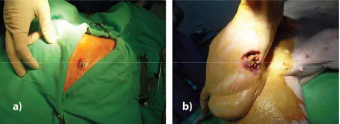
Fig. 1 – a) The entrance wound and b) exit wound (gauze packing). The dimensions of all exit wound openings are given in
Table 1. The largest exit openings were found in wounds number 6 and 14, which showed the maximum mass of devi-talized tissue, related to 1 mm length of the wound channel (m/l).
The clinical patterns of the wound
The primary surgery of the wound
The data obtained in this study were related to the extent and severity of the injuries in the affected tissues. The skin injury was assessed through the characteristics of the entrance and the exit wound openings (Figure 1). The en-trance wound openings were of regular round shape skin de-fects with the diameter of 8 8 mm. All edges of the hole were bruised with the contusion zone 2–3 mm wide.
The exit openings were irregular, star-like lacerations, with strips of ischemic skin, detached from the subcutaneous tissue, requiring excision. The skin injury was wider at the exit than at the entrance, with bruise size up to 10 mm from
the edge of the wound, which sometimes spreaded into the intracutaneous hematoma. Skin vitality estimation was easily made and a narrow zone of clearly mashed and avascular skin straps was excised.
The differences in the clinical aspect of necrosis in mu-scles during PS of the wound are shown in Table 2.
Table 2 The differences in the clinical aspect of necrosis during
primary surgery (PS) and after 24 h Animals (n) during PS after 24 h Necrosis (extent)
A B A B
0 0 0 4 3
1 4 5 3 4
2 3 2 0 0
3 0 0 0 0
Total 7 7 7 7
Vol. 72, No. 9 VOJNOSANITETSKI PREGLED Page 789
Djenić N, et al. Vojnosanit Pregl 2015; 72(9): 785–793.
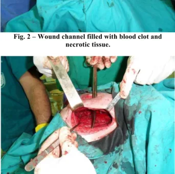
The muscles showed different degrees and extent of the injuries. The wound channels were filled with blood clots and necrotic tissue caused by tissue contusion due to direct bullet action (Figure 2). There was a circular zone of the de-vitalized muscle tissue around the wound channel. Devitali-zed muscle was of dark red color, not elastic consistency without contractility on touch with a pair of pincers and without capillary bleeding on the surface section. The line between the zone with clear signs of devitalization and the region of healthy muscle was quite clearly visible (Figure 3).
Fig. 2 – Wound channel filled with blood clot and necrotic tissue.
Fig. 3 – The completed primary surgery with clear zone of healthy muscles.
The numerous smaller or larger foci of hemorrhage were also found in muscles. A mass of devitalized excised tissue during PS was shown in the Table 1 for all wounds. This Table clearly presents the devastating effect of a bullet by measuring and comparison of the size of exit hole, the length of the wound channel and the mass of the excised de-vitalized tissue. A femoral blood vessel was injured and liga-ted in only one case. Drains were placed after PS through all the wounds. The wounds in the group B were covered with FGA along wound channel and primarily closed (Figure 4).
Thrombin and fibrinogen solutions were loaded into a double-barreled syringe that allowed them to mix and com-bine and injected in the wound channel.
Wound revision
All of 14 PS treated wounds underwent revision 24 h. The whole wound channel is longitudinally open that gave the possibility to well overview the clinical appearance of the wound.
The differences in the clinical aspect of necrosis after 24 hours are shown in Table 2.
The results of PS in all wounds fulfilled the clinical requirements, so the wounds from both groups were assessed as effectively treated (Table 2). Since there were no or minimal signs of necrosis, hemorrhage and infections (0–1) in animals from the group B, FGA was applied again, then the drain was placed and wounds were closed primarily (Figure 5).
Fig. 4 – Application of fibrin glue with antibiotics after pri-mary surgery.
Fig. 5 – After wound revision a drain was placed and fibrin glue with antibiotics applied.
The drain was removed 24 h after revision. Later, du-ring the experiment, the pigs were in good general condition, with body temperatures within the normal range and taking food and water normally. All animals survived the first 7 days after the injury.
Light microscopy
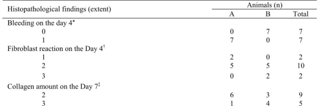
Table 3 The differences in histopathological findings on the days 4 and 7 after wounding
Animals (n) Histopathological findings (extent)
A B Total
Bleeding on the day 4*
0 0 7 7
1 7 0 7
Fibroblast reaction on the Day 4†
1 2 0 2
2 5 5 10
3 0 2 2
Collagen amount on the Day 7‡
2 6 3 9
3 1 4 5
For abbreviations see under Table 2.*Fisher’s exact test, p < 0.01. † Somers’d 2.84, p < 0.01.‡ Somers’d 2.1, p < 0.05.
Fig. 6 – a) The inlets of fibrin glue and bunched thick collagen fibers, and b) The fibrin glue and young granulation tissue with the numerous blood vessels (hematoxyllin and eosin, 100).
Fig. 7 – The presence of mature collagen connective tissue, lymphocyte and giant cell reaction around unabsorbed fibrin glue remains (Masson trichrome, 200).
absent or minimally spread necrosis of muscle tissue, while the degree of inflammatory reaction was usually minimal to mode-rate both in the group A and B. There were no statistically signi-ficant differences after 24 h between the groups in the degree of necrosis, bleeding and inflammation.
In samples taken on the postoperative day 4 with and without FGA, a moderate inflammatory reaction was registered with mixed cellular composition, composed of lymphocytes and macrophages, and less often of polymorphonuclears, while nec-rosis was absent or poorly spread. The young granulation tissue was found, rich in blood vessels, as well as the fibroblasts and the fibrocytes with the initial production of immature collagen of a moderate degree.
On the postoperative day 4 in the group B, acellular eosi-nophilic homogeneous material was observed, which represen-ted unabsorbed residues of the fibrin glue, locarepresen-ted on the surfa-ce of the wound or in the shape of small irregular islands, surrounded by the collagen connective tissue. The
mononuc-lear inflammatory cells, type of lymphocytes and macropha-ges, were little to moderately present, while the connective tissue cells, fibroblasts and fibrocytes were prevalent. The thick and dense collagen fibers arranged in the form of bun-dles, as well as the young granulation tissue with numerous blood vessels were observed (Figure 6).
The differences in bleeding after 4 days are shown in Table 3.
The statistically significant differences between the groups A and B after 4 days were found in the degree of ble-eding and fibroblast reaction (Table 3) (p < 0.01), whereas the differences in the degree of necrosis and inflammation, as well as in the produced amount of collagen and the number of giant cells were not statistically significant.
Vol. 72, No. 9 VOJNOSANITETSKI PREGLED Page 791
Djenić N, et al. Vojnosanit Pregl 2015; 72(9): 785–793.
Statistically significant differences after 7 days of surgery between the groups A and B were found in the amount of the produced collagen, while the differences in the degree of necro-sis, inflammation, and fibroblast reaction were not statistically significant (Table 3).
Discussion
More than 70% of all firearm injuries in modern wars and civil practice are injuries of extremities, and 60% of them are soft-tissue injuries 30, 35.
These injuries most often belong to the second degree of urgency. This implies that surgery may be postponed for a cer-tain period of time, without the fear of developing of adverse consequences because of the delay, if properly treated 4, 40.
Soft-tissue gunshot wounds of extremities are characteri-zed by the high morbidity and low mortality. Their high inci-dence is the problem for the second and third military level (ec-helon) of care 31, 32. The basic war surgery principles of aggressi-ve resuscitation, early and thorough debridement, short-duration damage-control surgical procedures, and rapid evacuation were critical in reduction of wound infection rates 35, 36. Nowadays the frequency of this kind of injuries is increasing because of crimi-nal and terroristic activities. A high incidence of soft tissue inju-ries and the difficult conditions for the surgical treatment raise the question: how to shorten hospital treatment and provide bet-ter surgical approach. The timing of wound closure is important. Delayed primary closure is wound closure performed within 4–7 days after the injury. The aim of DPS is to close the wound du-ring the fibroblastic phase of wound healing. This occurs between the days 3 and 6 following injury 15.
The original approach to treatment of gunshot wounds is applied in this experiment.
There are no data in the medical literature about the experimental monitoring of the healing process of shot wounds after PS treatment to the full healing, as well as the data about experimental application of FGAs within PS treatment. The available works only show the effectiveness of FG as hemostatic in war wounds 41–44. Fibrin sealants have several advantages. They speed up the formation of a stable clot; they can be appli-ed to very small blood vessels and to areas that are difficult to reach; they lower the risk of postoperative inflammation or in-fection; they provide slow-release delivery of medications (an-tibiotics) to tissues exposed during surgery and they are conveniently absorbed by the body during the healing process.
Greco et al. 45 presented that the delivered amount of each drug was enough to maintain the minimal inhibitory con-centration until the day 4 of culture for the most of antibiotics, resulting in a prolonged release of the drug. Kram et al. 46 psented that the addition of antibiotics to fibrin sealant clots re-sulted in continuous diffusion of antibiotics into the surroun-ding for up to 5 to 7 days. The antibacterial effects of fibrin sealant clots with antibiotics were significantly higher compa-red to fibrin sealant clots without antibiotics. In addition, the presence of fibrin sealant clots with antibiotic resulted in a re-duction in bacterial growth 46.
Fibronectin consisted in FG can locally support creation of fibrin polymers, adhesion of fibroblasts and reepithelization of
the tissue, and that speeds up the wound healing process 18, 41–44. Personal war experience and experimental works are the source of the idea how to improve the surgical approach and the treat-ment of the wounds caused by a projectile of a high initial velocity 16, 18, 26, 47.
The M70AB2 automatic rifle has the initial velocity of projectile M67 of 720 m/s, which is close to the initial high velocity level. For this reason and with the intention to avoid bone damage, shooting was done from the distance of 5 m 26. Projectile M67 after causing short straight channel (9 cm ± 3 cm) starts to destabilize. The shapes and dimensions of the exit wound are directly dependent on the quantity of the energy transferred from the projectile, which is proportional to the length of the wound channel and the degree of destabilization of the bullet 23, 48, 49. The wound channel with the length over 100 mm is characterized by large devitalization, which may be measured by the amount of excised tissue. The mass of devitali-zed tissue is increasing in proportion to the length of wound channel. The basic knowledge about ballistic gunshot wounds, as well as about destructive power of certain weapons, is needed to be known by the surgeon in order to assess the nature and extent of injury and to apply the most efficient treatment. Our study confirmed that the applied primary surgery of the wounds induced with a shot with a M70AB2 rifle was fully effecti-ve 16, 23, 26, 47.
The margins of the irreversible circulatory disturbances become clearly recognized only after several hours from injury, and they are ideal limits for the removal of necrotic tissues du-ring primary surgery. The signs of devitalization are much clea-rer after six than after one hour from injury. There are no signi-ficant differences in the extent of the irreversible changes between 6 and 12 h. The pathological changes after 24 h were more clearly expressed than after 12 h, but there was no increase of a necrotic zone. After 24 h an additional necrosis can be de-veloped only as a result of the impaired circulation or an infecti-on 50. For that reason, primary closure was performed after 24 h in the group B.
Histopathologically no statistically significant differences in the degree of necrosis, inflammation and bleeding were regis-tered between the groups A and B, after 24 h. There were signi-ficant differences in the degree of bleeding and fibroblast reacti-on between the groups A and B after 4 days and in the amount of produced collagen after 7 days. This suggests a local protecti-ve and general stimulating effect of FGA.
We gave 1 g of ceftriaxone preventively to all animals beca-use of its broad spectrum of antimicrobial activity and the simplicity of application. The use of antibiotics can prevent infec-tion and local metabolic disturbances, limit local tissue destructi-on, and reduce the amount of necrotic tissue in penetrating guns-hot wounds of soft tissue 30, 31, 51. Systemic use of antibiotics is necessary before starting primary surgical therapy that is a part of the Definitive Surgical Trauma Care in the days that follow 51, 52.
Conclusion
closed primarily with delayed suture without the risk of develo-ping complications if on revision, 24 h after primary surgery, there were no present necrotic tissues, hematoma, and any signs of infection when fibrin glue with antibiotics (ceftriaxone and clindamycin) was applied.
The use of this method should be limited to individual and strictly controlled cases in civil practice for now. The application of this method in echeloned military field me-dical systems dealing with casualties needs further scienti-fic confirmations.
R E F E R E N C E S
1. Friedrich PL. Antiseptic treatment of fresh wounds. Langenbecks Arch Klin Chir 1898; 57: 288−310. (German)
2. Stanojević V. The history of medicine. Belgrade-Zagreb: Medicinska knjiga; 1964. (Serbian)
3. Todorić M. Wounds by firearms projectiles. In: Dragovic M, Todoric M, editors. Emergency and war surgery. Belgrade: Velarta; 1998. p. 57−66.
4. Višnjić M. Mechanism of injury in war wounds. In: Višnjić M , edi-tor. War surgery. Niš: Kulturni centar; 2000. p. 17−26. (Serbian) 5. ManringMM, Hawk A, Calhoun JH, Andersen RC. Treatment of war
wounds: a historical review. Clin Orthop Relat Res 2009; 467(8): 2168−91.
6. Nikoliš G, Brecelj B, Dimković D, Gušić B, Kralj I, Lavrič B, et al. War Surgery I. Belgrade: SU JNA; 1953. p. 5−18. (Serbian)
7. Papo I, Piščević S, Funtek M, Đuknić M, Arneri V, Bervar M. War Sur-gery. Belgrade: Vojnoizdavački zavod; 1980. p. 50−4. (Serbian) 8. Danić R. War injuries - general pathology and therapy. Belgrade:
Vojno-Sanitetski Glasnik; 1939. (Serbian)
9. Pavlović M, AntićČ, Ćirić S, Đenić N, Vitas R, Popović-Filipović S. Mili-tary Hospital in Nis. Belgrade: Vojnoizdavački zavod; 2010. p. 25−6. (Serbian)
10. Đenić N, Ćirić S, Popović-Filipović S. Denić N, Cirić S, Popović -Filipović S. On 130th anniversary of Military Hospital in the town of Nis: January, 1878-January, 2008. Vojnosanit Pregl 2008; 65(1): 69−80. (Serbian)
11. Milanović M. Eminent Serbian physicians. Belgrade: Vojna štam-parija; 2005. (Serbian)
12. Giannou C, Baldan M. Mechanisms of injury during armed conflict. In: Giannou C, Baldan M, editors. War surgery: working with lim-ited resources in armed conflict and other situations of violence. Vol. 1. Geneva: International Committee of the Red Cross; 2009. p. 53-78
13. Szul AC, Davis LB. Walter Reed Army Medical Center Borden In-stitute.Emergency War Surgery: Third United States Revision, 2004 (Textbooks of Military Medicine). Washington, DC: United States Department of Defense; 2004.
14. Musalatov HA, Eliseev AT, Brovkin SV, Kostin VA. Soft Tissue Wounds. In: Musalatov HA, editor. Surgery of disasters: A text-book. Moscow RF: Meditsina; 1998. p. 237−57. (Russian) 15. Dufour D, Kromann Jensen S, Owen-Smith M, Salmela J, Stening GF,
Zet-terström B. Surgery for victims of war. 3rd ed. Geneva: International Committee of theRed Cross; 1998.
16. Todorić M. Primary surgical care of soft-tissue injuries caused by bullets from modern military rifles in an animal experiment. Vo-jnosanit Pregl 1985; 42(2): 83−7. (Serbian)
17. Balint B, Cernak I, Petakov M, Bugarski D, Malićević Z, Mandic-Radić S, et al. The use of single-donor fibrin glue prepared by recycled cryoprecipitation in experimental liver surgery. Haematologia (Bu-dap) 2002; 32(2): 135−45.
18. Stanojković Z, Stanojević G, Stojanović M, Milić D, Zivić S. Determina-tion of fibrin glue with antibiotics on collagen producDetermina-tion in colon anastomosis. Vojnosanit Pregl 2008; 65(9): 681−7.
19. Tredwell S, Jackson JK, Hamilton D, Lee V, Burt HM. Use of fibrin sealants for the localized, controlled release of cefazolin. Can J Surg 2006; 49(5): 347−52.
20.Spotnitz WD. Commercial fibrin sealants in surgical care. Am J Surg 2001; 182(2 Suppl): 8S−14S.
21.Thompson DF, Davis TW. The addition of antibiotics to fibrin glue. South Med J 1997; 90(7): 681−4.
22.Jackson MR. New and potential uses of fibrin sealants as an ad-junct to surgical hemostasis. Am J Surg 2001; 182(Suppl 2): S36−9.
23.Fackler ML. Wounding patterns of military rifle bullets. Int Def Rev 1989; 1: 59−64.
24.Berlin R, Gelin LE, Janzon B, Lewis DH, Rybeck B, Sandegård J, et al. Local effects of assault rifle bullets in live tissues. Acta Chir Scand Suppl 1976; 459: 1−76.
25.Berlin R, Janzon B, Rybeck B, Sandegärd J, Seeman T. Local effects of assault rifle bullets in live tissues. Part II. Further studies in live tissues and relations to some simulant media. Acta Chir Scand Suppl 1977; 477: 5−48.
26.Ćakić J, Dimitrijević J, Simović M, Nanusević O, Durdević D, Milovanović S. The effect of gammaphos on the course and out-come of surgical treatment in combined radiation injuries in pigs. Vojnosanit Pregl 1994; 51(3): 179−91. (Serbian)
27.Dufour D, Kromann JS, Owen SM, Salmela J, Stening GF, Zetter-strom B. Delayed primary closure and skin grafts. In: Molde A, editor. Surgery for victims of war. 3rd ed. Geneva: ICRC. 1994. p. 39−40.
28.Edlich RF, Rodeheaver GT, Thacker JG, Lin KY, Drake DB, Mason SS, et al. Revolutionary advances in the management of trau-matic wounds in the emergency department during the last 40 years: part I. J Emerg Med 2010; 38(1): 40−50.
29.Giannou C, Baldan M. Surgical management of war wounds. Dressings. In: Giannou C, Baldan M, editors. War surgery: working with limited resources in armed conflict and other sit-uations of violence. Vol. 1. Geneva: International Committee of the Red Cross; 2009. p. 225−6.
30.Jovanović Z, Popović Z. War injuries of extremities. In: Dragović M, Todorić M, editors. Emergency and war surgery. Beograd: Velarta; 1998. p. 792−800. (Serbian)
31.Nikolić D. War injuries of the extremities. Vojnosanit Pregl 2004; 61(5): 547−56.
32.Todorić M. War surgical principles of organization. In: Dragovic M, Todoric M, editors. Emergency and war surgery. Belgrade: Velarta; 1998. p. 76−80. (Serbian)
33.Gray R. War wounds: basic surgical management. Geneve: In-ternational Committee of the Red Cross; 1994. p. 24−31. 34.Dougherty PJ, Najibi S, Silverton C, Vaidya R. Gunshot wounds:
epidemiology, wound ballistics, and soft-tissue treatment. Instr Course Lect 2009; 58: 131−9.
35.Taylor N, Ingari J, Baechler M, Levin LS. Extremity War Injuries and Damage Control Orthopaedic Surgery. 65th annual
Meet-ing of the ASSH; 2010 October 7. Boston; Boston MA: ASSH Annual Meeting. 2010. Available from: http://www.assh.org/AnnualMeeting/program/Pages/Instru ctional-Course-Lectures.aspx
36.Mazurek MT, Ficke JR. The scope of wounds encountered in casualties from the global war on terrorism: from the battle-field to the tertiary treatment facility. J Am Acad Orthop Surg 2006; 14(10 Spec No.): 18−23.
Vol. 72, No. 9 VOJNOSANITETSKI PREGLED Page 793
Djenić N, et al. Vojnosanit Pregl 2015; 72(9): 785–793.
38.Giannou C, Baldan M. Red Cross wound score and classification system. In: Giannou C, Baldan M, editors. War surgery: working with limited resources in armed conflict and other situations of violence. Vol. 1. Geneva: International Committee of the Red Cross; 2009. p. 83−91.
39.Bartlett CS, Helfet DL, Hausman MR, Strauss E. Ballistics and gunshot wounds: effects on musculoskeletal tissues. J Am Acad Orthop Surg 2000; 8(1): 21−36.
40.Katoch R, Rajagopalan S. Warfare injuries: history, triage, trans-port and field hospital setup in armed forces. Med J Arm Force Ind 2010; 66(4): 304−8.
41.Alam HB, Burris D, DaCorta JA, Rhee P. Hemorrhage control in the battlefield: role of new hemostatic agents. Mil Med 2005; 170(1): 63−9.
42.Jackson MR. Fibrin sealants in surgical practice: An overview. Am J Surg 2001; 182(Suppl 2): 1S−7S.
43.Mankad PS, Codispoti M. The Role of Fibrin Sealants in hemo-stasis. Am J Surg 2001; 182(Suppl 2): 21S−8S.
44.Morikawa T. Tissue sealing. Am J Surg 2001; 182(Suppl 2): 29S−35S.
45.Greco F, de Palma L, Spagnolo N, Rossi A, Specchia N, Gigante A.
Fibrin-antibiotic mixtures: Anin vitro study assessing the pos-sibility of using a biologic carrier for local drug delivery. J Biomed Mat Res 1991; 25(1): 39−51.
46.Kram HB, Bansal M, Timberlake O, Shoemaker WC. Antibacterial effects of fibrin glue-antibiotic mixtures. J Surg Res 1991; 50(2): 175−8.
47.Albreht M, Sćepanović D, Ceramilac A, Milivojević V, Berger S, Ta-sić G, et al. Experimental soft tissue wounds caused by stan-dard military rifles. Acta Chir Scand Suppl 1979; 489: 185−98.
48.French RW, Callender RG. Ballistic Characteristics of Wound-ing Agents. In: Coates JB, Brayer JC, editors. Wound ballistics. Washington, DC: Office of the surgeon general, Department of the Army; 1962. p. 91−141.
49.Hopkinson DA, Marshall TK. Firearm injuries. Br J Surg 1967; 54(5): 344−53.
50.Wang ZG, Qian CW, Zhan DC, Shi TZ, Tang CG. Pathological changes of gunshot wounds at various intervals after wound-ing. Acta Chir Scand Suppl 1982; 508: 197−210.
51.Hospenthal DR, Murray CK, Andersen RC, Bell RB, Calhoun JH, Cancio LC, et al. Guidelines for the prevention of infections associated with combat-related injuries: 2011 update: en-dorsed by the Infectious Diseases Society of America and the Surgical Infection Society. J Trauma 2011; 71(2 Suppl 2): 210−34.
52.Boffard KD. Extremity trauma. In: Boffard KD, editor. Manual of Definitive Surgical Trauma Care. 3rd ed. London: Hodder Arnold; 2011. p. 154−62.