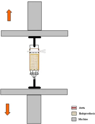
Biomechanical and histologic analysis in aortic endoprosthesis using fibrin glue
Texto
Imagem


Documentos relacionados
Apesar da amostragem por conveniência não probabilística não ter permitido um estudo de prevalência mais apurado, uma vez que a amostra não é representativa da
Transplantation of muscle-derived stem cells plus biodegradable fibrin glue restores the urethral sphincter in a pudendal nerve-transected rat
The patients were assigned to 3 groups with an equivalent number of participants each, according to the material used in the conjunctival graft fixation technique: Nylon Group (NG)
No caso e x p líc ito da Biblioteca Central da UFPb, ela já vem seguindo diretrizes para a seleção de periódicos desde 1978,, diretrizes essas que valem para os 7
É um período de grandes mudanças na Ciência e tem as principais características na obra de Galileu, inspirado pelas idéias de Francis Bacon (falecido em 1626) e Descartes
Esse trabalho mapeou os principais fatores críticos para o sucesso de projetos de implantação de ERP de manutenção industrial em usinas do ramo sucroalcooleiro, através
Foi realizado um estudo observacional, prospectivo, analítico, tipo coorte onde foram acompanhados recém-nascidos internados em Unidade de Terapia Intensiva Neonatal,
Neste trabalho o objetivo central foi a ampliação e adequação do procedimento e programa computacional baseado no programa comercial MSC.PATRAN, para a geração automática de modelos
