A Multi-Stage Algorithm for Enhanced
X-Ray Image Segmentation
ADITYA A. TIRODKAR
B.E. Student, Department of Computer Engineering,
Thadomal Shahani Engineering College, Mumbai University
29, Pragati Bhavan, Sadashiv Cross Lane,
Girgaum, Mumbai-400004
Cell. No: +91-9820492348
1. Abstract
With the ever increasing usage of empirical data collected from X-Ray and other Digital Imaging techniques, it has become imperative that this data be subjected to computer algorithms for speedy and more accurate diagnosis. Segmentation is one of the key techniques that are employed during the pre-processing stages of these algorithms for separating those details from the images that are required for analysis. There are currently a number of widespread techniques for segmentation, in use. Our proposed algorithm is a quick and more qualitatively efficient technique for segmentation that is optimized for X-Ray images. It applies Otsu’s algorithm to provide thresholding values that can be used for contrasting and binarizing the images. Also, an edge detection technique has been applied to better evince observations, allowing more fruitful extraction of information and the algorithm has itself been tested on a set of 40 images.
Keywords: X-Ray, Segmentation, Otsu, Edge Detection, Digital Imaging
2. Introduction
Digital Imaging is now a cornerstone in the provision of diagnostic data to medical practitioners. It involves the conveyance of information regarding various internal body organs which can be then used for determining the correct cause of any ailment and facilitating the provision the requisite treatment. Magnetic Resonance Imaging (MRI), computed tomography (CT), digital mammography and other imaging modalities are in ever increasing use as critical components in diagnostic techniques [4].
Computer Technology has been brought to the fore for bringing such effective and voluminous number of medical images possible. Computer algorithms play a major part in extricating data from a medical image such as facilitating and automating the delineation of anatomical structures, checking the tortuousness of bones, identifying bone cracks and various other biomedical applications [10]. Computer integrated surgery and computerised diagnostic techniques use pre—processed images. One major such technique lies in the realm of X-Ray image analysis.
• Study of Bone Structure • Identification of Bone Fracture • Measurement of Fracture Treatment • Treatment Planning Prior to Surgery [1]
The process of segmentation however, is one that is quite challenging in its nature. In fact, segmentation alone can determine the eventual success or failure of the analysis at hand. Segmentation involves working on a number of facets of an image which include noise and distortion, homogeneity of pixel intensities, closeness in grayscale levels and the perceivable lack of definition [1]. Thus, it involves obtaining a clear image, ensuring that pixels have clearly demarcated intensities and then obtaining thresholds in order to divide the image into a bright and a dark region to carry out segmentation.
3. Past Literary Work/ Literary Review:
Segmentation techniques can be basically categorized into two main groups, spread across three generations of algorithmic complexity [2]. The first generation of segmentation techniques involved low-level techniques which contain simple image analysis techniques such as heuristic edge tracing and region growing. The second generation involves uncertainty models and optimization methods which include c-means clustering, optimal graph search algorithms, neural networks etc. Lastly, the third generation involves techniques that are based on learning. Thus, they contain a certain amount of empirical knowledge about the segmentation process as it happens. Such methods that incorporated higher-level knowledge include atlases, tissue maps etc. which contain a priori information, models and expert-defined information about a desired object.
The two main groups compositing the segmentation techniques elaborated above are gray level feature based methods and texture feature based methods [1]. Gray level feature based techniques involve using the grayscale value of a particular image pixel. The gray level is calculated by:
Gray Level = (R+G+B) / 3
This gray level value can then be used to provide the threshold required for the segmentation of an image. Histogram based methods [3], Edge based methods [4] and region based methods [5] all fall under this category. The lacunae of these techniques are involved with the correct calculation of the image’s threshold. Noise and other distortion can ruin this calculation, rendering these techniques inefficacious.
Textural techniques are geared towards segmenting an image based on similar textural properties [2]. Models which incorporate active shape and appearance can be used. Atlas based techniques which invoke information from in-built resources on anatomy; shape, size etc. are also used for this purpose. However, these techniques require the expert implementation of the resource database. They are also slower in performance due to the manual tracing required and are not always accurate. Segmentation based on sonography images greatly uses these techniques.
Morphological and wavelet based transforms are also used to carry out segmentation on a smaller scale and involve reconstruction of the image. In fact, the next step posited in the usage of segmentation often involves the use of these transforms in order to compare images with a knowledge set of images that can point to certain ailments or conditions [1].
4. Proposed Algorithm:
4.1 Initial Pre-processing
• The initial step involves obtaining the image and converting it into an array-map containing pixel values. Since X-Ray images are inherently Black and White, we can state that all R, G, B values shall be equal. If not, the image can always be converted into Black and White. The array-map now contains the individual pixel values and locations of the X-Ray’s pixels.
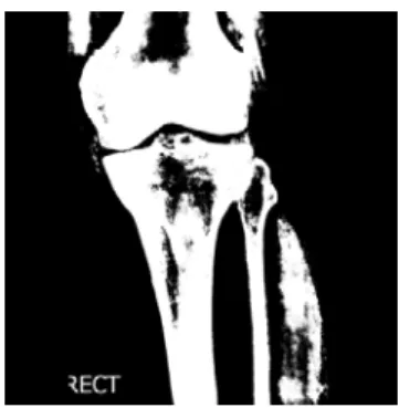
• It is evident that whenever a holistic transform incorporating all the pixels in the image is taken, the number of black pixels will be huge. Thus it can be surmised that any kind of thresholding at this juncture would lead to demarcation of only the majority dark and majority light regions. Thus, only the outline of the body part in the X-Ray would be seen which provides us with no definition at all as seen in the Figure 1.
Original Image Normal Thresholding Figure 1: Images obtained by Applying Otsu’s Thresholding Technique With no Pre-Processing
The algorithm thus, modify the original image to be taken for segmentation by removing the every single pixel in the image with a grayscale value less than 10. Thus, this leads to any superflous black pixels in the image getting replaced with white which entails that now, the variations in gray seen inside the body part that signify the bone shall actually be darker than the surrounding pixels. Thus, this should make the bone’s lighter pixels stand out. Such an image is seen in Figure 2.
Fig. 2 Modified Effective Image for Segmentation
4.2 Image Contrasting
Original Image Contrasted Image Figure 3: Contrasted Image with Threshold Obtained by Using Otsu’s Algorithm
4.3 Image Thresholding
• Thresholding is a technique that involves the calculation of a correct value that enables the division of an image into two sections disparate on grayscale value i.e. the background (darker part) and the foreground (brighter part) [6]. Thus, it allows the entire image to be converted into a monochrome image. Otsu’s algorithm is the primary technique that is used for this purpose. Otsu's thresholding method involves iterating through all the possible threshold values and calculating a measure of spread for the pixel levels each side of the threshold, i.e. the pixels that either fall in foreground or background [7]. The aim is to find the threshold value where the sum of foreground and background spreads is at its minimum. For this purpose, the property of variance is used and is implemented using a histogram calculated for the image. The histogram values are then calculated for arbitrary values of threshold using the formulae given below. The threshold value that gives us the lowest value of Within Class Variance or the Highest Value of Between Class Variance is taken as final.
• To do so, one first finds the mean of the histogram values below and above the threshold and also the weighted mean of the histogram values. The answers are then tabulated into a formula that calculates the two values stated above:
[7]
Another technique used is the one provided by Sauvola called Adaptive Segmentation [8]. It involves an iterative segmentation process which involves first obtaining the threshold for a fixed window of values in the image. The window is then increased and the threshold value is modified. Thus, at every step, the threshold calculated includes the threshold of the newer set of pixels as well as the threshold of the old window. It can thus be optimized for a particular part of an image. For the proposed algorithm, Otsu’s technique is used since the entire X-Ray is to be binarized.
Original Image Optimized Binarized Image Figure 4: Thresholded Images
4.4 Edge Detection
One unnecessary property of segmentation techniques is that the image may look distorted. This is because the picture is essentially removed from its original perspective. It is not a monochrome image which lacks definition other than its perceivable shape and size [2]. However, shape and size are exactly the two properties one wishes to extrude from an image by using segmentation. Thus, this entails that for further processing and obtaining the final image one should be able to remove the basic information from the image i.e. the shape by identifying the edges of the image. Thus, the final part in the proposed algorithm involves employing edge detection on the monochrome image [9]. Generally, operators such as Canny or Prewitt are used for the same. However, for the current purpose we used an original technique based on simple pixel values in an image. The technique involves the following steps:
Firstly, this technique involves the conversion of the black and white image into an intermediate image that contains three colours: black, white and yellow. The yellow pixels indicate black pixels that do not border any white pixels i.e. any black pixels that are not at an edge. To find out which pixel to convert into yellow, we have to take into consideration exactly what should be the number of bordering pixels for a pixel at an edge.
Step 1: In the image-map, for each pixel: first find the number or bordering pixels in the 8X8 region surrounding it that are white or black. If a particular pixel is surrounded by even a single white pixel, then it is kept as white.
Step 2: If the number of black pixels is found to be less than four and the number of surrounding yellow pixels is not zero, then the pixel is turned white.
Step 3: If the above condition is not satisfied and it is seen that the current pixel has a greater or equal number of yellow bordering pixels than white bordering pixels, then the pixel is turned yellow.
Binarized Image Edge-Detected, Final Image Figure 5: Edge Detection
Thus, the final image provides us with a simple, low complexity image that can easily be used for obtaining edge-wise or shape and size based testing. Techniques such as wavelet identification can be applied on these images. Any superfluous information can be easily removed by merely improving the original X-Ray image quality and obtaining an image with the bone at a much higher colour intensity that the surrounding muscle or tissue. Also, in the final image, one can notice that the longest string of black pixels corresponds to the bones themselves . Thus, one can take that as a valuable and standalone parameter obtained from pre-processing.
Original Image
Pre-Processed Image
Contrasted Image
Binarized Image
Final Image
Figure 6: Flowchart Figure 7: Generated Outputs
5. Results
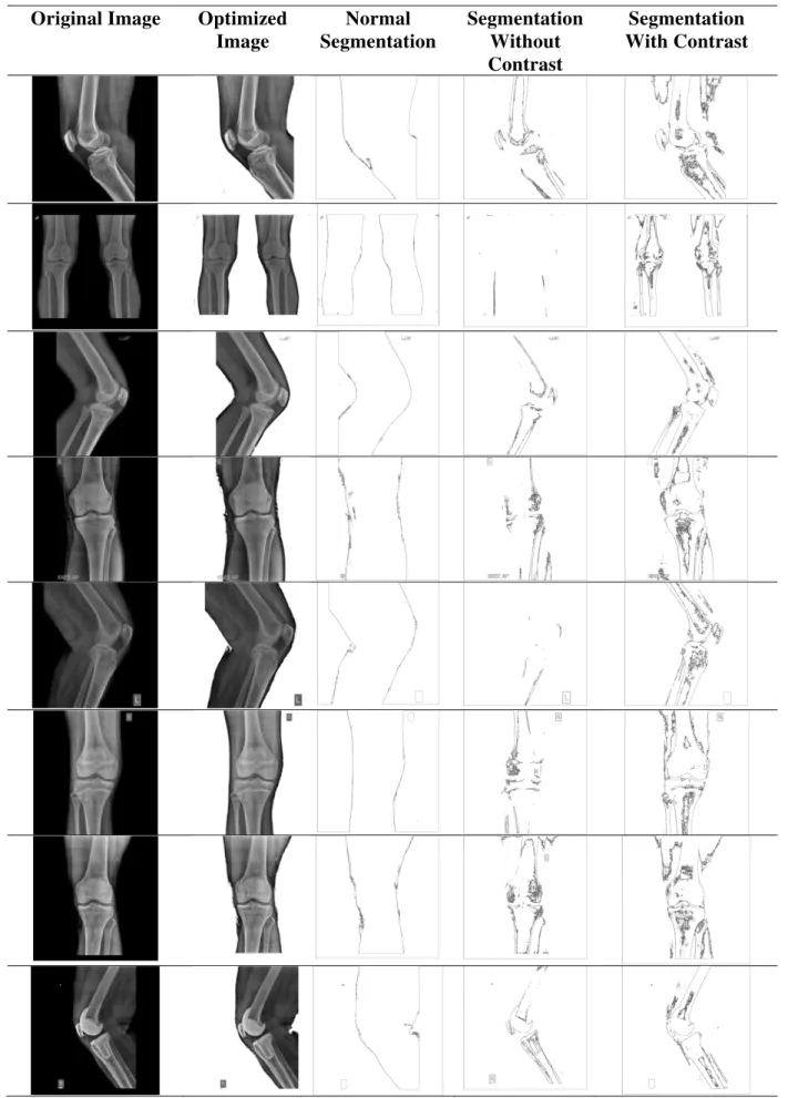
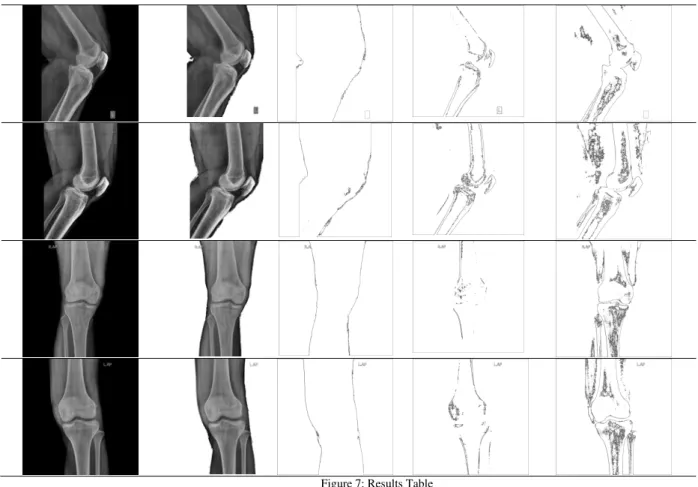
For further analysis, a set of radiology images has been obtained and compiled from a local eminent Radiologist and the proposed technique has been carried out on them. The set contains 40 images of left and right knee X-Rays of 20 patients. The results obtained were tabulated and compared for qualitative comparison between standard segmentation techniques and our technique. A sample set of 12 images has been demonstrated with the results thus obtained, is given for analysis:
Start
Pre-Processing
Contrasting
Thresholding
Edge Detection
Table 1: A Comparison of Results seen of the Test Images with Different Techniques
Original Image
Optimized
Image
Normal
Segmentation
Segmentation
Without
Contrast
Figure 7: Results Table
The above results were obtained on a Java Code run in the Netbeans IDE on an Intel i5-2400 3.10 GHz Computer with 4GB of RAM with 1GB Graphics Memory. The results were obtained in a matter of seconds with four intermediate images being generated in the process. As was seen, the contrasted segmentation better evinced the shape of the bone in the image. Without contrast, often the image was not at all clear albeit focussing on the bones more than tissue. Thus, if the X-Ray image itself is clearly defined, with a clear contrast between tissue and bone then the final result will also be more elaborate. Regardless, the conveyance of information in the final technique was found to be more in every single case than segmentation techniques such as those propounded in algorithms such as [1].
7. Conclusion
A qualitative comparison of the above images point out that the proposed algorithm provides us with a quick and easy method for segmentation. Also, due to the clear delineation of the steps required, it is easy to remove parts of the algorithm in order to obtain images that better suit the purpose. It brings to light the various fundamental modifications that much be brought about when working with X-Ray images. These modifications, when applied can much better help the pre-processing required by computer algorithms in cases such as fracture detection. Also, we can much better detect any changes in bone size or orientation since the shape and size is provided quite clearly. It thus, is a much more conducive algorithm for working with the segmentation of images than traditional segmentation techniques.
7. Acknowledgements
through his databank for providing the high quality X-Ray and sonography images which were used for testing the technique.
8. References
[1] Mahendran, S.K. and Baboo, S.S. Enhanced Automatic X-Ray Bone Image Segmentation Using Wavelets and Morphological Operators. IPCSIT vol.6 (2011) Pg.125-129.
[2] Withey, D.J. and Koles, Z.J. (2007) Three generations of medical image segmentation: Methods and available software, Int J Bioelectromag., Vol.9, Pp.67-68.
[3] Jung, C.R. and Scharcanski, J. (2005) Robust watershed segmentation using wavelets, Image and Vision Computing, Vol. 23, Issue 7, Pp. 661-669.
[4] Law, T., Itoh, H. and Seki, H. (1996) Image filtering, edge detection, and edge tracing using fuzzy reasoning, IEEE Trans PAMI, Vol. 18, Pp.481-491.
[5] Pham, D.L., Xu, C. and Prince, J.L. (2000) Current methods in medical image segmentation. Ann. Rev. Biomed. Engg. Vol. 2, Pp. 315-37.
[6] Schildt, Herbert. The Complete Java Reference, Seventh Edition. Chapter 25: Images Pg. 755-786.
[7] Otsu, Noboyuki. A Threshold Selection Method for Gray-Level Histograms, IEEE Transactions on Systems, Man and Cybernetics Vol. SMC-9 No. 1 Jan 1979. Pg. 62-66.
[8] Greensted, Andrew. The Lab Book Pages, Otsu Thresholding. 17 June 2010
http://www.labbookpages.co.uk/software/imgProc/otsuThreshold.html
[9] J. Sauvola*, M. PietikaKinen. Adaptive Document Image Binarization. Pattern Recognition 33 (2000) 225-236. [10] Rafael C. Gonzales, Richard E. Woods, Digital Image Processing, Addison Wesley, 1992.