Sexual reproduction in the Caribbean coral
genus
Isophyllia
(Scleractinia: Mussidae)
Derek Soto and Ernesto Weil
Department of Marine Science, Universidad de Puerto Rico, Recinto de Mayagu¨ez, Mayagu¨ez, Puerto Rico, United States
ABSTRACT
The sexual pattern, reproductive mode, and timing of reproduction of Isophyllia sinuosa andIsophyllia rigida, two Caribbean Mussids, were assessed by histological analysis of specimens collected monthly during 2000–2001. Both species are simultaneous hermaphroditic brooders characterized by a single annual
gametogenetic cycle. Spermatocytes and oocytes of different stages were found to develop within the same mesentery indicating sequential maturation for extended planulation. Oogenesis took place during May through April in I. sinuosaand from August through June inI. rigida. Oocytes began development 7–8 months prior to spermaries but both sexes matured simultaneously. Zooxanthellate planulae were observed inI. sinuosaduring April and inI. rigidafrom June through September. Higher polyp and mesenterial fecundity were found inI. rigida
compared toI. sinuosa. Larger oocyte sizes were found inI. sinuosathan inI. rigida, however larger planula sizes were found inI. rigida. Hermaphroditism is the exclusive sexual pattern within the Mussidae while brooding has been documented within the related generaMussa,ScolymiaandMycetophyllia. This study represents the first description of the sexual characteristics ofI. rigidaand provides an updated description ofI. sinuosa.
Subjects Developmental Biology, Marine Biology, Zoology
Keywords Caribbean, Mussidae, Coral reproduction, Hermaphroditic, Brooder
INTRODUCTION
Reproduction in corals consists of a sequence of events which include: gametogenesis, spawning (broadcasters), fertilization, embryogenesis, planulation (brooders), dispersal, settlement and recruitment (Harrison & Wallace, 1990). The success of the reproductive effort is determined largely by the timing, duration, frequency and intensity of the aforementioned events (Babcock et al., 1986). In corals, sexual pattern, mode of reproduction, fertilization, larval dispersal, recruitment and survivorship are key components in determining evolutionary fitness (Szmant, 1986;Edmunds, 2005;Vermeij, 2006; Weil, Croquer & Urreiztieta, 2009; Pinzon & Weil, 2011) which is defined as the product of sexual output (fecundity) and survivorship (Metz, Nisbet & Geritz, 1992). Consequently, the ability of coral species to adapt to modern-day environmental pressures depends greatly on the ability of species to reproduce effectively.
The reproductive characteristics of some scleractinian groups have been more thoroughly studied than others; however, little is known about the reproductive patterns
Submitted5 October 2015
Accepted7 October 2016
Published10 November 2016
Corresponding author
Derek Soto, derek.soto@upr.edu
Academic editor
Xavier Pochon
Additional Information and Declarations can be found on page 16
DOI10.7717/peerj.2665
Copyright
2016 Soto and Weil
Distributed under
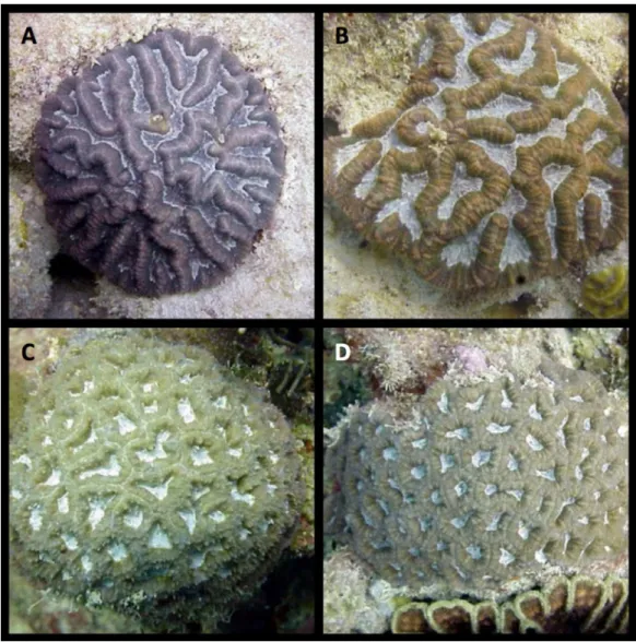
of many Caribbean coral species and some of the available information is conflictive or incomplete (Fadlallah, 1983;Harrison & Wallace, 1990;Harrison, 2011;Weil & Vargas, 2010;Pinzon & Weil, 2011). Of the approximately 60 Caribbean zooxanthellate coral species reported, thorough descriptions of their reproductive characteristics and cycles are available for 19 species; many other studies available provide partial or conflicting results (Weil, 2003;Weil & Vargas, 2010;Harrison, 2011). Reproductive studies of the sexual patterns ofI. sinuosawere among the first studies of such nature performed in the Caribbean (Duerden, 1902). These were limited to histological observations of oocytes in a few colonies ofI. sinuosa(Figs. 1Aand1B), therefore, the species is classified as gonochoric. This characterization contrasts with the reproductive mode of other studied Mussids which are classified as hermaphroditic. Currently, there is no information available on the reproductive biology ofI. rigida(Figs. 1Cand1D).
This study characterizes the reproductive biology ofI. rigidaandI. sinuosain terms of sexual pattern, mode of development, gametogenic cycles, and fecundity. These fundamental aspects of the physiology of this taxa are understudied. Knowledge of the reproductive biology and ecology of coral species is important for the interpretation of their population and ecological dynamics, their patterns/potential for dispersal, and their local and geographical distribution. The threats currently faced by coral reefs and the ongoing global effort to understand why corals are dying highlight the need to expand our understanding of basic coral physiology.
MATERIALS AND METHODS
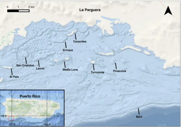
Sampling for this study was carried out at La Parguera Natural Reserve, off the southwest coast of Puerto Rico (Fig. 2). This complex reef environment is among the many regions experiencing deterioration by anthropogenic and environmental climate influences at local and global scales. Coral reefs in La Parguera are important local economic drivers, supporting artisanal and recreational fishing, tourism, recreational activities and also protect coastal settlements, seagrass communities and other wetland habitats from the effects of hurricanes and coastal erosion (Ballantine et al., 2008).
At least five unique sample cores were collected monthly for 14 months between March 2000 and May 2001 (Fig. 3A). A total of 89 samples of each species were collected. Colonies were selected by searching in a zig-zag pattern over the distributional range of both species (5–18 m). Samples were collected from San Cristobal reef (1755′24.88″N, 676′14.52″W), Caracoles reef (1757′46.02″N, 672′8.21″W), Media Luna reef
(1756′22.68″N, 672′43.26″W), Pinaculos (1756′1.13″N, 670′39.75″W), Turrumote reef (1756′13.56″N, 671′8.92″W), Beril (1752′47.85″N, 6659′1.40″W), El Palo (1755′50.2″N, 6705′36.9″W), Laurel (1755′50.2″N, 6705′36.9″W) and Enrique (1755′50.2″N, 6705′36.9″W) (Figs. 3Band4).
Sample cores were placed in Zenker Formalin (Helly’s solution) for 24 h, rinsed and then decalcified in 10% HCl solution. Tissues were then cleaned and placed in plastic tissue holders. Preserved samples were sequentially dehydrated in the rotary tissue processor under 70 and 95%, ethanol, Tissue Dry, and xylene solution (Tissue Clear III). Samples were embedded into Paraplast blocks then sectioned using a rotary microtome. The 8–10 strip sections (7–10mm) were obtained from each embedded block and placed
onto glass slides. Finished tissue slides were stained utilizing a modified Heidenhain’s Aniline-Blue method (Coolidge & Howard, 1979) to examine the maturation stages of gametocytes and embryos.
Slides were examined under an Olympus BX40 compound microscope coupled to an Olympus DP26 digital microscope camera. Images were captured utilizing Olympus cellSens 1.7 imaging software. The sexual pattern, gametogenic cycle and fecundity of
each species were determined by observing the gametocyte development throughout the collection year. Gamete stages were characterized according toSzmant-Froelich, Reutter & Riggs (1985). Oocyte sizes were obtained using cellSens, by taking perpendicular measurements at the cell’s widest points. Cell length and width measurements were used to calculate geometric area. Fecundity was assessed by counting oocytes per mesentery (I. sinuosan = 120;I. rigida n = 60) and per polyp (I. sinuosan = 10;I. rigida n = 5) on histologic cross-sections during months with the highest proportion of mature oocytes (I. sinuosaApril 2001 n = 5;I. rigidaMay 2001 n = 5).
In April 2012, several presumed gravid colonies of each species were collected and placed in an open seawater aquarium system to observe planulation. Two colonies of each species were placed within six-gallon aerated aquariums under continuously circulating seawater and daylight synchronized lights. Specimens were placed under mesh-lined PVC pipes allowing water to freely circulate. Traps were checked daily for larvae over a 90-day period.
Statistical analyses
Results are expressed as means ± standard error. All statistical tests were performed using the RStudio 0.99.484 software platform (R Studio Team, 2015) using the stats package (R Development Core Team, 2015). Normality was assessed using the Shapiro-Wilk test performed with the R function shapiro.test. Equality of variance was tested using the F test performed with the R function var.test. Differences in fecundity were tested by means of a Wilcoxon rank sum test with continuity correction performed with the R function wilcoxon.test.
Collection permit
All coral tissue samples were collected under a General Collection Permit granted by the Puerto Rico Department of Natural Resources (DNER) to the Faculty of the Department of Marine Sciences, University of Puerto Rico Mayaguez (UPRM).
RESULTS
I. sinuosaStage I oocytes are small (78.92 ± 13.15mm2), stain pink and are characterized by sparse
cytoplasm and prominent nuclei (Fig. 4A). Oocytes originate within the linings of the mesoglea in the central regions of the mesenteries. Stage II oocytes are larger than stage I cells (144.54 ± 43.19mm2), exhibit prominent nuclei and abundant cytoplasm (Fig. 4A).
Stage III oocytes are larger than stage II (264.51 ± 37.24mm2), tend to have a round
shape, stain pink or red, and are characterized by many cytoplasmic globules which produce a grainy appearance (Fig. 4B). Stage IV oocytes are larger and boxier than stage III (376.69 ± 73.20mm2). This stage is characterized by dark staining nuclei and large
globules in the cytoplasm (Figs. 4C–4E).
No stage I spermaries were found, suggesting this stage occurs briefly and/or is difficult to differentiate using the current method. Stage II spermaries form small poorly defined bundles which form in the mesenteries surrounding oocytes (Fig. 4C). Stage III
staining spermatids. Stage IV spermaries stain dark red and are larger than stage III. Tails visible on spermatozoa at high magnification are indicative of stage V spermaries (Fig. 4E). Spermary sizes were not measured.
Stage I planulae are approximately the same size as stage IVoocytes (404.07mm2) and stain
pink. During this stage, zooxanthellae become visible within the planulae. Stage II planulae (455.45 ± 32.84mm2) are characterized by an outer layer composed of columnar cells which
contain nematocysts and cilia (Fig. 4F). Developing mesenteries can be seen within the gastrodermis of stage III planula (501.98 ± 44.68mm2). Stage IV planula were not observed.
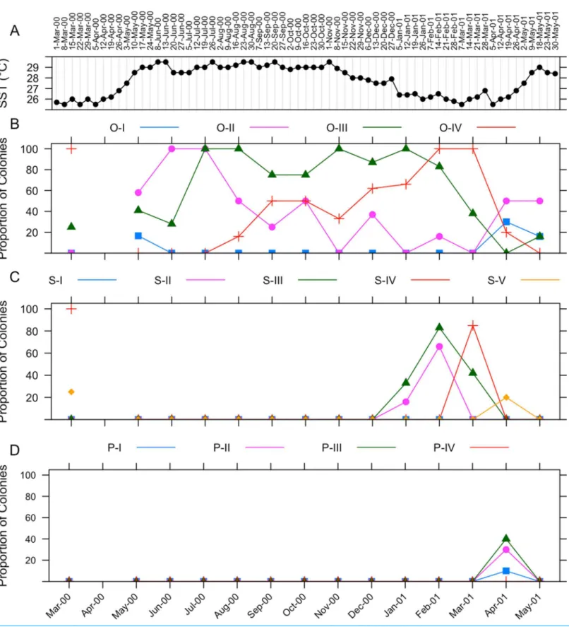
The gametogenic cycle ofI. sinuosais summarized inFig. 5. Weekly sea surface temperature measurements taken during the collecting period are included for reference (Fig. 5A). Oogenesis in I. sinuosalasts approximately 11 months (Fig. 5B). Onset of oogenesis was determined to occur during May 2000 and during April 2001. Onset of oogenesis was determined as the month of appearance of stage I and II oocytes after the culmination of the previous gametogenic cycle. Stage II oocytes were prevalent in tissues during all months sampled except during November 2000 and January 2001. Stage III oocytes were observed in all sampled months except April 2001. Stage IV oocytes were observed between August 2000 through May 2001.
Spermatogenesis takes places during four months (Fig. 5C). Onset of spermatogenesis was not determined because stage I spermaries were not identified. Stage II spermaries were observed during January through February 2001. Stage III spermaries were visible from January through March 2001. Stage IV spermaries were present in March 2001. Stage V spermaries were present in tissues in April 2001.
Stage I–III planulae were observed in histologic sections during April 2001 (Fig. 5D). The identification of planulae on tissue sections coincided with a sharp decrease in the proportion of colonies containing mature (IV) oocytes. No larvae were collected from specimens placed in aquaria for observation.
I. rigida
Stage I oocytes are very small (72.97 ± 15.75mm2) and are characterized by
sparse cytoplasm and a large nucleus. Stage II oocytes are larger than stage I cells (101.25 ± 23.09 mm2), are ovoid shaped and feature a prominent nucleus and nucleolus
(Fig. 6A). A pink-staining nucleus and red nucleolus can clearly be identified in many stage III oocytes (148.77 ± 49.35mm2) (Fig. 6B). Stage IV oocytes are large (190.40 ± 45.18
mm2), irregularly shaped and contain large vacuoles in the ooplasma which give it a
grainy appearance (Figs. 6Cand6D).
Stage I planulae are approximately the same size as stage IV oocytes (approximately 324.01 ± 71.64mm2), stain pink, and contain zooxanthellae in the epidermis.
Zooxanthellae were observed within planula beginning at this stage. Stage II planulae are
larger (521.27 ± 84.18 mm2) (Fig. 6F) and exhibit an epidermis consisting of columnar
epithelium similar toI. sinuosa. Stage III and stage IV larvae measure 818.91 ± 82.96mm2
and 951.78 ± 176.36mm2respectively, and show clear development of the mesenteries.
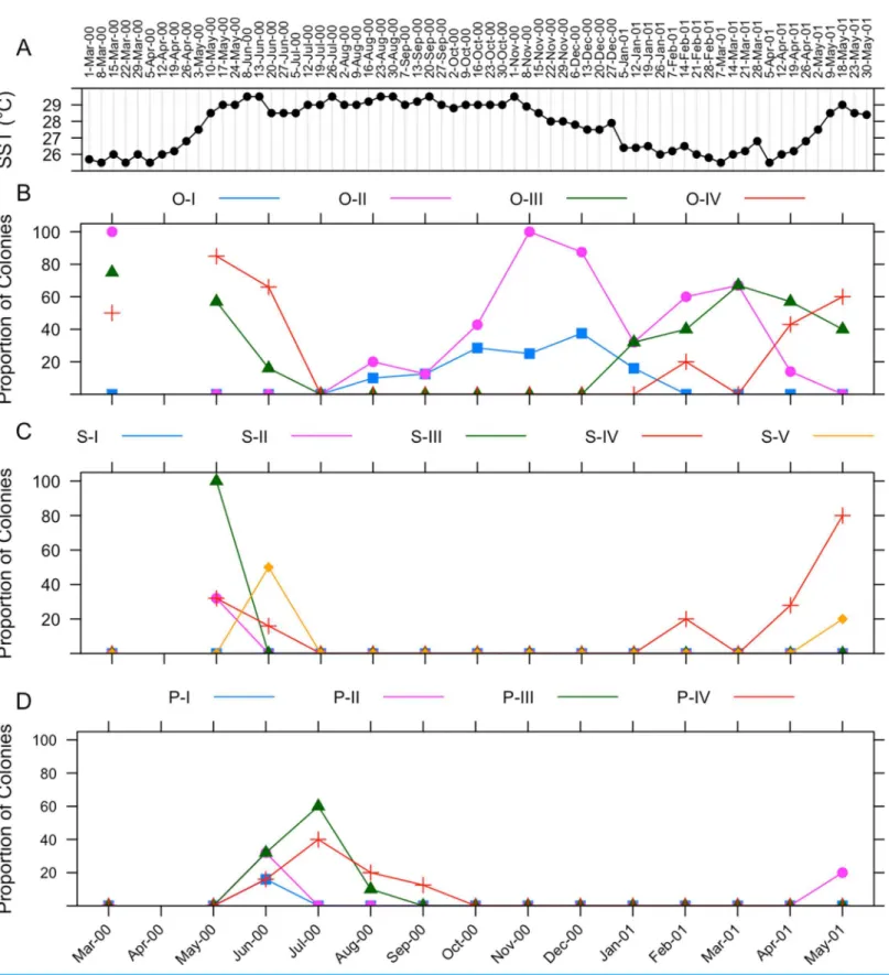
The gametogenic cycle ofI. rigida is summarized in Fig. 7. Weekly sea surface temperature measurements taken during the collecting period are included for reference (Fig. 7A). Oogenesis in I. rigida lasts approximately 10 months (Fig. 7B). Oogenesis began during August 2000. Stage II oocytes were observed in tissues in March 2000 and August 2000 to April 2000. Stage III oocytes were observed in March 2000, May and June 2000 and from January 2001 through May 2001. Stage IV oocytes were observed in samples collected during March, May and June 2000, and February, April and May 2001.
Spermatogenesis inI. rigida is estimated to last approximately 2–3 months (Fig. 7C). Onset of spermatogenesis was not determined because stage I spermaries were not identified. Stage II spermaries were observed in May 2000. Stage III spermaries were visible in May 2000. Stage IV spermaries were observed first in June 2000. Stage V spermaries were observed in May 2000.
Stage I planulae were observed in June 2000 indicating the onset of embryogenesis (Fig. 7D). The appearance of planulae coincided with a sharp decrease in the
proportion of colonies containing mature oocytes. Stage II planulae were observed during June 2000 and May 2001. Stage III planulae were observed from June through
August 2000. Stage IV planulae were observed in tissues from June throughout September 2000. No larvae were collected from specimens placed in aquaria for observation.
Fecundity
Mesenterial fecundity inI. sinuosa(11.13 ± 8.27 oocytes/mesentery) was significantly higher (Wilcoxon-rank sum test,W= 1,208,p< 2.210-16) than inI. rigida(1.70 ± 3.52 oocytes/mesentery) (Fig. 8A). Polyp fecundity inI. sinuosa(110.11 ± 96.33 oocytes/polyp) was significantly higher (Wilcoxon-rank sum test,W= 18,p= 0.018) compared to I. rigida (20.45 ± 23.91 oocytes/polyp) (Fig. 8B).
Oocyte size
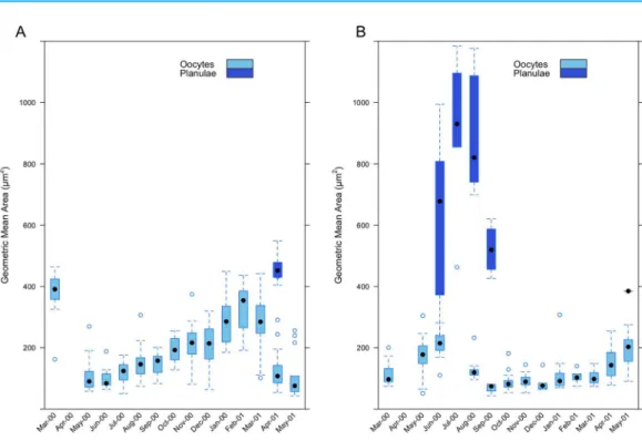
Measurements of oocyte geometric area inI. sinuosa(range 43.94–463.79mm2) show an
increase in the size of oocytes as maturity progresses from April through March (Fig. 9A). Mean geometric area is lowest during the month of June 2000 (97.22 ± 28.85mm2)
and greatest during February 2001 (333.95 ± 74.32mm2). The appearance of planulae
in histological sections during the month of April 2001 (459.07 ± 45.83mm2) (range:
404.07–548.49mm2) coincides with a sharp decrease in mean geometric area of oocytes
compared to the previous month (285.68 ± 96.46 vs. 143.28 ± 84.07mm2). Measurements
of oocyte geometric area inI. rigida(range 43.31–307.35mm2) also show a trend of
increasing oocyte size as maturity progresses from August through June (Fig. 9B). Mean geometric area is lowest during the month of September 2000 (68.35 ± 17.04mm2)
the month of July 2000 (909.48 ± 250.56mm2) and ranged from 241.66–1,183.96mm2.
Mean oocyte geometric area was greater in I. sinuosathan inI. rigida (Wilcoxon-rank sum test,W= 43,911,p< 2.1310-13); however, mean planulae geometric area
Figure 9 Monthly geometric mean oocyte and planulae areas in (A)I. sinuosaand (B)I. rigida.
was significantly higher in I. rigidacompared toI. sinuosa(Wilcoxon-rank sum test, W= 186,p= 0.008).
DISCUSSION
Microscopic observations indicate that both I. sinuosa and I. rigida are simultaneous hermaphrodites (gametes of both sexes are present in a single individual at the same time). Gametes of both sexes are produced adjacent within the same mesentery (dygonism) in both species. Both species are brooders (bear live young) which transfer endosymbiotic zooxanthellae directly from parent to offspring. Both species are characterized by a single annual gametogenic cycle. This study represents the first description of the sexual characteristics of I. rigidaand contradicts observations by
Duerden (1902) which label I. sinuosa as a gonochoric species. The incorrect
classification ofI. sinuosaas the sole gonochoric outlier within the traditional Mussidae was a contrasting element in a group which is otherwise uniformly hermaphroditic
Table 1 Comparison of reproductive characteristics ofMussidae(Clade XXI).
Subfamily Genus Species Sexual pattern Mode of
development
Source
Mussinae Mussa M. angulosa H Steiner (1993)
Isophyllia I. rigida H Brooding This study
I. sinuosa H Brooding Duerden (1902)andThis study
Mycetophyllia M. ferox H Brooding Szmant-Froelich (1984),Szmant (1986)and
Morales (2006)
M. aliciae H Brooding Morales (2006)
M. lamarckiana H Brooding Morales (2006)
M. danaana H Brooding Morales (2006)
M. reesi
Scolymia(Atlantic) S. cubensis H Brooding E. Weil (2016, unpublished data)
S. lacera H Brooding E. Weil (2016, unpublished data)
S. wellsi H Brooding Pires, Castro & Ratto (2002)
Faviinae Favia(Atlantic) F. fragrum H Broadcast Duerden (1902),Fadlallah (1983),Szmant (1986),
Richmond & Hunter (1990)andSoong (1991)
Colpophyllia C. amaranthus H Broadcast E. Weil (2016, unpublished data)
C. natans H Broadcast Steiner (1995),Hagman, Gittings & Deslarzes (1998),
Boland (1998)and E. Weil (2016, unpublished data)
Diploria D.labyrinthiformis H Broadcast Duerden (1902),Fadlallah (1983),Wyers, Barnes & Smith (1991)andWeil & Vargas (2010)
Pseudodiploria D. clivosa H Broadcast Soong (1991)andWeil & Vargas (2010)
D. strigosa H Broadcast Szmant (1986),Richmond & Hunter (1990),Soong, 1991,Steiner (1995)andWeil & Vargas (2010)
Manicina M. areolata H Brooding Duerden (1902),Fadlallah (1983),Richmond & Hunter (1990)andJohnson (1992)
Mussismilia M.hispida H Broadcast Neves & Pires (2002)andPires, Castro & Ratto (1999)
M. hartii H Broadcast Pires, Castro & Ratto (1999)
M. brazilensis H Broadcast Pires, Castro & Ratto (1999)
Note:
(Duerden, 1902; Fadlallah, 1983; Richmond & Hunter, 1990). This study confirms the dominant pattern of sexual reproduction described for Mussid corals (Baird, Guest & Willis, 2009) and provides further support for conserved reproductive patterns within coral families (Harrison, 2011).
Traditional morphology-based classifications are being restructured by designating systematic affinities using molecular methods in combination with morphometric analyses. The traditional Mussidae family has recently undergone extensive restructuring by separating Indo-Pacific Mussids from their Atlantic counterparts which are more closely related to some members of the family Faviidae (Fukami et al., 2004;Fukami et al., 2008;Budd et al., 2012). The resulting ‘modern’ Mussidae (clade XXI) is composed of the genera Mussa, Isophyllia, Mycetophyllia,andScolymia(Atlantic) under the Mussinae subfamily andFavia(Atlantic), Colpophyllia, Diploria, Pseudodiploria, Manicinaand Mussismiliaunder the Faviinae subfamily. Under the new classification, hermaphroditism has been exclusively documented within all genera of the subfamily Mussinae:
Mycetophillia(Szmant-Froelich, 1985;Morales, 2006),Scolymia(Pires, Castro & Ratto, 2002; E. Weil, 2016, unpublished data) andMussa(Steiner, 1993) and within the subfamily Faviinae:Favia(Soong, 1991), Colpophyllia(E. Weil, 2016, unpublished data), Diploria (Weil & Vargas, 2010)Pseudodiploria(Weil & Vargas, 2010), Manicina(Johnson, 1992), Mussismilia(Pires, Castro & Ratto, 1999) (Table 1). Mode of development within the modern Mussidae is mixed; both brooding and spawning species are present. Brooding has been documented within Mycetophyllia(Morales, 2006),Scolymia(Pires, Castro & Ratto (2002); E. Weil, 2016, unpublished data), andManicina(Johnson, 1992).
Broadcast spawning occurs inColpophyllia(E. Weil, 2016, unpublished data),Diploria (Weil & Vargas, 2010),Pseudodiploria(Weil & Vargas, 2010), andFavia(Soong, 1991). Sexual mode exhibits more plasticity than sexuality (Van Moorsel, 1983;Harrison, 1985): contrasting modes of development exist within families and even within genera
(Harrison, 2011).
Szmant (1986)suggested that sexual mode is potentially a function of habitat stability, where successful recruiters would be small, rapidly maturing species, which produce many offspring over short periods but subject to high mortality rates. Thus, the sexual modality of species occupying unstable habitats would gravitate towards brooding because it increases the chances of successful recruitment by reducing gamete and larval mortality even in low population densities.Edinger & Risk (1995)on noting a correlation between brooding and eurytopy, hypothesized that brooding corals may preferentially survive in unstable habitats due to higher recruitment success. The benefits provided by the brooding modality may partially explain why, in recent decades, brooding corals have begun to dominate some Caribbean reefs following degradation by natural and anthropogenic disturbances (Hughes, 1994;Mumby, 1999;Knowlton, 2001;Irizarry-Soto & Weil, 2009).
is thought to be advantageous in sessile hermaphrodites which are ecologically distant from other mates and may have limited access to gametes of the other sex, providing a viable alternative for successful fertilization (Ayre & Miller, 2004;Darling et al., 2012;
Sawada, Morita & Iwano, 2014). These corals may then switch to sexually produced larvae as population sizes increase (Ayre & Resing, 1986). Selfing has been documented in the brooding coralsSeriatopora hystrix(Sherman, 2008), Favia fragumandPorites astreoides (Brazeau, Gleason & Morgan, 1998).
The duration of the gametogenic cycle is similar inI. sinuosaandI. rigida (11 and 10 months, respectively). Long oocyte generation times, differential gamete maturation, and long brood retention times inIsophylliasuggest the possibility of multiple brooding events during a single gametogenetic cycle. This strategy may increase reproductive output due to space limitations within polyps. A single annual gametogenetic cycle is the dominant pattern in most broadcasting corals such asOrbicella, Montastraea,Diploria, Porites,Acropora,Siderastrea(Szmant, 1986; (Vargas-A´ngel & Thomas, 2002;Weil & Vargas, 2010) and brooding Caribbean corals likePoritesandMycetophyllia(Szmant, 1986;
Soong, 1993;Vermeij et al., 2004;Morales, 2006). Multiple spawning events have been documented inAcanthastrea lordhowensis(Wilson & Harrison, 1997) and cannot be ruled out in these species.
Both species differ in the timing of oogenesis and planulogenesis events by various months which suggests that opportunities for hybridization between both species are limited. The dates of onset of oogenesis in both species (May inI. sinuosa and August in I. rigida) coincide with warm local sea surface temperatures suggesting seasonal synchronization of the gametogenic cycle. In I. sinuosa,planulae were observed in histologic sections during April 2001 which suggests that fertilization occurred during early April (most recent Full Moon: April 9). InI. rigida,planulae were observed in June 2000 which suggests a fertilization date in late May (most recent Full Moon: May 6, 2001). Various environmental factors have been shown to correlate with coral reproductive cycles and may play a role in their synchronization, including sea
temperature, salinity, day length, light/dark cycles and tidal cycles (Harrison & Wallace, 1990).Van Woesik, Lacharmoise & Ko¨ksal (2006)showed experimentally that some coral spawning schedules correlate strongly with solar insolation levels prior to gamete release, however, water temperatures are highly influential in determining actual gamete maturity.van Woesik (2009)also demonstrated a positive correlation between the duration of regional wind calm periods and the coupling of mass coral spawnings. Studies with the brooding coral Pocillopora damicornisrevealed that synchronization of larval production was lost under constant artificial new moon and full moon conditions, demonstrating that planulation in some species is linked to nighttime irradiance (Jokiel, Ito & Liu, 1985).
Acquisition of the endosymbiontSymbiodiniumin Isophyllia occursdirectly from parent to offspring (vertical transmission), a characteristic strongly linked to the brooding modality (Baird, Guest & Willis, 2009). Vertical symbiont transmission may be
conditions (Padilla-Gamin˜o et al., 2012). Brooded larvae are capable of motility immediately or shortly after planulation (Fadlallah, 1983), in contrast to broadcast spawned propagules which are positively buoyant and may take between 12–72 h to become motile (Baird, Guest & Willis, 2009). By avoiding the surface, brooded larvae may better avoid exposure to high levels of solar radiation which may overwhelm the photosynthetic capacities of zooxanthellae producing oxygen radicals (Tchernov et al., 2004) and cause tissue damage and mortality (Lesser et al., 1990). However, under high temperature conditions, larvae of corals with vertical symbiont transmission may suffer higher oxidative stress and tissue damage, suggesting that these corals may be more vulnerable to the effects of ocean warming (Yakovleva et al., 2009).
There is increasing evidence that sexual reproduction in corals is highly susceptible to natural and anthropogenic stressors that reduce fecundity, fertilization success, and larval survival (Harrison & Wallace, 1990;Harrison, 2011). Increases in sea surface temperatures as a consequence of global warming have produced widespread coral bleaching events and disease outbreaks with massive mortality of susceptible individuals. This worldwide decline of coral reefs underscores the need for understanding sexual reproduction in corals as the only mechanism capable of safeguarding their future. Sexual recombination is an important prerequisite for the selection of individuals which are to be able to adapt to the pressures of a changing environment. A greater understanding of the mechanisms and variables in sexual reproduction in corals, in combination with knowledge of the taxonomy and variability of the species, is essential for any coral reef management strategy (Harrison & Wallace, 1990).
ACKNOWLEDGEMENTS
We would like to acknowledge I. Urreiztieta for her training and assistance with coral histology. The authors would like to acknowledge the Department of Marine Sciences, University of Puerto Rico Mayaguez (UPRM) for providing support for boat use, diving, and laboratory space. We also thank the reviewers for their helpful comments which greatly enhanced this manuscript.
ADDITIONAL INFORMATION AND DECLARATIONS
Funding
Funding was provided by Deep CRES-NOAA Coastal Ocean Program award
# NA09NOS4260223 to the University of Puerto Rico and a Sea Grant College Program grant (# R-101-1-98) to E. Weil provided partial funding, equipment and/or logistics. The funders had no role in study design, data collection and analysis, decision to publish, or preparation of the manuscript.
Grant Disclosures
The following grant information was disclosed by the authors:
Competing Interests
The authors declare that they have no competing interests.
Author Contributions
Derek Soto performed the experiments, analyzed the data, contributed reagents/
materials/analysis tools, wrote the paper, prepared figures and/or tables, reviewed drafts of the paper.
Ernesto Weil conceived and designed the experiments, performed the experiments,
contributed reagents/materials/analysis tools, wrote the paper, reviewed drafts of the paper.
Field Study Permissions
The following information was supplied relating to field study approvals (i.e., approving body and any reference numbers):
Corals were sampled under a General Collection Permit granted by the Puerto Rico Department of Natural Resources (DNER) to the Faculty of the Department of Marine Sciences UPRM.
Data Deposition
The following information was supplied regarding data availability: The raw data has been supplied asSupplemental Dataset Files.
Supplemental Information
Supplemental information for this article can be found online athttp://dx.doi.org/ 10.7717/peerj.2665#supplemental-information.
REFERENCES
Ayre DJ, Miller KJ. 2004.Where do clonal coral larvae go? Adult genotypic diversity conflicts with reproductive effort in the brooding coral,Pocillopora damicornis.Marine Ecology Progress Series277:95–105DOI 10.3354/meps277095.
Ayre DJ, Resing JM. 1986.Sexual and asexual production of planulae in reef corals.Marine Biology
90(2):187–190DOI 10.1007/BF00569126.
Babcock RC, Bull GD, Harrison PL, Heyward AJ, Oliver JK, Wallace CC, Willis BL. 1986.
Synchronous spawnings of 105 scleractinian coral species on the Great Barrier Reef.Marine Biology90(3):379–394DOI 10.1007/BF00428562.
Baird AH, Guest JR, Willis BL. 2009. Systematic and biogeographical patterns in the reproductive biology of scleractinian corals. Annual Review of Ecology, Evolution, and Systematics 40(1):551–571DOI 10.1146/annurev.ecolsys.110308.120220.
Ballantine DL, Appeldoorn RS, Yoshioka P, Weil E, Armstrong R, Garcia JR, Otero E, Pagan F, Sherman C, Hernandez-Delgado EA, Bruckner A, Lilyestrom C. 2008.
Biology and ecology of Puerto Rican coral reefs. In: Riegl B, Dodge RE, eds.Coral Reefs of the USA. Dordrecht: Springer, 375–406.
Boland GS. 1998.Spawning observations of the scleractinian coralColpophyllia natansin the northwest Gulf of Mexico.Gulf of Mexico Science16:226–227.
Budd AF, Fukami H, Smith ND, Knowlton N. 2012.Taxonomic classification of the reef coral family Mussidae (Cnidaria: Anthozoa: Scleractinia).Zoological Journal of the Linnean Society
166(3):465–529DOI 10.1111/j.1096-3642.2012.00855.x.
Coolidge BJ, Howard RM. 1979.Animal Histology Procedures. Bethesda: US Department of Health, Education, and Welfare, Public Health Service, National Institutes of Health.
Darling ES, Alvarez-Filip L, Oliver TA, McClanahan TR, Coˆte´ IM. 2012.Evaluating life-history strategies of reef corals from species traits.Ecology Letters15(12):1378–1386
DOI 10.1111/j.1461-0248.2012.01861.x.
Duerden JE. 1902.West Indian Madreporarian Polyps.Vol. 8. Washington, D.C.: US Government Printing Office.
Edinger EN, Risk MJ. 1995.Preferential survivorship of brooding corals in a regional extinction. Paleobiology21(2):200–219DOI 10.1017/S0094837300013208.
Edmunds PJ. 2005.Effect of elevated temperature on aerobic respiration of coral recruits. Marine Biology146(4):655–663DOI 10.1007/s00227-004-1485-5.
Fadlallah YH. 1983.Sexual reproduction, development and larval biology in scleractinian corals. Coral Reefs2(3):129–150DOI 10.1007/BF00336720.
Fukami H, Budd AF, Paulay G, Sole´-Cava A, Chen CA, Iwao K, Knowlton N. 2004.
Conventional taxonomy obscures deep divergence between Pacific and Atlantic corals. Nature 427(6977):832–835DOI 10.1038/nature02339.
Fukami H, Chen CA, Budd AF, Collins A, Wallace C, Chuang Y-Y, Chen C, Dai C-F, Iwao K, Sheppard C, Knowlton N. 2008. Mitochondrial and nuclear genes suggest that stony corals are monophyletic but most families of stony corals are not (Order Scleractinia, Class Anthozoa, Phylum Cnidaria).PLoS ONE3(9):e3222
DOI 10.1371/journal.pone.0003222.
Hagman DK, Gittings SR, Deslarzes KJP. 1998.Timing, species participation, and environmental factors influencing annual mass spawning at the Flower Garden Banks (Northwest Gulf of Mexico).Gulf of Mexico Science16(2):170–179.
Harrison PL. 1985.Sexual characteristics of scleractinian corals: systematic and evolutionary implications. In: Gabrie C, Salvat B, eds.Proceedings of the Fifth International Coral Reef Congress, 27 May–1 June 1985. Vol. 4. Tahiti: Symposia and Seminars (B), 337–342.
Harrison PL. 2011.Sexual reproduction of scleractinian corals. In: Dubinsky Z, Stambler N, eds. Coral Reefs: An Ecosystem in Transition. Dordrecht: Springer, 59–85.
Harrison PL, Wallace CC. 1990.Reproduction, dispersal and recruitment of scleractinian corals. In: Dubinsky Z, ed.Coral Reefs, Ecosystems of the World.Vol. 25. Amsterdam: Elsevier, 133–207.
Hughes TP. 1994.Catastrophes, phase shifts, and large-scale degradation of a Caribbean coral reef. Science-AAAS-Weekly Paper Edition265(5178):1547–1551.
Irizarry-Soto E, Weil E. 2009.Spatial and temporal variability in juvenile coral densities, survivorship and recruitment in La Parguera, southwestern Puerto Rico.Caribbean Journal of Science45(2–3):269–281DOI 10.18475/cjos.v45i2.a14.
Johnson KG. 1992.Synchronous planulation ofManicina areolata(Scleractinia) with lunar periodicity.Marine Ecology Progress Series87(3):265–273DOI 10.3354/meps087265.
Jokiel PL, Ito RY, Liu PM. 1985.Night irradiance and synchronization of lunar release of planula larvae in the reef coralPocillopora damicornis.Marine Biology88(2):167–174
DOI 10.1007/BF00397164.
Knowlton N, Jackson JB. 1993.Inbreeding and outbreeding in marine invertebrates.The Natural History of Inbreeding and Outbreeding. Chicago: University of Chicago Press, 200–249.
Lesser MP, Stochaj WR, Tapley DW, Shick JM. 1990.Bleaching in coral reef anthozoans: effects of irradiance, ultraviolet radiation, and temperature on the activities of protective enzymes against active oxygen.Coral Reefs8(4):225–232DOI 10.1007/BF00265015.
Metz JAJ, Nisbet RM, Geritz SAH. 1992. How should we define ‘fitness’ for general ecological scenarios? Trends in Ecology & Evolution7(6):198–202
DOI 10.1016/0169-5347(92)90073-K.
Morales JA. 2006.Sexual reproduction in the Caribbean coral genusMycetophyllia, in La Parguera Puerto Rico. Master’s Thesis, ProQuest Dissertations and Theses.
Mumby PJ. 1999.Bleaching and hurricane disturbances to populations of coral recruits in Belize. Marine Ecology Progress Series190:27–35DOI 10.3354/meps190027.
Neves E, Pires D. 2002.Sexual reproduction of Brazilian coralMussismilia hispida(Verrill, 1902). Coral Reefs21(2):161–168.
Padilla-Gamin˜o JL, Pochon X, Bird C, Concepcion GT, Gates RD. 2012.From parent to gamete: vertical transmission of Symbiodinium(Dinophyceae) ITS2 sequence
assemblages in the reef building coral Montipora capitata.PLoS ONE7(6):e38440
DOI 10.1371/journal.pone.0038440.
Pinzon J, Weil E. 2011.Cryptic species in the Atlantic-Caribbean scleractinian genusMeandrina: a multi-variable review of the taxonomy and description of the new speciesMeandrina jacksoni. Bulletin of Marine Science87(4):823–853.
Pires DO, Castro CB, Ratto CC. 1999.Reef coral reproduction in the Abrolhos Reef Complex, Brazil: the endemic genusMussismilia.Marine Biology135(3):463–471
DOI 10.1007/s002270050646.
Pires DO, Castro CB, Ratto CC. 2002.Reproduction of the solitary coral Scolymia wellsi Laborel (Cnidaria, Scleractinia) from the Abrolhos reef complex, Brazil. In: Moosa MK, Soemodihardjo S, Soegiarto A, Romimohtarto K, Nontji, A, Nontji A, Soekarnoand, Suharsono, eds.Proceedings of the Ninth International Coral Reef Symposium, Bali, 23–27 October 2000. Vol. 1. 382–384.
R Development Core Team. 2015.R: A Language and Environment for Statistical Computing. Vienna: R Foundation for Statistical Computing.Available athttps://www.R-project.org/.
R Studio Team. 2015.RStudio: Integrated Development for R. Boston: RStudio, Inc.Available at http://www.rstudio.com/.
Richmond RH, Hunter CL. 1990.Reproduction and recruitment of corals: comparisons among the Caribbean, the Tropical Pacific, and the Red Sea.Marine Ecology Progress Series
60(1–2):185–203DOI 10.3354/meps060185.
Sawada H, Morita M, Iwano M. 2014.Self/non-self recognition mechanisms in sexual reproduction: new insight into the self-incompatibility system shared by flowering plants and hermaphroditic animals.Biochemical and Biophysical Research Communications
450(3):1142–1148DOI 10.1016/j.bbrc.2014.05.099.
Sherman CDH. 2008.Mating system variation in the hermaphroditic brooding coral, Seriatopora hystrix.Heredity100(3):296–303DOI 10.1038/sj.hdy.6801076.
Soong K. 1991.Sexual reproductive patterns of shallow-water reef corals in Panama.Bulletin of Marine Science49(3):832–846.
Soong K. 1993.Colony size as a species character in massive reef corals.Coral Reefs12(2):77–83
Steiner SCC. 1993.Comparative ultrastructural studies on scleractinian spermatozoa (Cnidaria, Anthozoa).Zoomorphology113(2):129–136DOI 10.1007/BF00403090.
Steiner SCC. 1995.Spawning in scleractinian corals from SW Puerto Rico (West Indies).Bulletin of Marine Science56(3):899–902.
Szmant-Froelich A, Reutter M, Riggs L. 1985.Sexual reproduction ofFavia fragum(Esper): lunar patterns of gametogenesis, embryogenesis and planulation in Puerto Rico.Bulletin of Marine Science37(3):880–892.
Szmant AM. 1986.Reproductive ecology of Caribbean reef corals.Coral Reefs5(1):43–53
DOI 10.1007/BF00302170.
Szmant-Froelich A. 1984.Reef coral reproduction: diversity and community patterns. In: Advances in Reef Science. Coral Gables: University of Miami, 122–123.
Szmant-Froelich A. 1985.The effect of colony size on the reproductive ability of the Caribbean coralMontastrea annularis(Ellis and Solander). In: Gabrie C, Salvat B, eds. Proceedings of the Fifth International Coral Reef Congress,27 May–1 June 1985. Vol. 4. Tahiti: Symposia and Seminars (B), 295–300.
Tchernov D, Gorbunov MY, de Vargas C, Yadav SN, Milligan AJ, Ha¨ggblom M, Falkowski PG. 2004.Membrane lipids of symbiotic algae are diagnostic of sensitivity to thermal bleaching in corals.Proceedings of the National Academy of Sciences of the United States of America
101(37):13531–13535DOI 10.1073/pnas.0402907101.
Van Moorsel GWNM. 1983.Reproductive strategies in two closely related stony corals (Agaricia, Scleractinia).Marine Ecology Progress Series13:273–283
DOI 10.3354/meps013273.
van Woesik R. 2009.Calm before the spawn: global coral spawning patterns are explained by regional wind fields.Proceedings of the Royal Society of London B: Biological Sciences
277(1682):715–722DOI 10.1098/rspb.2009.1524.
Van Woesik R, Lacharmoise F, Ko¨ksal S. 2006. Annual cycles of solar insolation predict spawning times of Caribbean corals. Ecology Letters9(4):390–398
DOI 10.1111/j.1461-0248.2006.00886.x.
Vargas-A´ngel B, Thomas J. 2002.Sexual reproduction ofAcropora cervicornisin nearshore waters off Fort Lauderdale, Florida, USA.Coral Reefs21(1):25–26
DOI 10.1007/s00338-001-0208-3.
Vermeij MJA. 2006.Early life-history dynamics of Caribbean coral species on artificial substratum: the importance of competition, growth and variation in life-history strategy.Coral Reefs
25(1):59–71DOI 10.1007/s00338-005-0056-7.
Vermeij MJA, Sampayo E, Bro¨ker K, Bak RPM. 2004.The reproductive biology of closely related coral species: gametogenesis inMadracisfrom the southern Caribbean.Coral Reefs
23(2):206–214DOI 10.1007/s00338-004-0368-z.
Weil E. 2003.The corals and coral reefs of Venezuela.Latin American Coral Reefs. Amsterdam: Elsevier Science, 303–330.
Weil E, Croquer A, Urreiztieta I. 2009.Temporal variability and impact of coral diseases and bleaching in La Parguera, Puerto Rico from 2003–2007.Caribbean Journal of Science
45(2–3):221–246DOI 10.18475/cjos.v45i2.a10.
Weil E, Vargas WL. 2010.Comparative aspects of sexual reproduction in the Caribbean coral genusDiploria(Scleractinia: Faviidae).Marine Biology157(2):413–426
DOI 10.1007/s00227-009-1328-5.
International Coral Reef Symposium.Vol. 4. Panama: Smithsonian Tropical Research Institute, 533–538.
Wyers SC, Barnes HS, Smith SR. 1991.Spawning of hermatypic corals in Bermuda: a pilot study. Hydrobiologia216(1):109–116DOI 10.1007/BF00026450.
![Figure 3 Collection data for [i]I. sinuosa[i] and [i]I. rigida[i]. (A) Number of samples collected per month, (B) Number of samples collected per location.](https://thumb-eu.123doks.com/thumbv2/123dok_br/18133824.325628/4.918.277.866.135.747/figure-collection-samples-collected-number-samples-collected-location.webp)