Ken Tsumiyama1, Yumi Miyazaki1, Shunichi Shiozawa1,2,3,4*
1Department of Biophysics, Kobe University Graduate School of Health Science, Kobe, Japan,2Department of Medicine, Kobe University Graduate School of Medicine, Kobe, Japan,3The Center for Rheumatic Diseases, Kobe University Hospital, Kobe, Japan,4Global Center of Excellence (GCOE), Tokyo, Japan
Abstract
Background: The cause of autoimmunity, which is unknown, is investigated from a different angle, i.e., the defect in immune ‘system’, to explain the cause of autoimmunity.
Methodology/Principal Findings:Repeated immunization with antigen causes systemic autoimmunity in mice otherwise not prone to spontaneous autoimmune diseases. Overstimulation of CD4+T cells led to the development of autoantibody-inducing
CD4+T (
aiCD4+T) cell which had undergone T cell receptor (TCR) revision and was capable of inducing autoantibodies. The aiCD4+T cell was induced byde novoTCR revision but not by cross-reaction, and subsequently overstimulated CD8+T cells,
driving them to become antigen-specific cytotoxic T lymphocytes (CTL). These CTLs could be further matured by antigen cross-presentation, after which they caused autoimmune tissue injury akin to systemic lupus erythematosus (SLE).
Conclusions/Significance:Systemic autoimmunity appears to be the inevitable consequence of over-stimulating the host’s immune ‘system’ by repeated immunization with antigen, to the levels that surpass system’s self-organized criticality.
Citation: Tsumiyama K, Miyazaki Y, Shiozawa S (2009) Self-Organized Criticality Theory of Autoimmunity. PLoS ONE 4(12): e8382. doi:10.1371/journal. pone.0008382
Editor:Derya Unutmaz, New York University, United States of America
ReceivedSeptember 11, 2009;AcceptedNovember 30, 2009;PublishedDecember 31, 2009
Copyright:ß2009 Tsumiyama et al. This is an open-access article distributed under the terms of the Creative Commons Attribution License, which permits unrestricted use, distribution, and reproduction in any medium, provided the original author and source are credited.
Funding:This work is supported by the Global Center of Excellence (GCOE) Program grant from the Ministry of Education, Culture, Sports, Science and Technology of Japan, and the Japan Science and Technology Organization. The funders had no role in study design, data collection and analysis, decision to publish, or preparation of the manuscript.
Competing Interests:The authors have declared that no competing interests exist.
* E-mail: [email protected]
Introduction
Since ‘clonal selection theory of immunity’ of F. Macfarlane Burnet and subsequent molecular biological discoveries on V(D)J recombi-nation and the diversity and individuality of immune response, how autoimmunity arises remains unclear. Apart from the term ‘autoim-munity’ which is now ready-made, in the present study, we tried to see the pathogenesis of autoimmunity from different angle and test the integrity of immune ‘system’. The method we have chosen was to stimulate the system maximally by antigen to the levels far beyond its steady-state just like testing the capability of automobile. In a perfectly reproducible experiments in which the mice not prone to autoimmune diseases were immunized repeatedly with antigen, we have unexpect-edly and surprisingly discovered that overstimulation of immune system beyond its self-organized criticality inevitably leads to systemic autoimmunity. Subsequent detailed molecular analyses revealed in the first that autoantibodies are induced not by cross reaction to antigen but byde novoT cell receptor (TCR) revision. Second, final maturation of effector cytotoxic T lymphocyte (CTL)viaantigen cross-presenta-tion is sine qua non for generating autoimmune tissue injury. Most importantly, we now show that autoimmunity arises not from ‘auto-immunity’, but as a natural consequence of normal immune response when stimulated maximally beyond system’s self-organized criticality.
Results
Induction of Autoantibodies
Consistent with the common observation that T cells become anergic after strong stimulation with antigen [1], we observed
that 26 immunization with staphylococcus enterotoxin B
(SEB) caused SEB-reactive Vb8+
CD4+
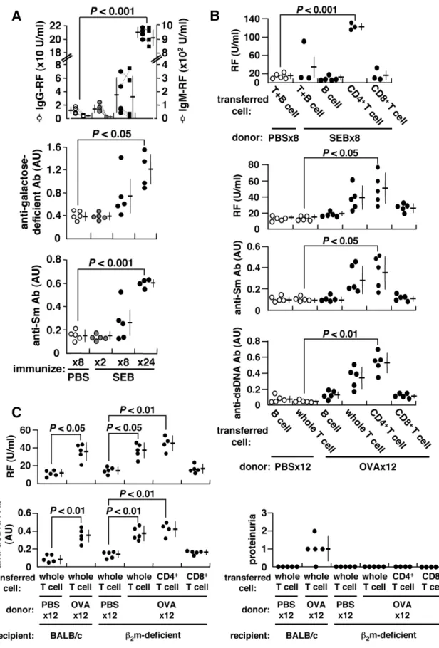
T cells from BALB/c mice to become anergized. However, these cells recovered from anergy to divide and produce IL-2 after further immuni-zation 86with SEB (Figure S1A). This was accompanied by the induction of autoantibodies, including IgG- and IgM-rheumatoid factor (RF), anti-Sm antibody, and in particular, RF reactive against galactose-deficient IgG, typically found in human autoimmunity [2] (Figure 1A). Autoantibodies can also be induced by other conventional antigens, including ovalbumin (OVA) or keyhole limpet hemocyanin (KLH) (Figure S2) as long as immunizing antigen is correctly presented to T cells (Figure S1B). CD4+
T cells of repeatedly-immunized mice become fully matured, expressing CD45RBlo, CD27loand CD122hi(data not shown), and these primed CD4+
T cells can confer RF generation in naı¨ve recipients following adoptive transfer (Figure 1B). The induction of autoantibodies is independent of CD8+T cells or
MHC class I-restricted antigen presentation for the following reasons. First, both RF and anti-dsDNA antibody can be consistently induced upon repeated immunization of b2
-micro-globulin (b2m)-deficient BALB/c mice with OVA.b2m-deficient
mice are deficient in CD8+
T cells, which are reduced to
,0.8% of splenic T cells [3] (Figure S3). Second, the ability to induce autoantibodies was transferable from OVA-immunized BALB/c mice to b2m-deficient mice solely via CD4+ T cells
(Figure 1C). Thus, CD4+
T cells from repeatedly-immunized mice acquire the ability to induce autoantibodies. We refer to these as autoantibody-inducing CD4+
T (aiCD4+
Figure 1. Induction of autoantibodies and proteinuria.BALB/c mice were repeatedly injected i.p. with 25mg SEB, 500mg OVA or PBS every 5 d.
(A) Serum IgG- and IgM-RFs, anti-galactose-deficient IgG and anti-Sm antibodies were measured using ELISA. The arbitrary unit (AU) of 1.0 is equivalent to the titer obtained from sera of prototypic autoimmune MRL/lpr mice. Data from each mouse are connected by dotted lines. (B) Adoptive transfer of splenic B, T, CD4+T or CD8+T cells of SEB-, OVA- or PBS-immunized BALB/c mice into naı¨ve BALB/c mice. The recipient mice were given single i.p. injection of 25mg SEB or 500mg OVA 24 h after cell transfer, and autoantibodies were measured 2 weeks later. (C) Adoptive transfer of cells from
OVA-immunized BALB/c mice intob2m-deficient mice.
Mechanism of Autoantibody Induction To further clarify the characteristics of aiCD4+
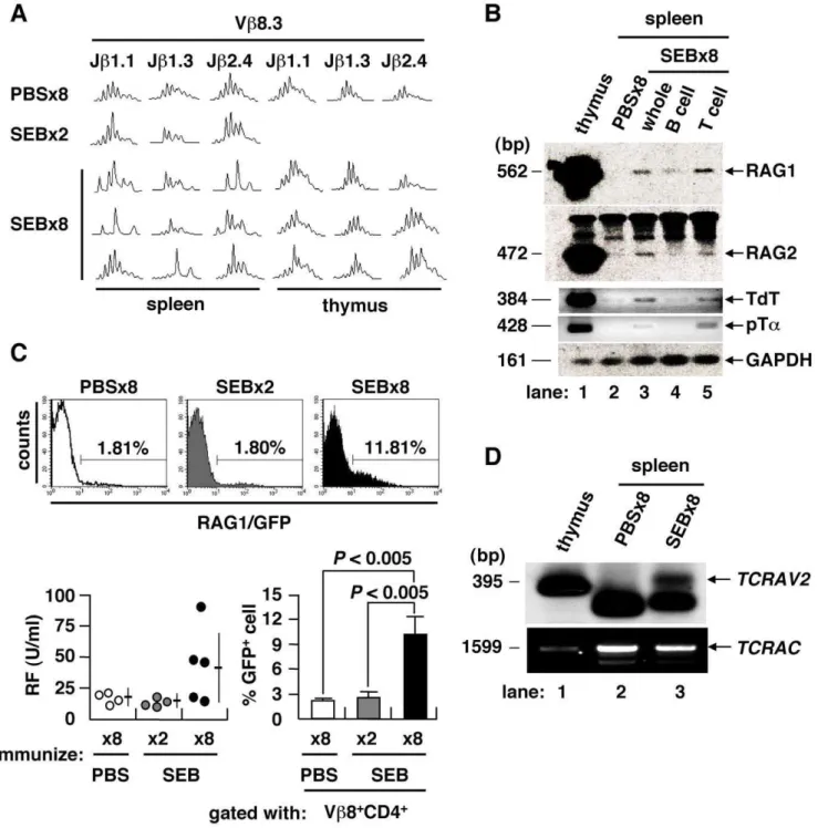
T cells, we examined their TCR repertoire by spectratyping of their complementarity determining region 3 (CDR3) [4]. Combinato-rial assessment of Vband Jbshowed that the CDR3 length profiles of CD4+
splenocytes in mice immunized either 86with PBS or 26
with SEB fit a normal Gaussian curve, typical of a diverse and unbiased TCR repertoire (Figure 2A). However, splenocytes, but
not thymocytes, from mice immunized 86 with SEB showed
skewed length profiles, suggesting that TCR revision was in progress at periphery of the spleen. Genes encoding components of the V(D)J recombinase complex were specifically re-expressed in mice immunized 86with SEB, including the recombination-activating genes 1 and 2 (RAG1/2), terminal deoxynucleotidyl transferase (TdT) and surrogate TCRachain (pTa) [5] (Figure 2B). The RAG1 gene is expressedin vivoafter immunization 86with
Figure 2. TCR revision upon repeated immunization with antigen.(A) TCR CDR3 length profiles of mice immunized 86with PBS, 26or 86 with SEB. TCR repertoire of splenic CD4+
T cell was skewed only after immunization 86with SEB. (B) Expression of V(D)J recombinase complex and related molecules in the spleen of PBS- or SEB-injected BALB/c mice. (C) GFP+
cells in the Vb8+ CD4+
T population ofrag1/gfpknock-in mice. IgG-RF as induced inrag1/gfpknock-in mice after immunization 86with SEB (lower left). The GFP+T cell fraction was also increased among Vb8+CD4+T cells (mean6SD, 4–5 mice/group). (D) TCRachain revision in the spleen of mice immunized 86with SEB was determined by LM-PCR detection of dsDNA breaks at the RSS flanking theTCRAV2, with PCR-amplified TCRaconstant region (TCRAC) as a DNA quality control.
SEB in rag1/gfp knock-in mice [6] (Figure 2C). In these mice, serum RF was increased in conjunction with an increase of GFP-expressing Vb8+
CD4+
T cells in the spleen. To directly prove that V(D)J recombination took place at the periphery in spleen, we used ligation-mediated PCR (LM-PCR) to detect blunt-end DNA fragments harboring a rearranged coding V region flanked by recombination signal sequences (RSS) [7,8]. We identified rearranged intermediates corresponding to the TCRa variable region 2 (TCRAV2) in the splenocytes of mice immunized 86with SEB (Figure 2D). These findings indicate that repeated immuni-zation with conventional antigen can induce the generation of aiCD4+
T cells which have undergone TCR revision and are capable of stimulating B cells [9]. This observation is in line with previous findings showing that such somatic mutations occur often in lymphocytes, a process which is considered to be a major stochastic element in the pathogenesis of autoimmunity [10,11]. Thus, overstimulation of CD4+T cells by repeated immunization
with antigen and induction of full maturation inevitably leads to the generation of aiCD4+
T cells which have undergone TCR revision and are capable of inducing autoantibodies. Importantly, the present study shows that suchaiCD4+
T cells are induced byde novoTCR revision but not by cross-reaction to antigen.
Induction of Autoimmune Tissue Injury
Repeated immunization with OVA can also lead to autoim-mune tissue injury and the production of autoantibodies reactive against IgG, Sm and dsDNA (Figure 3 and Figure S2A). Serum immune complex (IC), proteinuria, and the deposition of IC and OVA in the kidney were noted in mice immunized 126with OVA (Figure 3A). Typical diffuse proliferative glomerular lesions were seen in the kidney, and these glomeruli were infiltrated with CD8+
T cells. These observations resemble the clinical features observed in lupus patients, who typically exhibit an increase in CD8+
T cells in the peripheral blood and infiltration of CD8+
T cells in kidney [12,13]. Immunization of mice 126with OVA led to
re-expression of the V(D)J recombinase complex and enlargement of the spleen (Figure S4A), and an increase in anti-dsDNA antibody, which is uniquely linked to autoimmune tissue injury in lupus nephritis [14] (Figure S2A). Pathological findings included diffuse membranous (wire-loop) and/or proliferative glomerulonephritis in the kidney (Figure 3A), infiltration of plasma cells around hepatic bile ducts (Figure S4B), enlarged lymphoid follicles with marked germinal center in spleen (Figure S4B), occasional lymphocyte infiltration into the salivary glands (data not shown), lymphoid cell infiltration into the thyroid, and perivascular infiltration of neutrophils and macrophages into the skin dermis of the auricle (Figure S4B). The lupus band test, diagnostic of SLE, was positive in the skin at the epidermal-dermal junction (Figure 3B).
Mechanism of Autoimmune Tissue Injury
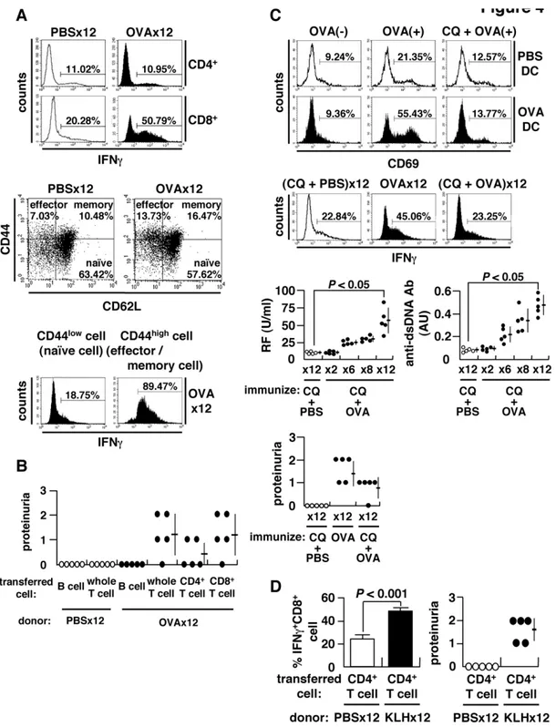
It has been shown previously that IFNc is increased in association with autoimmune tissue injury [15–17]. Consistent with this, we found that the number of IFNc+
CD8+
T cells, but not regulatory T or T helper 17 cells, was increased following immunization 126with OVA (Figure 4A and data not shown).
We also observed an expansion of IFNc-producing effector/ memory CD8+
T cells, which are necessary for adaptive immunity [18] (Figure 4A). These IFNc-producing CD8+ T cells were
observed to have infiltrated into OVA-deposited glomeruli of OVA-immunized mice (Figure 3A). CD8+
T cells are required for tissue injury based on the following observations. First, the transfer of CD8+
T cells can induce renal lesions in mice (Figure 4B), as well as the generation of new IFNc+
CD8+
T cells in the spleens of
recipient mice following cell transfer (Figure S5). Second, autoimmune tissue injury is not induced by the transfer of CD8+
T cells from OVA-immunized wild-type mice intob2m-deficient
mice (Figure 1C). And finally, CD8+ T cell transfer must be
accompanied by at least a 16booster immunization with OVA to
induce autoimmune tissue injury in the recipient mice (Figure S6). The findings indicate that full-matured, IFNc-producing effector CD8+T cells are required for the induction of autoimmune tissue
injury, provided that the relevant antigen is correctly presented on the target organs. These are well-established characteristic of CTL and not novel. We show, however, that (i) CTL is induced through an immune, but not ‘autoimmune’, process, and that (ii) autoimmune tissue injury inevitably occurs when CD8+T cells
are overstimulated to become matured effector CTLs. The latter means that regardless of how CTL is induced, the consequence of CTL over-induction is immune tissue injury.
Antigen Cross-Presentation
We next show that antigen cross-presentation is required for the induction of CTL and tissue injury. To test this, we co-cultured OVA-pulsed dendritic cells (DC) from mice immunized 126with OVA together with T cells from OVA-TCR transgenic DO11.10 mice exclusively expressing OVA-reactive TCR [19]. We show that OVA-reactive DO11.10 CD8+
T cells are activated upon co-culture with OVA-pulsed DCs (Figure 4C and Figure S7). Further, autoimmune tissue injury and the increase in IFNc+CD8+T cells,
but not of autoantibody generation, were both abrogated by adding chloroquine (CQ), an inhibitor of antigen cross-presenta-tion (Figure 4C). This indicates that antigen cross-presentacross-presenta-tion is required for the expansion of IFNc-producing CD8+T cells and
autoimmune tissue injury.
aiCD4+T Cell Helps CD8+T Cell to Induce Tissue Injury
Since CTL appear to play a rather passive role in autoimmu-nity, we next studied whether or notaiCD4+T cell help is required
for the induction of autoimmune tissue injury. Since anti-CD4 treatment almost abrogates generation of IFNc-producing CD8+
T cell and autoimmune tissue injury in OVA-immunized BALB/c mice (Figure S8), to test whether this CD4+T cell-mediated help is
mediated by aiT cells or antigen-specific T cells, we have transferred CD4+
T cells from mice immunized 126with KLH into CD4+
T-depleted BALB/c mice immunized 86with OVA (Figure 4D). Because full-matured IFNc+ CTLs do not develop
with less than 86 immunization with OVA (Figure S9), this
experiment can test the ability of aiCD4+
T cells that have undergone TCR revision to promote the maturation of OVA-specific CTL. The result showed that both autoimmune tissue injury and OVA-specific IFNc+CD8+T cells arose in these mice
after transfer, indicating that aiCD4+
T cells with de novoTCR revision are required for the maturation of CD8+
T cell and autoimmune tissue injury (Figure 4D).
Discussion
The present findings are consistent with the current consensus that CD4+
T cells normally die viaactivation-induced cell death (AICD) after repeated exposure to a single antigen, while naı¨ve CD4+T cells having a ‘cross-reactive’ TCR with lower affinity can
be activated through repeated exposure to the same antigen and survive due to weak TCR signaling, ultimately acquiring autoreactivity [20]. We show here, however, thataiCD4+
T cells are induced not by cross-reaction, but byde novo TCR revision. TheaiCD4+
T cells thus generated induce not only autoantibodies but also full-maturation of CD8+
Figure 3. Induction of autoimmune tissue injury.BALB/c mice were injected i.p. with 500mg OVA every 5 d. (A) Serum IC measured 2 d after
final immunization, expressed as AU. Proteinuria assessed 9 d after final immunization: grades 1, 2 and 3 represent 30–100 mg/dl, 100–300 mg/dl and 300–1000 mg/dl of urinary protein, respectively (upper left). Representative histopathology of kidneys from mice immunized 126with PBS or OVA (lower left) (H&E staining, bar = 20mm; original magnification6400): glomerular expansion with cellular infiltration including eosinophils seen under the same magnification. Immunohistochemistry for deposited IC, IgG, C3 and OVA (upper right) (bar = 50mm; original magnification6200), and cells infiltrated into glomeruli (bar = 20mm; original magnification6300), stained in serial tissue sections using anti-CD8a(53–6.7) and anti-IFNc(R4-6A2) monoclonal antibodies, in the specimens of mice immunized 126with OVA (lower right). (B) Lupus band test stained with anti-IgG and anti-C3 antibodies (bar = 20mm; original magnification6400).
Figure 4. Expansion of CD8+T cells and antigen cross-presentation.
(A) Spleen cells stimulated with 50 ng/ml phorbol myristate acetate (PMA) and 500 ng/ml ionomycin for 4 h in the presence of brefeldin A (10mg/ml) and stained for intracellular IFNc(upper). Subsets of CD8+T cells
categorized into naı¨ve (CD44lowCD62Lhigh), effector (CD44highCD62Llow), and memory (CD44highCD62Lhigh) fractions (middle). Flow cytometry of IFNc+ cells within naı¨ve or effector/memory CD8+
T cell populations. Spleen cells were separated into naı¨ve (CD44low) and effector/memory (CD44high) cells
using CD44 MACS beads, and IFNc+cells within the CD8+T population was evaluated (lower). (B) Adoptive transfer of splenocytes of OVA-immunized BALB/c mice into naı¨ve recipients. The recipients were injected with 500mg OVA 24 h after cell transfer, and proteinuria examined 2 weeks later. (C)
Cross-presentation of OVA to CD8+T cells. Splenic CD11c+DC from OVA-immunized or control mice were incubated in the presence (OVA(
+)) or
absence (OVA(2)) of 1 mg/ml OVA with or without chloroquine (CQ) (20mg/ml) for 3 h, followed by a co-culture with KJ1-26+CD8+T cells of DO11.10
transgenic mice for 24 h to examine surface expression of CD69 (upper). Inhibition of cross-presentationin vivoby administration of 250mg CQ per
mouse 3 h prior to immunization with OVA or PBS. IFNc+ CD8+
T cells (middle), autoantibodies and proteinuria (lower) after 126immunization. (D) Requirement of autoantibody-inducing CD4+T cells for CD8+T cell-mediated autoimmune tissue injury. BALB/c mice were immunized 12
6with KLH, and CD4+
T cells were isolated using MACS beads. Cells were transferred into the anti-CD4 antibody-treated recipient mice immunized 86with OVA. Percent matured CTL, i.e., IFNc+
CD8+
tissue injury akin to human SLE. Thus, induction of aiCD4+
T cells is a critical step, and subsequent induction of effector CTL is a critical next step in the development of autoimmunity [21,22]. The question of how autoimmunity is triggered can therefore be deduced to the quantitative response of host against immunizing antigen, i.e., the ability of host to present and/or cross-present antigen. It then follows that the ability of certain antigens such as measles virus to cause autoimmunity may be due to their ability, in conjunction with its ability to present antigen, to overstimulate CD4+and/or CD8+T cells of certain hosts beyond integrity of
their immune system. Living organisms are constantly exposed to a broad range of environmental antigens, as exemplified by the recent re-emergence of measles virus infection among a subpop-ulation of Japanese young adults who were not vaccinated against the virus. We therefore conclude that systemic autoimmunity necessarily takes place when host’s immune ‘system’ is overstim-ulated by external disturbance, i.e., repeated exposure to antigen, to the levels that surpass system’s self-organized criticality, and propose here ‘self-organized criticality theory’ explaining the cause of autoimmunity.
Materials and Methods
Ethics Statement
This study was approved by the Institutional Animal Care and Use Committee and carried out according to the Kobe University Animal Experimental Regulations.
Reagents
APC (allophycocyanin)-conjugated antibody against CD4 (RM4-5), and PE-conjugated antibodies against CD62L (MEL-14), CD69 (H1.2F3) and were purchased from BioLegend (San Diego, CA); FITC-conjugated antibodies against CD44 (IM7.8.1) and DO 11.10 clonotypic TCR (KJ1-26) and PE-conjugated rat IgG1 isotype control from CALTAG Laboratories (Burlingame, CA); PE-Cy5 (phycoerythrin-cyanin 5)-conjugated antibody against CD8a (53-6.7), PE-conjugated antibodies against Vb8 TCR (F23.1) and IFNc (XMG1.2) from BD PharMingen (San Diego, CA).
Animal Studies
Animal studies with BALB/c female mice (Japan SLC, Inc., Hamamatsu, Japan) and DO11.10 TCR transgenic mice [19] (Jackson Laboratory, Bar Harbor, ME), b2m-deficient mice [3]
and rag1/gfp knock-in mice [6] of BALB/c background were performed with the approval of the Institutional Review Board. Mice (8 weeks-old) were immunized with 25mg SEB (Toxin Technologies, Sarasota, FL), 500mg OVA (grade V; Sigma, St. Louis, MO), 100mg KLH (Sigma) or PBS by means of i.p.
injection every 5 d.
Frozen sections of kidney and dermis were stained for C3, IgG or OVA using goat anti-C3 (Bethyl laboratories, Inc., Montgom-ery, TX) and Alexa Fluor 488-conjugated anti-goat IgG antibodies (Molecular Probes, Eugene, OR), Alexa Fluor 594-conjugated anti-mouse IgG antibody (Molecular Probes), or rabbit anti-OVA antibody (Sigma). For CD8 or IFNcstaining, paraffin-embedded sections of kidney were stained with rat antibodies against CD8a
(53-6.7; BD PharMingen) or IFNc (R4-6A2; BD PharMingen), followed by reaction with VECTASTAIN Elite ABC rat IgG kit (Vector, Burlingame, CA).
To detect intracellular IFNc, cells (16106/ml) were stimulated with 50 ng/ml phorbol myristate acetate (PMA; Sigma) and 500 ng/ml ionomycin (Sigma) in the presence of brefeldin A (10mg/ml; Sigma). After 4 h, cells were stained with anti-CD8
antibody, followed by fixation with 2% formaldehyde, permeabi-lization with 0.5% saponin (Sigma) and stained for IFNc.
For adoptive cell transfer, B, T, CD4+T and CD8+T cells were
isolated from spleens to .90% purity using MACS beads (Miltenyi Biotec, Germany). The cells were transferred into naı¨ve BALB/c or b2m-deficient mice via i.p. (56106/mouse) or i.v.
(2.56107/mouse) injection. The recipients received a single i.p. injection of 25mg SEB or 500mg OVA 24 h after cell transfer, and sera, urine and organ of recipients were studied 2 weeks afterwards.
BALB/c mice were injected i.p. with 200mg anti-CD4 antibody (GK1.5; BioLegend) to deplete CD4+ T cell 24 h after
immunization 86with OVA. Four days later, CD4+T cells from
mice immunized 126with KLH were transferred to the CD4+
T-depleted mice. The recipient mice received a single i.p. injection of 100mg KLH 24 h after the cell transfer.
Assay for Mediators
Sera were assayed for anti-Sm antibody using Sm antigen (ImmunoVision, Springdale, AR), RF (Shibayagi Co., Gunma, Japan), RF for galactose-deficient IgG (Eisai Co., Ltd., Tokyo, Japan) and anti-dsDNA antibody using dsDNA (Worthington Biochemical Co., Lakewood, NJ) after digestion by S1 nuclease (Promega, Madison, WI). Serum IC was detected using goat anti-C3 antibody (Bethyl Lab.).
CDR3 Length Spectratyping cDNAs from thymocytes and CD4+
splenocytes were subjected to PCR amplification using Cb- and Vb8-specific primers. Amplified products were subjected to run-off reactions using three fluorophore-labeled Jb primers, Jb1.1, Jb1.3 and Jb2.4, and analyzed by GeneScan software (Perkin-Elmer Applied Biosys-tems, Emeryville, CA) [4].
RT-PCR
Total RNA was reversely transcribed to cDNA and amplified by PCR [23]. The products were fractionated by electrophoresis and transferred to nylon membranes (Roche Diagnostics, Mannheim, Germany). The membranes were hybridized to fluorescein end-labeled probes and visualized by alkaline phosphatase (ALP)-end-labeled anti-fluorescein antibody and Gene Images CDP-Star chemilumi-nescence reaction (Amersham Pharmacia Biotech, Piscataway, NJ). The primers and probes were: 59 -CCAAGCTGCAGACATTC-TAGCACTC-39(forward), 59 -CAACATCTGCCTTCACGTCG-ATCC-39 (reverse) and 59 -AACATGGCTGCCTCCTTGCCG-TCTACCCT-39(probe) for RAG1 [24]; 59 -CACATCCACAAG-CAGGAAGTACAC-39 (forward), 59 -GGTTCAGGGACATCT-CCTACTAAG-39 (reverse) and 59 -GCAATCTTCTCTAAAGA-TTCCTGCTACCT-39 (probe) for RAG2 [24]; 59 -GAACAAC-TCGAAGAGCCTTCC-39 (forward), 59 -CAAGGGCATCCGT-GAATAGTTG-39(reverse) and 59 -ATTCGGTCACCCACATT-GTGGCAGAGAAC-39(probe) for TdT; 59 -CAACTGGGTCAT-GCTTCTCC-39 (forward), 59-TGGCTGTCGAAGATTCCC-39
(reverse) and 59 -CCGTCTCTGGCTCCACCCATCACACTG-CT-39(probe) for pTa.
LM-PCR
Biosystems, Foster City, CA). A second PCR was performed using 1ml of the first PCR product (diluted 1/100), BW-1HR, and nested primer specific for 39flanking sequence ofTCRAV2(59 -TATTGTG-GATGCTAACAAGTGCTTTC-39). Amplified DNA was trans-ferred to membranes and visualized using fluorescein end-labeled probe specific forTCRAV2(59 -TAACATAAGAATGCACCGCT-TACACC-39) and ALP-labeled anti-fluorescein antibody. Primers for control TCRAC region were amplified using the primers 59 -CAGAACCCAGAACCTGCTGTG-39 and 59 -ACGTGGCAT-CACAGGGAA-39. Nomenclature of the TCRA gene segments was according to the ImMunoGeneTics (IMGT) database (http:// imgt.cines.fr).
Antigen Cross-Presentation OVA-reactive CD8+
T cells were isolated from spleens of DO 11.10 mice using MACS beads (Miltenyi Biotec). CD11c+
DCs (46105/well) were isolated using MACS beads (Miltenyi Biotec)
and incubated with 1 mg/ml OVA for 3 h, then co-cultured with DO11.10 CD8+ T (KJ1-26+CD8+) cells (2
6105/well) for 24 h,
and the expression of CD69 on DO11.10 CD8+ T cells was
examined. IL-2 and IFNcin culture supernatants were measured by ELISA (Biosource, Camarillo, CA).
To inhibit cross-presentation, mice were immunizedin vivowith 250mg of chloroquine (Sigma) 3 h prior to immunization with
500mg OVA or PBS every 5 d. Presence of autoantibodies was
analyzed 2 d after each immunization, and proteinuria and IFNc+
CD8+
T cells were examined 9 d after the final immunization.
Statistical Analysis
Statistical analyses were performed using Student’sttest, and the data are expressed as the mean6SD.
Supporting Information
Figure S1 Induction of autoantibodies depends on correct presentation of antigen to T cells. (A) BALB/c mice were repeatedly injected i.p. with 25mg of SEB or PBS every 5 d.
Sorted Vb8+
CD4+
splenocytes obtained 9 d after the final immunization were stimulated in vitrowith plate-bound 2mg/ml anti-CD3 (145-2C11; Cederlane, Ontario, Canada) and 5mg/ml anti-CD28 (37.51; BD PharMingen) antibodies for 24 h. Culture supernatant assayed for IL-2 (mean6SD, 5 mice/group), or the cells were labeled with carboxyfluorescein diacetate succinimidyl ester (CFSE; Molecular Probes) and further cultured for 72 h followed by flow cytometry. (B) Requirement of correct antigen presentation for induction of RF. Induction of RF after immunization 86 with SEB in B10.D2 and BALB/c mice
(efficient in presenting SEB) and in C57BL/6 (B6) mice (inefficient in presenting SEB).
Found at: doi:10.1371/journal.pone.0008382.s001 (1.17 MB TIF)
Figure S2 Generation of autoantibodies after repeated immu-nization with antigen. (A) The 8 week-old BALB/c mice were injected i.p. with 500mg OVA every 5 d, and serum RF and Sm, and dsDNA antibodies (upper), and serum IgG and anti-OVA antibodies (lower) were quantified by ELISA 2 d after respective immunization. An arbitrary unit (AU) of 1.0 is the equivalent titer in sera of MRL/lpr mice. Serum IgG was quantified by ELISA (Bethyl Laboratories), and anti-OVA antibody was quantified using mouse anti-OVA monoclonal antibody (OVA-14; Sigma) as reference. (B) BALB/c mice were immunized i.p. with 100mg KLH every 5 d. Serum RF and anti-Sm antibodies were measured by ELISA 2 d after respective
immunization, AU 1.0 = equivalent detected in sera of MRL/lpr mice.
Found at: doi:10.1371/journal.pone.0008382.s002 (1.00 MB TIF)
Figure S3 Induction of autoantibodies in CD8+T cell-deficient
mice.b2m-deficient mice were immunized with 500mg OVAvia
i.p. injection every 5 d, and IgG-RF, anti-dsDNA antibody, and proteinuria were measured.
Found at: doi:10.1371/journal.pone.0008382.s003 (0.69 MB TIF)
Figure S4 Expression of V(D)J recombinase complex and histopathology of OVA-immunized BALB/c mice. (A) Expression of V(D)J recombinase complex after immunization 126with OVA
as detected using RT-PCR (upper left). GFP+
cells in the CD4+
T cell ofrag1/gfpknock-in mice after immunization 126with OVA (lower left). Appearance and weights of spleens and a represen-tative low-magnification view of the spleens from PBS- and OVA-immunized mice (right, mean 6 SD, 9 mice/group). Enlarged lymphoid follicles with marked germinal centers were seen in mice immunized with OVA (H&E staining, bar = 200mm; original magnification 620). (B) Representative renal and extra-renal
histopathology in the mice immunized 126with OVA. A
wire-loop-like massive membranous glomerulonephritis in the kidney (upper left) (PAS staining, bar = 20mm; original magnification
6400), plasma cell infiltrates around bile ducts (upper middle) (bar = 20mm; original magnification6400), expansion of lymphoid
follicle in the white pulp of spleen (upper right) (bar = 200mm;
original magnification640), focal infiltrates of mononuclear cells to thyroid (lower left) (bar = 50mm; original magnification6100), and diffuse infiltration of inflammatory cells into auricular subcutaneous tissue (upper right) (bar = 50mm; original
magni-fication6200).
Found at: doi:10.1371/journal.pone.0008382.s004 (6.01 MB TIF)
Figure S5 The de novo generation of IFNc-producing CD8+
T cells in recipient mice after cell transfer. Percentage of IFNc+
cells within the CD8+T population of the recipient mice was examined
2 weeks after cell transfer (mean6SD, 5 mice/group).
Found at: doi:10.1371/journal.pone.0008382.s005 (0.73 MB TIF)
Figure S6 Transfer of the ability to induce anti-ds DNA antibody or tissue injury by transfer of CD4+
or CD8+
T cells, respectively. Adoptive transfer of cells from OVA-immunized mice into naı¨ve BALB/c mice, with or without 16booster injection of
OVA (500mg, 24 h post-transfer). Autoantibodies and proteinuria measured 2 weeks later.
Found at: doi:10.1371/journal.pone.0008382.s006 (0.70 MB TIF)
Figure S7 Antigen-specific activation of T cells and the expression of MHC class I on DC. (A) Spleen cells were cultured with or without 1 mg/ml of OVA for 24 h, and the expression of CD69 on CD4+
T or CD8+
T cells was examined by flow cytometry. (B) DC from PBS- or OVA-immunized mice (PBS DC or OVA DC) were incubated in the presence or absence of chloroquine (CQ) (20mg/ml) for 2 h and OVA (1 mg/ml) for 3h. OVA- and/or CQ-pulsed DCs were stained with biotin-conjugated anti-H-2kdantibody (SF1-1.1; BD PharMingen) and PE-conjugated streptavidin (BioLegend).
Found at: doi:10.1371/journal.pone.0008382.s007 (1.62 MB TIF)
Figure S8 Requirement of CD4+
T cell help for inducing autoimmune tissue injury. The mice were depleted of CD4+
T cells by treatment with 200mg anti-CD4 antibody (Ab) (GK1.5; BioLegend) 24 h prior to 66, 96and 126immunization with
OVA. Control mice were injected with 200mg rat IgG (CALTAG
Lab.). (A) A representative flow cytometry plot showing that CD4+
3.4261.02% in peripheral blood mononuclear cells (PBMC) 9 d after 3rd treatment with anti-CD4 Ab. (B) Mice were immunized 126with OVA with or without adding anti-CD4 antibodies, and the number of IFNc+
cells within the CD8+
T population (upper and lower left) (mean6SD, 5 mice/group) and proteinuria (lower right) were evaluated.
Found at: doi:10.1371/journal.pone.0008382.s008 (2.02 MB TIF)
Figure S9 Study on the requirement of autoantibody-inducing CD4+T cells for autoimmune tissue injury. Neither OVA-specific
matured IFNc+CD8+T cells or autoimmune tissue injury were
observed until BALB/c mice were immunized at least 106with
OVA. The percent splenic IFNc+CD8+T cells (left, mean 6SD, 4 or 5 mice/group) and proteinuria (right) were examined after immunization 66, 86, 106and 126with OVA.
Found at: doi:10.1371/journal.pone.0008382.s009 (0.67 MB TIF)
Acknowledgments
We dedicate this work to the Late Professor Emeritus Atsushi Okabayashi, a mentor to SS, who introduced us to this area of study. We thank Prof. Masaaki Miyazawa, Deartment of Immunology, Kinki University School of Medicine, and Prof. Nobuo Saaguchi, Department of Immunology, Kumamoto University Graduate School of Meicine, for useful advice, authorization of results and providing mice. We also thank Dr.Sachiyo Tsuji-Kawahara, Kinki University, for kindly providingb2m-deficient mice and Dr. Hideya Igarashi, Kumamoto University Graduate School of
Medicine, for kindly providing rag1/gfp knock-in mice. We thank Mai
Takimoto and Toshie Nakashima, graduate students of our department,
for studies ofrag1/gfpknock-in mice, CDR3 spectratyping and LM-PCR,
Dr. Akira Hashiramoto, Division of Rheumatology, Kobe University, for helpful discussions, and Dr. Marc Lamphier for reviewing the manuscript.
Author Contributions
Conceived and designed the experiments: SS. Performed the experiments: KT YM. Analyzed the data: KT YM SS. Wrote the paper: KT SS.
References
1. Kawabe Y, Ochi A (1990) Selective anergy of Vb8+ , CD4+
T cells in staphylococcus enterotoxin B-primed mice. J Exp Med 172: 1065–1070. 2. Parekh RB, Dwek RA, Sutton BJ, Fernandes DL, Leung A, et al. (1985)
Association of rheumatoid arthritis and primary osteoarthritis with changes in the glycosylation pattern of total serum IgG. Nature 316: 452–457. 3. Koller BH, Marrack P, Kappler JW, Smithies O (1990) Nomal development of
mice deficient inb2M, MHC class I proteins, and CD8+T cells. Science 248:
1227–1230.
4. Pannetier C, Cochet M, Darche S, Casrouge A, Zoller M, et al. (1993) The size of the CDR3 hypervariable regions of the murine T-cell receptorbchains vary as a function of the recombined germ-line segments. Proc Natl Acad Sci U S A 90: 4319–4323.
5. Kallenbach S, Doyen N, Fanton d’Andon M, Rougeon F (1992) Three lymphoid-specific factors account for all junctional diversity characteristics of somatic assembly of T-cell receptor and immunoglobulin genes. Proc Natl Acad Sci U S A 89: 2799–2803.
6. Kuwata N, Igarashi H, Ohmara T, Aizawa S, Sakaguchi N (1990) Absence of expression of RAG1 in peritoneal B-1 cells detected by knocking into RAG1 locus with green fluorescent protein gene. J Immunol 163: 6355–6359. 7. Fugmann SD, Lee AI, Shockett PE, Villey IJ, Schatz DG (2000) The RAG
proteins and V(D)J recombination: complexes, ends, and transposition. Ann Rev Immunol 18: 495–527.
8. Schlissel M, Constantinuescu A, Morrow T, Baxter M, Peng A (1993) Double-strand signal sequence breaks in V(D)J recombination are blunt, 59-phosphor-ylated, RAG-dependent, and cell cycle regulated. Genes Dev 7: 2520–2532. 9. Duty JA, Szodoray P, Zheng NY, Koelsch KZ, Zhang Q, et al. (2009)
Functional anergy in a subpopulation of naı¨ve B cells from healthy humans that express autoreactive immunoglobulin receptors. J Exp Med 206: 139–151. 10. Goodnow CC (2007) Multistep pathogenesis of autoimmune disease. Cell 130:
25–35.
11. Han JH, Akira S, Calame K, Beutler B, Selsing E, et al. (2007) Class switch recombination and somatic hypermutation in early mouse B cells are mediated by B cell and Toll-like receptors. Immunity 27: 64–75.
12. Blanco P, Pitard V, Viallard JF, Taupin JL, Pellegrin JL, et al. (2005) Increase in activated CD8+T lymphocytes expressing perforin and granzyme B correlates with disease activity in patients with systemic lupus erythematosus. Arthritis Rheum 52: 201–211.
13. Couzi L, Merville P, Deminiere C, Moreau JF, Combe C, et al. (2007) Predominance of CD8+T lymphocytes among periglomerular infiltrating cells
and link to the prognosis of class III and class IV lupus nephritis. Arthritis Rheum 56: 2362–2370.
14. Heinlen LD, McClain MT, Merrill J, Akbarali YW, Edgerton CC, et al. (2007) Clinical criteria for systemic lupus erythematosus precede diagnosis, and associated autoantibodies are present before clinical symptoms. Arthritis Rheum 56: 2344–2351.
15. Haas C, Ryffel B, Le Hir M (1997) IFN-c is essential for development of autoimmune glomerulonephritis in MRL/lprmice. J Immunol 158: 5484–5491. 16. Richards HB, Satoh M, Jennette JC, Croker BP, Yoshida H, et al. (2001) Interferon-cis required for lupus nephritis in mice treated with the hydrocarbon oil pristine. Kidney Int 60: 2173–2180.
17. Perez de Lema G, Maire H, Franz TJ, Escribese M, Chilla S, et al. (2005) Chemokine receptor Ccr2 deficiency reduces renal disease and prolongs survival in MRL/lpr lupus-prone mice. J Am Soc Nephrol 16: 3592–3601.
18. Chang TG, Palanivel VR, Kinjyo I, Schambach F, Intlekofer AM, et al. (2007) Asymmetric T lymphocyte division in the initiation of adaptive immune responses. Science 315: 1687–1691.
19. Murphy KM, Heimberger AB, Loh DY (1990) Induction by antigen of intrathymic apoptosis of CD4+
CD8+ TCRlo
thymocytes in vivo. Science 250: 1720–1722.
20. Paliard X, West SG, Lafferty JA, Clements JR, Kappler JW, et al. (1991) Evidence for the effects of a superantigen in rheumatoid arthritis. Science 253: 325–329.
21. Kurts C, Carbone FR, Barnden M, Blanas E, Allison J, et al. (1997) CD4+ T cell help impairs CD8+
T cell deletion induced by cross-presentation of self-antigens and favors autoimmunity. J Exp Med 186: 2057–2062.
22. Sun JC, Williams MA, Bevan MJ (2004) CD4+
T cells are required for the maintenance, not programming, of memory CD8+
T cells after acute infection. Nat Immunol 5: 927–933.
23. Aikawa Y, Morimoto K, Yamamoto T, Chaki H, Hashiramoto A, et al. (2008) Treatment of arthritis with a selective inhibitor of c-Fos/activator protein-1. Nat Biotechnol 26: 817–823.
24. Huang CY, Golub R, Wu GE, Kanagawa O (2002) Superantigen-induced TCRalocus secondary rearrangement: role in tolerance induction. J Immunol 168: 3259–3265.