Redescription of the poorly known planktonic copepod
Pontellopsis lubbockii (Giesbrecht, 1889) (Pontellidae)
from the Eastern Tropical Pacific with a key to species
Eduardo Suárez-Morales1,†, Eva Kozak2,‡
1 El Colegio de la Frontera Sur (ECOSUR), Av. Centenario Km 5.5, A.P. 424, Chetumal, Quintana Roo 70014, Mexico 2 Centro Universitario de la Costa Sur (CUCSUR), Universidad de Guadalajara. San Patricio Melaque, Jalisco, Mexico
Corresponding author:Eduardo Suárez-Morales (esuarez@ecosur.mx)
Academic editor:D. Defaye | Received 1 September 2012 | Accepted 18 October 2012 | Published 29 October 2012
Citation: Suárez-Morales E, Kozak E (2012) Redescription of the poorly known planktonic copepod Pontellopsis lubbockii (Giesbrecht, 1889) (Pontellidae) from the Eastern Tropical Paciic with a key to species. ZooKeys 234: 1–18.
doi: 10.3897/zookeys.234.3933
Abstract
During a survey of the epipelagic zooplankton carried out of the coast of the Mexican states of Jalisco and Colima, in the Eastern Tropical Paciic, female and male specimens of the poorly known calanoid copepod
Pontellopsislubbockii (Giesbrecht, 1889) were collected. Because previous descriptions and illustrations are largely incomplete and have caused some taxonomical confusion, this species is fully redescribed from specimens from the Mexican Paciic. he species has some characters that have been overlooked, but those related to the female genital double-somite are the most striking, it has two conical dorsal protuberances and a long ventral spiniform process unique of this species. he mouthparts of this species have not been hitherto described and igured, the lexible terminal setae of legs 3 and 4 is noteworthy. he male general morphology agrees in general with previous data, but new details of the leg 5 and geniculate antennule are added. Its mouthparts, with strong, serrate setae on the maxillae and maxillules, and a strong mandibular edge, suggest that this is a predator form. A dichotomous key for the identiication of males and females of the species of Pontellopsis known from the Eastern Tropical Paciic is included.
Keywords
Zooplankton, Mexican Paciic, taxonomy of copepods, biodiversity, pelagic crustaceans
www.zookeys.org
Copyright E. Suárez-Morales, E. Kozak. This is an open access article distributed under the terms of the Creative Commons Attribution License 3.0 (CC-BY), which permits unrestricted use, distribution, and reproduction in any medium, provided the original author and source are credited.
Eduardo Suárez-Morales & Eva Kozak / ZooKeys 234: 1–18 (2012)
2
introduction
he genus Pontellopsis Brady, 1883 currently contains up to 33 species (Boxshall and Halsey 2004; Razouls et al. 2012; Walter and Boxshall 2012). As other members of the family Pontellidae, species of Pontellopsis are usually recorded in surface waters (0–10 m) of tropical and warm temperate latitudes (Othman and Toda 2006). In general, pontellids are regarded as good indicators of water masses (Sherman 1963, 1964; Matsuo and Marumo 1982; Hernández-Trujillo 1989). Because of their mor-phological complexity and variability (Fleminger 1956, 1967b, 1975; Silas and Pil-lai 1973), their taxonomy is still in lux, partly caused by incomplete descriptions that have raised taxonomic confusion in diferent regions (Pillai 1977; Jeong et al. 2009). herefore, in some instances, it is necessary to revise and redescribe spe-cies following upgraded modern standards in order to facilitate the identiication of these species and related forms (Mulyadi 2002; El-Sherbiny and Ueda 2008). One of these poorly deined pontellid species is P. lubbockii (Giesbrecht, 1889), whose original description and subsequent illustrations by Giesbrecht (1893) Wil-son (1950), and Pillai (1977) are limited and lacking in detail. Several important characters of this species have been omitted, not only details of the taxonomically relevant characters, but of the mouthparts and legs 1-4, which still remain unde-scribed. Besides the occurrence of this species at the type locality of Columbia (Giesbrecht 1889), Wilson (1950) reported this species from the Eastern Paciic; the identity of some of Wilson’s specimens were revised by Pillai (1977), who noticed some inconsistencies both in its identiication and in the records related to this spe-cies. Wilson (1950) identiied and labeled female pontellids from the of Sri Lanka, in the South Paciic as P. lubbockii but Pillai (1977) noticed that these were in fact specimens of P. krämeri (Giesbrecht, 1896); in the same sample he found also cope-podites of Pontella sp. and of Labidocera acuta (Dana, 1849).
Pontellopsis lubbockii has been relatively rarely taken and is known as a neritic
equatorial species endemic to the Gulf of California and adjacent areas of the Eastern Tropical Paciic (ETP) (Brinton et al. 1986, Suárez-Morales and Gasca 1998) and extending to Ecuador (Pillai 1977). Overall, the pontellid copepod fauna of the area south of the inluence of the California Current and of the Mexican and Central American coasts of the Paciic is still poorly known. Previous regional surveys by Alameda-De la Mora (1980), Álvarez-Cadena (1985), Suárez-Morales and Gasca (1989), Morales-Ramírez (2001), Fernández-Álamo et al. (2000), Álvarez-Silva et al. (2003), and Morales-Ramírez and Suárez-Morales (2009) include only one species
of Pontellopsis in this area of the ETP and P. lubbockii was not recorded. In some
instances this could be a result of misidentiications or the rarity of the species. In this work we report and redescribe this poorly known pontellid based on female and male specimens collected during a series of zooplankton surveys carried out of the central Mexican Paciic coast. We also provide comments on the morphology of the mouthparts and a key for the identiication of the males and females of the species of
Material and methods
he zooplankton samples analyzed were obtained at twelve sites sampled during 27 months between December 1995 and December 1998 on board the R/V BIP-V and “León Marino”. Samples were collected at night time by oblique, semicircular trawls at diferent depths (10- 115 m) with a Bongo net (0.5 mm mesh size). he zooplankton samples were ixed and preserved with 4% formaldehyde bufered with sodium borate. Copepods were sorted from the original samples and transferred to 70% ethanol with a drop of glycerine for further analysis. Voucher specimens were deposited in the Zoo-plankton collection of El Colegio de la Frontera Sur, Chetumal, Mexico (ECO-CHZ).
systematics
Order Calanoida Sars, 1903 Family Pontellidae Dana, 1853
Pontellopsis Brady, 1883
Monops Lubbock, 1853
Pseudomonops Claus, 1892
P. lubbockii (Giesbrecht, 1889)
Monops lubbockii Giesbrecht, 1899
http://species-id.net/wiki/Monops_lubbockii
Figs 1–5
Type locality. Eastern Tropical Paciic (3–6°N, 80–82° W), about 400 km west of the coasts of Colombia and 320 km south of the Panama coast.
Material examined. Two adult females from the central Paciic of Mexico,
Eduardo Suárez-Morales & Eva Kozak / ZooKeys 234: 1–18 (2012)
4
developed, forming large spine-like processes (Fig. 1A, B). Processes straight, poste-riorly directed, reaching about halfway along urosome. Cephalosome with rounded forehead, dorsal lenses absent. Rostrum biid, with long, slender rostral ilaments, gap between rostral rami wide (Fig. 1H), in lateral view reaching halfway of second anten-nular segment (Fig. 1G). Urosome with two segments: genital double somite and anal somite. Genital double-somite representing about 55% of urosome length, excluding caudal rami; somite strongly asymmetrical, with pair of dorsal protuberances arising from distal margin of somite (Fig. 1C, D). In dorsal view, right protuberance subtri-angular, curved, posteriorly directed, reaching about half way along anal somite. Left process smaller, also posteriorly directed rounded tapering distally into strongly chi-tinized bulb-like process (Fig. 1C). Proximal margin of somite bearing lateral spine-like process on each margin, slightly asymmetrical, right one being longer. Ventral surface of genital double somite swollen, with sickle-shaped process arising anterior to genital operculum, posteriorly directed (Fig. 1D, E, F). Anal somite subrectangular, about 1.5 times wider than long, with rounded distomedial process between insertion points of caudal rami. Dorsal surface of anal somite swollen in lateral view, orna-mented with rows of minute spinules. Caudal rami weakly asymmetrical, left ramus slightly larger than right, both rami bearing 6 setae: 1 inner, 3 terminal, 1 outer setae plus short, slender dorsal seta.
Antennules (Fig. 2A) symmetrical, 16-segmented. Segments armed as follows (Arabic numbers= setae; Roman numerals= spines, aes=aesthetascs): 1 (I-III) (1), 2 (IV-VII) (9+aes), 3 (VIII-X) (6,I+aes), 4 (XI-XIII) (4,II+3aes), 5 (XIV) (1,I+aes), 6 (XV-XVI) (4,I+ 2aes), 7(XVII) (1+aes), 8(XVIII) (1+aes), 9 (XIX) (1+aes), 10 (XX) (1+aes), 11 (XXI) (1+aes), 12 (XXII) (1), 13 (XXIII) (1), 14 (XXIV) (1,I), 15 (XXV) (2+aes), 16 (XXVI-XXVIII) (4+aes). Larger and longer setae on segments 2, 4, 7, 8, and 13. Modiied, wide-based heavily setulated seta proximally inserted on segment 6; same segment with distally blunt, strongly chitinized spine reaching about 2/3 of way along succeeding segment 7 (Fig. 2A).
Antenna (Fig. 2B) biramous: coxa with short plumose distal seta. Basis and irst endopodal segment separated, basis bearing 2 setae, one short, one long. First endopo-dal segment elongate, armed with two small subdistal setae. Second endopoendopo-dal seg-ment with 9 and 7 setae on proximal and distal lobes, respectively; distal lobe armed with basal outer row of spinules; exopod 6-segmented, setation formula 1, 2, 1, 1, 1,2.
Mandible (Figs 2C–E) with wide, heavily chitinized gnathobase; mandibular palp biramous, basipod robust, subrectangular, armed with inner basipodal seta. Endopod 2-segmented, irst segment armed with 3 long and one short setae; second segment with 6 terminal setae. Exopod 5-segmented, setal formula as: 1, 1,1,1,2. Mandibular distal edge bearing 7 teeth: from ventral margin dentition includes one apical (a), one subapical (sa), two compound medial (med), and three basal (bas) (see Fig. 2C); me-dial teeth with rounded edges. Clusters of long and short spinules on base of meme-dial teeth; dorsal end of gnathobase with tight row of setae.
Eduardo Suárez-Morales & Eva Kozak / ZooKeys 234: 1–18 (2012)
6
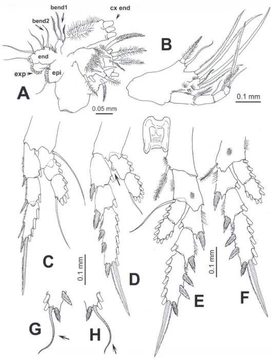
Figure 2. Pontellopsis lubbockii (Giesbrecht) from the Mexican Paciic. Adult female A antennule (in two sections) B antenna C mandible edge showing dentition, apical (a), subapical (sa), medial (med), and basal (bas) teeth D same, another view E mandibular palp F maxilla.
epipodite (epi); basis with 3 and 1 setae on proximal (bend1) and distal (bend2) en-dites, respectively; 1st and 2nd endopod segments, each with 2 setae, incorporated into basis, distal endopod segment with 5 apical setae; exopod with 8 setae.
3 setae. Basis with 2 setae; endopod 4-segmented, setal formula of endopod as: 2, 2, 1, 1. Basal and endopodal setae strongly serrate.
Maxilliped (Fig. 3B) uniramous, with praecoxa and coxa fused, three syncoxal endites well developed, with setal formula 2, 2, 3; endites setae strong, serrate. Inner lateral margin of third endite with rows of short setae. Basis fringed with medial row of 5-6 spinules and 2 distal setae. Endopod 4-segmented, setal formula of endopod as: 2, 1, 1, 2.
Leg 1 with 3-segmented endopod; legs 2-4 with 2-segmented endopods and 3-seg-mented exopods (Figs 3C-F). Coxae with plumose inner seta; basis of leg 4 with slen-der outer seta, medial patch of spinules on medial anterior margin of legs 3 and 4. First endopodal segment of second leg with inner rounded protuberance (arrowed in Fig. 3D). In one specimen examined, terminal exopodal spine of legs 3 and 4 modiied, represented by lexible seta (Italized in setal formula) (Fig. 3G,H). Seta and spine for-mula (Arabic numbers=setae, Roman numerals=spines) of legs 1-4 as:
Coxa Basis Exopod Endopod
Leg 1 0-1 0-0 I-1;I-1;II,I,4 0-1;0-2;1,2,3 Leg 2 0-1 0-0 I-1; I-1;III,I,5 0-3; 2,2,4 Leg 3 0-1 0-0 I-1; I-1; III,1,5 0-3; 2,2,4 Leg 4 0-1 1-0 I-1; I-1;III,1,5 0-3; 2,2,3
Leg 5 (Fig. 1I, J) biramous, slightly asymmetrical; coxa and intercoxal sclerite fused. Basis subrectangular, naked. Endopod distally bifurcate, about 0.3 times as long as exopodal ramus. Exopod of both legs 1-segmented, elongate, right leg with 3 outer spiniform processes and a large distal inner process; left leg smooth except for two subdistal outer spine-like setae.
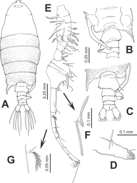
Male. Body (Fig. 4A) robust, slightly smaller than female (1.85–2.07 mm, average:
Eduardo Suárez-Morales & Eva Kozak / ZooKeys 234: 1–18 (2012)
8
Figure 3. Pontellopsis lubbockii (Giesbrecht) from the Mexican Paciic. Adult female A maxillule showing ar-mature of coxal endite (cx end distal spiniform elements cut short), proximal basal endite (bend1), distal basal endite (bend2), epipodite (epi), exopod (exp), and endopod (end) B maxilliped C leg 1 D leg 2 E eg 3 F leg 4 G variant form of leg 3 third exopodal segment with lexible terminal setal element (arrowed) h same, leg 4.
segments 9 and 10 with coarse double row of acuminate sharp teeth (Fig. 4F). Seg-ment 11 with proximal process forming fan-like row of strong spines plus two usual stout spines adjacent to segmental margin (Fig. 4G). Anterior margin of segments 10 and 11 with usual spiniform processes parallel to segmental margin. Left antennule as in female except for shorter spiniform process on segment 6 which is also relatively shorter than in female (Fig. 5A).
Leg 5 (Figs 5B–E) asymmetrical, typical of pontellids. Left leg 5 short; coxa quad-rate, basipod (bp) robust, cylindrical, naked. Exopod 3-segmented, segments 2–3 partly fused; irst segment cylindrical, with subtriangular process on outer distal mar-gin. Second exopodal segment (Fig. 5E) with medial surface covered by patch of long hair-like setae, segment with inner rounded expansion and subdistal seta on outer lat-eral margin; third segment with 2 unequal spines plus inner spiniform process. Right leg 5 basis with 2 unequal setae. Exopod with two segments, forming robust, widely open chela; irst segment (exp1) forming thumb of chela ending in short, strong pro-cess curving inward with inner surface armed with shallow cuticular ridges and small spinules (Fig. 5C). Second exopodal segment forming distal elongate inger, tapering distally, armed with two subequal proximal setae on outer surface plus one proximal and one distal setae inserted on inner surface of segment (Fig. 5D).
Remarks. Our specimens from the Mexican Paciic were identiied as P. lubbockii
by the females having acute, symmetrical posterolateral corners of the ifth pedigerous somite plus an asymmetrical genital double-somite as long as the anal somite and with two dorsal protuberances. Males have a long, curved process on the right side of the ifth pedigerous somite, a laterally directed process on the third urosomite combined with a pair of long stout setae on the right margin of the genital double somite. Fe-males of this species are easily distinguishable from its congeners by the structure and details of the genital double somite. It is unique in having two conical dorsal processes and also a ventral spine arising from the genital ield. One of these processes might have been overlooked in previous descriptions (Giesbrecht 1889; Pillai 1977) but its presence was conirmed in museum specimens from California (USNM-109384) and of Ecuador (USNM-80382). here are other species of Pontellopsis bearing dorsal processes, like P. inlatodigitata Chen & Shen, 1974, P. laminata Wilson, 1950, P.
herdmani hompson & Scott, 1903, P. scotti Sewell, 1932, P. macronyx Scott, 1909,
and P. yamadae Mori, 1937. Only one such dorsal process is illustrated in previous
Eduardo Suárez-Morales & Eva Kozak / ZooKeys 234: 1–18 (2012)
10
Eduardo Suárez-Morales & Eva Kozak / ZooKeys 234: 1–18 (2012)
12
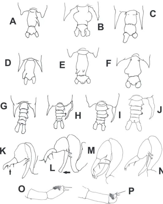
1974). A diferent pattern, with a single globose lateral process tapering distally into a spine was depicted for the same nominal species by Silas and Pillai (1973), but it also diverges from the pattern observed in P. lubbockii. he structure of the female genital double-somite of P. yamadae is probably the most similar to that of P. lubbockii and Figure 6.Schematic illustrations of characters used in the identiication key to species of Pontellopsis
in some cases both species may be confused, but the dorsal processes are quite distinct, digitiform, none of them reaching the dorsal margin of the somite (Mori 1937; Jeong et al. 2009). Both species also difer in the structure of the thoracic processes, short, rounded in P. yamadae and long, spiniform in P. lubbockii. he structure of the female leg 5 is also diferent in both species, with a much shorter and more robust outer ramus
in P. yamadae (see Mori 1937).
he extremely long spiniform ventral process present in the genital double-somite
of P. lubbockii, is a unique character of this species and has not been hitherto described
in or illustrated in previous works (Giesbrecht 1893; Pillai 1977). In only a few spe-cies of the genus a ventral process related to the genital ield has been described: in P.
albatrossi, P. armata, and P. villosa (Brady 1883) it is a short, curved spine arising from
the genital ield (Zheng et al. 1982). Yet another interesting character of P. lubbockii
is the modiication of the distal spines of the third exopodal segment of legs 3 and 4, they are lexible elements, thus contrasting with the usual pattern of stout, spiniform terminal setae. he data available to us from various descriptions suggest that this is a unique character among members of this genus.
he mandibular dentition found in our specimens agrees with the pattern de-scribed by Fleminger (1956) for this species and genus; dentition is quite uniform among species of Pontellopsis and its taxonomical value is weak. In addition, this species has the main characters described by Ohtsuka and Onbé (1991) as Type II specialized mouthparts for predation, with serrate maxillar setae, a relatively narrow mandibular edge armed with sharp, blade-like teeth, and clusters of setae and spinules near the base of the teeth. Overall, our analysis supports the notion that this species is a predator, as long known for other species of Pontellopsis (Lillelund and Lasker 1971).
Distribution of Pontellopsis in the Eastern Tropical Paciic
In the Eastern Paciic, particularly in the California Current region, only a few spe-cies of Pontellopsis have been recorded: Pontellopsis occidentalis Esterly, 1906, P.
re-galis (Dana, 1849), and P. lubbockii. Pontellopsis occidentalis is regarded as endemic
of southern California, the Gulf of California, and Baja California area.
Pontellop-sis regalis is frequently found in waters of the ETP (Fleminger 1967a; Brinton et al.
1986; Hernández-Trujillo 1989, 1994). Additional records of the genus are found south of the California Current region, of the southern sector of Baja California and the Mexican Paciic: P. armata (Giesbrecht, 1889), P. tenuicauda (Giesbrecht, 1889),
P. brevis (Giesbrecht, 1889), P. perspicax (Dana, 1849), and P. yamadae Mori, 1937
Eduardo Suárez-Morales & Eva Kozak / ZooKeys 234: 1–18 (2012)
14
Key to the species of Pontellopsis of the Eastern Paciic
Females
1A Posterolateral corners of ifth pedigerous somite with terminally rounded processes (Figs 6A, E) ...2
1B Posterolateral corners of ifth pedigerous somite forming acute spiniform processes (Fig. 6B,C,D,F) ...3
2A Genital double-somite elongate, with 2 acute dorsal processes of unequal size on posterior half of somite (Fig. 6E) ...P. yamadae
2B Genital double-somite with 2 unequal spiniform processes, one small, one long, in right side of posterior half of somite (Fig. 6A) ...P. tenuicauda
3A Spiniform processes of ifth pedigerous somite reaching the middle length of anal somite or beyond (Fig. 6C, D) ...4
3B Spiniform processes of ifth pedigerous somite not as long, barely reaching the posterior margin of the genital double-somite or even shorter (Fig. 6 B,F) ...5
4A Genital double-somite with strong, thumb-like process on left margin. Anal somite half the length of genital double-somite (Fig. 6D) ...P. villosa
4B Genital double-somite without distinct process. Anal somite as long as genital double-somite (Fig. 6C) ...P. armata
5A Genital double-somite as long as or slightly longer than anal somite, with processes or expansions on both margins or on dorsal surface ...6
5B Genital double-somite twice as long as anal somite, with lateral process on right margin only ...P. occidentalis
6A Genital double- somite with two dorsal conical unequal protuberances ... ...P. lubbockii
6B Genital double- somite with no such dorsal processes ...7
7A Both lateral margins of genital double-somite expanded forming nearly symmetrical rounded processes, that on the right side globular; anal somite strongly produced between caudal rami (Fig. 6B) ...P. perspicax
7B Genital double-somite with asymmetrical, rounded lateral processes, anal somite not strongly produced between caudal rami (Fig. 6F) ...P. regalis
Males
1A Posterolateral corners of ifth pedigerous somite with symmetrical or nearly symmetrical processes (Fig. 6G) ...2
1B Posterolateral corners of ifth pedigerous somite with strongly asymmetrical processes, with long, slender, curved process on the right side (Fig. 6H) ...3
process on irst exopodal segment of right leg 5 very short, distally blunt ... ...P. occidentalis
2B Second urosomite without such process on left margin; second exopodal seg-ment of left leg 5 globose, half as long as preceding segseg-ment (Fig. 6P), process on irst exopodal segment of right leg 5 short, distally acute ...P. villosa
3A Left posterolateral corner of ifth pedigerous somite forming short terminally rounded or broadly subtriangular process, not acute (Fig. 6I) ...4
3B Left posterolateral corner of ifth pedigerous somite forming relatively long acute process (Fig. 6H) ...5
4A Second and third urosomites with weak lateral expansions (Fig. 6I), process on irst exopodal segment of right leg 5 long, distally truncate (arrow in Fig. 6L) ...P. tenuicauda
4B Second and/or third urosomites with lateral expansion on right side, pro-cess on irst exopodal segment of right leg 5 long, tapering distally (Fig. 6 K, M, N) ...7
5A First urosomite symmetrical, armed with small unequal setae inserted on pos-terolateral margin ...6
5B First urosomite clearly asymmetrical, with rounded process on right lateral margin; process armed with two long, stout setae ...P. lubbockii
6A Right posterolateral corner of ifth pedigerous somite long, acute, tapering dis-tally (Fig. 6I); caudal rami as long as wide, distal segment of chela with protu-berance on medial position of inner margin (arrow Fig. 6K) ...P. regalis
6B Right posterolateral corner of ifth pedigerous somite long, slender from in-sertion, branch-like (Fig. 6H); caudal rami twice as long as wide, distal seg-ment of chela with low proximal expansion on inner margin ...P. armata
7A Second and third urosomites expanded laterally, process on irst exopodal segment of right leg 5 shorter than second exopodal segment (Fig. 6N) ... ...P. yamadae
7B Only third urosomite expanded laterally. Right leg 5 with inger-like process of irst exopodal segment longer than second exopodal segment (Fig. 6M) ... ...P. perspicax
Acknowledgements
Fron-Eduardo Suárez-Morales & Eva Kozak / ZooKeys 234: 1–18 (2012)
16
tera Sur (ECOSUR), Chetumal, Mexico. he comments and valuable suggestions of two referees allowed us to improve a previous version of this contribution, particu-larly in reference to the interpretation and description of morphological details of the mouthparts. he ine editorial work and processing of the manuscript by Danielle Defaye is deeply appreciated.
References
Alameda-De la Mora G (1980) Sistemática y Distribución de los Copépodos (Crustacea) del Golfo de Tehuantepec (México). Tesis de Licenciatura, Fac. de Ciencias, Universidad Na-cional Autónoma de México.
Álvarez-Cadena JN (1985) Composición y abundancia de los copépodos planctónicos de la Bahía de Mazatlán. Anales del Instituto de Ciencias del Mar y Limnología, Universidad Nacional Autónoma de México 12: 1–14.
Álvarez-Silva C, Miranda-Arce MG, De Lara-Isassi G (2003) Familia Pontellidae (Crustacea: Copepoda) en la Bahía La Ventosa, Oaxaca, México: Sistemática y ecología. Revista de Biología Tropical 51: 737–742.
Boxshall GA, Halsey SH (2004) An Introduction to Copepod Diversity. he Ray Society, Lon-don, 966 pp.
Brady GS (1883) Report on the Copepoda collected by H.M.S. Challenger during the years 1873– 1876. Reports of the Scientiic Results of the Voyage of the Challenger Zoology 8: 1–142. Brinton E, Fleminger A, Siegel-Causey D (1986) he temperate and tropical planktonic biotas
of the Gulf of California. CalCOFI Reports 27: 228–266.
Chen QC, Zhang SZ (1965) he planktonic copepods of the Yellow Sea and the East China Sea I. Calanoida. Studia Marina Sinica 7: 20–131.
Chen QC, Shen CJ (1974) he pelagic copepods of the South China Sea. II. Studia Marina Sinica 9: 125–137.
El-Sherbiny M, Ueda H (2008) Redescription of the poorly known calanoid copepod Pontella karachiensis Fazal-Ur-Rehman, 1973 from the Red Sea with notes on its feeding habits. Plankton & Benthos Research 3: 19–17.
Fernández-Álamo MA, Sanvicente-Añorve L, Alameda-De la Mora G (2000) Copepod as-semblages in the Gulf of Tehuantepec, Mexico. Crustaceana 73: 1139–1153. doi: 10.1163/156854000505137
Fleminger A (1956) Taxonomic and distributional studies on the epiplanktonic calanoid co-pepods (Crustacea) of the Gulf of Mexico. PhD hesis, Harvard University Library, Cam-bridge, 317 pp.
Fleminger A (1967a) Distributional atlas of calanoid copepods in the California Current re-gion, Part II. CalCOFI Atlas 7: 1–213.
Fleminger A (1975) Geographical distribution and morphological divergence in American coastal-zone planktonic copepods of the genus Labidocera. In: Cronin LE (Ed.) Estuarine research: 1: Chemistry, biology and the estuarine system. Academic Press, NY, 392–418. Fleminger A (1986) he Pleistocene equatorial barrier between the Indian and Paciic Oceans and
a likely cause for Wallace’s Line. UNESCO Technical Papers on Marine Science 49:84–97. Giesbrecht W (1889) Elenco dei Copepodi pelagici raccolti dal tenente di vascello G. Chierchia
durante il Viaggio della R. Corvetta: Vettor Pisani” negli anni 1882–1885 e dal tenente di vascello F. Orsini nel Mar Rosso, nel 1884. Atti Accad. naz Liucei. Rd., Ci. Sci. is. mat. nat. 4: 24–29.
Giesbrecht W (1893) Systematik und Faunistik der pelagischen Copepoden des Golfes von Neapel und der angrenzenden Meeres-Abschnitte. Fauna Flora Golfes Neapel 19: 1–831. Hernández-Trujillo S (1989) Copépodos de la familia Pontellidae en Baja California Sur
(1982–1984). Investigaciones Marinas CICIMAR 4: 225–232.
Hernández-Trujillo S (1994) Pontellidae copepods in the Paciic of Baja California, México, July 1988. Investigaciones Marinas CICIMAR 9: 55–58.
Hernández-Trujillo S, Palomares-García R, López-Ibarra GA, Esqueda-Escárcega G, Pacheco R (2004) Riqueza especíica de copépodos en Bahía Magdalena, Baja California Sur, Mé-xico. Anales del Instituto de Biología, Universidad Nacional Autónoma de México, Serie Zoología 75: 253–270.
Jeong HG, Suh HL, Soh HY (2009) Pontellopsis species (Copepoda, Pontellidae) in the Korean waters, with notes on the female genital structures and their zoogeography. Animal Cells and Systems 13: 187–203. doi: 10.1080/19768354.2009.9647211
Lillelund K, Lasker R (1971) Laboratory studies of predation by marine copepods on ish lar-vae. Fishery Bulletin 69: 655–667.
Matsuo Y, Marumo R (1982) Diurnal vertical migration of pontellid copepods in the Kuroshio. Bulletin of the Plankton Society of Japan 29:89–98.
Mori T (1937) he pelagic Copepoda from the neighboring waters of Japan. Soyo Company, Tokyo, 150 pp.
Morales-Ramírez A (2001) Biodiversidad marina de Costa Rica, los microcrustáceos: Subclase Copepoda (Crustacea: Maxillopoda). Revista de Biología Tropical 49(2): 115–133. Morales-Ramírez A, Suárez-Morales E (2009) Copepoda. In: Wehrtmann IS, Cortés J (Eds)
Marine Biodiversity of Costa Rica. Monographiae Biologicae 86, Springer Science, he Netherlands, 291–305. doi: 10.1007/978-1-4020-8278-8_27
Mulyadi (2002) he calanoid copepods family Pontellidae from Indonesian waters, with notes on its species-groups. Treubia 32:1–167.
Ohtsuka S, Onbé T (1991) Relationship between mouthpart structures and in situ feeding habits of species of the family Pontellidae (Copepoda: Calanoida). Marine Biology 111: 213–225. doi: 10.1007/BF01319703
Othman BHR, Toda T (2006) Pontellid copepods from Singapore. Coastal Marine Science 30: 305–319.
Eduardo Suárez-Morales & Eva Kozak / ZooKeys 234: 1–18 (2012)
18
Pillai PP (1977) On the identity of certain specimens of Pontellopsis Brady (Copepoda : Cala-noida) of the ‘Wilson Collection’ deposited in the US National Museum. Journal of the Marine Biology Association of India 19: 58–67.
Razouls C, de Bovée F, Kouwenberg J, Desreumaux N (2012) Diversity and geographic dis-tribution of marine planktonic copepods. Available at http://copepodes.obs-banyuls.fr/en Scott A (1909) he Copepoda of the Siboga Expedition. 1. Free-swimming, littoral and
semi-parasitic Copepoda. Siboga Expedition Monographs 29: 1–323.
Sherman K (1963) Pontellid copepod distribution in relation to surface water types in the central North Paciic. Limnology and Oceanography 8: 214–227. doi: 10.4319/lo.1963.8.2.0214 Sherman K (1964) Pontellid copepod occurrence in the central South Paciic. Limnology and
Oceanography 9:476–484. doi: 10.4319/lo.1964.9.4.0476
Silas EG, Pillai PP (1973) he calanoid copepod family Pontellidae from the Indian Ocean. Journal of the Marine Biology Association of India 15: 771–858.
Suárez-Morales E, Gasca R (1989) Copépodos calanoides epipelágicos del Domo de Costa Rica Junio-Agosto, 1982. Ciencias Marinas 15: 89–102.
Suárez-Morales E, Gasca R (1998) Updated checklist of the marine Copepoda (Crustacea) of Mexico. Anales del Instituto de Biología, Universidad Nacional Autónoma de México, Serie Zoología 69: 105–119.
hompson IC, Scott A (1903) Report on the Copepoda collected by Prof. Herdman at Ceylon in 1902. Ceylon Pearl Oyster Fisheries 7: 227–307.
Walter TC, Boxshall GA (2012) Pontellopsis Brady, 1883. World Copepoda database. Ac-cessed through: World Register of Marine Species at http://www.marinespecies.org/aphia. php?p=taxdetails&id=104211
Wilson CB (1950) Copepods gathered by the United States Fisheries Steamer “Albatross” from 1887 to 1909, chiely in the Paciic Ocean. Bulletin of the US National Museum 100: 141–441.