Maria Letícia Sperandéo de Macedo
Avaliação vascular não invasiva (NIVA) em
gestantes com diabete gestacional e com
hiperglicemia leve utilizando o SphygmoCor
®Tese apresentada ao Programa de Pós-Graduação em Ginecologia, Obstetrícia e Mastologia, Área de Concentração em Obstetrícia da Faculdade de Medicina de Botucatu - Unesp, para obtenção do título de doutor.
Orientadora:
Professora Titular Marilza Vieira Cunha Rudge
FICHA CATALOGRÁFICA ELABORADA PELA SEÇÃO TÉCNICA DE AQUISIÇÃO E TRATAMENTO DA INFORMAÇÃO
DIVISÃO DE BIBLIOTECA E DOCUMENTAÇÃO - CAMPUS DE BOTUCATU - UNESP
BIBLIOTECÁRIA RESPONSÁVEL: Selma Maria de Jesus
Macedo, Maria Letícia Sperandéo de.
Avaliação vascular não invasiva (NIVA) em gestantes com diabete gestacional e com hiperglicemia leve utilizando o SphygmoCor / Maria Letícia Sperandéo de Macedo. – Botucatu : [s.n.], 2007.
Tese (doutorado) – Faculdade de Medicina de Botucatu, Universidade Estadual Paulista, 2007.
Orientadora: Marilza Vieira Cunha Rudge Assunto CAPES: 40101150
1. Diabetes na gravidez
CDD 618.3
Aos meus pais Artur e Célia,
Pela presença que nos faz sentir sempre acompanhados.
Por nos ensinarem que as dificuldades devem ser enfrentadas
com naturalidade e que nada é tão difícil assim...
Pelo apoio constante na busca dos nossos ideais.
A carreira acadêmica traz-me grande satisfação. Se não fora o
exemplo dos senhores talvez tivesse escolhido outro caminho
que, provavelmente, não me traria a mesma alegria.
To my dear Daniele,
One day, you told me;
_ I couldn’t make it without you.
Today, is my turn to say;
_ It would have been impossible if you were not there with
Aos meus irmãos Arthur, Fernando e Caio,
Pelo apoio, pela alegria e pelo compartilhamento.
A convivência com vocês ajudou a lapidar minha
personalidade e fez de mim uma pessoa melhor.
Às minhas cunhadas, Alessandra e Renata,
Pela torcida sincera e pela amizade.
Aos meus sobrinhos; Marina, Sophia,
Rodrigo e Gustavo,
Al Sig Nucio e Sig.ra Susetta,
Mi avete accolto come una figlia,
anche se sono lontana di casa mi
sento in famiglia.
A Ivana,
À Professora Marilza Vieira Cunha Rudge,
Pelo seu espírito verdadeiramente acadêmico e empreendedor.
mas, sobretudo, por desejar e incentivar o progresso de seus
pares e tutelados. Isto é ser Professora Titular de verdade.
Eu agradeço por todo apoio e incentivo recebidos do início ao
Ao Departamento de Ginecologia e Obstetrícia, em especial à
disciplina de Obstetrícia, à Faculdade de Medicina de
Botucatu e, à UNESP pela minha formação como especialista
e pela pós-graduação.
Ao Dr. Peraçoli e Dra. Iracema,
Pelos ensinamentos da residência e de sempre. Os senhores são
exemplos de profissionais para todos nós que passamos pelo
departamento. É uma honra poder ter convivido com os
senhores.
À Profa. Dra. Maria Aparecida Mourão Brasil,
Pelo auxílio em todos os momentos da realização deste
trabalho. Pela disponibilidade com que sempre me atendeu,
pela amizade e pelo bom humor. Todos nós sabemos que uma
tese de doutorado não se faz sozinho, tantas pessoas nos
prestam auxílios inestimáveis para a sua realização.
Aos amigos, Cláudia, Roberto, Vera e Joélcio por tantas
palavras e gestos de amizade.
Ao setor de Pós-Graduação, em especial à Regina Spadin por
todo auxílio prestado.
Às minhas queridas amigas de sempre e para sempre; Lillyan,
Daniela, Renata e Fabiana.
À Faculdade de Medicina de Jundiaí, em especial ao
Departamento de Tocoginecologia, na pessoa do Dr. Nelson
Maia, pela amizade e pela confiança no nosso trabalho.
Ao Curso de Medicina da Universidade Cidade de São Paulo,
na pessoa do Dr. José Lúcio Machado, diretor do Curso de
Medicina, pelo apoio e amizade.
Ao Grupo de Apoio à Pesquisa - GAP da Faculdade de
Medicina de Botucatu - UNESP .
Ao Hélio Rúbens de Carvalho Nunes pela análise estatística.
Ao Departamento Harris Birthright Centre for Fetal
Medicine- King’s College Hospital- London School of
Medicine –University of London.
Ao Professor Kypros Nicolaides por nos receber em programa
cooperativo para realização deste trabalho e também pelo
treinamento em medicina fetal.
We are greatful to Prof. Nicolaides for having me in a
co-operative program to develop the study here presented and
for the training in fetal medicine.
À Dr. Makrina Savvidou pela co-orientação deste trabalho.
To Dr. Makrina Savvidou for being co-advisor in this work.
Aos companheiros de trabalho,
Daniele Luminoso, Christine Kayhura e James Anderson pelo
trabalho em equipe, tabulação e organização do banco de
To Daniele, Christine and James for the team work in
organizing the data base.
Agradeço especialmente também,
À Universidade Cidade de São Paulo na pessoa do Prof.
Rubens Lopes da Cruz, reitor da UNICID.
À UNICID e o Prof. Rubens foram a primeira instituição e
representante a prestarem apoio para a realização deste
trabalho.
Agradeço ao Prof. Rubens pela generosidade e pela atenção
que sempre me devota.
Capítulo I_______________________________________________ 16
Resumo... 18
Abstract... 19
Introdução... 20
Tonometria de aplanação... 21
Referências Bibliográficas... 35
Capítulo II______________________________________________ 42 Abstract... 48
Introduction... 50
Methods... 52
Results... 58
Discussion... 65
References... 71
Capítulo III______________________________________________ 76 Introdução... 78
Revisão de Literatura... 81
Objetivo... 85
Material e Métodos... 86
Referências Bibliográficas... 89
Capítulo I
Tonometria de Aplanação – Método Promissor e não invasivo
para avaliação da função endotelial na gravidez.
Applanation Tonometry - Non Invasive and Promising Method for Evaluation
of Endothelial Function in Pregnancy.
Maria Letícia Sperandéo de Macedo*/**
Daniele Luminoso***
Cláudia Garcia Magalhães*
José Carlos Peraçoli*
Iracema de Mattos Paranhos Calderon*
Marilza Vieira Cunha Rudge*
*Departamento de Ginecologia e Obstetrícia da Faculdade de Medicina de Botucatu-
UNESP.
**Departamento de Tocoginecologia da Faculdade de Medicina de Jundiaí.
***Departamento de Obstetrícia e Ginecologia da Universitá degli Studi di Cagliari-
RESUMO
A hipertensão gestacional está presente em cerca de 10% das gravidezes
e ainda é a primeira causa de mortalidade materna no Brasil.O diabetes
gestacional complica 7,6% das gestações no Brasil e está associado a
resultados perinatais insatisfatórios. Estas complicações cursam com
disfunção endotelial e alteração da elasticidade da parede vascular. A
tonometria de aplanação é um método não invasivo, portátil e de fácil
aprendizagem que avalia a função endotelial através do estudo da rigidez
arterial (perda da elasticidade arterial). Além de avaliar a função endotelial
este método oferece estudo indireto de vários parâmentros
cardiovasculares centrais. O grande número de informações que este
método obtém de maneira não invasiva, faz deste, um instrumento
valoroso em pesquisa. Apresenta grande potencial, especialmente, na
compreensão dos mecanismos fisiopatológicos que cursam com
comprometimento vascular na gravidez.
Palavras-Chave: Tonometria de Aplanação; Rigidez arterial; Disfunção
ABSTRACT
Gestational hypertension affects 10% of pregnancies and is still the first
cause of maternal mortality in Brazil. Gestational diabetes affects 7,6% of
pregnancies in Brazil and is associated with an unsatisfactory peri-natal
outcome. These complications are associated to endothelial dysfunction and
abnormal elasticity of the arterial wall. Applanation tonometry is a
non-invasive, portable and easy learning method that evaluates endothelial
function by the study of arterial stiffness (lost of arterial elasticity). Beyond
the endothelial function evaluation, this method gives, indirectly, several
central cardiovascular parameters. The great number of information
obtained non invasively by this method, makes of this, a valuable instrument
in research. It has special potential to help in the comprehension of the
mechanisms of those diseases that presents with vascular commitment in
pregnancy.
Keywords: Applanation tonometry; Arterial Stiffness; Endothelial
INTRODUÇÃO
A função endotelial alterada está implicada nas duas
complicações clínicas mais freqüentes da gestação: hipertensão e diabetes
(Porta et al., 1987, Ramsay et al., 2003, Montenegro et al., 2004)
As gestações complicadas por estas entidades são
consideradas de alto risco pela freqüência e pelas repercussões maternas e
perinatais. A hipertensão gestacional está presente em cerca de 10% das
gravidezes e ainda é a primeira causa de mortalidade materna no Brasil
(Peraçoli & Parpinelli, 2005, Bezerra et al., 2005).O diabetes gestacional,
está presente em 2% a 4 % das gestações nos Estados Unidos (Counstan
et al., 1995, Katz et al., 2002), e em até 7,6% das gestações no Brasil
(Ministério da saúde, 2001). Está associado a resultados perinatais
insatisfatórios, óbito fetal inexplicável e desenvolvimento futuro de diabete
tipo II após a 4 década em 50% das mulheres que desenvolveram diabetes
na gestação (Cunningham et al., 2001, Silva et al., 2003, Rudge et al.,
2007).
O endotélio vascular é uma camada de células que reveste a
parede interna dos vasos sanguíneos, é sensível às mudanças de forças
hemodinâmicas e responde à esses fatores liberando substâncias
vasoativas para garantir a homeostase (Verma et al., 2002 ). O óxido nítrico
é uma das mais potentes substâncias vasodilatadoras produzidas pelo
queda da resistência periférica. Quando sua produção é ineficiente ou sua
biodisponibilidade é inadequada, estamos frente a uma das vertentes da
chamada “disfunção endotelial“ (Verma et al., 2002, Cerqueira et al., 2002).
A adequada adaptação do organismo materno à gestação
cursa com aumento da produção de óxido nítrico e queda da resistência
vascular periférica com consequente queda da pressão arterial (Williams et
al., 1997, Savvidou et al., 2000). Nas gestações complicadas por
hipertensão ou diabete, há disfunção da célula endotelial e diminuição da
biodisponibilidade ou da produção de óxido nítrico (Garner et al., 1990,
Wilkinson et al., 2000, Savvidou et al., 2002, Savvidou et al., 2003,
Davidson et al., 2004,).
A disfunção endotelial está relacionada à rigidez arterial e
pode ser estudada por método físico não invasivo denominado tonometria
Tonometria de aplanação é o método no qual a morfologia
da onda de pressão arterial de determinada artéria pode ser estudada de
maneira não invasiva com o objetivo de obter o índice de rigidez
(elasticidade) arterial (Hayward et al., 2002). Esta técnica é baseada nos
princípios da tonometria ocular utilizada para medida da pressão intraocular
pela “aplanação “da superfície do globo ocular (Hayward et al., 2002).
O’Rourke et al., 1996 desenvolveram equipamento eletrônico
e portátil para realização de tonometria de aplanação arterial. É um sistema
de análise da onda de pulso, que avalia, de maneira não invasiva, a rigidez
do sistema arterial. Seu software é equipado com uma função de
transferência na qual através da leitura da onda de pulso de vaso periférico
são obtidos os parâmetros centrais (aorta) (Mackenzie et al., 2002).
O aparelho consta de um transdutor (tonômetro) com
formato e dimensões de uma caneta, que apresenta na ponta um sensor -
cristal piezzoelétrico - acoplado a um computador com o software
Figura 1- Fotografia do Equipamento SphygmoCor®.
A leitura da onda de pulso é feita pela colocação deste
tonômetro sobre o ponto mais forte de pulsação arterial de determinada
artéria; esta artéria é suavemente comprimida contra os tecidos mais
profundos como osso ou cartilagem, resultando em uma superfície
“aplanada” que possibilita ao sensor captar as variações da onda de pulso
(figura 2,3 e 4). Este sinal é captado e interpretado pelo software que
Figura 2- Ilustração representando a técnica para obtenção da onda de
pulso. Colocação do trandutor sobre o ponto máximo de pulsação
da artéria radial do paciente.
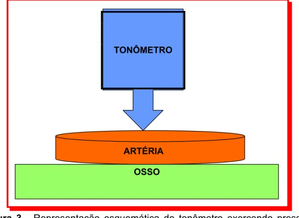
Figura 3 - Representação esquemática do tonômetro exercendo pressão
sobre a artéria e osso.
TONÔMETRO
ARTÉRIA
Figura 4 – Ilustração representando a aplanação da parede arterial quando
comprimida entre tonômetro e o osso.
Este sistema foi clinicamente validado e demonstrou ser
método reprodutível e com fácil aplicabilidade clínica (O’Rourke et al.,
1996).
O estudo da rigidez arterial vem ganhamdo muito interesse
nos últimos anos e sua importância está na relação existente entre os
fatores de risco (idade avançada, hipertensão arterial, obesidade,
hipercolesterolemia, diabetes e tabagismo) para doença cardiovascular e a
rigidez do sistema arterial (McEniery et al., 2006).
A rigidez arterial é a manifestação mais precoce destas
doenças, antes mesmo do aparecimento da placa de ateroma (Wilkinson et
al., 2000).
entre eles estão: os elementos estruturais da parede do vaso como elastina
e colágeno, a pressão de distensão vascular e o tônus do músculo liso
vascular. Mudanças no tônus muscular e quebra da integridade do
colágeno e elastina alteram a distribuição de forças na parede arterial
promovendo remodelamento da parede do vaso, tornando-o mais rígido.
Essas mudanças do tônus vascular estão diretamente relacionadas com a
produção local de óxido nítrico pelo endotélio vascular.
Estudos recentes utilizando tonometria de aplanação
demonstraram que existe relação direta entre rigidez da parede arterial e
disfunção endotelial. Estes fatores foram avaliados pela análise da onda de
pulso e pela velocidade da onda de pulso, duas técnicas obtidas pela
tonometria de aplanação (Hayward et al., 2002, Wilkinson et al., 2002,
Wilkinson et al., 2002, McEniery et al., 2006).
O sistema de tonometria de aplanação desenvolvido por
O’Rourke e cols é denominado comercialmente de Sphygmocor e estuda a
rigidez arterial por duas técnicas distintas; a velocidade da onda de pulso e
a análise da onda de pulso. A velocidade da onda de pulso (pulse wave
velocity) é o cálculo do tempo que a onda de pulso leva para percorrer
uma distancia conhecida. Para esta distância, o trecho mais utilizado é
aquele entre a artéria carótida e a artéria femural. Um eletrocardiograma,
que monitora a paciente, é acoplado ao microcomputador e as ondas de
pulso destas artérias (carótida e femoral) são obtidas separadamente pela
a base (pé) da onda de pulso para calcular o tempo e a velocidade em
metros por segundos que a onda de pulso leva para percorrer este trecho
da aorta.
Esta técnica aplicada neste segmento do corpo em questão
tem relação direta com a rigidez da aorta (figura 5 e 6).
Figura 5 – Leitura da onda de pulso da artéria carótida
Velocidades mais baixas são uma das características de
sistemas mais elásticos demonstrando que a artéria não perdeu a
capacidade elástica de acomodar (distensão) o volume de sangue e nem a
de absorver o impacto de pressão sobre a parede. Isso significa que a onda
representam o mecanismo contrário; a parede da artéria apresenta
remodelamento das fibras e aumento do tônus da musculatura lisa,
tornando-se rígida. Isto resulta em pouca capacidade de absorver o impacto
de pressão antes de transmitir a onda de pulso, fazendo com que este
pulso de pressão seja propagado quase que imediatamente (Mackenzie et
al., 2002, Wilkinson et al., 2003, Salvi et al., 2004).
A análise da onda de pulso (pulse wave analysis) estuda
a morfologia da onda de pulso. A leitura da onda de pulso é feita da mesma
maneira que a da velocidade da onda de pulso, porém neste caso os vasos
a serem estudados são a artéria radial, que já foi validada e a artéria
carótida. A morfologia da onda de pulso é obtida e registrada no
microcomputador e a função de transferência do software produz a
morfologia da onda de pulso da aorta ascendente derivada da onda de
pulso da artéria radial. Esta técnica avalia a rigidez arterial sistêmica e a
pressão arterial central de maneira indireta pela função de transferência.
A morfologia da onda de pressão é composta de uma onda
de pressão progressiva gerada pela contração ventricular e uma onda de
pressão retrógrada (refletida), gerada pela reflexão da onda de pressão ao
Figura 7 – Representação esquemática da formação da onda de pulso que
é uma somatória entre a onda progressiva e onda refletida
O conceito da onda refletida é fundamental para
compreensão e interpretação da morfologia da onda. Em sistemas
elásticos, a onda de pressão do pulso percorre o trajeto em questão em
baixa velocidade e a onda refletida retorna a raiz da aorta na diástole,
aumentando a pressão do pulso nesta fase do ciclo cardíaco e melhorando
a perfusão coronariana que ocorre na diástole. Em sistemas rígidos, a onda
Onda progressiva
Onda Progressiva + Onda Refletida Onda refletida
Sístole Diástole
refletida retorna ao coração ainda na sístole cardíaca, o que aumenta a
resistência periférica intravascular a ser vencida neste período (sístole),
aumentando a pressão de ejeção ventricular (trabalho cardíaco) o que
resultará em má perfusão das artérias coronárias (isquemia miocárdica) e
hipertrofia ventricular esquerda (HVE) (Figura 8). É importante ressaltar que
a HVE é um dos principais fatores de risco de morbidade e mortalidade de
causa vascular na população geral (Blacher et al., 1999, Mackenzie et al.,
2002, Wilkinson et al., 2003).
Figura 8 – Onda refletiva retorna ainda na sístole aumentando a pressão
O principal parâmetro da análise da onda de pulso é o índice
de amplificação (aumento). Este é medido pela diferença entre os dois
picos de pressão da onda progressiva e da onda refletida e é expresso
como porcentagem da pressão do pulso. A pressão do pulso é a diferença
de pressão entre o pico sistólico e a pressão diastólica final (Mackenzie et
al., 2002, Wilkinson et al., 2002) (figura 9)
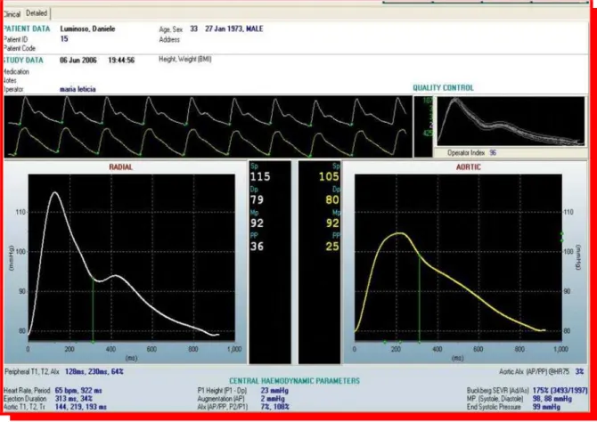
Figura 9 – Análise da Onda de Pulso obtido pelo SphygmoCor®,
demonstrando a morfologia da onda na artéria radial e na artéria
aorta. A onda da artéria aorta é obtida mediante função de
A tonometria de aplanação demonstrou ser método
reproduzível e confiável, foi validado em animais e em humanos e tem a
vantagem de utilizar técnica não invasiva, de fácil aprendizagem, é obtida
com o uso de equipamento portátil e de fácil aplicabilidade clínica (Hayward
et al., 2002).
Esta técnica foi explorada por vários grupos de pesquisa em
animais e humanos. Além de estudos sobre a fisiologia do sistema
vascular, foi demonstrada relação direta entre rigidez arterial e risco
aumentado de hipertensão, rigidez arterial e disfunção endotelial, rigidez
arterial e taxa de mortalidade por evento cardiovascular, e também de
rigidez arterial e hipercolesterolemia, diabetes tipo I, tipo II, insulinemia pos
prandial e estados hiperinsulinemicos (Blacher et al., 1999, Duanping et al.,
1999, Brooks et al.,1999, Brooks et al., 2001, Wilkinson et al., 2002,
Wilkinson et al., 2003, Westerbacka et al., 2005, Greenfield et al., 2006).
Na gravidez, foi estudada a relação entre a onda de pulso e
as alterações fisiológicas cardiovasculares da gravidez (Smith et al., 2004),
assim como, a relação entre o índice de amplificação e hipertensão
induzida pela gravidez.
Os resultados apontaram significativo aumento da rigidez
arterial na pré-eclampsia (Elvan- Taspinar et al., 2004, Spasojevic et
al., 2005). Outros trabalhos evidenciaram a correlação inversa entre
(Elvan-Taspinar et al., 2005).
Este método tem a vantagem de ser inócuo, não invasivo, e
portátil e ainda de fácil aprendizagem. Seu uso em pesquisa trará um
avanço no entendimento fisiopatológico da gestação e das patologias
maternas que cursam com comprometimento vascular. A sua aplicabilidade
clínica demanda mais investigações mas parece ser método promissor e de
REFERÊNCIAS BIBLIOGRÁFICAS
1. Porta M, La Selva M, Molinatti P, Molinatti GM.Endothelial cell function
in diabetic microangiopathy.Diabetologia 1987;30:601-9.
2. Ramsay JE, Simms RJ, Crawford Lynn, Greer IA, Lumsden MA, Sattar
N. Enhancement of Endothelial Function by pregnancy. Inadequate
response in women with type 1 diabetes. Diabetes Care 2003;26(2)
475-479.
3. Montenegro CAB, Leite SP, Castro P, Regattieri N, Lima MLA, Filho JR.
Predição e Prevenção da Toxemia Gravídica:2004. Femina 2004; 32 (6)
509-515.
4. Peraçoli JC, Parpinelli MA. Síndromes Hipertensivas da Gestação:
Identificação de Casos Graves. Rev Bras Ginecol Obstet. 2005;27(10):
627-34.
5. Bezerra EHM, Alencar Júnior CA, Feitosa RFG, Carvalho AAA.
Mortalidade Materna por Hipertensão: índice e análise de suas
caracterpisticas em uma maternidade-escola. Rev Bras Ginecol Obstet.
6. Counstan DR. “Diabetes in America” 2nd edition. National Diabetes Data
Group. National Institute of Diabetes and Digestive and Kidney
diseases.NIH Publications no 95 – 1468/ 1995.
7. Katz L, Amorin M, Coutinho I, Santos LC. Análise Comparativa de
Testes Diagnósticos para Diabete Gestacional. Rev Bras Ginecol
Obstet. 2002; 24(8): 527-33.
8. Brasil, Ministério da Saúde. Secretaria de Políticas de Saúde.
Departamento de Ações Programáticas Estratégicas. Plano de
Reorganização da Atenção à Hipertenção Arterial e Diabetes Mellitus-
Brasília: Ministério da Saúde- 2001.
9. Rudge MVC, Calderon IMP. A Responsabilidade do Obstetra Sobre o
Diagnóstico e o Tratamento do Diabete Melito Gestacional. Rev Bras
Ginecol Obstet. 2007; Editorial.
10. Cunningham FG, Gant NF, Leveno KJ, Gilstrap LC, Hauth JC,
Wenstrom KD. Diabetes. In Williams Obstetrics. McGraw-Hill:
USA-2001;1359-81.
11. Silva MRG, Calderon IMP, Gonçalves LC, Aragon FF, Padovani CR,
Pimenta WP. Ocorrência de Diabetes Melito em Mulheres com
Hiperglicemia em Gestação prévia. Rev Saúde Pública. 2003;37(3):
12. Verma S, Anderson TJ. Fundamentals of Endothelial Function for the
Clinical Cardiologist. Circulation 2002:105; 546-49.
13. Cerqueira NF, Yoshida WB. Óxido Nítrico. Revisão. Acta Cirúrgica
Brasileira 2002: 17(6); 417-23.
14. Savvidou MD, Kametas NA, Donald AE, Nicolaides KH. Non-Invasive
Assessment of Endothelial Function in Normal Pregnancy. Ultrasound
Obstet Gynecol 2000: 15;502-507.
15. Williams DJ, Vallance PJ, Neild GH, Spencer JAD, Imms FJ. Nitric
Oxide-Mediated Vasodilation in Human Pregnancy. AM J Physiol 1997:
272 (41); H748-52.
16. Davidson JM, Homuth V, Jeyabalan A, Conrad KP, Karumanchi A,
Quaggin S, Dechend R, Luft FC. New Aspects in the Pathophysiology of
Preeclampsia. J AM Soc Nephrol 2004: 15; 2440-48.
17. Savvidou MD, Hingorani Ad, Tsikas D, Frolich JC, Vallance P,
Nicolaides KH. Endothelial dysfunction and raised plasma
concentrations of asymmetric dimethylarginnine in pregnant women who
18. Savvidou MD, Geerts L, Nicolaides KH. Impaired vascular reactivity in
pregnant women with insulin-dependent diabetes mellitus. Am J Obstet
Gynecol 2002: volume 186, number 1.
19. Garner PR, D’alton ME, Dudley DK, Huard P, Hardie M. Preeclampsia in
Diabetic Pregnancies. Am J Obstet Gynecol 1990:163(2): 505-08.
20. Wilkinson IB, MacCallum H, Rooljmans DF, Murray GD, Cockcroft JR,
McKnight JA, Webb DJ. Increased Augmentation Index and Systolic
Stress in type 1 diabetes Mellitus. QJ Med 2000:93;441-48.
21. McEniery CM, Wallace S, Mackienzie IS, McDonnell B, Newby DE,
Cockcroft JR, Wilkinson IB. Endothelial Function is Associated with
Pulse Pressure, Pulse Wave Velocity, and Augmentation Index in
Healthy Humans. Hypertension 2006. Published on line before print
August 28, 2006.
22. Hayward CS, Kraidly M, Webb CM, Collins P. Assessment of
Endothelial Function Using Peripheral Waveform Analysis. A Clinical
Application. J Am Coll Cardiol 2002; 40:521-8.
23. O’Rourke MF, Gallagher DE. Pulse wave analysis. J Hypertens 1996;
24. Mackenzie IS, Wilkinson IB, Cockcroft JR. Assessment of arterial
stiffness in clinical practice. QJ Med 2002; 95: 67-74.
25. Salvi P, Lio G, Labat C, Ricci E, Pannier B, Benetos A. Validation of a
new non-invasive portable tonometer for determining arterial pressure
wave and pulse wave velocity:The PulsePen device .Journal of
Hypertension 2004; 22 (12) 2285-2293.
26. Wilkinson IB, MacCallum H, Rooijmans DF, Murray GD at all. Increased
augmentation index and systolic stress in type1 diabetes mellitus.Q J
Med 2000; 93:441-48.
27. Wilkinson IB, Qasem A, McEniery CM, Webb DJ at all.Nitric Oxide
regulates Local Arterial Distensibility In Vivo. Circulation 2002;
105:213-217.
28. Wilkinson IB, MacCallum H, Cockcroft JR, Webb DJ. Inhibition of basal
nitric oxide synthesis increases aortic augmentation index and pulse
wave velocity in vivo. Blackwell Science Ltd Br J Clin Pharmacol, 2002;
53:189-192.
29. Wilkinson IB. Arterial Stiffness, endothelial function and cardiovascular
disease. Green College, Oxford. Submitted for the degree of Doctor of
30. Blacher J, Guerin AP, Pannier B, Marchais SJ, Safar ME, London GM.
Impact of Aortic Stiffness on Survival in End-Stage Renal Disease.
Circulation 1999; 99:2434-39.
31. Lian D, Arnett DK, Tyroler HA, Riley WA, Chambless LE, at all. Arterial
Stiffness and the Development of Hypertension. The ARIC Study.
Hypertension 1999; 34:201-206.
32. Wilkinson IB, McEniery CM. Arterial Stiffness, Endothelial Function and
Novel Pharmacological Approches. Annual Scientific Meeting of
ASCEPT 2003.Clinical and Experimental Pharmacology and Physiology
2004; 31: 795-799.
33. Wilkinson IB, Prasad K, Hall IR, Thomas A at all. Increased Central
Pulse Pressure and Augmentation Index in Subjects With
Hypercholesterolemia. J AM Coll Cardiol 2002; 39(6):1005-11.
34. Greenfield JR, Samaras K, Chisholm DJ, Campbell LV. Effect of
Postprandial Insulinemia and Insulin Resistance on Measurement of
Arterial Stiffness ( augmentation index). Inter J Cardiol xx 2006 xxx-xxx.
Article In Press.
35. Brooks B, Molyneaux L, Yue DK. Augmentation of Central Arterial
36. Brooks BA, Molyneaux LM, Yue DK. Augmentation of Central Arterial
Pressure in Type 2 Diabetes. Diabetes UK. Diabetic Medicine 2001;
18:374-380.
37. Westerbacka J, Leinonen E, Salonen JT, Salonen R, at all. Increased
Augmentation of Central Blood Pressure is Associated with Increases in
Carotid Intima-Media Thickness in Type 2 Diabetic Patients.
Diabetologia 2005; 48:1654-1662.
38. Smith SA, Morris JM, Gallery EDM. Methods of Assessment of the
Arterial Pulse Wave in Normal Human Pregnancy. AM J Obst Gynecol
2004; 190: 472-6.
39. Spasojevic M, Smith SA, Morris JM, Gallery EDM. Peripheral Arterial
Pulse Wave Analysis in Women With Pre-Eclampsia and Gestational
Hypertension. BJOG: an International Journal of Obstetrics and
Gynaecology 2005; 112:1475-1478.
40. Elvan-Taspinar A, Franx A, Bots ML, Bruinse HW, Koomans HA. Central
Hemodynamics of Hypertensive Disorders in Pregnancy. Am J
Hypertension 2004; 17:941-46.
41. Elvan-Taspinar A, Franx A, Bots ML, Koomans HA, Bruinse HW. Arterial Stiffness and Fetal Growth in Normotensive Pregnancy. Am J
Capítulo II
Esta pesquisa foi realizada em um programa cooperativo entre o Harris Birthright Centre for Fetal Medicine – King’s College Hospital London – School of Medicine University of London e o Departamento de Ginecologia e Obstetrícia – Disciplina de Obstetrícia da Faculdade de Medicina de Botucatu – UNESP mediante bolsa sanduiche – PDEE – fornecida pela CAPES e apoio da Fetal Medicine Foundation – London.
Maternal arterial stiffness by Pulse Wave Analysis and Pulse
Wave Velocity: Ranges for Normal Pregnancies and Behavior
During Gestation
MLS Macedo 1,2 , D Luminoso 1,3 Savvidou MD1 and K Nicolaides1
1-Harris Birthright Centre for Fetal Medicine-King’s College London
2-Department of Gynecology and Obstetrics, Botucatu Medical School, Sao Paulo State
University, Brazil.
3-Department of Obstetrics and Gynaecology, Università degli Studi di Cagliari, Cagliari,
ABSTRACT
Background: Normal pregnancies are the result of a good maternal
adaptation to pregnancy. These changes in the maternal circulation are the
natural mechanism to compensate the demands of the new condition in
normal pregnancy. When the necessary changes fail to occur in pregnancy,
there is a great chance of the development of pregnancy induced
hypertension3. Maternal cardiovascular changes affect arterial stiffness and
this can be demonstrated by applanation tonometry, a non-invasive method
that provides useful information about maternal central hemodynamics
parameters. Objectives: The purpose of this study was to build a curve of
normal ranges for Augmentation Index in pregnancy and to demonstrate the
maternal cardiovascular behavior assessed by aplanation tonometry.
Methods: Pulse wave analysis and pulse wave velocity were obtained by
aplanation tonometry of 195 pregnant women. Measurements were
performed once in pregnancy and ranges were obtained through out
pregnancy from 11 to 41 weeks of gestation. The same measurements were
obtained from 25 nonpregnant healthy controls. Results: Augmentation
Index was significantly lower in pregnancy (p=0,000). Central systolic
(p=0,001), central diastolic (p=0,008) and central pulse pressures (p=0,002)
were lower than controls. Heart rate was increased (p=0,000) and ejection
duration (p=0,0002), heart cycle (p= 0,000), diastole and systole (p=0,000)
showed no significant difference. Conclusion: Applanation tonometry is a
good method to assess the cardiovascular function in pregnancy.
Augmentation Index is lower and changes with gestation. Normal
pregnancies presents with a drop in central systolic, diastolic and pulse
pressures and an increase in heart rate. Our results also showed that are no
differences in Pulse Wave Velocity between healthy controls and normal
INTRODUCTION
Normal pregnancies are the result of a good maternal
adaptation to pregnancy. The most important haemodynamic changes in the
maternal circulation for a successful outcome are the increase in the cardiac
output and blood volume, and the decrease in the peripheral vascular
resistance and consequently decrease in blood pressure1,2,3,4,5. These
events commence very early in normal pregnancies, about 6 to 8 weeks and
become significant by 12 weeks. Cardiac output, which is the product of
stroke volume and heart rate, increases 30 to 50% above non pregnant
values and reaches maximum values around 25 to 30 weeks.
Heart rate increases until 32 weeks at 15 to 20 bpm above
pre-gravid rate and stroke volume increases until 20 weeks of about 20 to
30 % of non-pregnant values.
Blood pressure (BP), which is the product of cardiac output
and systemic vascular resistance, decreases 5 to 10 mmHg, reaches the
nadir around 20 weeks and remains low until 30 -32 weeks, when it slowly
starts to return to near non-pregnant values by term3,6.
These changes in the maternal circulation are the natural
mechanism to compensate the demands of the new condition in normal
pregnancy. When these mechanisms fail to occur, there is a great chance of
development of pregnancy induced hypertension3. Maternal cardiovascular
changes affect arterial stiffness and this can be demonstrated by
information about maternal central hemodynamics parameters7,8.
Previous studies demonstrated the relationship between the
physiological changes in pregnancy and arterial stiffness as well as the
abnormal maternal cardiovascular behavior in hypertensive disorders8,9,10.
In the current study, we demonstrate the behavior of
maternal cardiovascular system assessed by two main techniques of
aplanation tonometry: pulse wave analysis (PWA) and pulse wave velocity
(PWV).
We used the SphygmoCor® (Atcor Medical, West Ryde,
Australia), which is a validated device by O’Rourke and Col11, that consists
of an applanation tonometer and an electrocardiogram device (ECG)
connected to a micro computer running a specific software. The tonometer
is a pencil shaped probe containing, on its tip, a micromanometer
(piezoelectric crystal of approximately 0,5mm x 1.0mm) that is placed, with
light pressure, over a superficial artery at the highest point of pulsation in
order to obtain and record the pressure within the arterial lumen.
The SphygmoCor® device is provided with a transfer function
by which the central blood pressure and central pulse wave form is derived
from the brachial blood pressure and the radial peripheral wave
respectively. This is used for the PWA measurement while, for the PWV
measurement, the ECG is simultaneous recorded and it is used to calculate
the time delay between the foot of the pulse wave and the R wave of the
METHODS
Study Population
The study protocol was approved by the NHS Research
Ethics Committee - King’s College Hospital (KCH), and all the patients who
participated in this study gave written informed consent form.
This is a cross sectional study in which 195 patients
(singleton pregnancies) attending the Harris Birthright Centre for Fetal
Medicine (KCH) or the antenatal care unit of King’s College Hospital, and 25
nonpregnant women were enrolled. Women were studied once in pregnancy
and measurements were performed since 11 weeks of gestation until 41
weeks while the non-pregnant women were studied during the first half of
the menstrual cycle. All the patients involved in the study were between 15
and 44 years of age.
The data collection included age, current weight, height,
blood pressure, life style, personal, obstetric and family history and
pregnancy outcome. The patients who did not fulfill the criteria of healthy
singleton pregnancy or that developed any complication during pregnancy
Arterial Stiffness Assessment
Initially, two measurements of the brachial blood pressure
were obtained from the right arm in sitting position with the clinically
validated device for blood pressure monitoring in pregnancy – microlife ®.
Secondly, after ten minutes rest in a 45° left lateral position, two new
measurements of the brachial blood pressure were obtained from the right
arm, and the mean recorded in the SphygmoCor® software.
The arterial stiffness measurements were performed by 4
different trained operators with no significant difference between and within
them (inter and intra operator variability p=0,912). The SphygmoCor® quality
control was respected for each measurement, as the software has
incorporated this facility for each reading.
The intra operator and the inter operator variability were
performed in 5 non pregnant women and in 15 pregnant women equally
divided in the three trimesters of pregnancy.
Pulse Wave Velocity (PWV)
The pulse wave velocity is the speed at which the forward
pressure wave is transmitted from the heart through the vascular tree 1,2. It
is calculated by measuring the time taken for the arterial waveform to pass
between two different points in the arterial tree and this provides a measure
of stiffness of the arterial segment under investigation. We measured the
applanation tonometry. In order to obtain velocity of the pulse wave, the
distances between the above mentioned points were taken. We used a
tape-meter to measure the distance between the carotid artery and a
common land mark point (sternal notch) and between the radial artery and
the sternal notch. For the distance between the femoral artery and the
sternal noctch, a wooden compass was specially designed and
manufactured in order to obtain the actual distance between these two
points without any over estimation due to increase size of the maternal
abdomen during pregnancy. The carotid artery is the closest external
reference point to the aorta.
PWV Carotid to Radial and Carotid to Femoral
With the patient lying in a 45° left lateral position the mean
between two measurements of the right brachial blood pressure and the
distances of the sites to be studied (sternal notch-right carotid artery and
sternal notch-right radial artery) were recorded in the SphygmoCor®
software. The patient was connected to the SphigmoCor ECG, and
simultaneously a sequence of wave forms from the carotid and the radial
artery were obtained separately. This measurement was obtained twice for
each of the arterial sites studied and the mean value between the two
The pulse wave velocity is calculated in meters per second
by the ratio of the distance traveled by the pulse wave and the foot-to-foot
time delay between the pulse waves and a fixed point in the cardiac cycle, in
this case the R wave of the ECG 7,12 .The same procedure was used to
calculate the PWV from the right carotid artery to the right femoral artery.
Pulse Wave Analysis (PWA)
The pulse analysis is the study of the arterial pressure
waveform, which is the result of the forward pressure wave created by left
ventricular contraction and a reflected wave coming from the peripheral
mainly branch points 12. Therefore, the contour of the wave form of the
arterial pulse is represented by two different pressure peaks: the first one is
the maximum pressure of the forward wave produced by the left ventricle
during systole and the second one is produced by the overlapping of the
reflected wave (backwards wave). Depending how fast the reflected wave
travels form the periphery, the second shoulder peak will enhance the
pressure in the systole or in the diastole period. Arterial stiffening increases
the pulse wave velocity, thus, in this case, the reflected wave arrives earlier
and adds to the central systolic pressure while, in elastic systems, the
reflected wave adds pressure to the diastole 13.
The waveforms are obtained by applanation tonometry on
peripheral arteries (radial and carotid) and a transfer function within the
software generates the central waveform. The shape of the arterial pressure
PWA Radial and Carotid arteries
With the patient lying in a 45° left lateral position the mean
between two measurements of the right brachial blood pressure was
recorded in the SphygmoCor® software. With the right arm extended and
supported laterally at the level of the heart, in a comfortable position, two
successive quality readings of the right radial artery waveforms were
obtained and recorded.
The software calculates the stiffness indices on the
peripheral waveform directly obtained and also on the generated central
waveform, as we have already mentioned.
The PWA of the carotid artery was obtained in a similar way
but, in this case, we asked the patient to extend the neck backwards on a
straight line and the waveforms of the right carotid artery was obtained from
the strongest pulsating point.
Many parameters can be derived from the SphygmoCor®
device for PWA. In this study, the main factors analyzed and commented
are: central systolic (CSBP) and diastolic blood pressure (CDBP), central
pulse pressure(PP), which is the difference between CSBP and CDBP,
heart rate, augmentation pressure (AP), defined as the difference in
pressure between the reflected wave peak (P2) and the left ventricle
contraction peak (P1), primary wave pressure (P1H) defined as the peak
(AI1),defined as the ratio in % between augmentation pressure and pulse
pressure (AP/PP), augmentation index corrected by heart rate(AI @75) , a
second augmentation index (AI2), defined as the ratio in % between P2 and
P1, the SEVR (Sub Endocardial Viability Ratio), which is an index of
coronary perfusion, and finally ejection duration (ED%) defined as the ratio
in % between systolic time and entire heart cycle time which is an index of
heart work.
Statistical Analysis
The variables for pregnant and non-pregnant women were
expressed as mean ± sd (standard deviation) or median and quartis. The
data were compared using t Student test and for non-parametric analysis
the Mann-Whitney test. The minimum of significance level was set at p ≤
RESULTS
All patients included in the study group of the 195 pregnant
women maintained normal blood pressure during the whole pregnancy. The
mean peripheral systolic BP obtained with the patient in left lateral position
(LLP) was 112,3 ± 9,8 mmHg and the mean diastolic BP was 65.5 ± 7,2
mmHg for this group. The 25 non pregnant women who participated of the
control group were all normotensive subjects and the mean peripheral
systolic BP was 114,1 ± 10,5 and the mean peripheral diastolic BP was 70,6
± 9,1. Measurements of pulse wave analysis and pulse wave velocity were
taken from all patients successfully and it was well tolerated by all of them.
A curve for Augmentation Index in pregnancy (11 to 41 weeks of gestation)
with weekly ranges was obtained from the readings of the pulse wave
Evolution of augumentation index in pregnancy -30 -20 -10 0 10 20 30 40
0 10 20 30 40 50
Weeks of gestation
5th Percentile 95th Percentile
Figure 1 – Variation of Augmentation Index (correct for 75bpm) during
pregnancy. Values expressed in mean (M) and side deviation
(SD)
Table 1 - Comparison of parameters between non pregnant women and
pregnant women in the first trimester of gestation.
Parameter Non Pregnant Pregnant p
Aix_M 126,9 ± 16,5 109,5 ± 17,6 0,000(1)
Aix_p75_M 19,5 (9,5 ; 22,0) 5,5 (-7,0 ; 14,0) 0,002(2) Aix_perc_M 19,8 ± 11,1 6,9 ± 13,9 0,000(1)
AP_M 6,7 ± 4,1 2,5 ± 4,6 0,001(1)
CSBP_M 103,4 ± 11,4 95,8 ± 10,0 0,009(1)
CDBP_M 71,4 ± 9,1 64,4 ± 8,2 0,003(1)
CPP_M 32,0 ± 6,1 31,3 ± 5,7 0,684(1)
HR_M 63,9 ± 8,6 69,8 ± 7,6 0,007(1)
(1) t Student test α = 0,05. Values expressed in mean and side deviation (SD) (2) Mann-Whitney test α = 0,05. Values expressed in mediana and quartile.
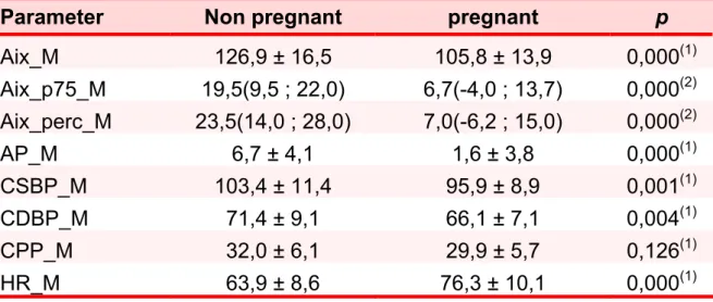
Table 2 - Comparison of parameters between non pregnant women and pregnant women in the second trimester of gestation.
Parameter Non pregnant pregnant p
Aix_M 126,9 ± 16,5 105,8 ± 13,9 0,000(1)
Aix_p75_M 19,5(9,5 ; 22,0) 6,7(-4,0 ; 13,7) 0,000(2) Aix_perc_M 23,5(14,0 ; 28,0) 7,0(-6,2 ; 15,0) 0,000(2)
AP_M 6,7 ± 4,1 1,6 ± 3,8 0,000(1)
CSBP_M 103,4 ± 11,4 95,9 ± 8,9 0,001(1)
CDBP_M 71,4 ± 9,1 66,1 ± 7,1 0,004(1)
CPP_M 32,0 ± 6,1 29,9 ± 5,7 0,126(1)
HR_M 63,9 ± 8,6 76,3 ± 10,1 0,000(1)
(1) t Student test α = 0,05. Values expressed in mean and side deviation (SD) (2) Mann-Whitney test α = 0,05. Values expressed in mediana and quartile.
Table 3 - Comparison of parameters between non pregnant women and
pregnant women in the third trimester of gestation.
Parameter Non pregnant pregnant p
Aix_M 131,0(116,5 ; 138,5) 104,5 (93,5 ; 115,5) 0,000(2)
Aix_p75_M 19,5 (9,5 ; 22,0) 7,0 (-3,0 ; 14,5) 0,003(2)
Aix_perc_M 19,8 ± 11,1 4,2 ± 12,7 0,000(1)
AP_M 6,7 ± 4,1 1,4 ± 3,7 0,000(1)
CSBP_M 103,4 ± 11,4 96,9 ± 8,4 0,003(1)
CDBP_M 71,4 ± 9,1 68,9 ± 7,0 0,166(1)
CPP_M 32,0 ± 6,1 28,0 ± 5,0 0,002(1)
HR_M 63,9 ± 8,6 81,1 ± 12,6 0,000(1)
Augmentation Index changed with gestation and was
significantly lower in mid-pregnancy when compared with controls. It started
to increase in the third trimester and reached pre-pregnancy levels in the
end of pregnancy (p= 0,00, figure 2 and tables 4 and 5).
Aix at 75 bpm vs GA
-30.0 -20.0 -10.0 0.0 10.0 20.0 30.0 40.0
0 10 20 30 40
GA
Figure 2 – Ilustration of the distribution of pregnant women through out
pregnancy and augmentation index values for pregnant and
non pregnant women (controls).
Aix at 75
Pregnant women
Non pregnant
Table 4 – Comparison of parameters between non pregnant women and pregnant women considering all gestational period.
Característica Não Grávidas Grávidas p
Aix_M 131(116,5 ; 138,5) 107(93,75 ;117) 0,000(2)
Aix_p75_M 19,5(9,5 ; 22,0) 6,75(-3,5 ;14,0) 0,000(2) Aix_perc_M 23,5(14 ; 28,0) 6,5(-6,2 ; 14,5) 0,000(2)
AP_M 7,0(4,0 ; 10,0) 1,75(-1,5 ; 4,5) 0,000(2)
CSBP_M 103,3 ± 11,3 96,3 ± 8,8 0,001(1)
CDBP_M 71,4 ± 9,1 66,9 ± 7,4 0,008(1)
CPP_M 32,5 (27,0 ; 36,5) 28,5 (25,0 ; 33,3) 0,055(2)
HR_M 64,0 (58,0 ; 68,5) 76,5 (69,0 ; 84,5) 0,000(2)
(1) t Student test α = 0,05. Values expressed in mean and side deviation (SD) (2) Mann-Whitney test α = 0,05. Values expressed in mediana and quartile.
Table 5 – Augmentation Index (AI) corrected by 75bpm in percentile in non
pregnant (controls) and in pregnant women through out gestation.
GA WEEKS 10th CENTILE 50th CENTILE 90th CENTILE
Controls 2.2 19.5 23.0
11-13 -8.5 9.0 20.9
14-16 -18.3 0.3 20.0
17-19 -8.7 6.8 14.9
20-22 -6.0 10.5 18.5
23-25 -11.7 5.5 15.3
26-28 -9.0 -3.0 23.5
29-31 -11.3 6.0 17.9
32-34 -8.0 6.5 18.6
35-37 6.0 13.5 26.9
38-41 -1.1 10.5 20.2
(3) Teste t de Student ao nível de significância α = 0,05. Valores expressos em média e desvio-padrão
Central systolic (p=0,001) and central diastolic (p=0,008)
blood pressures changed with gestation and it was lower than controls.
Central pulse pressure also changed with gestation and it was significantly
lower (p= 0,002) than controls in the third trimester. Heart rate was
significantly increased (p=0,000) when compared to controls and this
difference is evident since the first trimester of pregnancy, figure 3, 4 and 5
and tables 4 and 5). Ejection duration (p=0,0002), heart cycle (p= 0,000),
diastole and systole (p=0,000) time were shorter in pregnancy when
compared with controls (figure 5).
Radial and femoral pulse wave velocity in pregnant subjects
were lower than controls but this result showed no significant difference.
Cent Syst press vs GA
70.0 80.0 90.0 100.0 110.0 120.0
0 10 20 30 40
GA Sy s t pr e s
Figura 3 - Ilustration of the distribution of pregnant women through out
pregnancy and central systolic values for pregnant and non
pregnant (controls) women
Pregnant women
Non pregnant
Cent diast pres vs GA 45.0 50.0 55.0 60.0 65.0 70.0 75.0 80.0 85.0 90.0
0 10 20 30 40
GA Di a s t p re
Figura 4 - Ilustration of the distribution of pregnant women through out
pregnancy and central diastolic values for pregnant and non
pregnant (controls) women.
Heart rate vs GA
48.0 58.0 68.0 78.0 88.0 98.0 108.0
0 10 20 30 40
GA
H
R
Figura 5 - Ilustration of the distribution of pregnant women through out
pregnancy and heart rate values for pregnant and non pregnant
(controls) women.
Pregnant women
Non pregnant
-controls
Pregnant women
Non pregnant
DISCUSSION
In this study we used 2 techniques of applanation tonometry
to assess the behavior of the cardiovascular system in pregnancy. First, we
have established a curve for Augmentation Index (AI) in normal pregnancies
with weekly ranges from 11 weeks of gestation until 41 weeks, including 195
pregnant women distributed through all pregnancy. Although ranges for
Augmentation index in pregnancy have been established before8, this study
differs from that one in the sample size and by the fact that the ranges were
obtained weekly.
Our main findings show that AI changes with gestation and
that is lower than controls in mid pregnancy and increases in the third
trimester to reach pre-pregnancy levels at the end of gestation. These
findings are similar to previous study8 and demonstrates that AI follows a
similar curve of the blood pressure in pregnancy. The first systolic peak
(P1H) is higher in the first trimester and higher than controls; this is the
maximum pressure coming from the heart stroke and it is probably related to
the extra effort of the heart in accommodating the increase in blood volume.
This concept is also demonstrated by an increase in ejection duration
(ED%) during gestation starting form the first trimester.
We also demonstrated that central systolic and diastolic
pressures changes with gestation, being lower than controls and with
pressure also changes with gestation with greater fall in the second
trimesters. Heart rate increases in pregnancy and it is already evident in the
first trimester, consequently, the heart cycle is shorter and changes with
gestation, as diastole and systole time shorten, accompanying the changes
of heart rate.
Regarding the peripheral parameters of blood pressure, only
the brachial diastolic pressure showed changes with gestation being
significantly lower in the second trimester of gestation. All of these findings
demonstrated the known maternal cardiovascular adaptations to pregnancy
and that the method used in our study seemed to be reliable to assess
these changes and probably to assess the possible abnormalities of the
maternal adaptations to pregnancy.
Augmentation Index is related to central pulse pressure, and
this has been proved to be a better and more reliable parameter of
hemodynamics than brachial blood pressure(BP), as BP varies throughout
the arterial tree, due to vessel compliance, posture, exercise, age and the
phenomenon of wave reflection 16. It is known that blood pressure falls in
pregnancy due to the fall in the peripheral resistance to accommodate the
increase in blood volume and cardiac output1,2,3,4,5. However, the
mechanism of how these changes occur are not completely clarified.
Nitric Oxide (NO) liberated by the vascular endothelium is
arteriolar tone (vasodilatation) and consequently blood pressure14,17.
Recently, it was also demonstrated that NO also plays a role in the local
regulation of large artery stiffness18.
Assessment of endothelial function in pregnancy has been
done and it was demonstrated that normal pregnancies are associated with
enhanced endothelial function 19, 20.
It was also demonstrated in pregnant rats and in human
pregnancies that estrogen influences the bioavailability of endothelial
derived nitric oxide and relaxation of vascular smooth muscle cells21,22 .
Endothelial function was also proved to be associated, in healthy humans,
with pulse pressure, pulse wave velocity and augmentation Index, and
global endothelial function correlates more strongly to central pulse pressure
than to peripheral pulse pressure 23.
It seems that endothelial function and the production of
vasoactive substances plays an important role in the maternal
vasodilatation. The fact that applanation tonometry is able to measure non
invasively central pulse pressure and several indexes to assess endothelial
function, makes of it an important tool for the evaluation of pregnancies
affected by hypertension and diabetes, as these conditions are frequently
associated and appear to share a common etiology: endothelial dysfunction
Although the cardiovascular maternal adaptations to
pregnancy are well known, its mechanism are still unclear. Applanation
tonometry has proved to be a good method to assess this system during
gestation and may help us understand how physiological changes take
place in pregnancy, as well as to understand the mechanism of disease in
cases in which the maternal organism fails to adaptate properly to
pregnancy.
We have also performed Pulse Wave Velocity (PWV) from
Carotid artery to Radial artery and PWV from Carotid artery to Femoral
artery in both groups with no differences. Others9, have demonstrated
differences in PWV between hypertensive, preeclamptic and normotensive
pregnancies, however, it seams that between normotensive pregnancies
and healthy controls there are no differences in the PWV. This could be due
to the pregnant uterus compressing the vena Cava which alters the venous
return and in some way, could affect the results. Although, we have
performed the measurements with the patient in lateral position in order to
avoid this phenomenon.
The limitations of our work are basically that our study group
starts at 11 weeks of gestation and not at 6 or 8 weeks time in which the
vascular adaptations begin in pregnancy. We found it difficult recruiting
patients, as this work took place at a scanning department where the patient
came for a routine scan, consequently, our sample is more concentrated in
an even sample.
We would like to add that, although it is described that the
expertise in performing applanation tonometry comes with 20 scans, we
realized that after 20 exams the operator may be ready to perform it, but
there is still the need of more training to make it reproducible.
Applanation tonometry relies very much on the operator and
this is a limitation of the method.
In Summary, we demonstrated that applanation tonometry is
also a good method to assess the cardiovascular function in pregnancy.
Augmentation Index is lower and changes with gestation following a curve
that is similar to that one of the blood pressure in pregnancy; normal
pregnancies presents with a drop in central systolic, diastolic and pulse
pressures and this fall is more evident in the first and second trimesters of
pregnancy; heart rate is increased in normal pregnancies, changes with
gestation and is higher in the third trimester of pregnancy. Our results also
showed that are no differences in Pulse Wave Velocity between healthy
Acknowledgments:
This study was supported by the Fetal Medicine Foundation.
Maria Leticia S de Macedo had a scholarship from CAPES –
REFERENCES
1- Duvekot JJ,Peeters LLH. Very early changes in cardiovascular
physiology. In Clinical Physiology in Obstetrics, Chamberlain G, Pipkin
FB. (eds), foreword by Hytten F. Blackwell Science Ltd: London - 3rd ed.
1998; 3-32.
2- Swiet M. The cardiovascular system. In Clinical Physiology in
Obstetrics, Chamberlain G, Pipkin FB. (eds), foreword by Hytten F.
Blackwell Science Ltd: London - 3rd ed. 1998; 33-70.
3- Magness RR, Gant NF. Normal vascular adaptations in
pregnancy:potentials clues for understanding pregnancy-induced
hypertension. In Hypertension In Pregnancy, Walker JJ, Gant NF.
(eds).Chapman& Hall Medical: London-1997;5-26.
4- Chapman AB,Abraham WT, Zamudio S, Coffin C, Merouani A, Young D,
Johnson A, Osorio F, Goldberg C, Moore L, Dahms T and Schrier R.
Temporal relationships between hormonal and hemodynamic changes
in early human pregnancy. Kidney International 1998; 54: 2056-63.
5- Cunningham FG, Gant NF, Leveno KJ, Gilstrap LC, Hauth JC,
Wenstrom KD. Maternal adaptations to pregnancy. In Williams
6- Gordon MC. Maternal physiology in pregnancy. In Obstetrics- Normal
and Problem Pregnancies. Gabbe SG, Niebyl JR, Simpson JL.(eds).
Churchill Livingstone: USA-2002; 63-91.
7- Elvan-Taspinar A, Franx A, Bots ML, Koomans HA, Bruinse HW. Arterial
stiffness and fetal growth in normotensive pregnancy. Am J
Hypertension 2005; 18:337-341.
8- Smith SA, Morris JM, Gallery EDM. Methods of assessment of the
arterial pulse wave in normal human pregnancy. AM J Obst Gynecol
2004; 190:472-6.
9- Elvan-Taspinar A, Franx A, Bots ML, Bruinse HW, Koomans HA. Central
hemodynamics of hypertensive disorders in pregnancy. Am J
Hypertension 2004; 17:941-46.
10- Spasojevic M, Smith SA, Morris JM, Gallery EDM. Peripheral arterial
pulse wave analysis in women with pre-eclampsia and gestational
hypertension. BJOG: an International Journal of Obstetrics and
Gynaecology 2005; 112:1475-1478.
11-O’Rourke MF, Gallagher DE. Pulse wave analysis.J Hypertens
12- Mackenzie IS, Wilkinson IB, Cockcroft JR. Assessment of arterial
stiffness in clinical practice. QJ Med 2002; 95:67-74.
13- Wilkinson IB, Fuchs SA, Jansen LM, Spratt JC, Murray GD, Cockcroft
JR, Webb DJ. Reproducibility of pulse wave velocity and augmentation
index measured by pulse wave analysis. Journal of Hypertension 1998;
16:No 12(part2).
14- Hayward CS, Kraidly M, Webb CM, Collins P. Assessment of endothelial
function using peripheral waveform analysis. A Clinical Application. J
Am Coll Cardiol 2002; 40:521-8.
15- Wlkinson IB, Hall IR, MacCallum H, Mackienzie IS, McEniery CM, Arend
BJ, Shu Y-E, MacKay LS, Webb DJ, Cockcroft JR. Pulse-wave analysis.
Clinical Evaluationn of a noninvasive, widely applicable method for
assessing endothelial function. Arterioscler Thromb Vasc Biol. 2002;
1:147-152.
16- Wilkinson IB, Mohammad NH, Tyrrell S, Hall IR, Webb DJ, Paul VE,
Levy T, Cockcroft JR. Heart Rate Dependency of Pulse Pressure
Amplification and Arterial Stiffness. A Journ Hyperten .2002;15: 24-30.
17- Wilkinson IB, McEniery CM. Arterial Stiffness, Endothelial Function and
Novel Pharmacological Approaches. Clinical and Experimental
18- Wilkinson IB, Qasem A, McEniery CM, Webb DJ, Avolio AP, Cockcroft
JR. Nitric Oxide Regulates Local Arterial Distensibility In Vivo.
Circulation. 2002; 1: 213-17.
19- Savvidou MD, Kametas NA, Donald AE, Nicolaides KH. Non-Invasive
Assessment of Endothelial Function in Normal Pregnancy. Ultrasound
Obstet Gynecol. 2000; 15: 502-07.
20- Williams DJ, Vallance PJT, Neild GH, Spencer JAD, Imms FJ. Nitric
Oxide-Mediated Vasodilation in Human Pregnancy. Am J Physiol. 1997;
2 :748-52.
21- Mendelsohn ME, Karas RH. The Protective Effects of Estrogen on The
Cardiovascular System. N Engl J Med. 1999; 340(23):1801-11.
22- Zhang Y, Stewart KG, Davidge ST. Endogenous Estrogen Mediates
Vascular Reactivity and Distensibility in Pregnant Rat Mesenteric
Arteries. Am J Physiol Heart Circ Physiol. 2001; 280: 956-61.
23- McEniery CM, Wallace S, Mackenzie IS, McDonnell B, Yasmin, Newby
DE, COckcroft JR, Wilkinson IB. Endothelial Function Is Associated With
Pulse Pressure, Pulse Wave Velocity, and Augmentation Index in
24- Granger JP, Alexander BT, Llinas MT, Bennett WA, Khalil RA.
Pathophysiology of Hypertension During Preeclampsia Linking Placental
Ischemia With Endothelial Dysfunction. Hypertension. 2001;38(3):
718-22.
25- Shah DM. Preeclampsia: New Insights. Curr Opin Nephrol Hypertens.
2007;16 (3): 213-20.
26- Savvidou MD, Hingorai AD, Tsikas D, Frolich JC, Vallance P, Nicolaides
KH. Endothelial Dysfunction and Raised Plasma Concentration of
Asymetric Dimethylarginine in Pregnant Women Who Subsequently
Developed Pre-Eclampsia. Lancet.2003;3 (361):1511-7.
27- Vambergue A, Nuttens MC, Goeusse P, Biausque S, Lepeut M,
Fontaine P. Pregnancy Induced Hypertension in Women with
Gestational Carbohydrate Intolerance: The Diagest Study.
28- Roach VJ, Hin LY, Tam WH, Ng KB, Rogers MS. The Incidence of
Pregnancy – Induced Hypertension among patients with Carbohydrate
Intolerance. Hypertens Pregancy. 2000;19(2):183-9.
29- Barden A, Singh R, Walters BN, Richie J, Roberman B, Beilin LJ.
Factors Predisposing to Pre-eclampsia in Women with Gestational
Capítulo III
Avaliação vascular não invasiva (NIVA) em gestantes com
diabete gestacional e com hiperglicemia leve utilizando o
SphygmoCor
®.
Maria Letícia Sperandéo de Macedo1, Maria Aparecida Mourão Brasil2,
Marilza Viera Cunha Rudge3
1) Aluna de doutorado do Programa de Pós Graduação em Ginecologia,Obstetrícia e
Mastologia do Departamento de Ginecologia e Obstetrícia da Faculdade de Medicina
de Botucatu- UNESP. Professora Assistente do Departamento de Tocoginecologia da
Faculdade de Medicina de Jundiaí.
2) Superintendente técnica da FAMESP e Coordenadora Geral do Grupo de apóio à
Pesquisa (GAP) da Faculdade de Medicina de Botucatu - UNESP.
3) Professora Titular do Departamento de Ginecologia e Obstetrícia da Faculdade de
INTRODUÇÃO
O Programa de Pós-Graduação em Ginecologia, Obstetrícia
e Mastologia da Faculdade de Medicina de Botucatu – UNESP, desde
1983, tem como uma de suas linhas de pesquisa o estudo da Hiperglicemia
na Gestação (Rudge, 1983), que abrange aspectos clínicos e
experimentais.
Atualmente a área clínica desenvolve maior número de
pesquisas, em função da criação da Unidade de Pesquisa Clínica do
Hospital das Clínicas de Botucatu (Upeclin) ligada à Rede Nacional de
Pesquisa Clínica do Ministério da Saúde, originário de Chamada Pública
MCT/MS/FINEP no 04/2005.
No Brasil, 68% dos óbitos infantis ocorrem no primeiro mês
de vida, sendo 49% na primeira semana. A evolução dos coeficientes de
mortalidade infantil do Estado de São Paulo nas últimas décadas, mostra
diminuição acentuada de cifras superiores a 80 por mil nascidos vivos no
ano de 1975 para 14,25 em 2004. A diminuição da mortalidade ocorreu,
principalmente, no período pós-neonatal (28 dias a 11 meses completos),
com o controle das causas infecciosas e parasitárias responsáveis por
37,3% dos óbitos em 1975 e, por apenas, 4,8% em 2004. Os programas
preventivos e educacionais relacionados às políticas de saúde pública, com