Research Article
Structural Characterization and
In Vitro
Antioxidant
Activity of Kojic Dipalmitate Loaded W/O/W Multiple
Emulsions Intended for Skin Disorders
Maíra Lima Gonçalez, Diana Gleide Marcussi, Giovana Maria Fioramonti Calixto,
Marcos Antonio Corrêa, and Marlus Chorilli
Department of Drugs and Medicines, School of Pharmaceutical Sciences, UNESP, Rodovia Araraquara-Ja´u, Km 1, Campus, 14801-902 Araraquara, SP, Brazil
Correspondence should be addressed to Ma´ıra Lima Gonc¸alez; [email protected] and Marlus Chorilli; [email protected]
Received 24 November 2014; Accepted 16 January 2015
Academic Editor: Ajit S. Narang
Copyright © 2015 Ma´ıra Lima Gonc¸alez et al. his is an open access article distributed under the Creative Commons Attribution License, which permits unrestricted use, distribution, and reproduction in any medium, provided the original work is properly cited.
Multiple emulsions (MEs) are intensively being studied for drug delivery due to their ability to load and increase the bioavailability of active lipophilic antioxidant, such as kojic dipalmitate (KDP). he aim of this study was to structurally characterize developed MEs by determining the average droplet size (Dnm) and zeta potential (ZP), performing macroscopic and microscopic analysis and analyzing their rheological behavior andin vitrobioadhesion. Furthermore, thein vitrosafety proile and antioxidant activity of KDP-loaded MEs were evaluated. he developed MEs showed a Dnm of approximately 1 micrometer and a ZP of−13 mV, and no change was observed in Dnm or ZP of the system with the addition of KDP. KDP-unloaded MEs exhibited “shear thinning” low behavior whereas KDP-loaded MEs exhibited Newtonian behavior, which are both characteristic of antithixotropic materials. MEs have bioadhesion properties that were not inluenced by the incorporation of KDP. he results showed that the incorporation of KDP into MEs improved the safety proile of the drug. hein vitroantioxidant activity assay suggested that MEs presented a higher capacity for maintaining the antioxidant activity of KDP. ME-based systems may be a promising platform for the topical application of KDP in the treatment of skin disorders.
1. Introduction
Skin disorders such as skin aging and hyperchromia interfere negatively in the social behavior of people. hese disorders are mainly caused by reactive oxygen species induced, for example, by prolonged exposure to UV radiation, establishing a condition in which the oxidative attack of biomolecules is increased [1–6].
he current treatments include laser procedures, der-mabrasion and microderder-mabrasion, and chemical peels. Nevertheless, these treatments present several disadvantages such as persistent erythema, hypopigmentation, keloids, and hypertrophic scars [7].
Another common treatment modality is through the use of cosmetics with tretinoin and hydroquinone drugs;
however, many studies indicate that tretinoin can cause skin irritation (e.g., erythema, stinging, burning, and dryness) and hydroquinone can have carcinogenic and mutagenic efects [7].
herefore, developing new formulations to reduce these degenerative processes and increase people’s self-esteem is an ongoing goal. To this end, interest has grown in antioxidant drugs that interfere with the generation of free oxygen radicals and the reactions they trigger [8]. Among these antioxidant drugs, kojic acid (KA) has been highlighted because it presents antioxidant activity by chelating iron ions. KA also presents depigmentation activity by chelating the copper ion present in the active site of tyrosinase, which mediates the formation of melanin from the amino acid tyrosine [9–11].
O O O
O O
O
Figure 1: Structural formula of kojic dipalmitate (KDP) [12].
Nevertheless, KA is unstable at high temperatures (40∘C) and presents a labile oxidative property against light. here-fore, KA is used as kojic dipalmitate (KDP) (Figure 1), which is hydrolyzed by esterases present in skin cells to promote the
in siturelease of kojic acid [12].
KDP is a liposoluble white powder that is heat- and light-stable over a wide pH range [12].
Due to diiculties in solubilizing fat-soluble drugs such as KDP, a search for strategies to convey the drug is still required. KDP can be incorporated into colloidal carrier systems, such as multiple emulsions (MEs), a single colloidal system con-sisting of both water-in-oil and oil-in-water emulsion system types. MEs are classiied as oil-in-water-in-oil (O/W/O) or water-in-oil-in-water (W/O/W) depending on the dispersed and external phase compositions [13].
herefore, MEs are being intensively studied as a drug delivery system due to their ability to load lipophilic drugs to increase their bioavailability and protect such drugs against biological degradation and oxidation processes. his can prolong the drug release, which could possibly reduce the required dosages and application time of the formulation [14– 16].
hereby, ME can increase the efectiveness of KDP, acting as a promising drug delivery system to prevent and treat skin disorders [17].
However, MEs contain large and polydisperse droplets that are extremely thermodynamically unstable due to their easy and spontaneous separation into three distinct phases. MEs also tend to undergo coalescence, locculation, or creaming [18]. One way to obtain stable MEs is to reduce the droplet size through altering the method of preparation and monitoring the system over time to ensure that the droplet size does not increase. Another way to obtain the MEs stable over long periods of time is to choose the correct surfactant system that conserves the droplet size through changes in the interfacial tension between internal aqueous and oil phases in W/O/W emulsions [19].
he aim of this study was to perform the structural characterization of MEs through determining the average droplet size and zeta potential (ZP), performing macroscopic and microscopic analyses, and evaluating the rheological behavior andin vitrobioadhesion of the MEs. Furthermore,
the in vitro safety proile and antioxidant activity of
ME-incorporated KDP was evaluated.
2. Materials and Methods
2.1. Materials. For preparing the formulations, Kojic
dipal-mitate (KDP) from Via Pharmaceuticals, Brazil, sorbitan monooleate (Span 80) from Bold, Brazil, liquid petrolatum
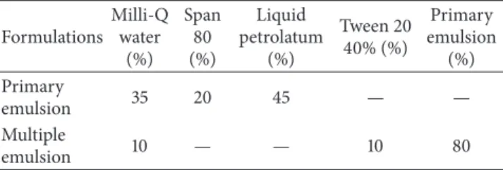
Table 1: Composition (%) of the KDP-unloaded ME formulations used in this study.
Formulations Milli-Q
water (%)
Span 80 (%)
Liquid petrolatum
(%)
Tween 20 40% (%)
Primary emulsion
(%) Primary
emulsion 35 20 45 — —
Multiple
emulsion 10 — — 10 80
from Teclab, Brazil, sorbitan monooleate ethoxylated 20 OE (Tween 20) from Vetec, Brazil, and water (Milli-Q) were used. In determining the in vitroantioxidant activity of the formulations, 2,2-diphenyl-1-picrylhydrazyl (DPPH) purchased from Fluka, Buchs, Switzerland, was employed.
2.2. Methods
2.2.1. ME Development. he ME W/O/W system was
devel-oped by a two-step process. Initially, the primary W/O emul-sion composed of 20% Span 80 (hydrophobic surfactant, HLB (hydrophile-lipophile balance) = 4.3), 45% liquid petrolatum, and 35% water (Milli-Q) was obtained, in which the liquid petrolatum and Span 80 were heated to approximately 40∘C and dispersed in water heated to the same temperature. he heat was maintained while the mixture was agitated using magnetic stirrer for 1 minute. he primary emulsion was dispersed into an aqueous solution of Tween 20 (hydrophilic surfactant−HLB = 15) to generate the W/O/W ME composed of 80% of the primary emulsion, 10% of the solution in 40% Tween 20, and 10% water (Milli-Q) [20].
Table 1 shows the components and their concentrations used in the preparation of the KDP-unloaded MEs. To obtain KDP-loaded MEs, 1.25% of the total quantity of liquid petrolatum was reduced from the primary emulsion and an equivalent percentage of KDP was incorporated.
he particle size distribution of a dispersed system depends on the speed of agitation between the dispersed and dispersing phases during the emulsiication process and the rate of addition of one phase over another. hese parameters were adjusted to obtain a proper system [21].
2.2.2. Physicochemical Characterizations of ME
(1) Macroscopic and Microscopic Analysis.he macroscopic
appearance, organoleptic properties, and homogeneity of the MEs were evaluated ater 24 hours of preparation. he MEs were also observed using an optical Leitz DM RXE microscope (LeicaTM) at25±0.5∘C. he samples were placed on glass slides and covered with a cover slip, and images were captured at 100x magniication.
( 2) Droplets Distribution and Quantiication
(A) Optical Microscope.MEs were placed on a glass slides,
Captured images were scanned and analyzed using the Motic Images Plus 2.0 sotware.
(B) Dynamic Light Scattering. he size and distribution
of droplets were determined by an optical particle ana-lyzer (Zetasizer Nano-ZS ZEN3600, Malvern Instruments) through the technique of dynamic light scattering. he samples were diluted in Milli-Q water at a 1 : 20 ratio and placed in the equipment. he analyses were performed in triplicate.
(3) Determination of pH.For the determination of pH, 1 g of
formulation was diluted into 10 mL of Milli-Q water. hen, the pH was checked using a digital pH meter (DM-Digimed 23). his assay was performed in triplicate.
(4) Determination of Electrical Conductivity.Ater the
con-ductivity (Digimed DM-32) was calibrated with KCl 0.1 N, the electrical conductivity was determined by directly inserting the electrode into the samples. his assay was performed in triplicate.
(5) Determination of the Zeta Potential.Zeta potential values
were obtained from the derivation of the electrophoretic mobility using the Zetasizer Nano-ZS ZEN3600 optical particle analyzer (Malvern Instruments). he samples were diluted in distilled water at a 1 : 20 ratio and placed in the equipment to be analyzed. he analyses were performed in triplicate.
(6) Rheological Study.Continuous low was analyzed at32 ±
0.1∘C and in triplicate on a controlled-stress AR2000 rheome-ter (TA Instruments, New Castle, DE, USA) equipped with parallel plate geometry (40 mm diameter) and a sample gap of 200�m. Samples of the systems were carefully applied to the lower plate, ensuring that sample shearing was minimized, and allowed to equilibrate for 3 min prior to analysis.
Continual testing was performed using a controlled shear rate ranging from 0.01 to 100 s−1 and back. Each stage lasted 120 s, with an interval of 10 s between the curves. he consistency index and low index were determined using the power law described in(1) for quantitative analysis of low behavior:
� = � ⋅ ��, (1)
where “�” is shear stress; “�” is shear rate; “�” is the consistency index; and “�” is the low index.
(7) In Vitro Bioadhesion Test. In vitro bioadhesion of the
systems was evaluated using TA.XT Plus texture analyzer (Stable Micro Systems, Surrey, England). A “Hold Until Time” test was used to evaluate the force required for the formulation to leave the animal skin.
he ear skin of domestic pigs was used as an experimental model. he ear skin of domestic pigs purchased from a local slaughterhouse was used as experimental model. he skin was separated from the cartilage with a scalpel and a 500�m thick layer stratum corneum and epidermis was separated
from the adipose tissue with a dermatome (Nouvag TCM 300, Goldach, USA), prior to analysis. Before the test, the skins were maintained in 0.9% NaCl solution for 10 minutes and were aixed to the probe with the aid of rubber rings.
MEs (7.5 g) were placed in Falcon (BD) centrifuge tubes with a capacity of 45 mL and centrifuged at 3500 rpm (Sorval, DuPont Model TC 6) for 3 minutes to eliminate air bubbles and obtain smooth surfaces. he Falcon tube was ixed below the probe. he sample was placed 10 mm below the analytical probe, which was lowered at a constant speed (1 mm/s) until it reached the sample (detected by a 2 mN triggering force) and remained in contact with the formulation for 60 seconds. hen, the probe was removed slowly at a speed of 0.5 mm/s. he force exerted by the probe on the skin surface was determined using the Texture Exponent Lite sotware to obtain a result curve for the bioadhesive forceversustime. Assays were performed in triplicate.
2.2.3. In Vitro Biological Assays
(1) Erythrocyte Hemolysis.he erythrocyte hemolysis assay
was performed using the experimental procedure described by Jumaa et al., 1999 [22], and Huang et al., 2008 [23]. Briely, before use, freshly collected human blood (O positive) was washed three times with a 7.4 pH solution of 0.01 M Tris-HCl containing 0.15 M NaCl (Tris-saline). A suspension containing 1% (v/v) erythrocytes was prepared with packed red blood cells resuspended in Tris-saline. KDP-loaded MEs, KDP-unloaded MEs, and free KDP were dissolved in Tris-saline to a inal concentration of 27�M. As a positive control (100% lysis), a 1% (v/v) Triton X-100 solution was used. Ater incubation for 1 hour at 37∘C, the samples were centrifuged at 3000×g for 2 minutes. Aliquots of 100�L of the supernatant were transferred to 96-well microplates and the absorbance was determined at 405 nm using a BioRad Model 3550-UV (USA) microplate reader. he assay was performed in triplicate. he percentage of hemolysis was calculated using the following equation: % hemolysis = (absorbance of the test sample/absorbance at 100% lysis)×100.
(2) In Vitro Nonspeciic Cytotoxicity. In vitro testing was
he concentrations were tested in triplicate using six addi-tional controls (cells in medium). Cell viability was calculated using the following equation: Cell viability (%) = [OD560 (sample)/OD560(control)]×100.
2.2.4. In Vitro Antioxidant Activity. Free radical
scav-enging activity was evaluated using the 2,2-diphenyl-1-picrylhydrazyl (DPPH) test with modiications [24]. One hundred microliters of free KDP, ME formulations, or control (ethanol; 10–60�g/mL) was added to 3.9 mL of DPPH solu-tion in ethanol (60�M). Ater 30 minutes of storage in a dark place, the absorbance was measured using a spectrophotome-ter at 517 nm. All measurements were repeated three times. Free radical scavenging activity was calculated using the following formula: % inhibition of DPPH= [(�0− �1)/�0×
100], where�0= absorbance of control and�1= absorbance of sample. he IC50 value was determined by plotting the concentration of formulationsversusthe percentage of DPPH that was maintained at a steady state [24].
2.2.5. Statistical Analyses. Data were analyzed using the
means and standard deviation and compared by analysis of variance (ANOVA). Tukey’s test was used to assess signiicant diferences between samples, where values of � < 0.05 were considered statistically signiicant. he Origin 7.0 SRO program was used for the treatment of the data.
3. Results and Discussion
3.1. Physicochemical Characterization of ME. he MEs were
viscous, white, and opaque with a characteristic odor. By microscopic analysis (Figure 2), larger droplets of less than 10�m of diameter were observed. At certain times, smaller droplets were observed within the larger droplets, character-istic of ME formulations. Moreover, there was no change in the appearance of the ME formulations with the addition of KDP.
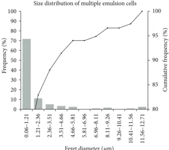
Figure 3shows the size of droplets and the calculated size distribution of droplets of the KDP-unloaded MEs.
he droplets had a maximum diameter of 12.487�m and a minimum diameter of 0.056�m, with an average diameter of 1.534�m. 72% of the droplets in the formulation were observed to have diameters ranging from 0.06 to 1.21�m. he addition of KDP did not cause statistically signiicant changes in droplet size (� > 0.05).
Diferent sizes of droplets can be obtained depending on the method of emulsiication selected, explaining the method by which the stability of the system is inluenced [25]. Because the droplet size within the emulsion determines the probability that phenomena such as locculation and coalescence will occur, in general, the smaller the size of the dispersed droplets, the greater the stability of the system [25]. he dynamic light scattering technique showed that KDP-unloaded MEs had two populations of droplets of diferent sizes. he results showed that 73.3% of the formulation droplets had an average diameter of 1.016�m and the remain-ing 23.7% had an average diameter of 350 nm, as could be observed morphologically using an optical microscope.
Figure 2: Microscopy of KDP-unloaded MEs (100x) at25 ± 0.5∘C.
80 85 90 95 100 0 10 20 30 40 50 60 70 80 90 100 F req uenc y (%) C u m u la ti ve f req uenc y (%) 0. 06 – 1. 21 1. 21 – 2. 3 6 2. 3 6 – 3. 51 3. 51 – 4. 66 4. 66 – 5. 8 1 5. 8 1 – 6. 96 6. 96 – 8. 11 8. 11 – 9. 2 6 9. 2 6 – 10. 4 1 10. 4 1 – 11. 5 6 11 . 56 – 12 . 71
Size distribution of multiple emulsion cells
Feret diameter (�m)
Figure 3: Size distribution of droplets of KDP-unloaded MEs analyzed by optical microscope.
hrough the statistical ANOVA test, the average pH values of the unloaded MEs (pH = 4.62) and KDP-loaded MEs (pH = 4.55) were found to be equal (� > 0.05), leading to the conclusion that the incorporation of KDP induced no signiicant change in pH. hese pH values are well tolerated by the skin, which presents a slightly acid pH (4.5 to 5.5) [20].
he pH is an important parameter to consider because large variations in the pH values of the formulations can cause chemical incompatibilities between the ingredients of the formulation, therefore compromising the eicacy and safety of the active compounds. In addition to being a parameter related to the intrinsic stability of the formulation, it is essen-tial to control the pH of topical application products to ensure that over time they do not cause changes in the microlora of the skin or induce irritation or skin sensitization, which could degrade the product and reduce its activity [21].
hrough the statistical ANOVA test, the average electrical conductivity of KDP-unloaded MEs (34.2�S/cm) and that of KDP-loaded MEs (31.90�S/cm) were found to be equal (� >
induced no signiicant change in the electrical conductivity of the ME system.
In terms of conductivity, the electrical properties of oil and water are extremely distinct and can be used to distin-guish whether the ME external phase is aqueous or oily. In accordance with the results obtained, electrical conductivity superior to 30�S/cm, it was conirmed that the external phase is aqueous and that the ME formulation is a W/O/W type of emulsion [26].
he KDP-unloaded MEs showed an average ZP of−13 mV. he addition of KDP did not cause statistically signiicant changes in the ZP (� > 0.05).
he ZP results from the electric charges on the surface of the particle and is related to the physical stability of the colloidal system [27]. At greater absolute values (more than +30 mV or less than−30 mV), the ZP of a system indicates the tendency of the particles to repel each other, which may indicate that the system is stable. If, however, the absolute values for the ZP are low or zero, the particles tend to agglomerate and the system can easily locculate [28]. In this case, the formulation should be monitored over time to check whether the intermediate ZP values (−13 mV) indicate an unstable system.
Semisolids products can exhibit a wide range of rheologi-cal behaviors. A good understanding of formulation rheology is very important to the design, selection, and operation of the equipment involved in evaluating the release performance and administration characteristics of the drug. Furthermore, rheological studies can provide useful information on the sta-bility and microstructure of emulsions. MEs can display low characteristics depending on the fraction volume, droplet size and distribution, and the viscosity of the dispersed phase [29– 32].
he relationship between the shear rate (Pa) and shear stress (1/s) of MEs is presented inFigure 4.
From the data obtained using(1)and shown inTable 2, KDP-unloaded MEs were demonstrated to exhibit “shear thinning” low behavior (� < 1), whereas KDP-loaded MEs exhibited Newtonian behavior (� = 1).
he KDP-unloaded ME formulation behaved as a pseu-doplastic shear thinning luid because its viscosity (�∗) decreased with increasing stress. his may be due to the breaking of organized structures that promote the formation of droplets, which are less organized structures [33].
However, the incorporation of the KDP drug had a strong inluence on the formulation, leading the KDP-loaded ME formulation to behave as a low-viscosity Newtonian luid. his phenomenon is related to the intense reduction in the droplet size with the introduction of KDP, which thinned the low and resulted in a higher difusion coeicient than when larger oil droplets are present.
Furthermore, the rheogram showed that the up and down curves did not overlap. he up curve was below the down curve, which is typical and characteristic of antithixotropic materials.
Antithixotropic properties occur because the steady application of shear stress increases the apparent viscosity, whereas cessation of the applied stress decreases the viscosity back to its initial value in a time-dependent manner [30].
0 20 40 60 80 100
0 50 100 150 200 250 300 350
KDP-loaded-ME KDP-unloaded ME
Shear rate (1/s)
Sh
ea
r str
ess (P
a),
�
∗ (P
a
·
s)
�∗KDP-loaded ME
�∗KDP-unloaded ME
Figure 4: Flow rheograms of KDP-unloaded ME (I) and
KDP-loaded ME (◻). Closed symbol represents up curve and open symbol represents down curve.�∗(◊) is viscosity. Standard deviations have been omitted for clarity; however, in all cases, the coeicient of variation of triplicate analyses was less than 10%. Data were collected at32 ± 0.25∘C.
Table 2: Consistency index (�) and low index (�) of KDP-loaded MEs and KDP-unloaded MEs.
Formulations � �
KDP-loaded ME 0.63 1.02
KDP-unloaded ME 9.42 0.76
he increase in the apparent viscosity of the ME formu-lations with time at a constant shear was most likely due to the reorganization of the droplets by strong forces. he high viscosity of the MEs could retard the free motion of the droplets, thus delaying creaming, locculation, and coa-lescence of the system [33]. he conclusion from these results is that stable MEs can be obtained with the components used in this study only under the application of vigorous stirring during preparation so that viscosity is incorporated and the destabilization of the system is avoided.
he KDP-unloaded MEs and KDP-loaded MEs were evaluated for their changes in bioadhesion to determine whether the addition of KDP alters the bioadhesive ability of the ME system.
Table 3 shows the bioadhesion test results of KDP-unloaded MEs and KDP-loaded MEs.
he results demonstrate that the incorporation of KDP did not inluence the bioadhesion of the MEs. Moreover, the formulations showed values very close to the bioadhesion of polyacrylic acid bioadhesive hydrogels used in previous studies [32].
hus, MEs have properties that favor the interaction between its components and the epithelial cells of the skin.
Table 3: Bioadhesion of KDP-unloaded MEs and KDP-loaded MEs.
Formulations Bioadhesive
work (N⋅s) Bioadhesive force (N) KDP-unloaded MEs 0.0166±0.0016 0.0103±0.0029 KDP-loaded MEs 0.0163±0.0025 0.0099±0.0006
because bioadhesive formulations remain in contact with the biological substrate for longer periods of time, which may lead to a gradient of drug concentration at the site of action and therefore improve the clinical performance of the treatment.
3.2. In Vitro Biological Assays. Before exposing users to the
product, the potential irritation of a new topical preparation should be investigated to avoid inducing skin irritation. For ethical, legal, and inancial reasons,in vivotests were banned for testing cosmetics and formulations. hus, in recent years, various in vitro assays have been conducted to test the products prior to realizing therein vivosafety proiles [34].
Erythrocyte-induced hemolysisin vitrois considered to be a simple and reliable measure for estimating the membrane damage causedin vivobecause erythrocytes are easily isolated by centrifugation [35].
In vitro cytotoxicity assays using the hemolysis of red
blood cells were used to assess the safety proile of the formulations and they are presented inFigure 5.
Free KDP caused lysis of4.09 ± 0.13% of the erythrocyte membranes. KDP-unloaded MEs caused lysis of 1.57% ±
0.47% of the erythrocyte membranes. he incorporation of KDP in ME was 2.98 ± 1.12% and showed decreased erythrocyte lysis compared to the free KDP. hus, all systems showed a tolerable hemolysis of erythrocytes. he positive control was represented by 100% hemolysis of erythrocytes using Triton X-100, a known hemolytic agent. he study results indicate that the treatment developed using the ME system showed low toxicity and may therefore be a potential alternative to current therapeutic applications [23].
In vitro cytotoxicity was performed using J-774 mouse
macrophages as a cellular model. he data are shown as the percentage of cell viability (Figure 6).
he cell viability assays showed that more than 93% of the normal macrophages treated with free KDP survived. Moreover, neither the unloaded MEs nor the KDP-loaded MEs showed toxic activity. herefore, the results indicate the safety and biocompatibility of the formulations and free KDP with eukaryotic cells [36–38].
3.3. In Vitro Antioxidant Activity. he formulations with or
without the addition of KDP were evaluated for theirin vitro
antioxidant activity over a period of 28 days. he results obtained are shown inFigure 7.
he antioxidant activity was measured based on the methodology of Blois, 1958 [24], in which the stable radical DPPH is reduced by antioxidants. he results of this study are expressed as the percent inhibition of DPPH (%).
Over the period of 28 days, there was a decrease in the antioxidant power of all the experimental groups, with
H
emo
lysis (%)
0 5 80 90 100 110
Free KDP KDP-unloaded ME
KDP-loaded ME Triton X-100
Figure 5: Percentage of hemolysis of red blood cells ater treatment with free KDP, KDP-unloaded MEs, and KDP-loaded MEs.
86 88 90 92 94 96 98 100
1 5 10 18.6
C
ell
ula
r via
b
ili
ty (%)
Free KDP KDP-unloaded ME KDP-loaded ME
Concentration (�M)
Figure 6: Percentage of cell viability ater treatment with free KDP, KDP-unloaded MEs, and KDP-loaded MEs.
the most marked decrease for the free KDP. Diferences between the samples were signiicant (� < 0.05) and the lesser destabilization of the samples is most likely due to the increased stabilization of the KDP-loaded ME formulation.
herefore, the ME formulation maintained the antiox-idant properties of KDP, which makes MEs a promising vehicle for the incorporation of KDP to facilitate its topical application in the treatment of skin aging.
4. Conclusion
0 10 20 30 40 50 60 70
0 10 20 30
D
PPH inhib
it
io
n (%)
Days
Free KDP KDP-loaded ME KDP-unloaded ME
Figure 7: Percent inhibition of the 2,2-diphenyl-1-picrylhydrazyl (DPPH) radical by the formulations over a period of 28 days.
the KDP drug strongly inluenced the formulation, leading the KDP-loaded MEs to behave as a Newtonian luid. his phenomenon can be related to the intense reduction in the droplet size in the presence of KDP, which thinned the low. However, both low types are characteristic of antithixotropic materials. he properties of MEs favor the interaction between the ME system and the epithelial cells of the skin. KDP incorporation did not inluence the bioadhe-sion of the MEs, and KDP-loaded MEs showed low toxicity and a good antioxidant activity. hese results indicated that this is a promising system for using KDP in the treatment of skin aging.
Conflict of Interests
he authors declare that there is no conlict of interests regarding the publication of this paper.
Acknowledgments
his work was inancially supported by Fundac¸˜ao de Amparo `a Pesquisa do Estado de S˜ao Paulo (FAPESP), Conselho Na-cional de Desenvolvimento Cient´ıico e Tecnol´ogico (CNPq), and Programa de Apoio ao Desenvolvimento Cient´ıico (PADC-FCF).
References
[1] G. Benech, “Novo ativo clareador extra´ıdo de c´ıtricos,”
Cosmet-ics & Toiletries, vol. 14, pp. 51–53, 2002.
[2] S. Iriyama, T. Ono, H. Aoki, and S. Amano, “Hyperpigmentation in human solar lentigo is promoted by heparanase-induced loss of heparan sulfate chains at the dermal-epidermal junction,”
Journal of Dermatological Science, vol. 64, no. 3, pp. 223–228,
2011.
[3] K. Miyamoto, Y. Inoue, K. Hsueh et al., “Characterization of comprehensive appearances of skin ageing: an 11-year longitu-dinal study on facial skin ageing in Japanese females at Akita,”
Journal of Dermatological Science, vol. 64, no. 3, pp. 229–236,
2011.
[4] C. Longo, A. Casari, F. Beretti, A. M. Cesinaro, and G. Pellacani, “Skin aging: in vivo microscopic assessment of epidermal and dermal changes by means of confocal microscopy,”Journal of
the American Academy of Dermatology, vol. 68, no. 3, pp. e73–
e82, 2013.
[5] J. Labat-Robert and L. Robert, “Longevity and aging. Role of free radicals and xanthine oxidase. A review,”Pathologie Biologie, vol. 62, no. 2, pp. 61–66, 2014.
[6] M. B. Oliveira, A. H. D. Prado, J. Bernegossi et al., “Topical application of retinyl palmitate-loaded nanotechnology-based drug delivery systems for the treatment of skin aging,”BioMed
Research International, vol. 2014, Article ID 632570, 7 pages,
2014.
[7] M. O. Visscher, B. S. Pan, and W. J. Kitzmiller, “Photodamage. Treatments and topicals for facial skin,”Facial Plastic Surgery
Clinics of North America, vol. 21, no. 1, pp. 61–75, 2013.
[8] R. F. Pytel, L. V. N. Silva, A. S. Nunes, J.-L. Gesztesi, and A. da Costa, “Estudo in vivo de atividade anti-radicalar por quantiicac¸˜ao de per´oxidos cutˆaneos,” Anais Brasileiros de
Dermatologia, vol. 80, pp. S323–S328, 2005.
[9] J.-M. Noh, S.-Y. Kwak, H.-S. Seo, J.-H. Seo, B.-G. Kim, and Y.-S. Lee, “Kojic acid-amino acid conjugates as tyrosinase inhibitors,”
Bioorganic and Medicinal Chemistry Letters, vol. 19, no. 19, pp.
5586–5589, 2009.
[10] M. Rendon and S. Horwitz, “Topical treatment of hyperpigmen-tation disorders,”Annales de Dermatologie et de V´en´er´eologie, vol. 139, supplement 4, pp. S153–S158, 2012.
[11] M. L. Gonc¸alez, M. A. Corrˆea, and M. Chorilli, “Skin delivery of kojic acid-loaded nanotechnology-based drug delivery systems for the treatment of skin aging,”BioMed Research International, vol. 2013, Article ID 271276, 9 pages, 2013.
[12] A. Balaguer, A. Salvador, and A. Chisvert, “A rapid and reli-able size-exclusion chromatographic method for determination of kojic dipalmitate in skin-whitening cosmetic products,”
Talanta, vol. 75, no. 2, pp. 407–411, 2008.
[13] S. Y. Tang, M. Sivakumar, and B. Nashiru, “Impact of osmotic pressure and gelling in the generation of highly stable single core water-in-oil-in-water (W/O/W) nano multiple emulsions of aspirin assisted by two-stage ultrasonic cavitational emulsii-cation,”Colloids and Surfaces B: Biointerfaces, vol. 102, pp. 653– 658, 2013.
[14] M. Chorilli, P. S. Prestes, R. B. Rigon et al., “Structural charac-terization andin vivoevaluation of retinyl palmitate in non-ionic lamellar liquid crystalline system,”Colloids and Surfaces
B: Biointerfaces, vol. 85, no. 2, pp. 182–188, 2011.
[15] J. Kuntsche, J. C. Horst, and H. Bunjes, “Cryogenic transmission electron microscopy (cryo-TEM) for studying the morphology of colloidal drug delivery systems,” International Journal of
Pharmaceutics, vol. 417, no. 1-2, pp. 120–137, 2011.
[16] C.-X. Zhao, “Multiphase low microluidics for the production of single or multiple emulsions for drug delivery,”Advanced
Drug Delivery Reviews, vol. 65, no. 11-12, pp. 1420–1446, 2013.
m´ultiplas A/O/A e O/A/O acrescidas de iltros qu´ımicos e manteiga de karit´e,”Acta Farmac´eutica Bonaerense, vol. 28, no. 6, pp. 936–940, 2009.
[18] M. C. Garc´ıa, J. Mu˜noz, M. C. Alfaro, and J. M. Franco, “Physical characterization of multiple emulsions formulated with a green solvent and diferent HLB block copolymers,” Colloids and
Surfaces A: Physicochemical and Engineering Aspects, vol. 458,
pp. 40–47, 2014.
[19] T. Ohwaki, K. Nitta, H. Ozawa et al., “Improvement of the for-mation percentage of water-in-oil-in-water multiple emulsion by the addition of surfactants in the internal aqeous phase of the emulsion,”International Journal of Pharmaceutics, vol. 85, no. 1–3, pp. 19–28, 1992.
[20] G. Leonardi and P. M. Campos, “Estabilidade de formulac¸˜oes cosm´eticas,” International Journal of Pharmaceutical
Com-pounding, vol. 3, pp. 154–156, 2001.
[21] S. A. Ansari, A. O. Barel, M. Paye, and H. I. Maibach, “Skin pH and skin lora,” inHandbook of Cosmetic Science and Technology, pp. 221–231, 2009.
[22] M. Jumaa, P. Kleinebudde, and B. W. M¨uller, “Physicochemical properties and hemolytic efect of diferent lipid emulsion formulations using a mixture of emulsiiers,” Pharmaceutica
Acta Helvetiae, vol. 73, no. 6, pp. 293–301, 1999.
[23] Z.-R. Huang, S.-C. Hua, Y.-L. Yang, and J.-Y. Fang, “Develop-ment and evaluation of lipid nanoparticles for camptothecin delivery: a comparison of solid lipid nanoparticles, nanostruc-tured lipid carriers, and lipid emulsion,”Acta Pharmacologica
Sinica, vol. 29, no. 9, pp. 1094–1102, 2008.
[24] M. S. Blois, “Antioxidant determinations by the use of a stable free radical,”Nature, vol. 181, no. 4617, pp. 1199–1200, 1958. [25] M.-W. Jeong, S.-G. Oh, and Y. C. Kim, “Efects of amine and
amine oxide compounds on the zeta-potential of emulsion droplets stabilized by phosphatidylcholine,”Colloids and
Sur-faces A: Physicochemical and Engineering Aspects, vol. 181, no.
1–3, pp. 247–253, 2001.
[26] H. Masmoudi, Y. L. Dr´eau, P. Piccerelle, and J. Kister, “he evaluation of cosmetic and pharmaceutical emulsions aging process using classical techniques and a new method: FTIR,”
International Journal of Pharmaceutics, vol. 289, no. 1-2, pp. 117–
131, 2005.
[27] W. M. Obeidat, K. Schwabe, R. H. M¨uller, and C. M. Keck, “Preservation of nanostructured lipid carriers (NLC),”
Euro-pean Journal of Pharmaceutics and Biopharmaceutics, vol. 76,
no. 1, pp. 56–67, 2010.
[28] C. Freitas and R. H. M¨uller, “Efect of light and temperature on zeta potential and physical stability in solid lipid nanoparticle (SLN) dispersions,”International Journal of Pharmaceutics, vol. 168, no. 2, pp. 221–229, 1998.
[29] M. L. Bruschi, O. de Freitas, E. H. G. es Lara, H. Panzeri, M. P. D. Gremi˜ao, and D. S. Jones, “Precursor system of liquid crystalline phase containing propolis microparticles for the treatment of periodontal disease: development and characterization,”Drug
Development and Industrial Pharmacy, vol. 34, no. 3, pp. 267–
278, 2008.
[30] R. Pal, “Rheology of simple and multiple emulsions,”Current
Opinion in Colloid & Interface Science, vol. 16, no. 1, pp. 41–60,
2011.
[31] F. C. Carvalho, G. Calixto, I. N. Hatakeyama, G. M. Luz, M. P. D. Gremi˜ao, and M. Chorilli, “Rheological, mechanical, and bioadhesive behavior of hydrogels to optimize skin delivery systems,”Drug Development and Industrial Pharmacy, vol. 39, no. 11, pp. 1750–1757, 2013.
[32] G. Calixto, A. C. Yoshii, H. Rocha e Silva, B. S. F. Cury, and M. Chorilli, “Polyacrylic acid polymers hydrogels intended to topical drug delivery: preparation and characterization,”
Pharmaceutical Development and Technology, 2014.
[33] G. B. R. F. da Silva, M. V. Scarpa, G. Rossanezi, E. S. T. do Egito, and A. G. de Oliveira, “Development and charac-terization of biocompatible isotropic and anisotropic oil-in-water colloidal dispersions as a new delivery system for methyl dihydrojasmonate antitumor drug,” International Journal of
Nanomedicine, vol. 9, no. 1, pp. 867–876, 2014.
[34] C. F. Zanatta, V. Ugartondo, M. Mitjans, P. A. Rocha-Filho, and M. P. Vinardell, “Low cytotoxicity of creams and lotions formulated with Buriti oil (Mauritia lexuosa) assessed by the neutral red release test,”Food and Chemical Toxicology, vol. 46, no. 8, pp. 2776–2781, 2008.
[35] S. V. P. Malheiros, N. C. Meirelles, and E. de Paula, “Path-ways involved in triluoperazine-, dibucaine- and praziquantel-induced hemolysis,”Biophysical Chemistry, vol. 83, no. 2, pp. 89– 100, 2000.
[36] F. Kolenyak-Santos, R. N. de Oliveira, C. Garnero et al., “Nanostructured lipid carriers as a strategy to improve the in vitro schistosomiasis activity of praziquantel,” Journal of
Nanoscience and Nanotechnology, vol. 14, no. 1, pp. 1–12, 2014.
[37] M. H. Oyafuso, F. C. Carvalho, L. A. Chiavacci, M. P. D. Gremi˜ao, and M. Chorilli, “Design and characterization of silicone and surfactant based systems for topical drug delivery,”
Journal of Nanoscience and Nanotechnology, vol. 15, no. 1, pp.
817–826, 2015.
[38] M. B. Oliveira, G. Calixto, M. Graminha, H. Cerecetto, M. Gonz´alez, and M. Chorilli, “Development, characterization,
and in vitro biological performance of luconazole-loaded
Submit your manuscripts at
http://www.hindawi.com
Pain
Research and Treatment
Hindawi Publishing Corporation
http://www.hindawi.com Volume 2014
World Journal
Hindawi Publishing Corporationhttp://www.hindawi.com Volume 2014
Hindawi Publishing Corporation http://www.hindawi.com
Volume 2014
Toxins
Journal of
Vaccines
Journal ofHindawi Publishing Corporation
http://www.hindawi.com Volume 2014
Hindawi Publishing Corporation
http://www.hindawi.com Volume 2014
Antibiotics
Toxicology
Journal of
Hindawi Publishing Corporation
http://www.hindawi.com Volume 2014
Stroke
Research and Treatment
Hindawi Publishing Corporation
http://www.hindawi.com Volume 2014
Drug Delivery
Journal of Hindawi Publishing Corporationhttp://www.hindawi.com Volume 2014
Hindawi Publishing Corporation
http://www.hindawi.com Volume 2014 Advances in Pharmacological Sciences
Tropical Medicine
Hindawi Publishing Corporationhttp://www.hindawi.com Volume 2014
Medicinal ChemistryInternational Journal of
Hindawi Publishing Corporation
http://www.hindawi.com Volume 2014
Addiction
Journal ofHindawi Publishing Corporation
http://www.hindawi.com Volume 2014
Hindawi Publishing Corporation
http://www.hindawi.com Volume 2014
BioMed
Research International Emergency Medicine International
Hindawi Publishing Corporation
http://www.hindawi.com Volume 2014
Hindawi Publishing Corporation
http://www.hindawi.com Volume 2014
Diseases
Hindawi Publishing Corporation
http://www.hindawi.com Volume 2014
Anesthesiology Research and Practice
Scientifica
Hindawi Publishing Corporation
http://www.hindawi.com Volume 2014
Journal of
Hindawi Publishing Corporation
http://www.hindawi.com Volume 2014
Pharmaceutics
Hindawi Publishing Corporation
http://www.hindawi.com Volume 2014