Keywords:
PPG-5-CETETH-20; Transdermal drug delivery system; Sporotrichosis treatment; Fluconazole; Phase behaviour; Surfactant systems; Liquid crystalsIntroduction
Sporotrichosis is the most prevalent subcutaneous mycosis and is caused by a dimorphic fungus, Sporothrix schenckii, which has been isolated from soil, decaying vegetation, plants, timber, hay, and moss. he disease usually begins ater trauma that inoculates the microorganism into the skin and subcutaneous tissue [1]. Sporotrichosis has a worldwide distribution, although it is more frequent in tropical and subtropical areas with warm and humid climates. In South America, the estimated annual incidence is 48 to 60 cases per 100,000 population [2].
Several modalities have been used to treat cutaneous Sporotrichosis, such as local hyperthermia, cryotherapy, and antifungals (potassium iodide, itraconazol, amphotericin B, terbina and luconazole), which are mainly administered by intravenous or oral route for a systemic efect [2,3]. However, most of them cause side-efects, including gastric irritation, diarrhoea, nausea, vomiting and stomach pain, hypothyroidism, fever, chills, headache, impairment of renal function, and anaemia [3].
One strategy for avoiding gastrointestinal side efects is transdermal delivery [4]. here are several instances in which the most convenient drug intake method (the oral route) is not feasible, and thus, alternative routes must be investigated [5]. Although the intravenous route may avoid gastrointestinal side-efects, it is invasive, and few patients are compliant. hese disadvantages have encouraged research on alternative strategies, and the transdermal route has great potential for optional drug delivery [5]. Transdermal drug delivery not only avoids the digestive system, it also efectively reduces the applied dose [4].
Drug difusion through the skin may limit the drug action of transdermally delivered drugs. Surfactant systems such as microemulsions and liquid crystals have great potential as topical
*Corresponding author: Maria Palmira Dalon Gremião, Universidade Estadual Paulista UNESP, School of Pharmaceutical Sciences, Drugs and medicines Department, Rodovia Araraquara-Jaú, Km 01, 14801-902, Araraquara, SP, Brazil, Tel: 55-16-33016975; Fax: 55-16-33220073; E-mail: [email protected] ReceivedAugust 16, 2014; AcceptedOctober 06, 2014; PublishedOctober 14, 2014
Citation:Silva HR, Luz GM, Satake CY, Correa BC, Sarmento VHV, et al. (2014) Surfactant-based Transdermal System for Fluconazole Skin Delivery. J Nanomed Nanotechnol 5: 231. doi: 10.4172/2157-7439.1000231
Copyright: © 2014 Silva HR, et al. This is an open-access article distributed under the terms of the Creative Commons Attribution License, which permits unrestricted use, distribution, and reproduction in any medium, provided the original author and source are credited.
Surfactant-based Transdermal System for Fluconazole Skin Delivery
Hilris Rocha e Silva1, Gabriela Marielli da Luz1, Cínthia Yuka Satake1, Bruna Carolina Correa1, Victor Hugo Vitorino Sarmento2, Georgino
Honorato de Oliveira1, Flávia Chiva Carvalho1,3, Marlus Chorilli1 and Maria Palmira Dalon Gremião1*
1School of Pharmaceutical Sciences, Universidade Estadual Paulista UNESP, Araraquara, SP, Brazil
2Department of Chemistry, Federal University of Sergipe, Av. Vereador Olimpio Grande s/n, Centro – Itabaiana-SE, Brazil 3Department of Food and Medicines, Federal University of Alfenas, UNIFAL-MG, Alfenas, MG, Brazil
Abstract
The development of a controlled-release dosage form of antifungals is of crucial importance in view of the side-effects of conventional oral and intravenous treatments of Sporotrichosis. In this study, systems composed of polyoxypropylene (5) polyoxyethylene (20) cetyl alcohol (PPG-5-CETETH-20) as a surfactant, oleic acid as an oil phase, and water were developed as a possible luconazole transdermal drug delivery system. The systems were characterised by polarised light microscopy (PLM), SAXS, and rheological analysis, followed by cellular and histological analyses, in vitro release assays, and ex vivo skin permeation and retention studies using porcine ear tissue and a Franz diffusion cell. PLM and SAXS results indicated that the mixtures of surfactant, oil and water formed micellar and lamellar phases. The incorporation of luconazole in these systems was greater than in water and conventional dosage forms. Micellar systems behave as Newtonian luids, being more viscous than elastic in rheological analysis, and lamellar phases behave as pseudoplastic luids with high elastic moduli. In vitro and in vivo biological assays showed that the formulations did not affect normal cell macrophages and did not promote skin irritation. The release proile indicated that luconazole could be released in a controlled manner. It was found that the systems enhanced drug permeation and skin retention by changing only the composition of the components in the formulations. Therefore, PPG-5-CETETH-20-based systems have great potential as transdermal systems with different structural and rheological characteristics for Sporotrichosis treatment using antifungal drugs.
vehicles. Surfactants are able to stabilise mixtures of components with diferent polarities, thus creating additional regions for hydrophilic and lipophilic solubilisation of drugs. he drug can be protected against the dissolution medium due to the interfacial membrane, which is composed of the surfactant layer that the drug must pass through to be released. In this manner, surfactant systems also confer reservoir properties. he phase behaviour of surfactants enable the formation of microemulsions and liquid crystals, which is extremely attractive for nanotechnology because these structures are organised on the nanometric scale. In addition to the advantage of increased stability, their production can be scaled up easily. Varying the composition and proportions of surfactant, oil, and aqueous phase, it is possible to control the viscosity and texture of the systems above, which could lead to the immobilisation or localisation of the formulation at speciic sites in the body and routes of administration. herefore, surfactant systems have great potential as transdermal nanostructured drug delivery systems [6].
such as lamellar, hexagonal, and cubic phases, depending on the proportion of the components and the oily phases [6]. his capacity has been exploited as a technological platform to develop new drug delivery systems. Zidovudine (AZT)-loaded PPG-5-CETETH-20 systems were shown to enhance the permeation of the drug across the nasal mucosa in ex vivo tests employing a difusion chamber and excised porcine nasal mucosa. he in vivo results of the nasal administration of AZT-loaded PPG-5-CETETH-20 systems in rats revealed fast absorption (Cmax= 6.67 min) compared to commercial oral formulations (Cmax= 0.5–1.5 h) [7]. Another study of a Propolis microparticle-loaded PPG-5-CETETH-20-based system designed for periodontal pocket administration showed a prolonged release (more than 7 days) [8].
herefore, the objective of this study was to develop and characterise PPG-5-CETETH-20-based systems for transdermal administration of antifungals. Fluconazole was used as the drug model because it is a triazole antifungal agent with broad-spectrum activity. It is efective for deep seated mycosis caused by Candida, Cryptococcus spp., and others and also for supericial infections caused by dermatophytes such as Microsporum and Trichophyton spp [9].
he systems were characterised by polarised light microscopy (PLM), small-angle X-ray scattering (SAXS), and rheological measurements. he safety was also tested using an in vitro cellular assay and in vivo histopathological analysis. In addition, ex vitro studies were conducted to examine the extent of the binding of luconazole-loaded PPG-5-CETETH-20-based systems to the skin and their transdermal behaviour by in vitro permeation experiments using porcine ears and a Franz difusion cell.
Methodology
Materials
he surfactant PPG-5-CETETH-20 is available commercially as Procetyl AWS (Croda, Campinas, Brazil). Oleic acid was acquired from Synth (Diadema, Brazil), and luconazole was acquired from Purifarma, São Paulo, Brazil. High-purity water was prepared with a Millipore Milli-Q plus puriication system.
Preparation of the formulations
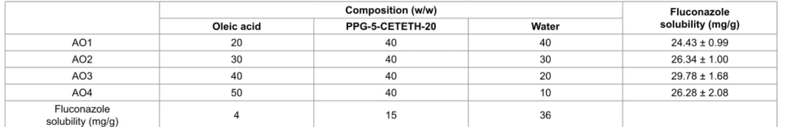
he formulations were prepared by manual stirring using PPG-5-CETETH-20 as a surfactant, oleic acid as an oily phase, and water as an aqueous phase in a beaker. he proportions of the constituents are indicated in Table 1. he formulations were prepared 24 h before the experiments and let at room temperature to allow the system to reach complete equilibrium. Fluconazole was incorporated by dissolving the drug powder directly into the systems.
Evaluation of the luconazole solubility in the formulations
he solubility of luconazole in the formulations was investigated by adding crescent speciic amounts of the drug powder (0.01% w/w) to 2 g of each system and to their isolated components. he mixtures
were manually homogenised using a glass rod and maintained at room temperature for 24 h. his procedure was repeated until precipitation was observed. Loaded samples were named AO1F, AO2F, AO3F, and AO4F.
Fluconazole was analysed by HPLC, with the UV detector set at 210 nm (Varian Pro Star 330). A reverse-phase Varian Chromsep C18 column (250×4.6 mm i.d., 5 µm pore size) was used. he mobile phase was a mixture of methanol/water (1:1 v/v), the low rate was set at 1 mL/min, and the injection volume was 20 µL. A calibration curve was constructed by preparing working solutions of luconazole in the mobile phase at concentrations ranging from 20 to 400 µg/mL.
Samples were prepared by diluting the formulation into the mobile phase to obtain a drug concentration of 150 µg/mL. he dilution was cleaned up by solid phase extraction (SPE) using 500 mg per 3 mL C18 cartridge. he SPE procedure included conditioning the samples with 1 mL of the mobile phase. he spiked samples (2.5 mL) were then loaded into the conditioned cartridge, and the analytes were collected, iltered through 0.45 µm-pore membranes, and injected into the HPLC system.
Studies of phase behaviour by polarised light microscopy
(PLM) and small-angle X-ray scattering (SAXS)
PLM was performed in an optical Jenamed 2, Carl Zeiss (Jena, Germany) microscope. he isotropic or anisotropic behaviour of the samples was observed, and pictures were taken at a 20,000 × magniication. Samples were prepared by placing a drop of formulation between a cover slip and a glass slide. hey were then examined under polarised light at room temperature.
SAXS data were collected at the Synchrotron SAXS beam line of the National Laboratory of Synchrotron Light (LNLS, Campinas, Brazil), equipped with an asymmetrically cut and bent Si (1 1 1) monochromator (λ=1.608 Å) that yields a horizontally focused beam. A vertical position-sensitive X-ray detector and a multichannel analyser were used to record the SAXS intensity, I(q), as a function of the modulus of the scattering vector q (q = (4π/λ)sin(ε/2)), with ε being the scattering angle. he samples were placed in a cell at 25°C. he parasitic scattering produced by the slits was subtracted from the total scattering intensity.
Rheological studies
he rheological measurements were taken with a controlled stress Rheostress RS1 rheometer (Haake, Karlsruhe, Germany) and analysed with Rheowin 3.5 sotware. Plate–plate geometry was used with a gap of 200 µm between plates 35 mm in diameter. Samples were carefully applied to the lower plate, ensuring that sample shearing was minimised, and were allowed to equilibrate for at least 3 min prior to analysis.
he low properties were determined with shear rates within the range of 0.01–100 s-1, which was chosen on the basis of the strength of resistance to the applied stresses. he rheological measurements were
Composition (w/w) Fluconazole
solubility (mg/g)
Oleic acid PPG-5-CETETH-20 Water
AO1 20 40 40 24.43 ± 0.99
AO2 30 40 30 26.34 ± 1.00
AO3 40 40 20 29.78 ± 1.68
AO4 50 40 10 26.28 ± 2.08
Fluconazole
solubility (mg/g) 4 15 36
performed on both the up and down curves. he low curves were itted to a power law model using the program Origin 7.0. All rheological determinations were carried out at 25 ± 0.25°C.
Oscillatory analysis of each sample was performed ater determination of its linear viscoelastic region at 25°C, where stress was directly proportional to strain and the storage modulus remained constant. Frequency sweep analysis was performed over the frequency range of 0.1–10 Hz at a constant stress of 1 Pa. he systematic error in the frequency of the rheometer was approximately 0.01 Hz.
Safety evaluation of the formulations
In vitro unspeciic cytotoxicity: Mammal cytotoxicity of the formulations was studied in vitro using J-774 mouse macrophages as the cellular model. Cells were seeded at a density of 2.5-10.0×10⁵ cells/ well in 96-well flat bottom microplates (Nunclon) and exposed for 48 h to diferent doses of the formulations and free-drug (18.6, 10, 5 and 1 µM) for control. Ater treatment, the compounds were removed, and the cells washed once with PBS. Cell viability was then colorimetrically assessed by measuring the mitochondrial-dependent reduction of MTT to formazan. For this purpose, the cells and MTT (0.4 mg/mL) were incubated in air at 37°C for 3 h. Ater the incubation period, the supernatant was removed, and formazan crystals were dissolved with DMSO (180 µL). he plates were shaken for 10 min, and the optical densities were measured at 560 nm in a multiwell spectrophotometer. Each concentration was assayed three times, and six additional controls (cells in medium) were used in each test. Data were exhibited in per cent of cellular viability.
In vivo evaluation:
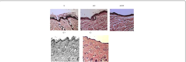
Animals: During the experiment, the animals were maintained in accordance with the guidelines established by Olfert et al. [10] and the Statement of Principles adopted by the Federation of American Societies for Experimental Biology (FASEB) Board. he animals had free access to food and water. Twenty-four hours before the start of treatment, areas of 4 cm x 5 cm were outlined on the back of each animal. he raw materials and formulation AO3F were tested. Each experimental group consisted of ive rats. he animals were given the following treatments: oleic acid (OA), PPG-5-CETETH-20 (PROC), formulation AO3F, benzalkonium chloride (BC) solution 15% (positive control, C+), and propanol (negative control, C-). All areas, except the control area, were subjected to a daily treatment with the indicated formulations for 21 days, always at the same time, accompanied by frictional circular massage.
Histological procedures: he animals were anesthetised with xylazine hydrochloride / ketamine hydrochloride and sacriiced in a CO2 chamber, ater which the areas of treated skin were removed, ixed in Bouin’s solution for 48 hours, rinsed, and treated routinely for embedding in parain (Histosec, Merck). Five non-serial histological sections (5 µm) were cut for each slide, with three slides per treatment area. he sections were stained in hematoxylin and eosin (HE) for histomorphometric and histopathological analyses.
Histomorphometric analysis was performed by capturing images of random cuts through an optical microscope Leica DMZ 2000 connected to a computer with the sotware Motic Images Advanced. hirty areas were randomly chosen, and the thickness of the epidermis and dermis for each treatment area was measured. From these measurements, a correction for the micrometre scale was calculated for an objective of 20x, which yielded the mean thickness and standard deviation for each experimental group.
Histopathological analysis consisted of counting the number of leukocytes and ibroblasts using a Leica DMZ 2000 optical microscope connected to a computer with the sotware Motic Images Advanced 3.2. For each experimental group, three slides with 5 nonserial sections per slide were obtained. Six areas of 250 µm2 of skin per experimental group, always in the region of the papillary dermis, were measured. herefore, each treatment comprised a total area of 30,000 µm2 per animal. From these results, the mean and standard deviation for each cell type was determined for each treatment.
Statistical analysis was performed using the Kruskal-Wallis statistical test, followed by the Wilcoxon-Mann-Whitney test with Bioestat version 5.0. he level of signiicance adopted was 5% (p<0.05).
Proof of concept of the formulations by drug release, drug
skin permeation, and drug skin retention studies
hese studies comprise drug release, skin permeation, and skin retention assays [11]. he loaded-formulations and loaded-oleic acid containing luconazole were tested at 10 mg/mL.
In the drug release study, a 45 µm-pore size cellulose acetate membrane was used to separate the formulations from the release medium. For drug permeation and retention studies, porcine ear skin obtained from healthy 6-month-old Brazilian pigs of a local slaughterhouse and prepared for the test as described elsewhere [12] were used. he ears were cleaned with water (25 ± 0.5°C), and the ears with injury were discarded. he undamaged skins were removed from the cartilage with a scalpel, and a 500 μm thick layer stratum corneum and epidermis were separated from the adipose tissue with a dermatome (Nouvag TCM 300, Goldach, USA). he prepared skins were frozen at −20°C and stored no longer than 4 weeks. On the day of the experiment, the skin was thawed in physiological saline solution containing 0.9% (w/v) NaCl (Merck, Germany) at 25 ± 0.5°C for 30 min; then, its hair was cut with a scissor.
he cellulose acetate membranes or skin pieces were placed between the donor and receptor chambers of a Franz difusion cell (Microette Plus, Hanson Research, Chatsworth, EUA) equipped with difusion cells with an exposition area of 1.77 cm2 for formulation deposition. he receptor chambers of the cells (7 mL) were illed with 10 mM monobasic bufer (pH 7.4) stirred at 300 rpm. he samples (400 mg) were dropped on the donor chambers in contact with the receptor chambers. he total test time was 24 h, and 2 mL of the receptor luid was collected and replaced by an equal volume of fresh receptor solution. he assays were carried out on six replicates for each sample.
Fluconazole was quantiied by HPLC using the same conditions described for the luconazole solubility study. A calibration curve was constructed by preparing working solutions of luconazole in 10 mM monobasic phosphate bufer (pH 7.4) at concentrations ranging from 1 to 250 µg/mL. Samples of collected receptor luid were iltered and injected into the HPLC system.
he results are represented by a graph of the cumulative mass of the drug that passes through a unit area of the membrane or skin versus time, calculated according to equation 1:
Q=Ct× +Vr
∑
Vc×Cc (1)For retention studies, the tape stripping method [13-15] was performed. he skins derived ater 6 h and 12 h of permeation assay were cleaned using sot paper. he stratum corneum was removed from sixteen tapes (Scotch 750, 3M), and retained luconazole was evaluated by solvent extraction. he tapes were placed in an assay tube containing 5 mL of methanol and vortexed for 2 min. hen, the sample was immersed in an ultrasonic bath for 30 min. he solvent was iltered through a 0.45 µm membrane and injected into the HPLC system. he remaining skin was cut into small pieces, placed in a tube containing 5 mL of methanol, and tested according to the same procedure.
Results and Discussion
Preparation of the formulations
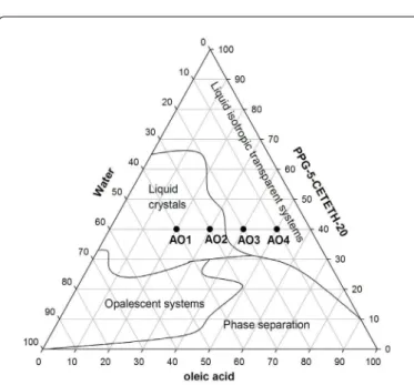
he composition of the formulations was chosen based on previous studies in which the ternary phase diagram of oleic acid,
PPG-5-CETETH-20 and water mixtures was obtained (Figure 1). For this study, thermodynamically stable systems, characterised by their spontaneous formation, transparency and high stability, were selected. Samples AO1 and AO2 correspond to semisolid transparent systems, and AO3 and AO4 correspond to liquid and transparent systems. heir compositions are given in Table 1. For AO1-4, the concentration of the surfactant was ixed at 40% (w/w), whereas the other component concentrations were varied. hese compositions were chosen to avoid the translucent dispersion and phase separation areas.
Evaluation of luconazole solubility in the formulations
he incorporation of luconazole into the individual oil, surfactant, water, and formulations was measured, and the results are displayed in Table 1. he formulations incorporated more drug than water and surfactant but less drug than oleic acid by itself. hese results conirm the lipophilic character of luconazole. It is worth highlighting that the formulations incorporated more luconazole than conventional topical pharmaceutical formulations (10 mg/g) due to the mixture of components with diferent polarities, namely, oil and water, in the presence of a surfactant that forms aggregates and allows additional regions of drug solubilisation.
Phase behaviour study by PLM and SAXS
he semisolid samples (AO1 and AO2) were anisotropic under polarised light, which is characteristic of liquid crystals that are birefringent as a result of their molecular ordering. he images obtained by PLM for AO1 and AO2 are shown in Figure 2, in which Maltese crosses characteristic of lamellar phases can be seen.
he transparent liquid systems investigated (AO3 and AO4) showed a dark background under PLM, and thus, they are isotropic systems that are characteristic of micellar systems, microemulsions, or cubic phases. Cubic phases are well known by their high stifness, and AO3-AO4 lowed easily. herefore, they may not be organised structurally as a mesophase.
he phase behaviour of the systems was conirmed by the SAXS measurements. he data that were collected from the scattering proiles were plotted as intensity, (I), versus the scattering vector module, q (Å -1). he resulting curves are shown in Figure 3.
Surfactants can form diferent aggregate types, which can be identiied by the peak positions on the scattering vector axis. From the scattering proile of loaded and unloaded AO1-AO2 formulations, two peaks were observed, possibly because of a degree of structural organisation. he ratio between the irst and second peak is 2:1, which is characteristic of lamellar liquid crystals [16]. his result correlates with the PLM experiments because Malta crosses were visualised for the AO1 and AO2 systems. For the AO3 and AO4 formulations, a maximum intensity value of q ≠ 0 followed by a long tail was observed, which is associated with short-range 3D spatial correlation and may be characteristic of micellar structures [17].
Rheological studies
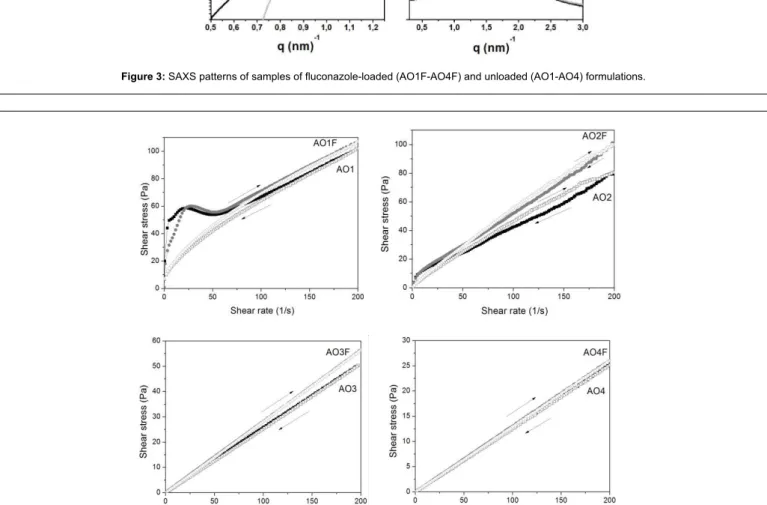
he low properties of these systems are displayed graphically in Figure 4, in which the shear stress is plotted versus the shear rate. In the continuous shear rheometry assay, both unloaded and loaded AO3-AO4 systems exhibited Newtonian behaviour. his result was expected because the samples consisted of isotropic liquid systems. he curves of AO1-AO2 showed a nonlinear relationship between shear stress and shear rate, thus exhibiting a non-Newtonian low characteristic. hese results are in accord with literature data showing that liquid crystalline phases are found to exhibit shear-thinning behaviour.
Figure 1: Ternary phase diagram of PPG-5-CETETH 20, oleic acid, and water, obtained from previous studies by Carvalho et al. [6].
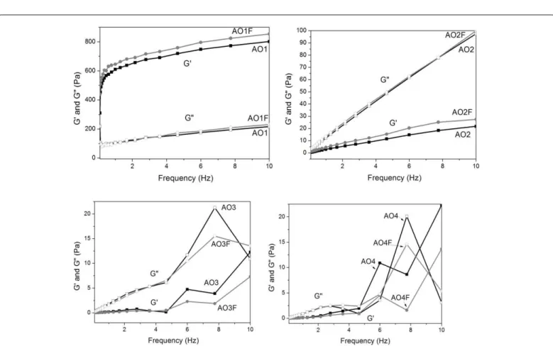
he oscillatory rheological data are displayed in Figure 5 as the storage modulus (G’) and loss modulus (G”) as a function of the oscillatory frequency. he storage modulus is a measure of the energy stored and recovered per deformation cycle and represents the solid-like component of a viscoelastic material. he storage modulus is large when a sample is predominantly elastic or highly structured. he loss
modulus is a measure of the energy dissipated per cycle and represents the liquid-like component [18]. he frequency sweep analyses suggest that AO3-AO4 are more viscous and less elastic at the selected frequency because G” > G’ and because both moduli are frequency dependent. For AO1-AO2, the G’ values were high and relatively independent of frequency. he slopes of the lamellar phase formulations (AO1-AO2) Figure 3: SAXS patterns of samples of luconazole-loaded (AO1F-AO4F) and unloaded (AO1-AO4) formulations.
were lower than the slopes of the liquid isotropic systems AO3-AO4, indicating a more structured character for the lamellar phases.
Safety evaluation of the formulations
In vitro unspeciic cytotoxicity: In vitro unspeciic cytotoxicity studies of the formulations were performed using J-774 mouse macrophages as the cellular model. he data (Figures 6 and 7) were exhibited in per cent of cellular viability. he assay results of cellular viability showed that the unloaded samples (AO, AO1, AO2, AO3, AO4) and loaded samples (AOF, AO1F, AO2F, AO3F, AO4F) were unable to kill normal cell macrophages. All samples exhibited cellular viability greater than that 92%, and therefore, it is suggested that the formulations and the drug do not present toxic activity or cytotoxicity.
In vivoevaluation:
Histomorphometric analysis of the epidermis and dermis: he results for the thickness of the epidermis and dermis are presented in Table 2. Analysis of variance (ANOVA) veriied a normal distribution of data, and the experiment followed a completely randomised design. he F test was used to detect diferences between treatments. he means of groups were compared, two by two, by the Tukey test.
he data suggest that the groups AO, PPG-5-CETETH-20, and formulation AO3F caused signiicantly higher thickening of the epidermis and dermis than the control (Figure 8). his result suggests an increase of skin hydration that would be revealed by an increase in the interstitial spaces. It is known that some cosmetic formulations, with or without active substances, may promote skin hydration, to the beneit of the skin [19].
According to Libardi [20], what makes the skin stay healthy and sot, with good lexibility and elasticity is the maintenance of skin hydration and the ability of the organism not only to stimulate cell renewal but also to synthesise components of the epidermis. For good functioning of the mechanism of skin hydration, the stratum corneum should be able to retain water so that its rate of evaporation is limited to a normal level. Proksch et al. [21] claim that skin hydration is intrinsically related to the integrity of the stratum corneum.
he properties of occlusion and moisturising of the components were observed for the formulation AO3F, characterised as a micellar system. he occlusive activity conferred by nano-systems has been cited in other works such as Brinon et al. [22] hose authors suggest that this
Figure 5: Frequency sweep proile of the storage modulus (G’ – closed symbols) and loss modulus (G” – opened symbols) of luconazole-loaded (grey symbols) and unloaded formulations AO1-AO4 (black symbols) at 25°C.
action is related to the characteristic colloidal structure of the emulsiied system, in which the organisation of the surfactant interfacial layer is similar to that of the lipid bilayers that join keratinocytes, increasing cohesion and, consequently, the ability to form a continuous ilm [23].
Histopathological analysis of the dermis: he presence of ibroblasts and leukocytes in larger amounts during skin irritation can be used to predict irritation. he number of ibroblasts and leukocytes counted in the papillary dermis of the various experimental groups is given in Table 2. By ANOVA, the distribution of the data for the number of ibroblasts and leukocytes per area was found to be non-normal, so the Kruskal-Wallis test (p<0.05) was used to analyse the results.
he most common adverse reactions of topical formulations is skin irritation, which can be seen as local intolerance and may elicit minor discomfort, as well as acute reactions that vary in intensity from
burning to itching. his irritation may lead to corrosion and tissue destruction. All of these reactions are restricted to the area in direct contact with the product. Generally, the degree of irritation is related to the concentration of an ingredient in the inal product and the cosmetic formulation [24].
It can be said that irritation causes a skin condition similar to that produced by a lesion. Lesions in skin tissue are characterised as any events that impair the structure or function of the tissue. hey produce changes in the ability of cells to perform their normal homeostatic functions, and they trigger various histochemical and biological processes that lead to vasomotor action to mobilise all the cells responsible for ighting infection and/or the ofending agent. hese processes are intended to replace losses of substances as quickly as possible and to restore the physiological balance and contribute to the survival of the organism [25].
Figure 7: Per cent cellular viability of luconazole-loaded formulations (AO1F, AO2F, AO3F, AO4F) and loaded oleic acid (AOF).
Group Epidermis (µm) Dermis (µm) Number of ibroblasts Number of leukocytes
Average SD Average SD Average SD Average SD
S 32.92 1.66 1136.76 37.20 12.47 2.30 2.6 1.65
AO 35.56 5.79 1097.83 47.64 12.27 2.30 1.87 1.53
AO3F 30.24 3.63 1014.82 66.79 12.67 2.59 0.27 0.52
C+ 37.58 3.00 1269.11 53.04 7.83 1.68 6.10 3.00
C- 23.95 1.62 672.95 51.48 9.10 1.65 0.67 0.88
Table 2: Thickness of the epidermis and dermis (µm). Number of ibroblasts and leukocytes in 250 µm2 of the papillary dermis in each experimental group. Key: (S)
PPG-5-CETETH-20, (AO) oleic acid, (AO3F) luconazole-loaded tested formulation, (C+) positive control, (C-) negative control, (SD) standard deviation.
S AO AO3F
C+
C+
At an early stage, an irritation process releases inlammatory mediators and mobilises a sequence of cells, especially neutrophils and lymphocytes, to combat the ofending agent or to start the immune processes that facilitate healing [26]. Inlammatory mediators act as chemotactic signals and growth factors for the inlammatory cells, which are represented by polymorphonuclear leukocytes (PMNs), macrophages, and lymphocytes. hese cells also release growth factors for endothelial cells, ibroblasts and keratinocytes. he release of all of these factors can lead to the formation of granulation tissue [27].
Although the results of the leukocyte count of PPG-5-CETETH-20 and oleic acid were higher than those obtained for the control, these counts still remain low. Importantly, these results may have occurred through the use of pure raw materials in the animal’s skin.
he results obtained for the formulation AO3F were not signiicantly diferent from those obtained for the control group, suggesting no occurrence of inlammatory processes when administering a formulation containing 40% oil, 40% surfactant and 20% water.
Other promising data are related to the count of ibroblasts. It was observed that the number of ibroblasts was signiicantly higher for the various experimental groups than for the control (Figure 9). his result indicates that the formulations may also improve properties related to
skin support and cell regeneration because these cells will originate collagen and elastin ibres.
he ibroblast is the main cell involved in the formation of granulation tissue, synthesising hyaluronic acid, ibronectin, collagens type I and III, elastin and proteases, such as collagenase [25]. he formulation AO3F increased the production of ibroblasts. his, in turn, increased the production of collagen, which is responsible for the strength and resistance of greater thickness and dermal trauma, promoting satisfactory skin restructuring [28,29].
Proof of concept of the formulations
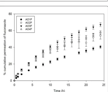
he drug release assay enables in vitro analysis of drug movement across a membrane using a two-compartment model. he donor compartment contains the formulation, and a non-rate-limiting membrane separates the compartments and supports the dose. he amount of luconazole released was expressed as a percentage, which was plotted against time in Figure 10. In addition, the pseudo-steady-state lux (J, µg/min cm) derived from Fick’s law was calculated as the gradient of the linear portion of the drug release curve [30]. he results are listed in Table 3.
his study revealed that AO3F-AO4F released more luconazole than AO1F-AO2F. his phenomenon most likely occurs because the latter formulations present a liquid crystalline network structure with higher elasticity than that of AO3F-AO4F. he less ordered AO3F-AO4F systems with reduced viscosity allowed a higher degree of mobility of drug molecules in their matrices.
he OA sample showed the highest value of lux (J), followed by AO3F. he other formulations did not present a signiicant diference. Because the AO sample presents no structural organisation, such as the liquid crystalline samples or the micellar system, the drug was released faster. he lux of AO3F could have been higher due to its position in the phase diagram of Figure 1. his sample was placed in a phase transition area, and therefore, its structure may not control drug release as much as the AO1F, AO2F or AO4F systems. AO1F was shown to prolong drug release to the greatest extent. his result may be due to the highest elastic modulus in AO1F, which increases the stifness of the liquid crystalline matrix. In addition to the diferences between the drug release rates, all formulations released luconazole in a controlled manner because an exponential pattern governed drug difusion, as seen in the curves of Figure 10. herefore, this study showed that structural modiications of surfactant systems can modify the rate and proile of drug delivery.
Permeation studies were performed in Franz difusion cells using the skin of porcine ear. he cumulative permeation of luconazole incorporated into formulations AO1F-AO4F was calculated for 6 h and 12 h of experiment, as was the drug retention in the stratum corneum, dermis, and epidermis. he values obtained are listed in Table 3.
he sample AOF promoted the highest drug permeation, followed by AO4F, which contains the largest quantity of oleic acid among the other formulations. his result indicates the enhancing efect of oleic acid on the in vitro permeability of luconazole. Oleic acid may reduce the lipid packing order in epithelial cell membranes. his efect could increase the luidity of the lipid layer and decrease the difusion resistance, thus assisting the permeation of compounds through the skin [31]. herefore, AO4F may be explored for a systemic efect because it promoted drug transportation across the skin. his suggests that the drug may reach the blood circulation.
(A) (B)
Figure 9: Photomicrographs showing the papillary dermis in areas: (A) C-; (B) picture representative of the treatments with S, AO, AO3F and C+, where a greater number of ibroblasts are seen in the dermis (1000x).
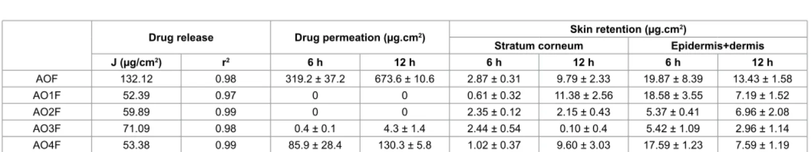
Drug release Drug permeation (µg.cm2) Skin retention (µg.cm 2)
Stratum corneum Epidermis+dermis
J (µg/cm2) r2 6 h 12 h 6 h 12 h 6 h 12 h
AOF 132.12 0.98 319.2 ± 37.2 673.6 ± 10.6 2.87 ± 0.31 9.79 ± 2.33 19.87 ± 8.39 13.43 ± 1.58
AO1F 52.39 0.97 0 0 0.61 ± 0.32 11.38 ± 2.56 18.58 ± 3.55 7.19 ± 1.52
AO2F 59.89 0.99 0 0 2.35 ± 0.12 2.15 ± 0.43 5.37 ± 0.41 6.96 ± 2.08
AO3F 71.09 0.98 0.4 ± 0.1 4.3 ± 1.4 2.44 ± 0.54 0.10 ± 0.4 5.42 ± 1.09 2.96 ± 1.14 AO4F 53.38 0.99 85.9 ± 28.4 130.3 ± 5.8 1.02 ± 0.37 9.60 ± 3.03 17.59 ± 1.23 7.59 ± 1.19 Table 3: Flux (J) and correlation coeficient (r2) derived from drug release curves, drug permeated through the skin at 6 h and 12 h, and drug retained in the stratum
corneum and in the epidermis + dermis at 6 h and 12 h. Data were collected for luconazole-loaded formulations (AO1F, AO2F, AO3F, AO4F) and loaded oleic acid (AOF), all containing 10 mg/mL of drug. The values represent the average ± standard deviation of six replicates.
In contrast, even though AO1-AO3 had retained more drug in the epidermis plus dermis than in the stratum corneum barrier, there was no drug permeation for these systems, which leads us to conclude that these samples promoted skin retention. In comparing AO1F-AO3F, an increase in the drug concentration in the stratum corneum with time was observed. AO1F and AO2F are lamellar phases, but AO1F exhibited a higher elastic modulus, which is characteristic of a more structurally organised system. herefore, it can be concluded that the higher organisation of the liquid crystalline structure of AO1F contributed to controlling drug release.
It is reported that the concentration of antifungal drug attained in the skin is an important factor in the treatment of dermatomycosis and that the presence of the therapeutically active form in the skin is closely related to the eicacy of the drug. However, this evidence was found for oral treatments [9]. Considering the topical application of the formulations studied here, it is possible to promote drug permeation or skin retention by changing only the composition of the components in the formulations. hese results open the way to considering PPG-5-CETETH-20-based transdermal systems for antifungal skin delivery for Sporotrichosis treatment.
Conclusions
he indings of this work noted the great potential of PPG-5-CETETH-20-based transdermal systems for either systemic or local action because it was found that both formulations enhanced drug permeation and skin retention. In vitro and in vivo biological assays showed that the formulations did not afect normal cell macrophages and did not promote skin irritation. herefore, the results presented here provide new possibilities for transdermal systems with diferent structural and rheological characteristics for the treatment of Sporotrichosis using antifungal drugs.
Acknowledgment
The authors thank the LNLS (The Brazilian Synchrotron Light Laboratory) for SAXS measurements and FAPESP (São Paulo Research Foundation), CNPq (National Council for Scientiic and Technological Development) and PADC-UNESP (Programa de Apoio ao
Desenvolvimento Cientíico) for the inancial support.
References
1. Bonifaz A, Saúl A, Paredes-Solis V, Fierro L, Rosales A, et al. (2007) Sporotrichosis in childhood: clinical and therapeutic experience in 25 patients. Pediatr Dermatol 24: 369-372.
2. Vásquez-del-Mercado E, Arenas R, Padilla-Desgarenes C (2012) Sporotrichosis. Clin Dermatol 30: 437-443.
3. De Araujo T, Marques AC, Kerdel F (2001) Sporotrichosis. Int J Dermatol 40: 737-742.
4. Yang F, Kamiya N, Goto M (2012) Transdermal delivery of the anti-rheumatic agent methotrexate using a solid-in-oil nanocarrier. Eur J Pharm Biopharm 82: 158-163.
5. Naik A, Kalia YN, Guy RH (2000) Transdermal drug delivery: overcoming the skin’s barrier function. Pharm Sci Technolo Today 3: 318-326.
6. Carvalho FC, Rocha e Silva H, da Luz GM, Barbi Mda S, Landgraf DS, et al. (2012) Rheological, mechanical and adhesive properties of surfactant-containing systems designed as a potential platform for topical drug delivery. J Biomed Nanotechnol 8: 280-289.
7. Carvalho FC, Campos ML, Peccinini RG, Gremião MP (2013) Nasal administration of liquid crystal precursor mucoadhesive vehicle as an alternative antiretroviral therapy. Eur J Pharm Biopharm 84: 219-227.
8. Bruschi ML, Freitas O, Lara EHG, Panzeri H, Gremião MPD, et al. (2008) Precursor system of liquid crystalline phase containing propolis microparticles for the treatment of periodontal disease: development and characterization. Drug Develop Ind Pharm 34: 267-278.
9. Sobue S, Sekiguchi K, Nabeshima T (2004) Intracutaneous distributions of luconazole, itraconazole, and griseofulvin in Guinea pigs and binding to human stratum corneum. Antimicrob Agents Chemother 48: 216-223.
10. Olfert ED, Cross BM, McWilliam AA (1993) Guide to the care and use of experimental animals. Canadian Council on Animal Care, Ottawa, Canada. 11. Polacow MLO, Pires-de-Campos MSM, Leonardi GR, Carvalho LS, Ribeiro
MCAP, et al. (2004) Eleito do ultra-som na permeação cutânea do tiratricol: análise histológica. Rev Bras Fisioter 8: 53-60.
12. Dick IP, Scott RC (1992) Pig ear skin as an in-vitro model for human skin permeability. J Pharm Pharmacol 44: 640-645.
13. Hosmer J, Reed R, Bentley MVLB, Nornoo A, Lopes LB (2009) Microemulsions containing medium-chain glycerides as transdermal delivery systems for hydrophilic and hydrophobic drugs. AAPS Pharm Sci Tech 10: 589-596. 14. Herai H, Gratieri T, Thomazine JA, Bentley MV, Lopez RF (2007) Doxorubicin
skin penetration from monoolein-containing propylene glycol formulations. Int J Pharm 329: 88-93.
15. Lopez RF, Bentley MV, Begoña Delgado-Charro M, Guy RH (2003) Optimization of aminolevulinic acid delivery by iontophoresis. J Control Release 88: 65-70. 16. Holmqvist P, Alexandridis P, Lindman B (1997) Modiication of the microstructure
in poloxamer block copolymer-water-“oil” systems by varying the “oil” type. Macromolecules 30: 6788-6797.
17. Fanun M (2008) A study of the properties of mixed nonionic surfactants microemulsions by NMR, SAXS, viscosity and conductivity. J Mol Liquids 142: 103-110.
18. Barnes HA, Hutton JF, Walter K (1989) An introduction to rheology. Elsevier, New York, USA.
19. Leonardi GR, Gaspar LR, Maia Campos PM (2002) Application of a non-invasive method to study the moisturizing effect of formulations containing vitamins A or E or ceramide on human skin. J Cosmet Sci 53: 263-268. 20. Libardi FS (1998) Lactato de amônio. Cosmet Toiletries 11: 50-53.
21. Proksch E, Nissen HP, Bremgartner M, Urquhart C (2005) Bathing in a magnesium-rich Dead Sea salt solution improves skin barrier function, enhances skin hydration, and reduces inlammation in atopic dry skin. Int J Dermatol 44: 151-157.
22. Brinon L, Geiger S, Alard V, Doucet J, Tranchant JF, et al. (1999) Percutaneous absorption of sunscreens from liquid crystalline phases. J Control Release 60: 67-76.
24. http://portal.anvisa.gov.br/wps/portal/anvisa/home
25. Kitchen S, Young S (1998) Reparo dos tecidos. In: Kitchen S, Bazin S. Eletroterapia de.
26. Andrews JM (1995) Cirurgia plástica, Atheneu, São Paulo.
27. http://www2.fcfar.unesp.br/Home/Pos-graduacao/CienciasFarmaceuticas/ MarlusChorilliDO.pdf
28. Chorilli M, Prestes PS, Rigon RB, Leonardi GR, Chiavacci LA, et al. (2011) Structural characterization and in vivo evaluation of retinyl palmitate in non-ionic lamellar liquid crystalline system. Surf B Biointerfaces 85: 182-188.
29. Shapiro SS, Saliou C (2001) Role of vitamins in skin care. Nutrition 17: 839-844.
30. Williams AC (2003) Transdermal and topical drug delivery. Pharmceutical Press, London, UK.
31. Nicolazzo JA, Reed BL, Finnin BC (2005) Buccal penetration enhancers--how do they really work? J Control Release 105: 1-15.
Citation:Silva HR, Luz GM, Satake CY, Correa BC, Sarmento VHV, et al. (2014) Surfactant-based Transdermal System for Fluconazole Skin Delivery. J Nanomed Nanotechnol 5: 231. doi: 10.4172/2157-7439.1000231
Submit your next manuscript and get advantages of OMICS Group submissions
Unique features:
• User friendly/feasible website-translation of your paper to 50 world’s leading languages
• Audio Version of published paper
• Digital articles to share and explore
Special features:
• 350 Open Access Journals
• 30,000 editorial team
• 21 days rapid review process
• Quality and quick editorial, review and publication processing
• Indexing at PubMed (partial), Scopus, EBSCO, Index Copernicus and Google Scholar etc
• Sharing Option: Social Networking Enabled
• Authors, Reviewers and Editors rewarded with online Scientiic Credits
• Better discount for your subsequent articles