Telomere Fragment Induced Amnion Cell
Senescence: A Contributor to Parturition?
Jossimara Polettini1,2, Faranak Behnia1, Brandie D. Taylor3, George R. Saade1, Robert N. Taylor4, Ramkumar Menon1*
1Division of Maternal-Fetal Medicine and Perinatal Research, Department of Obstetrics and Gynecology, The University of Texas Medical Branch at Galveston, Galveston, Texas, United States of America, 2Department of Pathology, Botucatu Medical School, UNESP–Univ. Estadual Paulista, Botucatu, Sao Paulo, Brazil,3Department of Epidemiology & Biostatistics, Texas A&M University System Health Science Center, College Station, Texas, United States of America,4Department of Obstetrics and Gynecology, Wake Forest University, Winston Salem, North Carolina, United States of America
Abstract
Oxidative stress (OS)-induced senescence of the amniochorion has been associated with parturition at term. We investigated whether telomere fragments shed into the amniotic fluid (AF) correlated with labor status and tested if exogenous telomere fragments (T-oligos) could induce human and murine amnion cell senescence. In a cross-sectional clinical study, AF telomere fragment concentrations quantitated by a validated real-time PCR assay were higher in women in labor at term compared to those not in labor.In vitrotreatment of primary human amnion epithelial cells with 40μM T-oligos ([TTAGGG]2) that mimic telomere
frag-ments, activated p38MAPK, produced senescence-associated (SA)β-gal staining and increased interleukin (IL)-6 and IL-8 production compared to cells treated with complemen-tary DNA sequences (Cont-oligos, [AATCCC]2). T-oligos injected into the uteri of pregnant
CD1 mice on day 14 of gestation, led to increased p38MAPK, SA-β-gal (SAβ-gal) staining in murine amniotic sacs and higher AF IL-8 levels on day 18, compared to saline treated con-trols. In summary, term labor AF samples had higher telomere fragments than term not in labor AF.In vitroandin situtelomere fragments increased human and murine amnion p38MAPK, senescence and inflammatory cytokines. We propose that telomere fragments released from senescent fetal cells are indicative of fetal cell aging. Based on our data, these telomere fragments cause oxidative stress associated damages to the term amniotic sac and force them to release other DAMPS, which, in turn, provide a sterile immune response that may be one of the many inflammatory signals required to initiate parturition at term.
Introduction
Signals that initiate normal labor are still unclear [1] although multitudes of putative biochemi-cal mediators and their pathways have been suggested as initiators [2,3]. The best documented signals occur in both maternal and fetal compartments and include endocrine (Corticotrophin relasing hormone [CRH], Adrenocorticotropic hormone [ACTH], functional progesterone withdrawal), immune (leukocyte and leukotriene activation) and mechanical factors (enhanced OPEN ACCESS
Citation:Polettini J, Behnia F, Taylor BD, Saade GR, Taylor RN, Menon R (2015) Telomere Fragment Induced Amnion Cell Senescence: A Contributor to Parturition? PLoS ONE 10(9): e0137188. doi:10.1371/journal.pone.0137188
Editor:Kang Sun, Shanghai Jiaotong University School of Medicine, CHINA
Received:May 27, 2015
Accepted:August 13, 2015
Published:September 23, 2015
Copyright:© 2015 Polettini et al. This is an open access article distributed under the terms of the
Creative Commons Attribution License, which permits unrestricted use, distribution, and reproduction in any medium, provided the original author and source are credited.
Data Availability Statement:All relevant data are within the paper.
Funding:This study is supported by development funds to Dr. R Menon by the Department of Obstetrics and Gynecology, The University of Texas Medical Branch, Galveston, TX, USA. Coordenação de Aperfeiçoamento de Pessoal de Nível Superior-CAPES, Brazil supported the primary author JP, CAPES/PGCI 12415-12-0.
uterine stretching and amniochorionic membrane disruption). These factors cause an inflam-matory activation (mostly mediated by cytokines), and prostaglandin production to transform a quiescent myometrium to an active contractile state at term [2,4–8]. Pathological activation of myometrial contractility by cytokines and prostaglandins also has been implicated in spon-taneous preterm birth (PTB) [9–11]. Identification of the critical signals and understanding their molecular mechanisms that initiate parturition is essential for reducing the risk of PTB, a major pregnancy complication.
We have proposed that fetal signals to initiate parturition arise from senescent fetal mem-brane cells. Senescence is characterized by irreversible growth arrest of cells and is a mecha-nism associated with aging [12]. Senescence of fetal cells is a natural physiologic process that occurs throughout gestation [13] and is particularly noticeable at term [13,14]. Increased senes-cence is likely due to enhanced oxidative stress (OS) generated by the growing fetus, uterine stretch or other still unknown factors [14]. OS-induced damage to cellular elements causes structural and functional alterations, resulting in senescence [15]. Morphologic (enlarged cells, and round and swollen organelles) and biochemical features (senescence associated β-Galactosidase [SAβ-Gal], of senescence are evident in fetal membranes from women in term labor compared to term not in labor [16]. AF from term labor also had dysregulated inflamma-tory markers compared to gestational age-matched not in labor samples, suggesting sterile inflammation (inflammation in the absence of infectious agent) and its associated senescence associated secretory phenotype (SASP), a unique set of inflammatory markers, that include cytokines, chemokines, growth factors, matrix degrading enzymes, inhibitors and various other agionists and antagonists [16]. We posit that the inflammatory milieu generated by senescent cells [17,18] activates functional progesterone withdrawal, produces uterotonins and signals parturition.In vitro, we recapitulated these findings in primary human amnion epithelial cells from term not in labor specimens. Amniotic epithelial cells exposed to OS developed SASP via activation of p38MAPK [19] resembling such changes seen in membranes from women in term labor [14]. Therefore, it is likely that senescence inducing risk factors of PTB cause patho-logic activation of senescence and SASP that lead to preterm labor.
It has been suggested that increased levels of cell-free fetal DNA in the maternal circulation, released as a result of placental senescence, can activate parturition [20]. A potential source of cell-free DNA is from the telomeres, the chromosome end caps that stabilize the genome in humans and other long-lived mammals [21]. Accordingly, we have reported that telomere length reduction occurs in fetal compartments throughout gestation, with the shortest telo-meres seen in term fetal membranes, suggesting a natural in utero aging process [13]. In human cells, telomeres range from 8,000–10,000 bp in length with a single-strand TTAGGG 3’
overhangs of 100–400 bases [22,23]. This terminal triplet of guanines is highly vulnerable to OS damage and single-strand breaks in this region are more resistant to nucleotide excision repair compared to the general genome [24]. The conversion of guanine into 8-oxoguanine (8-oxoG) is the most lethal OS induced lesion and the expectedly G-rich telomeres are highly susceptible to this damage [25]. We recently reported increased OS-induced DNA damage, predominated by 8-oxoG, due to reduced base excision repair by 8-oxoG glycosylase (OGG1) in human fetal membranes [26].
p38MAPKin vitroandin situin human amnion epithelial cells and in pregnant murine mod-els, respectively.
Materials and Methods
Institutional review board approval of the study
Amniotic fluid (AF) samples used for this study were from the Nashville Birth Cohort Biobank, established to study genetic and biomarker differences contributing to racial disparity in pre-term birth. Samples were collected at Centennial Medical Center Nashville, TN, USA from 2008–2011. The study protocols for recruitment and collection of AF samples were approved by the Western Institutional Review Board, Seattle, WA; the reuse of samples for preterm birth related projects was approved by the Institutional Review Board (IRB) at The University of Texas Medical Branch (UTMB), Galveston, TX, USA. The authors complied with the World Medical Association Declaration of Helsinki regarding ethical conduct of research involving human subjects. Informed written consent was obtained from subjects prior to sample collec-tion. Enrollment occurred at the time of admission for delivery.
Subject recruitment and phenotype definitions
In this nested cross-sectional analysis, pregnant women between the ages of 18–40 years pro-vided amniotic samples. Term specimens were obtained from women with a gestation age370/7weeks; labor was defined as the presence of spontaneous, regular uterine contrac-tions at a minimum frequency of 2 contraction/10 minutes, leading to delivery (term in labor group) and cervical dilatation. Women at term but not in labor (NIL) also were recruited. Details of this cohort and samples can be found in our other publications [29–33].
Amniotic fluid sample collection
For vaginal deliveries, AF samples were collected during labor immediately before artificial rupture of the membranes by transvaginal amniocentesis of intact membranes using a 22 gauge needle through the dilated cervical os. In cases undergoing cesarean delivery, samples were collected by transabdominal amniocentesis. In order to isolate the telomere fragments from intact telomere repeat sequences from amniocytes and other cells in the AF, samples were immediately centrifuged three times at 3000 x g to remove all cells and particulate debris (amniotic sludge) [34] and supernatant aliquots were processed rapidly and stored in the dark at -80°C in filled tubes to minimize auto-oxidation during storage.
Demographic data were collected from patient interviews and clinical data were extracted from the patient medical records. Data collection included age, ethinicity, socioeconomic status (education, annual income and marital status), smoking, pre-pregnancy body mass index, and a complete medical and obstetrical history.
Quantitation of telomere fragments in amniotic fluid
using 5 ng DNA for each sample were carried out in a 20μL volume using 2x DNA Master
SYBR Green kit (Applied Biosystems (ABI), Foster City, CA, USA) on an ABI 7500 real-time PCR machine with SDS software, version 1.3.1. Primers for telomere (tel1b, 5'-CGG TTT GTT
TGG GTT TGG GTT TGG GTT TGG GTT TGG GTT-3'; andtel2b, 5'-GGC TTG CCT TAC
CCT TAC CCT TAC CCT TAC CCT TAC CCT-3') were added to the final concentration of 0.2μM. The thermal cycling profiles were as follows: 95°C for 10 min, followed by 20 cycles of
95°C for 5 s, 56°C for 10 s, and 72°C for 60 s. Template controls were included in all plate reac-tions. The relative number of telomere fragments in each specimen was normalized to the ref-erence sample [2-(ΔCt(sample)–ΔCt(control)= 2-ΔΔCt] andβ-Globin was used as for internal control gene.
Primary human amnion cell cultures
Fetal membrane collection. Fetal membranes were dissected immediately after placental delivery from women undergoing elective repeat cesarean section for uncomplicated pregnan-cies at term, not in labor, at the John Sealy Hospital at UTMB, TX, USA. The IRB approval for discarded tissues was obtained prior to sample collection. The amnion layer was peeled from the underlying choriodecidua, washed in warm saline and small pieces (0.5 cm2) were digested twice with trypsin (1 mg/mL) and collagenase (0.5 mg/mL) for 30 minutes at 37°C. The diges-tion buffer was inactivated by DMEM complete media [(DMEM/F12 (Sigma-Aldrich, Saint Louis, MO, USA) supplemented with 15% fetal bovine serum (Sigma-Aldrich) and antibiotics (100 U/ml penicillin and 100 mg/ml streptomycin, (Sigma-Aldrich)] and the cells were col-lected by centrifugation. Cells were counted with a hemocytometer, and 1.5–2.0K cells were seeded in 10 cm culture flasks with DMEM complete media, at 37°C in a humidified atmo-sphere containing 5% CO2. The purity of the epithelial cells was greater than 95%, as
deter-mined by staining with cytokeratin antibodies (Pan-Cytokeratin, Abcam, Cambridge, MA, USA, #ab80826) [35,36]. The culture media were replaced every 48h. To control for effects of replicative senescence, all experiments were performed 8–10 days after primary culture. First passage cells were exposed to 40μM telomere overhang mimetic sequence (T-oligos,
[TTAGGG]2) or 40μM control oligonucleotides (Cont-oligos, [AATCCC]2) for 48 hours. This
working concentration was based on previously reported cytotoxic and/or cytostatic effects [37–40]. The oligonucleotides were purchased from Midland Certified Reagent Co. (Midland, TX, USA). Untreated cells were analyzed as a control. Results are representative from 5 inde-pendent cultures.
Cell viability assay. Cell viability was quantified based on a fluorescence assay. The mem-brane-impermeable dye propidium iodide (Sigma-Aldrich, #P4864) stains the nuclei of non-viable cells with red fluorescence, whereas the nuclei of all cells are stained with membrane-permeable Hoechst 33342 dye (Invitrogen, Carlsbad, CA, USA #H1399) [41]. Confluent cul-tures of amnion cells growing on glass chamber slides were evaluated for cell viability after 48h-treatment using 2.0 mg/ml propidium iodide and 1.0 mg/ml Hoechst 33342 for 20 minutes at 37°C under an Olympus BX43 fluorescence microscope with a URFL-T digital camera, and QCapture Pro software (Micropublisher 6.0, Burnaby, BC, Canada).
Assessment of DNA damage
(Dye Light 488, Abcam #ab96875) for 20min at RT, washed again, counter stained with 4’ -6-diamidino-2-phenylindole (DAPI) and mounted with mount media. Images were acquired and analyzed under 40x magnification.
Western Blot
—
p53, p38
Cultured amnion cells were lysed in RIPA lysis buffer with freshly added protease and phos-phatase inhibitors (0.01%). The lysate was collected after scraping the culture plate and the insoluble material was removed by centrifugation at 10,000 rpm for 20 min at 4°C. The concen-tration of protein in each lysate was determined by using the BCA protein assay kit (Pierce BCA Protein Assay Kit, Thermo Scientific, Waltham, MA, USA). Equal protein (30μg) from
each sample was loaded onto a 10% SDS-PAGE gel and electrophoresed at 120 V. The resolved proteins were transferred to a PVDF membrane using the iBlot transfer apparatus (Bio-Rad Laboratories, Hercules, CA, USA). The membranes were blocked in Tris-Buffered Saline (TBS) containing 0.1% Tween 20 (TBS-T) and 5% skim milk for 2h at room temperature. Blots were incubated separately with antibodies against total p38MAPK (Cell Signaling, Danvers, MA, USA, #9212), phosphorylated (P)-p38MAPK (Cell Signaling, #9211S), p53 (Abcam, #ab1101), P-p53 (Abcam, #ab1431) orβ-actin (Sigma-Aldrich, #A5441) at 4°C and shaken overnight. Blots were washed three times with TBS-T and incubated with appropriate peroxidase-conju-gated IgG secondary antibody for 1h at RT. All blots were developed using chemiluminescence reagents ECL Western Blotting Detection System (Amersham Piscataway, NJ, USA), in accor-dance with the manufacturer’s recommendations, followed by autoradiography.
Senescence-associated
β
-galactosidase (SA
β
-gal) assay
The SAβ-gal activity, a senescent cell marker [42], was evaluated using a commercial histo-chemical staining assay, following the manufacturer’s instructions (Senescence Cells Histo-chemical Staining Kit; Sigma-Aldrich). Briefly, cells cultured in chamber slides were washed twice in PBS, fixed for 6–7 min with the provided Fixation Buffer, washed again in PBS and incubated for 1h at 37°C with freshβ-gal solution. Following incubation, cells were evaluated using a standard light microscope. The number ofβ-gal positive cells was scored by counting at least 300 cells per representative field and expressed as percentage of total cells [41].
RNA isolation, cDNA preparation, and quantitative reverse transcription
PCR
RNA was extracted from amnion cells using Direct-zol RNA Mini Prep kits (Zymo-Research, Irvine, CA, USA), according to the manufacturer’s instructions. The quality and concentration of extracted total RNA were measured by using Gen 5 Software, version 2.1 (Biotek Synergy H4 Hybrid Reader, Winooski, VT, USA) and the RNA samples (0.1 mg/mL) were subjected to reverse transcription using the High-Capacity cDNA Archive Kit (Applied Biosystems, Carls-bad, CA, USA), in accordance with the manufacturer’s instructions. The cDNA was used to quantify gene expression using TaqMan-validated primers and TaqMan MGB probes (Applied Biosystems) were used to amplify IL-6, IL-8, Toll like receptor (TLR)-9 and 16S (reference) genes (ID Hs00174131_m1, Hs00174103_m1, Hs00152973_m1 and Hs99999901_s1, respec-tively). The comparative 2-ΔΔCtmethod was used for calculating relative gene expression.
Luminex immunoassay for IL-6 and IL-8
Corporation, Austin, TX, USA). Standard curves were developed with duplicate samples of known quantities of recombinant proteins that were provided by the manufacturer. Sample concentrations were determined by relating the samples absorbances to the standard curve by linear regression analysis. Concentrations below the assay detection limits (IL-6 = 5.89 pg/mL and IL-8 = 5.93 pg/mL) were considered as half of each value.
Inhibition of p38MAPK induced senescence by SB203580 (p38MAPK
inhibitor)
Considering the results regarding p38MAPK expression, additional experiments were performed using SB203580, a p38MAPK inhibitor, in order to verify the influence of p38MAPK activation on senescence profiles. Primary amnion cells were seeded for 24 hours before pretreatment with 30μM SB203580 (Sigma-Aldrich, #S8307) for 6h. Subsequently, T-oligos, Control-oligos or
complete media (untreated control) were added to the cells as described above. Senescence asso-ciatedβ-gal staining and IL-6 and IL-8 cytokine production were analyzed as described above.
Confirmation of p53 inducibility in amnion epithelial cells
We verified the inducibility of p53 in primary amnion cells by treatment with 100μM
etopo-side (Sigma-Aldrich, #E1383) for 24h. The drug was dissolved in 0.01% DMSO in DMEM complete media, and the control cells were treated with the same DMSO-media without the addition of etoposide [43]. Western blots were used to detect p53 expression.
Murine model of telomere fragment exposure
To test induction of senescence by T-oligos,in situstudies were conducted using pregnant CD-1 mice (Charles River Laboratories, Wilmington, MA, USA). Animals were shipped on day CD-10 of gestation and acclimated in a temperature-and humidity-controlled facility with automati-cally controlled 12:12 hour light and dark cycles. Mice were allowed to consume regular chow and drinking solution ad libitum. The Institutional Animal Care and Use Committee (IACUC) at the University of Texas Medical Branch at Galveston, TX, USA approved the study protocol. On day 14 of pregnancy, the mice (n = 5/group) were subjected to mini-laparotomy and each uterine horn was injected with saline (control), 60 nM T-oligo or Cont-oligo diluted in saline, either with or without 30μM of SB 203580 in a final volume of 150uL. The T-oligo concentration
was based on previous experimental data that demonstrated senescence in tumor cells after T-oligo injections [44]. Injections were done in between 2–3 gestational sacs (those most proxi-mal to the cervix) on the left side of gravid uteri as previously described in the infection aniproxi-mal model [45]. Animals were allowed to recover in a warm environment and daily monitoring. We sacrificed the animals on day 18 by carbon dioxide inhalation according to the IACUC and American Veterinary Medical Association guidelines. Fetal weight, demise/absorption was recorded. Maternal serum, AF and amniotic sacs were collected from each animal and stored at -80°C. Amniotic sacs were analyzed regarding oxidative stress marker staining (3-nitrotyrosine modified proteins, 3-NT) by immunohistochemistry, p38MAPK activation by western blot and SAβ-gal by specific immunostaining (as described above). Maternal serum and AF were ana-lyzed for IL-6 and IL-8 by Luminex assay (as described above).
Statistical analysis
when the cell size was less than 5. For continuous variables which were non-normally distrib-uted, Mann-Whitney test was used to test for equality of the medians. For telomere analysis, telomere fragments were transformed to the square root. Means and confidence intervals were back-transformed for reporting. Representative means and standard deviation (SD) of amplifi-able telomere fragments were assessed with t-tests and p<0.05 was used for significance.
Based on group means, standard deviation and effect size (f = 0.46), a post-hoc power analy-sis revealed that we had>80% power for our t-test to detect a difference in amplifiable
telo-mere fragments between groups at a 0.05 significant level. Square root transformation of the data was used due to the skewed distribution.
Data analysis from in vitro and in situ experiments. GraphPad Prism (version 5) soft-ware was used to calculate significant differences regarding densitometric quantitation of p38MAPK activation, percentage of SAβ-gal positive cells and mRNA and protein expressions. ANOVA followed by Tukey's Multiple Comparison post-hoc test, or Kruskal-Wallis test, were used for comparison among the studied groups according to normal or non normal distribu-tions, respectively.
Results
Clinical demographics
We compared demographic and clinical characteristics between 50 women in term labor and 51 women at term NIL. Women in term labor had a lower median maternal age, were less likely to be married and less likely to have a gravidity>2 when compared to term NIL, while no
dif-ferences were seen in the other variables between the groups (Table 1). We used samples that are gestational age matched to assure that the effect we report in this study are not impacted by gestational age differences. Gestational age for term labor and term NIL women at delivery were, respectively, 39 (1.6) and 39 (0.8) (median, IQR); p = 0.55 (Table 1).
Similarities and differences in telomere fragment levels
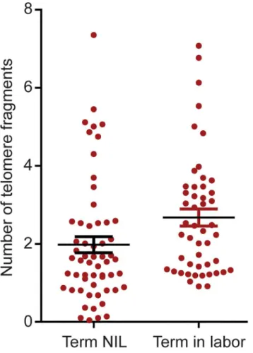
The mean levels of amplifiable AF telomere fragments were higher in term in labor than NIL [mean 2.4±0.2 (standard deviation SD)vs.mean 1.8±0.3; p = 0.04) (Fig 1).
Telomere fragments are not cytotoxic to human amniotic epithelial cell
cultures
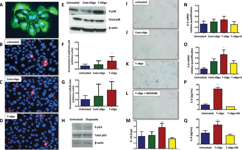
Concentrations of telomere fragments circulating in AF are higher under conditions that we previously documented to be associated with increased OS and short cellular telomeres, partic-ularly term labor [13,14]. To test our hypothesis that telomere fragments might be toxic to pri-mary human amnion epithelial cells (Fig 2A), we incubated synthesized telomere mimetic oligonucleotides (T-oligos), with amnion cells and evaluated their viability. As shown inFig 2B–2D, there were no differences in propidium iodide exclusion (red staining) among untreated, Cont-oligo and T-oligo treated cells, confirming their viability after 48h in culture. We interpret these findings to indicate that the subsequent results reflect experimentally induced changes that are not originating from general loss of cell membrane integrity in culture.
Amnion cell p38MAPK activation by T-oligo treatment
P-Table 1. Demographic, obstetric and clinical characteristics of studied patients according to the preg-nancy outcome.
Variable Term labor (n = 50) Term not in labor (n = 51) p value
**Maternal Age Median (IQR) 25 (7) 29 (8) 0.0032 Black Race n (%)
No 33 (67.4) 34 (68.0) 0.9446
Yes 16 (32.7) 16 (32.0)
Missing n = 3 Married n(%)
No 24 (49.0) 12 (24.5) 0.0119
Yes 25 (51.0) 37 (75.5)
Missing n = 5
Educational Grade achieved n(%)
<12 6 (12.5) 2 (4.2) 0.2678
12 42 (87.5) 46 (95.8)
Missing n = 7 Unemployed n(%)
No 26 (57.8) 20 (41.7) 0.1204
Yes 19 (42.2) 28 (58.3)
Missing n = 11 Income n(%)
$50k+ 8 (16.3) 14 (28.6) 0.0342
$30–50k 12 (25.0 19 (38.8)
$15–30k 17 (34.7 6 (12.2)
<$15k 12 (25.0) 10 (20.4)
Missing n = 4 **BMI
Median(IQR) 25 (10.5) 27.5 (9.3) 0.1565
Missing n = 4 Smoked n(%)
No 43 (87.8 46 (93.9 0.4865
Yes 6 (12.2) 3 (6.1)
Missing n = 3 Gravidity n(%)
<2 19 (40.3) 5 (10.6) 0.0009
2–5 28 (59.6) 42 (89.4)
Missing n = 8 Infant Sex n(%)
Female 25 (52.1) 29 (63.0) 0.2827
Male 23 (47.9) 17 (37.0)
Missing n = 7 APGAR n(%)
<7 1 (2.1) 3 (6.0) 0.2659
7–9 46 (97.9) 47 (94.0)
Missing n = 4
**GA at delivery (median, IQR) 39 (1.6) 39 (0.8) 0.5528 **Birth Weight (median, IQR) 3361.5 (572.5) 3355.0 (566.0) 0.8528
P-values were derived by Pearson Chi-square test or Fishers exact.
**P-value for maternal age, body mass index, gestational age at delivery and birth weight were derived by Mann-Whitney test.
p38MAPK than unstimulated controls (p = 0.02, ANOVA), but statistically significant differ-ences were not achieved between T-oligo and Cont-oligo treated cells. Active (phosphorylated) p53 (P-p53) was not seen in amnion cells after treatment with T- or Cont-oligos. This raises a question of p53 inducibility in primary human amnion cells, as we have not seen p53 activation using any stimulants that cause OS and senescence. However, we did verify that p53 could be activated in these cells with 100μM etoposide treatment for 24 h, a well-documented activator
of the anticancer agent p53 tumor suppressor. (Fig 2H).
T-oligos induce senescence phenotype and increase sterile
inflammatory markers in human amnion epithelial cell cultures
Senescence was tested by SAβ-gal staining after treatment with either or Cont-oligos. T-oligo treatment resulted in 1.7- and 1.6- fold increases in SAβ-gal positive cells compared to Cont-oligo and untreated cells respectively (p = 0.004) (Fig 2I–2K). Although, we noticed some p38MAPK activation after Cont-oligo treatment, it did not result in development of senescence phenotype. To verify that senescence activation was mediated by p38MAPK, incubation with the p38 inhibitor, SB203580, was performed. As shown inFig 2L, co-treatment with SB203580 decreased SA-β-gal positive cells compared to T-oligo treatment alone. The data are summa-rized inFig 2M.
Fig 1. Quantitation of telomere fragments in human amniotic fluid.Scatter plot representing the number of telomere fragments detected in amniotic fluid from normal term not in labor (NIL) and term in labor samples. The distribution of telomere fragments significantly differs between groups (p = 0.04; t-test).
Senescence associated secretory phenotype (SASP) activation
Inflammatory activation in senescing cells can modify the cellular environment. The expres-sion of two inflammatory cytokines, interleukin (IL)-6 and IL-8, was studied in response to T-oligo treatment. A slight increase in IL-6 mRNA expression was noted, but did not reach statis-tical significance (Fig 2N), while IL-8 gene expression was significantly stimulated in T-oligo treated cells relative to Cont-oligo or untreated cultures (Fig 2O). Co-treatment with SB203580 significantly reduced IL-8 expression, confirming p38MAPK mediation. We further verified
Fig 2. Human amniotic cells primary cultures.(A) Immunofluorescent staining of cytokeratin positive amnion epithelial cells. Inset,a.cytokeratin positive cells andb.nuclear staining DAPI. Original magnification x40. (B-D) Cell viability. Representative fluorescence photomicrographs of merged propidium iodide and Hoechst 33342-stained amnion cells.B.Untreated cells,C.Cont-oligo treated cells andD.T-oligo treated cells. Original magnification x40. (E-G) Representative image of Western blot analysis and densitometric quantitation of p38MAPK activation in amnion cells. E. Top panel = phosphorylated (P)-p38MAPK, middle panel = total p38MAPK and bottom panel =β-actin in untreated, Cont-oligo and T-oligo treated cells, respectively. F. Quantitation of P-p38MAPK densitometry normalized to total P-p38MAPK. T-oligo treatment produced significant (*) increase in P-p38MAPK compared to both untreated and Cont-oligo treated cells. G. Densitometric quantitation of P-p38MAPK normalized toβ-actin. Post hoc tests indicated that T-oligo treatment produced significant (*) increase in P-p38MAPK compared to untreated control, but was not significant compared to Cont-oligo treatment. (*ANOVA, p<0.05). (H)
Representative image of Western blot analysis of p53 activation in human amnion cells. Top panel = P-p53, middle panel = total p53 and bottom panel = β-actin in untreated and etoposide treated amnion cells, respectively. (I-M) Senescence associatedβ-galactosidase (SA-β-gal) staining of amnion cells. Single blue stained cells indicate positiveβ-gal activity. I. Untreated cells, J. Cont-oligo treated cells, K. T-oligo treated cells and L. T-oligo+SB203580 (p38MAPK inhibitor) treated cells. M. Quantification of the positive SA-β-gal cells. Bar graphs represent the differences in the percentage of SA-β-gal staining cells in each group. T-oligo treatment produced a significant increase (*) in the number of senescing cells, which was inhibited by SB203580 treatment. (*ANOVA, p<0.001). (N-Q) Senescence associated sterile inflammation in amnion cells. N. Relative quantification of IL-6 mRNA (p>0.05),O.Relative quantification of IL-8 mRNA in amnion cells (*p = 0.02), P. Protein concentration of IL-6 in conditioned media (*p<0.0001), and Q. Protein concentration of IL-8 in conditioned media (*p = 0.001). The production of IL-8 and IL-6 was inhibited by simultaneous treatment with SB203580.*ANOVA, T-oligo treated samples significantly higher compared with untreated, cont-oligo or T-oligo+SB samples. (Results are representative from 5 amnion cultures/ group. Telomere mimetic overhang sequence (T-oligo, [TTAGGG]2); control oligonucleotides (Cont-oligo, [AATCCC]2); untreated cells (Control CTR).
the release of IL-6 and IL-8 proteins from treated cells. Both cytokine levels were significantly higher following T-oligo treatment compared to all other groups, and levels in the conditioned media were reduced to untreated concentrations when co-incubated with SB203580 (Fig 2P and 2Q).
Evidence of
γ
-H2AX formation
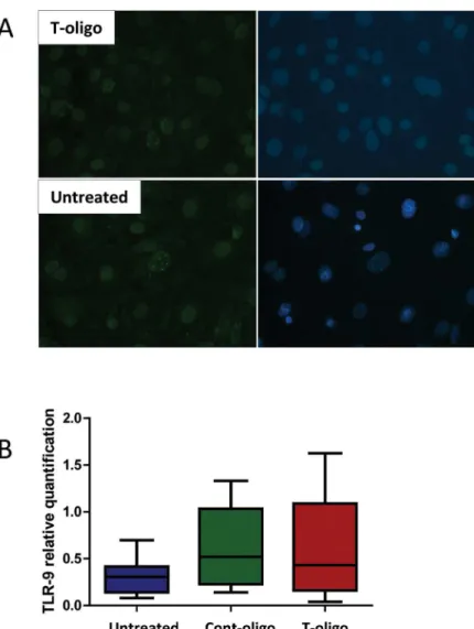
With the purpose of demonstrating the activation of DNA damage repair, we performed immunofluorescence staining of phosphorylated (γ)-H2AX formation at so-called DNA dam-age foci (DDF). We found the DDF to be more pronounced in cells treated with T-oligo com-pared to untreated cells (Fig 3A), verifying more DNA repair activation.
TLR-9 expression
In order to address a possible mechanistic pathway by which T-oligos activate intracellular sig-naling, we quantified TLR-9 mRNA. TLR-9 is known to trigger maternal immune cells activa-tion in response to placenta-derived DNA [47]. However, we did not observe any difference in TLR-9 expression in amnion cells treated with T-oligos compared to controls (untreated and Cont-oligo samples) (Fig 3B).
T-oligos induce fetal membrane senescence in pregnant mice
In order to validate our findings in anin vivomodel, T-oligos were injected into the intrauter-ine compartment of pregnant CD-1 mice on day 14 of gestation. Specimens of amniotic fluid and amniotic sac were collected after sacrificing the dams on day 18.Table 2depicts the descriptive general data regarding studied animals according to the treatment groups. There were no significant differences regarding placenta weight, animal or pup weight among the studied groups. The numbers of fetal demises and/or resorptions were significantly higher in T-oligo treated animals compared to vehicle (saline), Cont-oligo and T-oligo cotreatment with SB203580 (p = 0.001); however, preterm delivery was not observed in the studied animals. We cannot rule out a possible effect on late preterm delivery, as the injections were performed at ~70% of the colony gestational period and sacrifice was performed on day 18.
Evidence of OS induction in murine fetal membranes by T-oligos
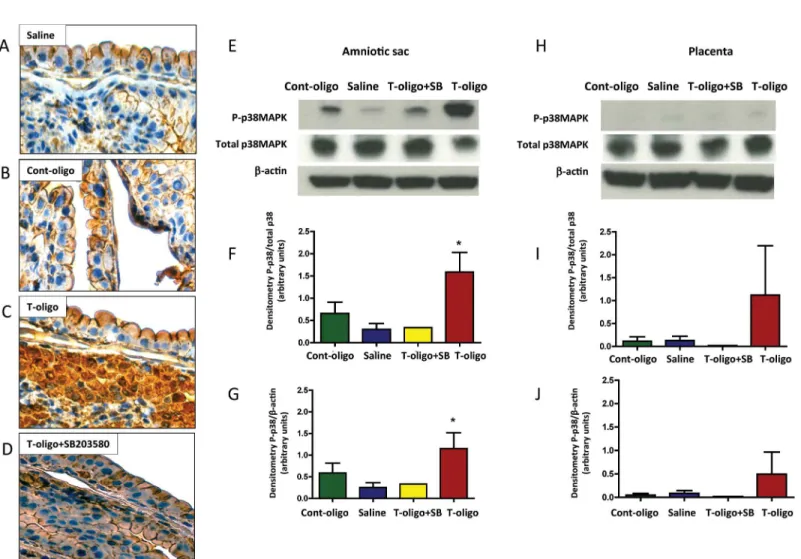
In animals treated with vehicle (saline) or Cont-oligos, microscopic examination of the fetal membranes showed minimal evidence of OS, as expected in healthy metabolizing tissues (Fig 4A and 4B). However, we observed intense staining of 3-nitrotyrosine (3-NT) modified pro-teins confirming OS induced by T-oligos (Fig 4C). Co-treatment with T-oligos and SB203580 reduced the 3-NT staining intensity (Fig 4D).
T-oligos activate p38MAPK in murine fetal membranes
As shown inFig 4E–4G, T-oligo-injected mice showed increased P-p38MAPK in the amniotic sac compared to saline, which was reduced to control levels in animals simultaneously treated with SB203580.
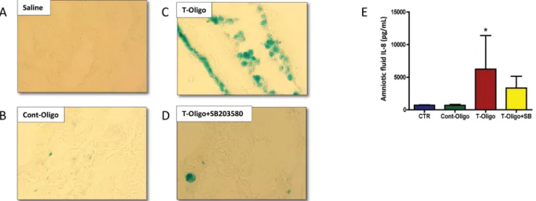
T-oligos cause senescence of murine amnion
T-oligos increase biomarkers of sterile inflammation, indicators of SASP
The activation of sterile inflammation by T-oligos, as a result of amniotic sac senescence, was examined by measuring maternal serum and amniotic fluid cytokine levels in the mouse model. Amniotic fluid from animals injected with T-oligos manifested increased concentra-tions of all evaluated cytokines (IL-6 and IL-8), however, only IL-8 reached statistical signifi-cance (Fig 5E). Co-treatment with SB203580 decreased cytokine concentrations. No
differences in cytokine levels were observed in maternal serum samples from the same dams.
Fig 3. DNA damage foci and Toll like receptor (TLR)-9 expression in human amnion cells.(A) Immunofluorescence staining of phosphorylated (γ) H2AX, a marker for DNA damage response activation. Top panel = T-oligo treated amnion cells, bottom panel = untreated cells. Left panel =γH2AX. Right panel = merged images with DAPI nuclear stain. The bright nuclear dots represent DNA damage foci and are more pronounced in cells treated with T-oligo. (B) Relative quantification of TLR-9 mRNA expression in amnion cells in the studied groups, untreated cells, Cont-oligo and T-oligo treated cells, respectively. Box plots represent the quantification relative to endogenous 16S RNA. Kruskal-Wallis test, p>0.05.(control
oligonucleotides (Cont-oligo, [AATCCC]2); Telomere mimetic overhang sequence (T-oligo, [TTAGGG]2).
Table 2. Descriptive data from studied CD-1 pregnant mice according to treatments groups (n = 5 animals/group): Saline, Cont-Oligo, T-oligo and T-oligo co-treatment with SB203580.
Saline Cont-oligo T-oligo T-oligo + SB203580 P*
Number of fetal demise and/or resorption sites 1.66 (±0.57) 0.33 (±0.57) 3.33 (±1.15)* 0.60 (±0.54) 0.001
Pup weight (g) 1.26 (±0.14) 1.22 (±0.39) 1.41 (±0.98) 1.05 (±0.20) 0.87
Placenta weight (g) 0.11 (±0.02) 0.10 (±0.02) 0.16 (±0.01) 0.09 (±0.04) 0.25 Animal weight Day 14 (g) 42.93 (±4.35) 39.50 (±0.85) 42.14 (±1.15) 39.63 (±2.46) 0.21 Animal weight Day 18 (g) 51.34 (±7.93) 49.50 (±0.51) 47.47 (±9.5) 46.43 (±5.47) 0.77
(g: grams; SB203580: p38MAPK inhibitor)
*Anova, Tukey's Multiple Comparison Test, p = 0.001.
doi:10.1371/journal.pone.0137188.t002
Fig 4. Animal models of T-oligo induced senescence.(A-D) Representative image of 3-nitrotyrosine (3-NT) modified proteins, an oxidative stress marker, in murine fetal membranes. Intrauterine injection of pregnant CD-1 mice were performed with either: A. Saline, B. Cont-oligo, C. T-oligo and D. T-oligo +SB23580 (p38MAPK inhibitor).(E-G) Representative image of Western blot analysis and densitometric quantitation of p38MAPK activation in murine amniotic sac. E. Top panel = phosphorylated (P)-p38MAPK, middle panel = total p38MAPK and bottom panel =β-actin in Cont-oligo, saline, T-oligo +SB203580 (p38MAPK inhibitor) and T-oligo treated mice, respectively. F. Densitometric quantitation P-p38MAPK normalized to total p38MAPK in amniotic sac tissue or G. normalized toβ-actin. T-oligo treatment produced a significant (*) increase in P-p38MAPK compared to saline and Cont-oligo treated groups. The co-treatment of T-oligo and SB203580 showed similar results to controls (saline and Cont-oligo). (*ANOVA, p<0.05). Results are representative from 3
animals/ group. (Telomere mimetic overhang sequence (T-oligo, [TTAGGG]2); control oligonucleotides (Cont-oligo, [AATCCC]2).
Discussion
The initiation of parturition is a complex process, whose precise signals remain unclear. Several systems maintain pregnancy through homeostatic balance including the endocrine, nervous, immune, hematological, microbiome, matrix metabolism and electro-physiological; antago-nism of any of these tends to promote labor [2,7,9,48–50]. Breakdown of these balanced sys-tems leads to cervical ripening, proteolysis, weakening and rupture of the fetal membranes and myometrial contraction leading to parturition [4,5]. Besides the well reported endocrine initia-tors, two general effectors of term parturition are oxidative stress and sterile inflammation, likely resulting from the augmented metabolic demands and depleted antioxidant reserves of the growing fetus [11,49,51]. This report opens a novel inquiry into the role of OS in the physi-ology of human parturition. The search for parturition triggers and prior data led us to hypoth-esize that OS at term signals fetal maturity and physiological aging of the fetal membranes, causing telomere shortening and sterile inflammation resulting in parturition [14,19,51]. This study provides insights into the telomere-dependent mechanism of senescence and inflamma-tory activation. The principal findings from this study are as follows: 1) The concentration of telomere fragments was higher in term labor than in term NIL samples. 2) T-oligos, that mimic shed telomeres, induced primary amnion epithelial cell senescencein vitrothrough the activa-tion of p38MAPK and SASP, manifested by increased SAβ-Gal and IL-8 gene and protein expression. Each of these steps can be mitigated using a p38MAPK inhibitor. 3) T-oligos caused murine fetal membrane OS, p38MAPK-mediated senescence and sterile inflammation (also reflected by elevated IL-8 concentrations). OS and cellular damage occur persistently dur-ing feto-placental growth [49,52,53], while cellular antioxidant proteins and repair responses prevent or minimize these damages, to avoid development of pathology. Diminished antioxi-dant capacity, or overwhelming OS, compromise tissue function and integrity and prompt aging [54]. Based on these principles, we propose a novel pathway whereby OS-induced DNA damage and telomere shortening in the fetal membranes accelerate senescence-associated
Fig 5. Senescence and inflammation induced by T-oligos in CD-1 pregnant mice.(A-D) Senescence associatedβ-galactosidase (SA-β-gal) staining of murine amniotic sac. Single blue stained cells indicateβ-gal activity. A. saline, B. Cont-oligo, C. T-oligo and D. T-oligo+SB203580 (p38MAPK inhibitor) treated mice. SA-β-gal staining is pronounced in T-oligo treated mice. (E) Concentration of interleukin (IL)-8 protein in murine amniotic fluid. Higher levels of IL-8 were found in T-oligo treated animals compared to controls (saline and Cont-oligo). The production of IL-8 was inhibited by simultaneous treatment with SB203580. (*ANOVA, p<0.05). Results are representative from 3 animals/ group. (Telomere mimetic overhang sequence (T-oligo, [TTAGGG]2); control
oligonucleotides (Cont-oligo, [AATCCC]2).
inflammation, which acts as a fetal signal for parturition. In support of this hypothesis, OS related risk factors (e.g., cigarette smoking and low grade infection) contribute to telomere-dependent premature aging and inflammation as seen in a subset of early PTB and pPROM cases [51].
Do telomere fragments trigger parturition?
The dose of telomere fragments used in ourin vitroandin situstudies or design of our study did not demonstrate that telomere fragments can induce labor; instead, the telomere fragments presence is brought up as a possible stimulator of partiturion signals, since a feedback loop they cause oxidative stress associated damages to the term amniotic sac and forces them to release other Damage Associated Molecular Patterns (DAMPs) and SASP factors. We believe that this is one of the many mechanisms that can force amnion membrane to undergo further damage and send signals to the neighboring layers to initiate parturition process. These signals can force changes in decidua (activation of leukocytes) and myometrium (functional progester-one withdrawal). Telomere reduction is a natural consequence of repeated cell division [55]. Chronic OS increases the rate of telomere shortening and reduces a cell’s replicative life span [56,57]. Telomere shortening was previously observed in fetal leukocytes and placental mem-branes from term and pPROM pregnancies [13]. In line with this, here we demonstrate that more telomere fragments are shed into the amniotic fluid in term labor, compared to NIL sub-jects.In vitro, telomere shortening and replicative senescence can be accelerated using OS inducers like cigarette smoke [41], which exerts its effects through p38MAPK activation in amnion cells [19]. We suggest that the differences in cases of term laborvs. NIL status repre-sents a buildup of OS mediators, inducing a senescent state. Premature senescence also can be induced by telomere-independent mechanisms [58].
Telomere shortening, sensed as DNA damage, can lead to further senescence by phosphory-lation of histone H2AX (γ-H2AX), a highly conserved histone family member that encodes a DNA repair and transcription regulator [59,60]. To confirm that telomere fragments might induce DNA damage in isolated amnion cells, we tested the phosphorylation of H2AX. T-oli-gos activated DNA damage repair byγ-H2AX expression at so-called DNA damage foci (DDF) [61]. DDF, therefore, could be a surrogate for oxidative stress-induced telomere shortening in feto-placental tissues as previously reported in certain pathologic pregnancies [13,62,63].
DNA oligonucleotides homologous to the telomere 30
externalization, nor do they induce lipid peroxidation. Instead, etoposide acts as an antioxidant against H2O2-induced phospholipid peroxidation in HL-60 cells [77,78]. These data indicate
that the etoposide effect in our cells is not related to OS induction. Studies also show that p38MAPK activation represents an alternate senescence mechanism [46,65,79–81]. Intrave-nous administration of T-oligos rescued mice from a fatal inoculum of human breast cancer [66] and lung cancer cells treated with T-oligos showed reduced tumor volume through senes-cence pathways that are not dependent on p53 (56). Accordingly, our previous results indicated that p38MAPK triggers senescence in oxidatively stressed human fetal membranes and amnion epithelial cells [14,19]. Furthermore, attenuation of inflammatory cytokines after p38MAPK inhibitor treatment confirms the primacy of this pathway in the generation of sterile inflamma-tion following T-oligo treatment in fetal cells and tissues.
The p38MAPK pathway is a major network of inflammation and stress responses [82] and in pregnancy, mediates IL-1β-induced MMP-9 in the fetal membranes [83], leading to labor. Recent findings from our laboratory reinforce the participation of p38MAPK in adverse preg-nancy outcomesin vitroandin vivo[14,19]. Three p38 isoforms (α,χandδ) activate distinct downstream cascades leading to DNA damage responses, whereby only p38δis reported to be p53- and p16-independent [82]. However, the SB203580 inhibitor, which effectively reverses the senescence phenotype in our models, is believed to be selective for the p38αandχproteins. We did not explore the specific p38 isoforms expressed in amnion cells, hence we cannot dis-card the hypothesis that multiple p38δisoforms might be activated in our cells.
Previous experiments using human breast carcinoma cells showed that T-oligos are effi-ciently taken up by cells within 30 to 60 minutes afterin vitroadministration and localize to the nucleus [66], where they are inherently more stable than non-G-quadruplex structures [84]. The Toll like receptor (TLR)-9 is a candidate receptor for T-oligo uptake, as it senses microbial DNA and endogenous cell-free-DNA [85,86]. However, we did not observe any difference in TLR-9 expression in amnion cells treated with T-oligos compared to controls (untreated and Cont-oligo samples). Thus, further experiments are needed to address the specific mechanism by which telomere fragments activate intracellular signaling in fetal cells.
Our data indicate that in addition to telomere shortening caused by ROS, the intracellular release of telomere fragments contributes to senescence of amnion cells via p38MAPK activa-tion. The telomere fragments themselves can amplify fetal cell senescence triggering an inflam-matory cytokine signature (SASP), which, in turn, can activate uterotonins and promote parturition. Activation of this axis prematurely, for example by excessive OS, may trigger pre-term labor. A better understanding of the pathways activated by telomere fragments, their bio-chemistry and their contribution to fetal membrane senescence should contribute to the design of more effective labor assessment (perhaps including PTB risk), and direct diagnostic and therapeutic interventions for labor induction or prevention.
Acknowledgments
Authors acknowledge support by all the staff from the Maternal-Fetal Medicine and Perinatal Research Laboratory, University of Texas Medical Branch at Galveston (UTMB), TX, USA.
Author Contributions
References
1. Romero R, Dey SK, Fisher SJ. Preterm labor: One syndrome, many causes. Science. 2014; 345: 760–
765. doi:10.1126/science.1251816PMID:25124429
2. Kamel RM. The onset of human parturition. Arch Gynecol Obs. 2010; 281: 975–982. doi:10.1007/ s00404-010-1365-9
3. Norman JE, Bollapragada S, Yuan M, Nelson SM. Inflammatory pathways in the mechanism of parturi-tion. BMC Pregnancy Childbirth. 2007; 7 Suppl 1: S7. doi:10.1186/1471-2393-7-S1-S7PMID: 17570167
4. Peltier MR. Immunology of term and preterm labor. Reprod Biol Endocrinol. 2003; 1: 122. doi:10.1186/ 1477-7827-1-122PMID:14651749
5. Orsi NM, Tribe RM. Cytokine networks and the regulation of uterine function in pregnancy and parturi-tion. J Neuroendocrinol. 2008; 20: 462–469. doi:10.1111/j.1365-2826.2008.01668.xPMID:18266939 6. McLean M, Bisits A, Davies J, Woods R, Lowry P, Smith R. A placental clock controlling the length of
human pregnancy. Nat Med. 1995; 1: 460–463. doi:10.1038/nm0595-460PMID:7585095
7. Christiaens I, Zaragoza DB, Guilbert L, Robertson SA, Mitchell BF, Olson DM. Inflammatory processes in preterm and term parturition. J Reprod Immunol. 2008; 79: 50–57. doi:10.1016/j.jri.2008.04.002 PMID:18550178
8. Pawelec M, Pałczyński B, Krzemieniewska J, Karmowski M, KoryśJ, Lątkowski K, et al. Initiation of pre-term labor. Adv Clin Exp Med. 2013; 22: 283–288. PMID:23709385
9. Romero R, Espinoza J, Kusanovic JP, Gotsch F, Hassan S, Erez O, et al. The preterm parturition syn-drome. BJOG. 2006; 113: 17–42. doi:10.1111/j.1471-0528.2006.01120.xPMID:17206962
10. Goldenberg RL, Culhane JF, Iams JD, Romero R. Epidemiology and causes of preterm birth. Lancet. 2008; 371: 75–84. doi:10.1016/S0140-6736(08)60074-4PMID:18177778
11. Challis JR, Lockwood CJ, Myatt L, Norman JE, Strauss JF, Petraglia F. Inflammation and pregnancy. Reprod Sci. 2009; 16: 206–215. doi:10.1177/1933719108329095PMID:19208789
12. Campisi J, d’Adda di Fagagna F. Cellular senescence: when bad things happen to good cells. Nat Rev Mol Cell Biol. 2007; 8: 729–40. doi:10.1038/nrm2233PMID:17667954
13. Menon R, Yu J, Basanta-Henry P, Brou L, Berga SL, Fortunato SJ, et al. Short fetal leukocyte telomere length and preterm prelabor rupture of the membranes. PLoS One. 2012; 7: e31136. doi:10.1371/ journal.pone.0031136PMID:22348044
14. Menon R, Boldogh I, Hawkins HK, Woodson Michael Polettini J, Syed TA, Fortunato SJ, et al. Histologi-cal evidence of oxidative stress and premature senescence in preterm premature rupture of the human fetal membranes recapitulated in vitro. Am J Pathol. American Society for Investigative Pathology; 2014; 184: 1740–1751. doi:10.1016/j.ajpath.2014.02.011PMID:24832021
15. Brandl A, Meyer M, Bechmann V, Nerlich M, Angele P. Oxidative stress induces senescence in human mesenchymal stem cells. Exp Cell Res. Elsevier Inc.; 2011; 317: 1541–1547. doi:10.1016/j.yexcr. 2011.02.015PMID:21376036
16. Behnia F, Taylor B, Woodson M, Hawkins H, Kacerovsky M, Fortunato S, et al. Chorioamniotic mem-brane senescence: A signal for parturition? AJOG. 2015; in press. doi:10.1016/j.ajog.2015.05.041. [Epub ahead of print]
17. Rodier F, Muñoz DP, Teachenor R, Chu V, Le O, Bhaumik D, et al. DNA-SCARS: distinct nuclear
struc-tures that sustain damage-induced senescence growth arrest and inflammatory cytokine secretion. J Cell Sci. 2011; 124: 68–81. doi:10.1242/jcs.071340PMID:21118958
18. Rodier F, Campisi J. Four faces of cellular senescence. J Cell Biol. 2011; 192: 547–556. doi:10.1083/ jcb.201009094PMID:21321098
19. Menon R, Boldogh I, Urrabaz-Garza R, Polettini J, Syed TA, Saade GR, et al. Senescence of primary amniotic cells via oxidative DNA damage. PLoS One. 2013; 8: e83416. doi:10.1371/journal.pone. 0083416PMID:24386195
20. Phillippe M. Cell-free fetal DNA-a trigger for parturition. N Engl J Med. 2014; 370: 2534–2536. doi:10. 1056/NEJMcibr1404324PMID:24963574
21. Morgan CC, Mc Cartney AM, Donoghue MT a, Loughran NB, Spillane C, Teeling EC, et al. Molecular adaptation of telomere associated genes in mammals. BMC Evol Biol. BMC Evolutionary Biology; 2013; 13: 251. doi:10.1186/1471-2148-13-251PMID:24237966
22. Blackburn EH. Switching and signaling at the telomere. Cell. 2001; 106: 661–673. PMID:11572773 23. Stewart S a, Ben-Porath I, Carey VJ, O’Connor BF, Hahn WC, Weinberg R a. Erosion of the telomeric
24. Von Zglinicki T. Oxidative stress shortens telomeres. Trends Biochem Sci. 2002; 27: 339–344. PMID: 12114022
25. Rhee DB, Ghosh A, Lu J, Bohr VA, Liu Y. Factors that influence telomeric oxidative base damage and repair by DNA glycosylase OGG1. DNA Repair (Amst). 2011; 10: 34–44. doi:10.1016/j.dnarep.2010. 09.008
26. Menon R, Polettini J, Syed TA, Saade GR, Boldogh I. Expression of 8-oxoguanine glycosylase in human fetal membranes. Am J Reprod Immunol. 2014; 72: 75–84. doi:10.1111/aji.12220PMID: 24589083
27. Hewitt G, Jurk D, Marques F, Correia-Melo C, Hardy T, Gackowska A, et al. Telomeres are favoured targets of a persistent DNA damage response in ageing and stress-induced senescence. Nat Commun. 2012; 3: 708. doi:10.1038/ncomms1708PMID:22426229
28. Xu Z, Duc KD, Holcman D, Teixeira MT. The length of the shortest telomere as the major determinant of the onset of replicative senescence. Genetics. 2013; 194: 847–857. doi:10.1534/genetics.113. 152322PMID:23733785
29. Fortunato SJ, Menon R, Velez DR, Thorsen P, Williams SM. Racial disparity in maternal-fetal genetic epistasis in spontaneous preterm birth. Am J Obstet Gynecol. 2008; 198: 666.e1–666.e–10. doi:10. 1016/j.ajog.2008.02.003
30. Menon R, Williams SM, Fortunato SJ. Amniotic fluid interleukin-1beta and interleukin-8 concentrations: racial disparity in preterm birth. Reprod Sci. 2007; 14: 253–259. doi:10.1177/1933719107301336 PMID:17636239
31. Menon R, Velez DR, Morgan N, Lombardi SJ, Fortunato SJ, Williams SM. Genetic regulation of amni-otic fluid TNF-alpha and soluble TNF receptor concentrations affected by race and preterm birth. Hum Genet. 2008; 124: 243–253. doi:10.1007/s00439-008-0547-zPMID:18807256
32. Menon R, Fortunato SJ, Edwards DRV, Williams SM. Association of genetic variants, ethnicity and pre-term birth with amniotic fluid cytokine concentrations. Ann Hum Genet. 2010; 74: 165–183. doi:10. 1111/j.1469-1809.2010.00562.xPMID:20369436
33. Menon R, Velez DR, Simhan H, Ryckman K, Jiang L, Thorsen P, et al. Multilocus interactions at mater-nal tumor necrosis factor-alpha, tumor necrosis factor receptors, interleukin-6 and interleukin-6 receptor genes predict spontaneous preterm labor in European-American women. Am J Obstet Gynecol. 2006; 194: 1616–1624. doi:10.1016/j.ajog.2006.03.059PMID:16731080
34. Pedro KJ, Espinoza J, Romero R, Gonçalves LF, Nien JK, Soto E, et al. Clinical significance of the presence of amniotic fluid“sludge”in asymptomatic patients at high risk for spontaneous preterm deliv-ery. Ultrasound Obs Gynecol. 2007; 30: 706–714.
35. Erkhembaatar LO, Kotani T, Sumigama S, Tsuda H, Mano Y, Hua L, et al. Increased expression of sphingosine kinase in the amnion during labor. Placenta. Elsevier Ltd; 2013; 34: 353–359. doi:10. 1016/j.placenta.2013.01.014PMID:23462226
36. Moore RM, Silver RJ, Moore JJ. Physiological apoptotic agents have different effects upon human amnion epithelial and mesenchymal cells. Placenta. 2003; 24: 173–180. PMID:12566244
37. Lee MS, Yaar M, Eller MS, Rünger TM, Gao Y, Gilchrest BA. Telomeric DNA induces p53-dependent reactive oxygen species and protects against oxidative damage. J Dermatol Sci. 2009; 56: 154–162. doi:10.1016/j.jdermsci.2009.08.008PMID:19906512
38. Marwaha V, Chen Y-H, Helms E, Arad S, Inoue H, Bord E, et al. T-oligo treatment decreases constitu-tive and UVB-induced COX-2 levels through p53- and NFkappaB-dependent repression of the COX-2 promoter. J Biol Chem. 2005; 280: 32379–32388. doi:10.1074/jbc.M503245200PMID:16046401 39. Li G-Z, Eller MS, Firoozabadi R, Gilchrest B a. Evidence that exposure of the telomere 3’overhang
sequence induces senescence. Proc Natl Acad Sci U S A. 2003; 100: 527–31. doi:10.1073/pnas. 0235444100PMID:12515865
40. Pitman RT, Wojdyla L, Puri N. Mechanism of DNA damage responses induced by exposure to an oligo-nucleotide homologous to the telomere overhang in melanoma. Oncotarget. 2013; 4: 761–771. PMID: 23800953
41. Yu AL, Birke K, Burger J, Welge-Lussen U. Biological effects of cigarette smoke in cultured human reti-nal pigment epithelial cells. PLoS One. 2012; 7: e48501. doi:10.1371/journal.pone.0048501PMID: 23155386
42. Harbo M, Koelvraa S, Serakinci N, Bendix L. Telomere dynamics in human mesenchymal stem cells after exposure to acute oxidative stress. DNA Repair (Amst). 2012; 11: 774–779. doi:10.1016/j.dnarep. 2012.06.003
44. Puri N, Pitman RT, Mulnix RE, Erickson T, Iness AN, Vitali C, et al. Non-small cell lung cancer is sus-ceptible to induction of DNA damage responses and inhibition of angiogenesis by telomere overhang oligonucleotides. Cancer Lett. 2014; 343: 14–23. doi:10.1016/j.canlet.2013.09.010PMID:24041868 45. Uchida K, Nakahira K, Mimura K, Shimizu T, De Seta F, Wakimoto T, et al. Effects of Ureaplasma
par-vum lipoprotein multiple-banded antigen on pregnancy outcome in mice. J Reprod Immunol. 2013; 100: 118–127. doi:10.1016/j.jri.2013.10.001PMID:24238827
46. Lamy E, Herz C, Lutz-Bonengel S, Hertrampf A, Márton M-R, Mersch-Sundermann V. The MAPK path-way signals telomerase modulation in response to isothiocyanate-induced DNA damage of human liver cancer cells. PLoS One. 2013; 8: e53240. doi:10.1371/journal.pone.0053240PMID:23382840 47. Hahn S, Giaglis S, Buser A, Hoesli I, Lapaire O, Hasler P. Cell-free nucleic acids in (maternal) blood:
any relevance to (reproductive) immunologists? J Reprod Immunol. 2014; 104–105: 26–31. doi:10. 1016/j.jri.2014.03.007PMID:24815811
48. Seol H-J, Oh M-J, Lim J-E, Jung N-H, Yoon S-Y, Kim H-J. The role of CD4+CD25 bright regulatory T cells in the maintenance of pregnancy, premature rupture of membranes, and labor. Yonsei Med J. 2008; 49: 366–371. doi:10.3349/ymj.2008.49.3.366PMID:18581584
49. Cindrova-Davies T, Yung H-W, Johns J, Spasic-Boskovic O, Korolchuk S, Jauniaux E, et al. Oxidative stress, gene expression, and protein changes induced in the human placenta during labor. Am J Pathol. American Society for Investigative Pathology; 2007; 171: 1168–1179. doi:10.2353/ajpath.2007. 070528PMID:17823277
50. Ganu R, Ma J, Aagaard KM. The role of microbial communities in parturition: Is there evidence of asso-ciation with preterm birth and perinatal morbidity and mortality? Am J Perinatol. 2013; 30: 613–624. doi: 10.1055/s-0032-1329693PMID:23161352
51. Menon R. Oxidative stress damage as a detrimental factor in preterm birth pathology. Front Immunol. 2014; 5: 1–14.
52. Watanabe K, Mori T, Iwasaki A, Kimura C, Matsushita H, Shinohara K, et al. Increased oxygen free rad-ical production during pregnancy may impair vascular reactivity in preeclamptic women. Hypertens Res. Nature Publishing Group; 2013; 36: 356–360. doi:10.1038/hr.2012.208PMID:23324862 53. Pereira AC, Martel F. Oxidative stress in pregnancy and fertility pathologies. Cell Biol Toxicol. 2014; 30:
301–312. doi:10.1007/s10565-014-9285-2PMID:25030657
54. Campisi J, Vijg J. Does damage to DNA and other macromolecules play a role in aging? If so, how? Journals Gerontol—Ser A Biol Sci Med Sci. 2009; 64: 175–178. doi:10.1093/gerona/gln065 55. Olovnikov AM. A theory of marginotomy. The incomplete copying of template margin in enzymic
syn-thesis of polynucleotides and biological significance of the phenomenon. J Theor Biol. 1973; 41: 181–
190. doi:10.1016/0022-5193(73)90198-7PMID:4754905
56. Von Zglinicki T. Role of oxidative stress in telomere length regulation and replicative senescence. Ann N Y Acad Sci. 2000; 908: 99–110. PMID:10911951
57. Von Zglinicki T, Pilger R, Sitte N. Accumulation of single-strand breaks is the major cause of telomere shortening in human fibroblasts. Free Radic Biol Med. 2000; 28: 64–74. doi:10.1016/S0891-5849(99) 00207-5PMID:10656292
58. Ramirez RD, Herbert B-S, Vaughan MB, Zou Y, Gandia K, Morales CP, et al. Bypass of telomere-dependent replicative senescence (M1) upon overexpression of Cdk4 in normal human epithelial cells. Oncogene. 2003; 22: 433–444. doi:10.1038/sj.onc.1206046PMID:12545164
59. Ohtani N, Hara E. Roles and mechanisms of cellular senescence in regulation of tissue homeostasis. Cancer Sci. 2013; 104: 525–530. doi:10.1111/cas.12118PMID:23360516
60. d’Adda di Fagagna F, Reaper PM, Clay-Farrace L, Fiegler H, Carr P, Von Zglinicki T, et al. A DNA dam-age checkpoint response in telomere-initiated senescence. Nature. 2003; 426: 194–198. doi:10.1038/ nature02118PMID:14608368
61. Chen JH, Hales CN, Ozanne SE. DNA damage, cellular senescence and organismal ageing: Causal or correlative? Nucleic Acids Res. 2007; 35: 7417–7428. doi:10.1093/nar/gkm681PMID:17913751 62. Biron-Shental T, Sukenik-Halevy R, Sharon Y, Goldberg-Bittman L, Kidron D, Fejgin MD, et al. Short
telomeres may play a role in placental dysfunction in preeclampsia and intrauterine growth restriction. Am J Obstet Gynecol. 2010; 202: 381.e1–7. doi:10.1016/j.ajog.2010.01.036PMID:20350645 63. Hallows SE, Regnault TRH, Betts DH. The long and short of it: the role of telomeres in fetal origins of
adult disease. J Pregnancy. 2012; 2012: 638476. doi:10.1155/2012/638476PMID:23094159 64. Sarkar S, Faller D V. Telomere-homologous G-rich oligonucleotides sensitize human ovarian cancer
cells to TRAIL-induced growth inhibition and apoptosis. Nucleic Acid Ther. 2013; 23: 167–174. doi:10. 1089/nat.2012.0401PMID:23634944
66. Yaar M, Eller MS, Panova I, Kubera J, Wee LH, Cowan KH, et al. Telomeric DNA induces apoptosis and senescence of human breast carcinoma cells. Breast Cancer Res. 2007; 9: R13. doi:10.1186/ bcr1646PMID:17257427
67. Klungland A, Bjelland S. Oxidative damage to purines in DNA: role of mammalian Ogg1. DNA Repair (Amst). 2007; 6: 481–488. doi:10.1016/j.dnarep.2006.10.012
68. Gnanasekar M, Thirugnanam S, Zheng G, Chen A, Ramaswamy K. T-oligo induces apoptosis in advanced prostate cancer cells. Oligonucleotides. 2009; 19: 287–292. doi:10.1089/oli.2009.0179 PMID:19642913
69. Longe HO, Romesser PB, Rankin AM, Faller D V, Eller MS, Gilchrest B a, et al. Telomere homolog oli-gonucleotides induce apoptosis in malignant but not in normal lymphoid cells: mechanism and thera-peutic potential. Int J Cancer. 2009; 124: 473–482. doi:10.1002/ijc.23946PMID:19003960
70. Rankin AM, Faller D V, Spanjaard R a. Telomerase inhibitors and“T-oligo”as cancer therapeutics: con-trasting molecular mechanisms of cytotoxicity. Anticancer Drugs. 2008; 19: 329–338. doi:10.1097/ CAD.0b013e3282f5d4c2PMID:18454043
71. Itahana K, Dimri G, Campisi J. Regulation of cellular senescence by p53. Eur J Biochem. 2001; 268: 2784–2791. PMID:11358493
72. Li GZ, Eller MS, Hanna K, Gilchrest BA. Signaling pathway requirements for induction of senescence by telomere homolog oligonucleotides. Exp Cell Res. 2004; 301: 189–200. doi:10.1016/j.yexcr.2004. 08.019PMID:15530855
73. Maier B, Gluba W, Bernier B, Turner T, Mohammad K, Guise T, et al. Modulation of mammalian life span by the short isoform of p53. Genes Dev. 2004; 18: 306–319. doi:10.1101/gad.1162404PMID: 14871929
74. Edwards MG, Anderson RM, Yuan M, Kendziorski CM, Weindruch R, Prolla T a. Gene expression pro-filing of aging reveals activation of a p53-mediated transcriptional program. BMC Genomics. 2007; 8: 80. doi:10.1186/1471-2164-8-80PMID:17381838
75. Cha J, Hirota Y, Dey SK. Sensing senescence in preterm birth. Cell Cycle. 2012; 11: 205–206. doi:10. 4161/cc.11.2.18781PMID:22189716
76. Hirota Y, Daikoku T, Tranguch S, Xie H, Bradshaw HB, Dey SK. Uterine-specific p53 deficiency confers premature uterine senescence and promotes preterm birth in mice. J Clin Invest. 2010; 120: 803–815. doi:10.1172/JCI40051PMID:20124728
77. Kagan VE, Kuzmenko AI, Tyurina YY, Shvedova AA, Matsura T, Yalowich JC. Pro-oxidant and antioxi-dant mechanisms of etoposide in HL-60 Cells: role of myeloperoxidase. Cancer Res. 2001; 61: 7777–
7784. PMID:11691792
78. Tyurina YY, Serinkan FB, Tyurin VA, Kini V, Yalowich JC, Schroit AJ, et al. Lipid antioxidant, etoposide, inhibits phosphatidylserine externalization and macrophage clearance of apoptotic cells by preventing phosphatidylserine oxidation. J Biol Chem. 2004; 279: 6056–6064. doi:10.1074/jbc.M309929200 PMID:14630936
79. Freund A, Patil CK, Campisi J. p38MAPK is a novel DNA damage response-independent regulator of the senescence-associated secretory phenotype. EMBO J. Nature Publishing Group; 2011; 30: 1536–
1548. doi:10.1038/emboj.2011.69
80. Aoshiba K, Tsuji T, Kameyama S, Itoh M, Semba S, Yamaguchi K, et al. Senescence-associated secre-tory phenotype in a mouse model of bleomycin-induced lung injury. Exp Toxicol Pathol. Elsevier GmbH.; 2013; 65: 1053–1062. doi:10.1016/j.etp.2013.04.001PMID:23688655
81. Cargnello M, Roux PP. Activation and function of the MAPKs and their substrates, the MAPK-activated protein kinases. Microbiol Mol Biol Rev. 2011; 75: 50–83. doi:10.1128/MMBR.00031-10PMID: 21372320
82. Xu Y, Li N, Xiang R, Sun P. Emerging roles of the p38 MAPK and PI3K/AKT/mTOR pathways in onco-gene-induced senescence. Trends Biochem Sci. 2014; 39: 268–276. doi:10.1016/j.tibs.2014.04.004 PMID:24818748
83. Lappas M, Riley C, Lim R, Barker G, Rice GE, Menon R, et al. MAPK and AP-1 proteins are increased in term pre-labour fetal membranes overlying the cervix: regulation of enzymes involved in the degrada-tion of fetal membranes. Placenta. 2011; 32: 1016–1025. doi:10.1016/j.placenta.2011.09.011PMID: 21963187
84. Cao Z, Huang CC, Tan W. Nuclease resistance of telomere-like oligonucleotides monitored in live cells by fluorescence anisotropy imaging. Anal Chem. 2006; 78: 1478–1484. doi:10.1021/ac0517601 PMID:16503597