ISSN 1553-3668
© 2005 Science Publications
Corresponding Author: Gabriele Baier-Bitterlich, University Prof., Ph. D., Biocenter Innsbruck, Division of Neurobiochemistry, Med. University of Innsbruck, Fritz Pregl Str. 3, A-6020 Innsbruck, Austria Tel: ++43/(0) 512-507-3272, Fax: ++43/(0) 512-507-2874
Early Cellular Responses of Purine Nucleoside-mediated Protection of
Hypoxia-induced Injuries of Neuronal PC12 Cells
Bettina Tomaselli
1,Valerie Podhraski
1,Günther Böck
2and Gabriele Baier-Bitterlich
11
Biocenter Innsbruck, Division of Neurobiochemistry, Medical University of Innsbruck
Fritz Pregl Str. 3, A-6020 Innsbruck, Austria
2
Biocenter Innsbruck, Division of Experimental Pathophysiology and Immunology
Medical University of Innsbruck Fritz Pregl Str. 3, A-6020 Innsbruck, Austria
Abstract: Hypoxia in brain may lead to cell death by apoptosis and necrosis. In parallel adenosine, a powerful endogenous neuroprotectant is formed. We wanted to investigate the effect of adenosine and its purine nucleoside relatives, inosine and guanosine on early cellular responses to hypoxia. O2 -sensitive neuronal PC12-cells were subjected to chemical hypoxia induced with rotenone, an inhibitor of mitochondrial complex I. Loss of viability after hypoxic insult was impressively rescued by adenosine, guanosine and inosine. PC12-cells mainly express the A2A adenosine receptor. Its inhibition with a specific antagonist (CSC) induced cell death of PC12-cells, which could be salvaged by adenosine but not with guanosine or inosine. We have previously demonstrated the important role of mitogen activated protein kinases 1/2 (p42/44 MAPK) in purine-mediated rescue. In this study we were interested in the involvement of protein kinases whose activities mediate these processes, including protein kinase A (PKA), phosphoinositide 3-kinase (PI3-K) and protein kinase C-related kinases(PRK 1/2).Pharmacological inhibition of PKA and PI3-K increased hypoxia-induced toxicity and likewise also affected the rescue by purine nucleosides. Nerve growth factor(NGF) andpurine nucleosides induced an activation of PRK 1/2, which to our knowledge indicates for the first time that these kinases are potentially involved in purine nucleoside-mediated rescue of hypoxic neuronal cells. Results suggest that A2A receptor expressing cells are mainly dependent on the purine nucleoside adenosine for their rescue after hypoxic insult. In addition to PKA, PI3-K is an important effector molecule in A2A-mediated signaling and for the rescue of PC12-cells after hypoxic insult.
Key words: Hypoxia, neuronal cell death, adenosine, neuroprotection, phosphoinositide 3-kinase, protein kinase C-related kinases, mitogen activated protein kinases
INTRODUCTION
Adenosine, the final metabolite in the stepwise dephosphorylation of ATP, is produced and released in the central nervous system in response to ischemia and hypoxia molecules[1, 2] and stimulation of adenosine receptors was hypothesized to result in an effective treatment of stroke[3-4], for a review[5]. Likewise, inosine and guanosine were shown to induce neurite outgrowth and preserve glial cell viability[6-8]. Despite vigorous studies, many aspects of the mechanisms involved in purine-based protection are still unclear. In this study we wanted to investigate the effect of purine nucleosides on early cellular responses to hypoxia, induced by rotenone, a mitochondrial complex I inhibitor[9-12].
For a test model O2-sensitive clonal rat pheochromocytoma (PC12)-cells, which are widely used as a model system for sympathetic ganglion-like neurons[13,14] and exposures to hypoxia[15] were used.
PC12- cells express abundant A2A adenosine receptors[16-18], which have been shown to affect these cellular responses to hypoxia[3,19]. PC12-cells differentiate into sympathetic neurons upon treatment with NGF[20], which requires activation of p42/44 MAPK and induction of gene expression[21]. Under certain circumstances purine nucleosides were shown to cooperate with NGF–mediated pathways[22-24]. Recent data in our hands revealed that p42/p44 MAPK was strongly activated by purine nucleosides especially adenosine. Hence we have put a special emphasis on the study of the impact of purine nucleosides on the early signaling elements leading to the activation of p42/44 MAPK following hypoxic insult.
MATERIALS AND METHODS
horse-serum, 0.625 % fetal-calf-serum, 1 % Pen/Strep and 1 % L-glutamine). Within 30 min[10] after addition of rotenone (10 µM), cells were stimulated with 500 µM adenosine, guanosine and inosine (Sigma). For inhibitor studies, nucleoside transport inhibitor S- (4-Nitrobenzyl)-6-thioinosine (NBTI, 10 µM, Sigma), adenosine A2A receptor antagonist 8-(3-Chlorostyryl) caffeine (CSC, 10 µM Sigma), A1 receptor antagonist 8-Phenyltheophylline (8-PT, 100 nM, Sigma), the cAMP-dependent protein kinase inhibitor H89 (5, 10 and 20 µM, Sigma) and the PI3-Kinase inhibitor LY294002 (25 µM, Sigma) were used. For CSC experiments, cells were starved in LM one day before stimulation. All pharmacological inhibitors were added 2 h, CSC 30 min prior to tests.
Fluorescence analysis: Cells were stained after 24 h. For microscopic analysis cells were fixed with 4 % PFA before staining and for FACS scan after staining with 2 % PFA. Primary antibodies: A2A, A2B, A1 (Chemicon); secondary antibody: Alexa Fluor 488 F(ab´) fragment of goat-anti-rabbit (Eubio).
Calculation of neurite outgrowth: Cells were stimulated with NGF-β (5 ng mL1), purines (500 µM), rotenone (10µM) and LY294002 (25 µM). On day 3 neurite-bearing-cells (neurites longer than one time cell diameter) were counted and averaged from 3 microscopic fields.
Immunoblots: Detection was done with following antibodies: phospho-PRK 1/2 and phospho-p42/44 MAPK (New-England-Biolabs), total-PRK 1 (BD Transduction Laboratories) and total-p42/44 MAPK (New-England-Biolabs). For quantification, blots were scanned with Molecular Dynamics Personal Densitometer SI scanner and ratios (active divided through total) were calculated.
Statistical analysis: All values in figures were expressed as the mean ± SEM. One-tailed Mann-Whitney-test was used. p-values of < 0,05 were considered statistically significant.
RESULTS AND DISCUSSION
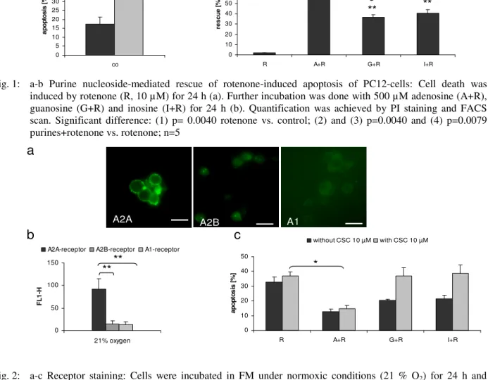
Chemical hypoxia induced apoptosis of neuronal PC12-cells (Fig.1a). In accordance with previous studies, purine nucleosides, especially adenosine, impressively counteracted this process (Fig.1b). Recent results in the literature indicated that hypoxia-induced membrane responses of PC12-cells are likely to be mediated via activation of the A2A adenosine receptors[16, 19]. In line with these findings cells express high levels of A2A but only marginal amounts of A2B and A1 receptors (Fig. 2a-b). Pharmacological inhibition of the A2A receptor by CSC enhanced cell death of hypoxic PC12-cells. While addition of
adenosine potently rescued cells, guanosine and inosine were ineffective (Fig. 2c). We therefore suppose that adenosine is the most important neuroprotectant for neuronal PC12-cells. However, there may be circumstances under which inosine can activate at least some of the receptors[25] and provides a larger activation than adenosine[5,11,26]. In line with these findings inosine was the only purine nucleoside used in this study, which was affected by the A1 receptor antagonist 8-PT (Table 1). Guanosine, does not bind to the adenosine receptor, but recent studies showed the existence of specific G-coupled receptors for guanosine[27]. Others[28] however, observed, that the mitogenic activity of guanosine was partly inhibited by antagonists of A1 and A2B adenosine receptors. Alternatively, inhibition of nucleoside transport by NBTI improved viability of cells (data not shown) and significantly increased adenosine-mediated rescue of hypoxic cells (Table 1), presumably due to the fact that inhibition of adenosine uptake via nucleoside transport increased A2A receptor-mediated signaling[29]. Our own results therefore confirm the hypothesis that adenosine mainly acts via adenosine receptor-mediated signaling[30], whereas many aspects of the mechanisms involved in inosine – and guanosine- based protection still remain unclear.
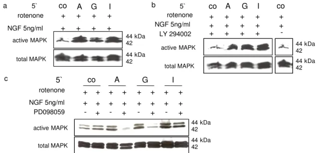
NGF triggers intracellular signaling cascades including MAPK and PI3-K, reviewed in:[31] and promotes the differentiation of neuronal cells e.g. PC12-cells, a well- studied model of growth factor actions[13,20,32-34]. Like NGF, the ability of PKA to differentiate PC12-cells is associated with a sustained activation of MAPK. As seen earlier, neurite outgrowth is down regulated under hypoxia and purine nucleosides support NGF –mediated rescue. Co localization of NGF receptors and A2A receptors suggests a potential cross-interaction between their signaling pathways[22]. Along with previous findings[23,35-39], earlier results by our lab, have demonstrated that purine nucleosides rapidly activate p42/44 MAPK and in combination with NGF led to a further additive enhancement. Under hypoxic conditions we could also observe the cooperative effects of NGF and purine nucleosides on p42/44 MAPK activation (Fig. 3a). PC12-cells were further treated with LY294002 and for control purpose with PD098059, a specific p42/44 MAPK inhibitor[40]. Administration of LY294002 led to the inhibition of adenosine-mediated p42/44 MAPK activation, whereas inosine- and guanosine-mediated activation of p42/44 MAPK was less effected (inosine) or even enhanced (guanosine) (Fig. 3b). Our results are in line with earlier data[39], which showed that PI3-K is required for activation of Rap1, which may represent an integral part of a direct path to p42/44 MAPK phosphorylation. As expected, MEK-1 inhibitor PD098059 reduced the purine nucleoside-mediated activation of p42/44 MAPK (Fig. 3c).
0 5 10 15 20 25 30 35 40 45
co
a
p
o
p
to
s
is
[
%
]
without R with R
a
**
10 10 20 30 40 50 60 70 80
R A+R G+R I+R
re
s
c
u
e
[
%
]
b
**
2
**
3
**
4
Fig.2 a-c
0 50 100 150
21% oxygen
F
L
1
-H
A2A-receptor A2B-receptor A1-receptor
**
**
a
0 10 20 30 40 50
R A+R G+R I+R
a
p
o
p
to
s
is
[
%
]
without CSC 10 µM with CSC 10 µM
b
*
A2A A2B A1
c
Table 1: Effect of pharmacological inhibitors on purine-mediated rescue
Treatment A+R G+R I+R
--- 63,55 ± 5,52 % (n=5) 37,55 ± 3,59 % (n=5) 42,48 ± 3,19 % (n=5) 8-PT 100 nM 70,58 ± 4,93 % (n=5) 40,20 ± 5,43 % (n=5) 32,40 ± 7,22 % (n=5) NBTI 10 µM 79,95 ± 1,84 % (n=5)(1) 48,53 ± 8,64 % (n=5) 39,50 ± 10,86 % (n=5)
Cells were incubated with NBTI, 8-PT, rotenone (R, 10 µM) and together with adenosine (A+R), guanosine (G+R) and inosine (I+R) in a concentration of 500 µM. FACS scan followed. Statistical difference (1) p=0,0079 NBTI+A+R vs. A+R
Fig. 1: a-b Purine nucleoside-mediated rescue of rotenone-induced apoptosis of PC12-cells: Cell death was induced by rotenone (R, 10 µM) for 24 h (a). Further incubation was done with 500 µM adenosine (A+R), guanosine (G+R) and inosine (I+R) for 24 h (b). Quantification was achieved by PI staining and FACS scan. Significant difference: (1) p= 0.0040 rotenone vs. control; (2) and (3) p=0.0040 and (4) p=0.0079 purines+rotenone vs. rotenone; n=5
Fig. 2: a-c Receptor staining: Cells were incubated in FM under normoxic conditions (21 % O2) for 24 h and stained for A2A, A2B or A1. Cells were analyzed on a Zeiss Axioplan-2-Fluorescence-Microscope; scale bar, 10µm (a). For quantification, FACS analysis was used. Fluorescence intensity (FL1-H) of unspecific binding was subtracted from values obtained for primary antibody (b). Cells were stimulated with CSC (10 µM), rotenone (R, 10 µM) and purines 500 µM: adenosine (A+R), guanosine (G+R) and inosine (I+R) (c). After 24 h, PI staining and FACS scan followed. Significant difference: (b) double-asterix (**) p=0.0079; (c) asterix (*) p=0.0143; (b) n=3, (c) n=5
The importance of PKA for PC12-cell viability was studied by use of the pharmacological inhibitor H89 (5, 10 and 20 µM). With increasing concentration H89 induced a rising toxicity and inhibition of the purine-mediated rescue. At a concentration of 10 µM, toxicity could still be reduced to some extent by adenosine and
0 10 20 30 40 50 60 70
R A+R G+R I+R
a p o p to s is [ % ]
without H89 H89 5 µM H89 10 µM H89 20 µM
a
0 5 10 15 20 25 30 35 40R A+R G+R I+R
a p o p to s is [ % ]
without LY 25µM with LY 25µM
*
b
0 10 20 30 40 50 60 70R A+R G+R I+R
a p o p to s is [ % ]
without H89 H89 5 µM H89 10 µM H89 20 µM
a
0 5 10 15 20 25 30 35 40R A+R G+R I+R
a p o p to s is [ % ]
without LY 25µM with LY 25µM
*
b
c
0 2 4 6 8NGF+R NGF+R+A NGF+R+G NGF+R+I
n e u ri te b e a ri n g c e ll s [ %
] without LY 25 µM with LY 25µM
c
0 2 4 6 8NGF+R NGF+R+A NGF+R+G NGF+R+I
n e u ri te b e a ri n g c e ll s [ %
] without LY 25 µM with LY 25µM
increased rotenone-mediated toxicity and significantly inhibited adenosine -mediated rescue of hypoxic cells (Fig. 4b). In accordance with these data, LY294002 decreased adenosine- and guanosine- yet not inosine-mediated rescue of neurites (Fig. 4c). This observation is in line with studies mentioned before, suggesting that
the PI3-K/Rac pathway is one of the early signals that mediate NGF-induced neuronal differentiation in PC12-cells[41], for a review[22].
In PC12-cells, a not yet elucidated inhibitory signaling link between NGF receptors and RhoA was suggested, reviewed in[42].
Fig. 3: a-c Effect of LY294002 and PD098059 on p42/44 MAPK activity in hypoxic PC12-cells: Cells were stimulated in serum-free medium for 5 min with NGF-ß 5 ng mL1 and rotenone 1 µM (co), additional with 500 µM adenosine (A), guanosine (G) and inosine (I) (a), +/- LY294002 25 µM (b) and +/- PD098059 (c)
Fig. 4: a-c Effect of H89 and LY294002: For inhibitor studies (a - b), cells were treated in LM with the inhibitors ((a) H89 5, 10 and 20 µM; (b) LY294002 25 µM) for 2 h. Stimulation with rotenone (R, 10 µM) and purine nucleosides 500 µM: adenosine (A+R), guanosine (G+R) and inosine (I+R) followed. PI staining and FACS scan was done. Statistical difference: asterix (*) p=0.0476; n=6. For the neurite outgrowth assay, stimuli were added together to adherent cells. On day 3 neurite positive cells were counted and evaluated (c)
Their data indicate that NGF –mediated activation of the TrkA receptor stimulates PI3-K, which in turn increases Rac1 activity to induce transient RhoA inactivation during the initial phase of neurite outgrowth. Among direct Rho effectors are the ‘protein
kinases N‘ (PKNs), termed also ‘protein kinase C-related kinases’ (PRK 1/2), reviewed in:[43]. Authors further showed that activation of PRK 1/2 involves 3-phosphoinositide-dependent protein kinase-1 (PDK1) and that the interaction of PRK 1/2 with PDK1 was
44 kDa 42 44 kDa 42 NGF 5ng/ml rotenone LY 294002 + -+ 5` NGF 5ng/ml A G co I rotenone + + + + 44 kDa 42 44 kDa 42 + + + + + + + + + + + + active MAPK total MAPK active MAPK total MAPK + + + +
5` co A G I co
a b
c 5`
NGF 5ng/ml
A G I
0 0,2 0,4 0,6 0,8 1 ra ti o [ a c ti v e /t o ta l]
control NGF 5 ng/ml NGF 50 ng/ml
0 0,2 0,4 0,6 0,8 1 ra ti o [ a c ti v e /t o ta l]
control NGF 5 ng/ml NGF 50 ng/ml
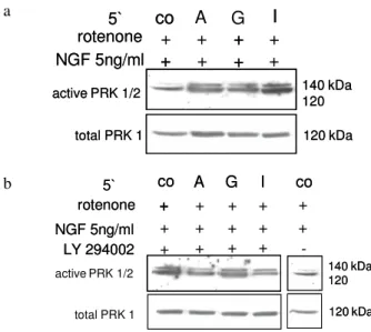
shown to be dependent upon Rho. Consistent with the finding that PKN and PRK 2 are effectors for Rho is the observation that PKN and PRK 2 can enhance or mediate changes in the actin cytoskeleton and gene transcription[44]. We were therefore interested in the role of PRK 1/2 (p120 /140). At first we studied, whether PRK 1/2 activity may be influenced by NGF. Indeed, as revealed in immunoblots, addition of NGF led to the concentration dependant activation of PRK 1/2 (Fig. 5a).
a
b
c
Fig. 5a-c: PRK 1/2 activation in PC12-cells: Normoxic cells were stimulated with/without NGF-ß 5 and 50 ng mL1 for 5 min (a, n=6). In addition cells were incubated with serum-free medium (co), with 500 µM purine nucleosides: adenosine (A), guanosine (G), inosine (I) and +/- rotenone 1 µM for 5 min (b – c, n=5). Immunoblots were analyzed with specific antibodies to PRK 1/2
In view of the fact discussed above that NGF apparently induces transient RhoA inactivation this result appears paradoxical. Similarly purines led to the activation of PRK 1/2, whereby inosine was the most potent activating component (Fig. 5b). Under hypoxia a higher basic and guanosine-mediated activity of PRK 1/2 was observed, whereas adenosine- and inosine-mediated kinase phosphorylation was reduced (Fig. 5c). NGF activated PRK 1/2 in hypoxic PC12-cells and the activation was further increased by a combination of NGF with purine nucleosides (Fig. 6a). Inhibition of
PI3-K with LY294002 led to the reduction of NGF+adenosine- and NGF+inosine- but not NGF+guanosine-mediated PRK1/2 activation (Fig. 6b). These data are in line with our findings that LY294002 inhibited adenosine and inosine but not guanosine – mediated MAPK activation. As discussed in previous publications it was suggested that inosine and guanosine exert its receptor-independent effects probably via PKN that has been implicated in mediating NGF-induced differentiation[45,46] and induction of gene
a
b
Fig. 6: a-b Effect of PI3-K inhibition on PRK 1/2 activation in hypoxic PC12-cells: Cells were treated with rotenone 1 µM. Additional stimulation was achieved by using NGF-ß 5 ng mL1, 500 µM adenosine (A), guanosine (G) and inosine (I) (a). Furthermore LY294002 25 µM (b) was used
expression[47-48]. PKN termed by this group[46,49] is a protein kinase of an apparent molecular mass of 45-47 kDa, which is rapidly activated by NGF and other agents and was selectively inhibited by purine analogs. We could however not detect an inhibiting effect of the inhibitor 6-TG on purine-mediated PRK 1/2 activation. Therefore it remains questionable, whether the earlier detected PKN (p 45-47) and the PKN alias PRK (p120/140) molecules are exactly the same kinases, apart from the fact that both of them are activated by NGF. This interesting question will be clarified in future studies.
In summary results in this study demonstrated that in PC12-cells, adenosine and guanosine strongly promote viability and in combination with NGF also neurite outgrowth after hypoxic insult. In line with previous findings results revealed that PI3-K and PKA are essential survival kinases for neuronal PC12-cells. For adenosine-mediated rescue of viability and neurite outgrowth MAPK activation is essential. Guanosine-and inosine-mediated rescue however, is apparently more independent of MAPK activation. PRK 1/2 are rotenone
140 kDa 120
5`
active PRK 1/2
A G
co I
rotenone
total PRK 1
5` 140 kDa 120 120 kDa - - - -A G co I + + + +
active PRK 1/2
total PRK 1
rotenone
140 kDa 120
120 kDa
5`
active PRK 1/2
A G
co I
rotenone
total PRK 1
5` 140 kDa 120 - - - -- - - -A G co I + + + + + +
active PRK 1/2
5` NGF 5ng/ml
A
G
co
I
rotenone 140 kDa 120 120 kDa+
+
+
+
active PRK 1/2
total PRK 1
+
+
+
+
5` NGF 5ng/mlco
I
rotenone 140 kDa 120 120 kDa+
+
+
active PRK 1/2
total PRK 1
+
+
+ + NGF 5ng/ml rotenoneactive PRK 1/2
total PRK 1
5` LY 294002 A G co I + -co + 140 kDa 120 120 kDa + + + + + + + + + + + + NGF 5ng/ml rotenone 5` LY 294002 A G
co I co
140 kDa 120
120 kDa
activated by NGF and purine nucleosides and might be involved in the regulation of the cytoskeleton[43]. Results, showing the positive impact of inosine, adenosine and guanosine on viability and neurite outgrowth of PC12-cells, support current studies on the possibility of novel neuronal cell regeneration therapies and confirm the important role of purine nucleosides in neuronal pathologies involving hypoxic insult.
ACKNOWLEDGEMENTS
This work was supported by a grant of the Austrian FWF P15823. We are grateful to Dr.G.Baier and Dr. C. Bandtlow for helpful discussion and support.
REFERENCES
1. Winn, H.R., R. Rubio and R.M. Berne, 1981. Brain adenosine concentration during hypoxia in rats. Am. J. Physiol., 241: H235-42.
2. Zetterstrom, T. et al., 1982. Purine levels in the intact rat brain. Studies with an implanted perfused hollow fibre. Neurosci. Lett., 29: 111-5.
3. Kobayashi, S. and D.E. Millhorn, 1999. Stimulation of expression for the adenosine A2A receptor gene by hypoxia in PC12 cells. A potential role in cell protection. J. Biol. Chem., 274: 20358-65.
4. Huang, N.K. et al., 2001. Activation of protein kinase A and atypical protein kinase C by A(2A) adenosine receptors antagonizes apoptosis due to serum deprivation in PC12 cells. J. Biol. Chem., 276: 13838-46.
5. Dunwiddie, T.V. and S.A. Masino, 2001. The role and regulation of adenosine in the central nervous system. Annu. Rev. Neurosci., 24: 31-55.
6. Chen, P. et al., 2002. Inosine induces axonal rewiring and improves behavioral outcome after stroke. Proc. Natl. Acad. Sci. U.S.A., 99: 9031-6. 7. Gysbers, J.W. and M.P. Rathbone, 1996. Neurite
outgrowth in PC12 cells is enhanced by guanosine through both cAMP-dependent and -independent mechanisms. Neurosci. Lett., 220: 175-8.
8. Jurkowitz, M.S. et al., 1998. Adenosine, inosine and guanosine protect glial cells during glucose deprivation and mitochondrial inhibition: correlation between protection and ATP preservation. J. Neurochem., 71: 535-48.
9. Tai, K.K. and D.D. Truong, 2002. Activation of adenosine triphosphate-sensitive potassium channels confers protection against rotenone-induced cell death: therapeutic implications for Parkinson's disease. J. Neurosci. Res., 69: 559-66. 10. Litsky, M.L. et al., 1999. Inosine and guanosine
preserve neuronal and glial cell viability in mouse spinal cord cultures during chemical hypoxia. Brain Res., 821: 426-32.
11. Bocklinger, K. et al., 2004. Purine nucleosides support the neurite outgrowth of primary rat cerebellar granule cells after hypoxia. Eur. J. Cell Biol., 83: 51-4.
12. Schafer, M. et al., 2003. Signaling of hypoxia-induced autonomous proliferation of endothelial cells. Faseb. J., 17: 449-51.
13. Greene, L.A. and A.S. Tischler, 1976. Establishment of a noradrenergic clonal line of rat adrenal pheochromocytoma cells which respond to nerve growth factor. Proc. Natl. Acad. Sci. U.S.A., 73: 2424-8.
14. Seta, K.A. et al., 2002. Responding to hypoxia: lessons from a model cell line. Sci STKE, 146: RE11.
15. Kobayashi, S., H. Zimmermann and D.E. Millhorn, 2000. Chronic hypoxia enhances adenosine release in rat PC12 cells by altering adenosine metabolism and membrane transport. J. Neurochem., 74: 621-32.
16. Arslan, G., B. Kull and B.B. Fredholm, 1999. Signaling via A2A adenosine receptor in four PC12 cell clones. Naunyn Schmiedebergs Arch. Pharmacol., 359: 28-32.
17. Hide, I., et al., 1992. A2A adenosine receptors from rat striatum and rat pheochromocytoma PC12 cells: characterization with radioligand binding and by activation of adenylate cyclase. Mol. Pharmacol., 41: 352-9.
18. van der Ploeg, I. et al., 1996. Functional characterization of adenosine A2 receptors in Jurkat cells and PC12 cells using adenosine receptor agonists. Naunyn Schmiedebergs Arch. Pharmacol., 353: 250-60.
19. Kobayashi, S. et al., 1998. Adenosine modulates hypoxia-induced responses in rat PC12 cells via the A2A receptor. J. Physiol., 508: 95-107.
20. Marshall, C.J., 1995. Specificity of receptor tyrosine kinase signaling: transient versus sustained extracellular signal-regulated kinase activation. Cell, 80: 179-85.
21. Marais, R., J. Wynne and R. Treisman, 1993. The SRF accessory protein Elk-1 contains a growth factor-regulated transcriptional activation domain. Cell, 73: 381-93.
22. Cheng, H.C., H.M. Shih and Y. Chern, 200. Essential role of cAMP-response element-binding protein activation by A2A adenosine receptors in rescuing the nerve growth factor-induced neurite outgrowth impaired by blockage of the MAPK cascade. J. Biol. Chem., 277: 33930-42.
23. Charles, M.P. et al., 2003. Induction of neurite outgrowth in PC12 cells by the bacterial nucleoside N6-methyldeoxyadenosine is mediated through adenosine A2a receptors and via cAMP and MAPK signaling pathways. Biochem. Biophys. Res. Commun., 304: 795-800.
25. Hasko, G. et al., 2000. Inosine inhibits inflammatory cytokine production by a posttranscriptional mechanism and protects against endotoxin-induced shock. J. Immunol., 164: 1013-9.
26. Jin, X. et al., 1997. Inosine binds to A3 adenosine receptors and stimulates mast cell degranulation. J. Clin. Invest., 100: 2849-57.
27. Traversa, U. et al., 2003. Rat brain guanosine binding site. Biological studies and pseudo-receptor construction. Bioorg. Med. Chem., 11: 5417-25.
28. Ciccarelli, R. et al., 2000. Cultured astrocyte proliferation induced by extracellular guanosine involves endogenous adenosine and is raised by the co-presence of microglia. Glia, 29: 202-11.
29. Parkinson, F.E. et al., 2000. Effects of nitrobenzylthioinosine on neuronal injury, adenosine levels and adenosine receptor activity in rat forebrain ischemia. J. Neurochem., 75: 795-802. 30. Fredholm, B.B. et al., 1999. Actions of caffeine in the brain with special reference to factors that contribute to its widespread use. Pharmacol. Rev., 51: 83-133.
31. York, R.D. et al., 2000. Role of phosphoinositide 3-kinase and endocytosis in nerve growth factor-induced extracellular signal-regulated kinase activation via Ras and Rap1. Mol. Cell Biol., 20: 8069-83.
32. Tischler, A.S. and L.A. Greene, 1975. Nerve growth factor-induced process formation by cultured rat pheochromocytoma cells. Nature, 258: 341-2.
33. Cowley, S. et al., 1994. Activation of MAP kinase kinase is necessary and sufficient for PC12 differentiation and for transformation of NIH 3T3 cells. Cell, 77: 841-52.
34. Kao, S. et al., 2001. Identification of the mechanisms regulating the differential activation of the mapk cascade by epidermal growth factor and nerve growth factor in PC12 cells. J. Biol. Chem., 276: 18169-77.
35. Sexl, V. et al., 1997. Stimulation of the mitogen-activated protein kinase via the A2A-adenosine receptor in primary human endothelial cells. J. Biol. Chem., 272: 5792-9.
36. Arslan, G. and B.B. Fredholm, 2000. Stimulatory and inhibitory effects of adenosine A(2A) receptors on nerve growth factor-induced phosphorylation of extracellular regulated kinases 1/2 in PC12 cells. Neurosci. Lett., 292: 183-6.
37. Fredholm, B.B. et al., 2001. International Union of Pharmacology. XXV. Nomenclature and classification of adenosine receptors. Pharmacol. Rev., 53: 527-52.
38. Schulte, G. and B.B. Fredholm, 2003. The G(s)-coupled adenosine A(2B) receptor recruits divergent pathways to regulate ERK1/2 and p38. Exp. Cell. Res., 290: 168-76.
39. Schulte, G. and B.B. Fredholm, 2003. Signalling from adenosine receptors to mitogen-activated protein kinases. Cell Signal, 15: 813-27.
40. Dudley, D.T. et al., 1995. A synthetic inhibitor of the mitogen-activated protein kinase cascade. Proc. Natl. Acad. Sci. U.S.A., 92: 7686-9.
41. Yasui, H. et al., 2001. Differential responses to nerve growth factor and epidermal growth factor in neurite outgrowth of PC12 cells are determined by Rac1 activation systems. J. Biol. Chem., 276: 15298-305.
42. Nusser, N. et al., 2002. Nerve growth factor signals through TrkA, phosphatidylinositol 3-kinase and Rac1 to inactivate RhoA during the initiation of neuronal differentiation of PC12 cells. J. Biol. Chem., 277: 35840-6.
43. Flynn, P. et al., 2000. Rho GTPase control of protein kinase C-related protein kinase activation by 3-phosphoinositide-dependent protein kinase. J. Biol. Chem., 275: 11064-70.
44. Sun, W. et al., 2000. MEK kinase 2 binds and activates protein kinase C-related kinase 2. Bifurcation of kinase regulatory pathways at the level of an MAPK kinase kinase. J. Biol. Chem., 275: 24421-8.
45. Volonte, C. et al., 1989. Differential inhibition of nerve growth factor responses by purine analogues: correlation with inhibition of a nerve growth factor-activated protein kinase. J. Cell. Biol., 109: 2395-403.
46. Greene, L.A., C. Volonte and A. Chalazonitis, 1990. Purine analogs inhibit nerve growth factor-promoted neurite outgrowth by sympathetic and sensory neurons. J. Neurosci., 10: 1479-85.
47. Benowitz, L.I. et al., 1998. Axon outgrowth is regulated by an intracellular purine-sensitive mechanism in retinal ganglion cells. J. Biol. Chem., 273: 29626-34.
48. Petrausch, B. et al., 2000. A purine-sensitive pathway regulates multiple genes involved in axon regeneration in goldfish retinal ganglion cells. J. Neurosci., 20: 8031-41.