SEPTIN2 and STATHMIN Regulate
CD99-Mediated Cellular Differentiation in
Hodgkin's Lymphoma
Wenjing Jian1☯, Lin Zhong2☯, Jing Wen1, Yao Tang1, Bo Qiu1, Ziqing Wu1, Jinhai Yan1, Xinhua Zhou1*, Tong Zhao2*
1Department of Molecular and Tumor Pathology Laboratory of Guangdong Province, School of Basic Medical Science, Southern Medical University, Guangzhou, China,2Department of Pathology, the Third Affiliated Hospital, Southern Medical University, Guangzhou, China
☯These authors contributed equally to this work.
*[email protected](TZ); [email protected](XHZ)
Abstract
Hodgkin’s lymphoma (HL) is a lymphoid neoplasm characterized by Hodgkin’s and Reed-Sternberg (H/RS) cells, which is regulated byCD99. We previously reported thatCD99 downregulation led to the transformation of murine B lymphoma cells (A20) into cells with an H/RS phenotype, whileCD99upregulation induced differentiation of classical Hodgkin’s lymphoma (cHL) cells (L428) into terminal B-cells. However, the molecular mechanism re-mains unclear. In this study, using fluorescence two-dimensional differential in-gel electro-phoresis and matrix-assisted laser desorption/ionization time of flight mass spectrometry (MALDI-TOF MS), we have analyzed the alteration of protein expression followingCD99 upregulation in L428 cells as well as downregulation of mouseCD99antigen-like 2 (mCD99L2) in A20 cells. Bioinformatics analysis showed thatSEPTIN2andSTATHMIN, which are cytoskeleton proteins, were significantly differentially expressed, and chosen for further validation and functional analysis. Differential expression ofSEPTIN2was found in both models and was inversely correlated withCD99expression.STATHMINwas identified in the A20 cell line model and its expression was positively correlated with that ofCD99. Im-portantly, silencing ofSEPTIN2with siRNA substantially altered the cellular cytoskeleton in L428 cells. The downregulation ofSTATHMINby siRNA promoted the differentiation of H/ RS cells toward terminal B-cells. These results suggest thatSEPTIN2-mediated cytoskele-tal rearrangement andSTATHMIN-mediated differentiation may contribute to changes in cell morphology and differentiation of H/RS cells withCD99upregulation in HL.
Introduction
Hodgkin’s lymphoma (HL) is one of the most common malignant neoplasms affecting the lymphoid and hematological systems. Classical Hodgkin’s lymphoma (cHL) is characterized by Hodgkin cells and multinucleated Reed-Sternberg cells (H/RS) [1]. Accumulating evidence
OPEN ACCESS
Citation:Jian W, Zhong L, Wen J, Tang Y, Qiu B, Wu Z, et al. (2015) SEPTIN2 and STATHMIN Regulate CD99-Mediated Cellular Differentiation in Hodgkin's Lymphoma. PLoS ONE 10(5): e0127568. doi:10.1371/journal.pone.0127568
Academic Editor:Tim Thomas, The Walter and Eliza Hall of Medical Research, AUSTRALIA
Received:August 5, 2014
Accepted:April 16, 2015
Published:May 22, 2015
Copyright:© 2015 Jian et al. This is an open access article distributed under the terms of theCreative Commons Attribution License, which permits unrestricted use, distribution, and reproduction in any medium, provided the original author and source are credited.
Data Availability Statement:All relevant data are within the paper and its Supporting Information files.
Funding:This study was supported by a grant from National Natural Science Foundation of China (Grant numbers: 81071941, 81302051).
suggests that H/RS cells are derived from clonal B-cells with loss of their B-cell phenotype [2]. Mature B-cells lacking B-cell receptors (BCR) normally die via apoptosis, suggesting that H/RS cells must have developed mechanisms to maintain survival. H/RS cells present a complex immunophenotype. For example, H/RS cells usually express markers associated with the mye-loid lineage (CD15) and markers associated with plasma cells (CD138, MUM-1) [3,4], but rarely B-cell markers, such as CD20, Oct-2, Ig, or components of the BCR (CD79aandCD79b) [5]. The cause of the aberrant expression of a large number of B-cell genes is currently unclear. It is proposed that B-cell markers are lost due to aberrant gene regulation and cytoskeletal rear-rangements [6]. H/RS cells are also characterized by abortive plasma cell differentiation [7], al-though, the underlying molecular mechanism is largely unknown.
CD99, a transmembrane glycoprotein encoded by theMIC2gene, is broadly expressed in hematopoietic cells, such as B-cells, T-cells, mononuclear cells, and neutrophils [8].CD99is highly expressed in non-Hodgkin lymphoma, including acute lymphoblastic lymphoma [9], but rarely expressed in H/RS cells in cHL, with the mechanism still elusive. Several studies indi-cate that the generation of H/RS-like cells might be related to the downregulation ofCD99[10,
11]. Kim et al [12] transfected IM9 (Ig-secreting lymphoblast) and BJAB (Burkitt’s lymphoma) cell lines with antisenseCD99and found that downregulation ofCD99led to the generation of cells with an H/RS phenotype. We previously reported that upregulation ofCD99in L428 cell line (L428-CD99) resulted in loss of H/RS cells morphology [13]. In addition, downregulation of mouseCD99antigen-like 2 (mCD99L2) in murine A20 cells led to induction of some H/RS-cell like morphologies [14]. The mCD99L2 antigen shows sequence homology to humanCD99
[15]. A20 is a murine cell line derived from a spontaneously arising tumor in an aged BALB/c mouse with the characteristic pathology of human diffuse large B-cell lymphoma (DLBCL) [16,17]. Taken together, these findings suggest thatCD99plays a critical role in H/RS cellular differentiation.
To investigate the underlying mechanism by whichCD99regulates H/RS cell differentia-tion, we used two-dimensional differential in-gel electrophoresis (2D-DIGE) combined with matrix-assisted laser desorption/ionization time of flight mass spectrometry (MALDI-TOF MS) to identify the changes in protein expression followingCD99upregulation of L428 cells, and downregulation of mCD99L2 in A20 cells. We found that 21 proteins were upregulated and 17 downregulated in L428 cells with ectopic overexpression ofCD99. Meanwhile, 21 upre-gulated proteins and 20 downreupre-gulated proteins were identified in A20 cells following silencing of mCD99L2 with siRNA. Among these identified proteins,SEPTIN2andSTATHMIN, which mediate cytoskeletal reorganization and play an important role in cellular differentiation, were selected for further validation and functional analysis.
Materials and Methods
Cell culture and transfection
Fluorescent dye labeling, 2D-DIGE, and MALDI-TOF MS
The cells were lysed on ice in lysis buffer (Cell Signaling Technology, Boston, MA, USA) sup-plemented with protease and phosphatase inhibitor (Roche), and 1 mM PMSF (Sigma), and centrifuged at 15,000 ×gfor 30 min at 4°C. A total of 50μg of protein was labeled with one of
three CyDye DIGE Fluors (GE Healthcare). Protein samples from four different groups
(L428-CD99, L428-CTR, A20-mCD99L2-, and A20-CTR) were labeled with Cy3 and Cy5, respective-ly. The internal standard contained equal amounts of each sample labeled with Cy2. CyDyes were combined with samples at a ratio of 400 pmol of CyDye to 50μg of sample. The labeling
reaction was performed on ice and in the dark for 30 min. The reaction was then quenched by incubating with 1.5μL of 10 mM lysine on ice and in the dark for 10 min. Both groups of
pro-teins (L428-CD99vs L428-CTR and A20-mCD99L2- vs A20-CTR) were pooled for 2-D gel electrophoresis for protein identification. 50μg of each pooled protein sample was diluted in
450 mL of rehydration buffer for isoelectric focusing on Ettan IPGphor IEF System (GE Healthcare) following the manufacturer's instructions with IPG strips (24 cm, pH 3–10 NL, GE Healthcare). The IPG strips were rehydrated prior to use at 50 V for 10 h, followed by focusing at 100 V for 2 h, 300 V for 2 h, 600 V for 2 h, 1000 V for 2 h, 3000 V for 2 h, 9000 V for 2 h, and maintained at 500 V for a total of 90,000 V h. The two-dimensional separation was trans-ferred onto 12% SDS-polyacrylamide gel electrophoresis (SDS-PAGE) on Ettan-DALT twelve perpendicular gel electrophoresis system (GE Healthcare). All CyDye-labeled gels were stained with Coomassie Blue R350 (GE Healthcare). The Cy2, Cy3, and Cy5-labeled images were scanned by a FLA5100 scanner system (GE Health) at the excitation/emission of 480 nm/530 nm (Cy2), 540 nm/590 nm (Cy3), and 620 nm/680 nm (Cy5).
Labeled image files were analyzed by DeCyder 2D 6.0 difference analysis software (GE Healthcare). All the protein spots from each gel were analyzed by Differential In-gel Analysis. The same protein spots on different gels were matched using an internal standard and analyzed using the DeCyder software (also known as BVA, Biological Variance Analysis). Differentially expressed protein spots of interest were excised from the stained gels and pooled together. The gel pieces were washed in 25 mM NH4HCO3, 50% acetonitrile (ACN) for 30 min, and then
de-hydrated in 100% ACN for 10 min. The proteins were digested using sequencing-grade trypsin (Promega Corporation) overnight at 37°C. Peptides were extracted in 5% TFA and 50% ACN, and dried using a Speedvac. The peptides were resuspended in 0.3% TFA, and co-crystallized byα-cyano-4-hydroxycinnamic acid (CHCA) matrix on a MALDI target. The proteins were
identified using an ABI 4800 Proteomic Analyzer MALDI-TOF MS mass spectrometer (Ap-plied Biosystems). Mass spectrometry spectra were identified in the Swiss Prot database using Global Proteome Server Explorer software (Applied Biosystems).
SiRNA Transfection
Transfection of siRNA was carried out using Lipofectamine2000 Transfection Reagent (Invi-trogen, CA) following the manufacturer’s protocols. For each transfection, 10μg of siRNA
oli-gos were used for 2 ×10⁶cells. The siRNA sequences are listed inS1 Table. The transfection efficiency was determined by quantitative real-time PCR (qRT-PCR) in triplicate.
RNA isolation, reverse transcription, and qRT-PCR analysis
sec. The relative mRNA levels were calculated using the 2-44Ctmethod. The qRT-PCR experi-ments were repeated independently three times.
Western blot
Cells were harvested and washed twice with cold PBS. Cell lysates were prepared, and equal amounts of protein (50μg) were separated on 8% SDS-PAGE, and transferred onto
polyvinyli-dene difluoride (PVDF) membranes (Hercules, CA, USA). Membranes were incubated with 5% skim milk in TBS-0.1% Tween-20 for 2 h to block the residual binding sites followed by im-munoblotting overnight at 4°C with appropriately diluted antibody. The antibodies used in this study are listed inS3 Table. Specific binding was revealed by mouse HRP-conjugated anti-rabbit IgG (Santa Cruz) and an enhanced chemiluminescence system (ECL-Plus; Amersham Biosciences, Piscataway, NJ, USA).
Patients: sample selection and ethical statement
Formalin-fixed, paraffin-embedded archival specimens of cHL and reactive lymphoid hyper-plasia (RH) were obtained from the Department of Pathology at the Nanfang Hospital affiliat-ed to Southern Maffiliat-edical University from March 2009 to December 2013. All samples were reviewed and classified according to the World Health Organization criteria (2008). The study was scrutinized and approved by the Medical Ethics Committee of Southern Hospital of South-ern Medical University. Written informed consent was obtained from each patient.
Immunohistochemistry and immunocytochemistry analyses
Immunohistochemistry (IHC) and immunocytochemistry (ICC) analyses were performed as previously described [20]. The antibodies used are listed inS3 Table. Evaluation of the immu-nohistochemical staining results was conducted independently by two pathologists (T.Z. and XH.Z.) who were blinded to the clinical data. Staining was scored as positive if at least 10% of the tumor cells were immunoreactive, and then scored as weak (1+), moderate (2+), or strong (3+) according to staining intensity.
Preparation of paraffin-embedded cell blocks
L428 cells (>5×10⁷/mL) were collected, washed twice with cold PBS, and then fixed in 10%
formaldehyde overnight at room temperature without suspension. Next day, the cell block was packaged with a lens paper and placed in the paraffin-embedded box, followed by IHC.
Immunofluorescence
Cells (2.0×10⁵/ml) were inoculated into each well of 6-well plates (Corning, NY, USA) and cul-tured in complete medium for 48 h followed by in serum-free medium for another 24 h. After deposition, fixation and permeabilization, the cells were labeled with appropriate antibodies. The antibodies used are listed inS3 Table. Negative controls were performed by replacing the primary antibodies with PBS. The cells were observed under a fluorescence microscope (Tokyo, Japan).
Flow cytometry
antibodies applied are listed inS4 Table. Data were analyzed in FCS Express V3 software (DeNovo Software, Canada).
Statistical analysis
All images, including Western blots and flow cytometry, were representative of at least three independent experiments. Quantitative RT-PCR assays were carried out in triplicate for each experiment. The data shown are expressed as the mean ± SD. Statistical analysis was performed using SPSS version 13.0 software. All comparisons were paired two-sample Student’s t-tests, except that qRT-PCR experiments withSTATHMIN-siRNAs orSEPTIN2-siRNAs were One-way ANOVA. P-value of<0.05 was considered statistically significant. Proteomic database
analysis to identify the proteins was based on peptide mass fingerprinting (PMF) data by searching MASCOT (http://www.matrixscience.com) software using a MASCOT Distiller, which detects peaks by fitting an ideal isotopic distribution to the experimental data. The pa-rameters were set as follows: mass tolerance 250 ppm, number of missed cleavage sites allowed 1, cysteine residue modified as carbamidomethylcys, variable oxidative modifications (M), minimum number of matched-peptides 5, species selected asHomo sapiens(human) orMus musculus(mice), with monoisotopic mass values. The mass and protein scores were obtained by searching MASCOT.
Results
Proteomic analysis: differential expression of proteins
We previously found that upregulation ofCD99in H/RS cells induced terminal B-cell differen-tiation [13], while downregulation of mCD99L2 in BALB/c mice led to the transformation of A20 cells into H/RS-like cells [14]. To explore the mechanism underlying theCD99-mediated differentiation of H/RS cells, we conducted differential proteome analyses of the L428-CD99
cells vs L428-CTR cells using 2D-DIGE with MALDI-TOF MS. A number of spots were found to be significantly altered (P<0.05) after upregulation ofCD99(Fig 1andS1 Fig).
Thirty-eight single spots were successfully identified using PMF (S5 Table). Of the 38 spots, 21 were upregulated and 17 were downregulated in L428-CD99cells compared with L428-CTR cells. To determine the potential functions of the differentially expressed proteins, we performed protein congregation analysis based on the Gene Ontology (GO) software suite (http://61.50. 138.115/GOfact/) in the terms of cellular component, biological process and molecular func-tion (S2 Fig), and identified several closely related proteins that were potentially involved in
CD99-mediated cell differentiation (S6 Table).
We also compared the protein expression profile of A20-mCD99L2- cells vs A20-CTR cells using the same methods. Results of 2D-DIGE, PMF, and Mascot-score maps are illustrated in
Fig 1andS1 Fig. Forty-one differentially expressed proteins were identified by PMF. Of these, 21 were upregulated and 20 were downregulated in the A20-mCD99L2- cells compared to the A20-CTR cells (S7 Table). Protein congregation analysis with Gene Ontology identified several proteins closely related to mCD99L2-mediated cell differentiation (S8 Table).
Differential expression of
SEPTIN2
and
STATHMIN
in L428-CD99 and
L428-CTR cells
We next validated the expression status ofSEPTIN2andSTATHMINin L428-CD99and L428-CTR cells by qRT-PCR, Western blot, ICC, and immunofluorescence. Compared with L428-CTR cells, both the mRNA (Fig 2A) and protein (Fig 2B) levels ofSEPTIN2were signifi-cantly decreased, while those ofSTATHMINwere increased in L428-CD99cells. Similar down-regulation ofSEPTIN2and upregulaton ofSTATHMINin L428-CD99cells were revealed by ICC (Fig 2C) and immunofluorescence (Fig 2D). In addition, ICC and immunofluorescence images showed thatSEPTIN2andSTATHMINwere primarily found in the cytoplasm (Fig 2E). These results showed thatSEPTIN2expression was negatively correlated withCD99, whereas a synergistic expression betweenCD99andSTATHMINwas observed, which was fur-ther validated in the Burkitt’s lymphoma cell line BJAB following silencing ofCD99by siRNA (S1 Text).
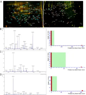
Fig 1. Representative 2D-DIGE, PMF and Mascot score maps of L428 cells and A20 cells. (A)Left panel: Protein spots expressed differentially between L428-CD99cells and L428-CTR cells were marked on the maps and further identified by MALDI-TOF MS. Right panel: Protein spots expressed differentially between A20-mCD99L2- cells and A20-CTR cells were marked on the maps and further identified by MALDI-TOF MS. (B)Left panel: MALDI-TOF mass spectrum of spot B15 obtained from L428 cells (spot B15 corresponds to SEPTIN2) after trypsin digestion. Right panel: The Mascot score of spot B15 confirmed by MALDI-TOF MS was 292. (C)Left panel: MALDI-TOF mass spectrum of spot A8 obtained from A20 cells (spot A8
corresponds toSEPTIN2) after trypsin digestion. Right panel: The Mascot score of spot A8 confirmed by MALDI-TOF MS was 58.(D)Left panel: MALDI-TOF mass spectrum of spot A28 obtained from A20 cells (spot A28 corresponds toSTATHMIN) after trypsin digestion. Right panel: The Mascot score of spot A28 confirmed by MALDI-TOF MS was 156.
Differential expression of
SEPTIN2
and
STATHMIN
in cHL and RH
tissues
The foregoing results suggest that upregulation ofCD99leads to decreasedSEPTIN2and in-creasedSTATHMINlevels in L428 cells. Therefore, we examined the expression ofSEPTIN2
andSTATHMINin cHL (n = 20) and RH (n = 5) tissues by IHC. Representative examples of positive immunostaining are shown inFig 3. Strong positive staining forSEPTIN2and
STATHMINwas detected in 4/20 and 8/20 cHL samples, respectively (S9 Table). In RH tissues, strongSEPTIN2staining was not detected in any of the 5 cases; however, moderately positive staining was detected in 4/5 (80%) and weakly positive was found in 1/5 (20%). Strong
Fig 2. Expression ofSEPTIN2andSTATHMINin L428-CD99and L428-CTR cells.Expression levels of SEPTIN2andSTATHMINwere detected by qRT-PCR (A), Western blot (B), ICC (C), and Confocal microscopy (D, E) (original magnification ×400).
doi:10.1371/journal.pone.0127568.g002
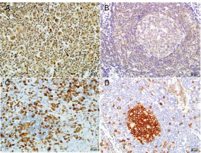
Fig 3. Detection ofSEPTIN2andSTATHMINby IHC in cHL and RH tissues.(A) Expression ofSEPTIN2 in cHL. (B) Expression ofSEPTIN2in RH. (C) Expression ofSTATHMINin cHL. (D) Expression ofSTATHMIN in RH. Original magnification ×400. Positive staining appears reddish brown.
STATHMINstaining was detected in 3/5 cases (60%), with the remainder (40%) showing mod-erate to intermediate staining.
Moreover, in RH tissues, there was strong staining forSTATHMINin the GC, and less dense, more scattered staining in the mantle zones. UnlikeSTATHMIN,SEPTIN2showed weak staining in the GC, but stronger expression in the mantle zones. In capp:ds:hodgkin lym-phoma(hl)HL tissues, histologically-confirmed H/RS cells showed strong to intermediate
STATHMINexpression, and variable backgroundSTATHMINexpression. In contrast, SEP-TIN2showed intermediate to strong expression in the cytoplasm of H/RS cells, but slightly stronger expression in background Non-H/RS cells. We also observed stronger staining for
STATHMINin the RH cases than in the cHapp:ds:hodgkin lymphoma(hl)L, but weaker ex-pression ofSEPTIN2.
SiRNA silencing of
SEPTIN2
results in cytoskeletal reorganization
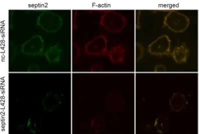
Bioinformatics analysis indicated thatSEPTIN2was associated with cytoskeletal organization (S6andS8Tables). The differential expression ofSEPTIN2in L428-CTR and L428-CD99cells suggests thatSEPTIN2might play a role in the regulation of the cytoskeletal dynamics in cHL. To test this hypothesis, we silencedSEPTIN2with siRNA and visualizedSEPTIN2andF-actin
in L428 cells by immunofluorescence. As shown inFig 4,SEPTIN2colocalized withF-actinin the cytoplasm of the control cells with a punctate distribution at the cell-substratum interface. SiRNA-mediated downregulation ofSEPTIN2reduced the expression ofSEPTIN2andF-actin
in L428 cells. Moreover,SEPTIN2downregulation led to fewer filopodia and thinner cortical filamentous actin at the cell surface. Thus, downregulation ofSEPTIN2may induce cytoskeletal rearrangement in L428 cells. The findings were consistent with our previous study showing thatCD99-upregulated L428 cells underwent terminal B-cell differentiation and displayed cy-toskeleton reorganization, disappearance of filopodia and thinning of cortical filamentous actin [13]. TheCD99-upregulated L428 cells showed reduced expression ofSEPTIN2. Taken together, these results suggest thatSEPTIN2plays a role in maintaining H/RS cell morphology andSEPTIN2-induced cytoskeletal reorganization may contribute toCD99-mediated differen-tiation of H/RS cells.
Fig 4. Transfection ofSEPTIN2siRNA resulted in cytoskeleton change in L428 cells.The Rhodamine-phalloidin was applied for visualizingF-actin. The overlay shows co-localization in yellow.
Dynamic expression of
STATHMIN
is correlated with B-cell lymphoma
differentiation
STATHMINplays an important role in cellular differentiation [22–24]. We previously showed that overexpression ofCD99promoted the differentiation of lymphoma cells into terminal B-cells [13]. The differential expression ofSTATHMINin L428-CTR and L428-CD99cells sug-gests thatSTATHMINmay contribute to the differentiation of HL. To test this, we evaluated
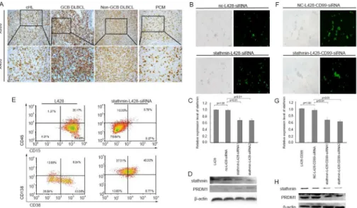
STATHMINexpression by IHC in 65 lymphoid neoplasms, including 20 cHL (S10 Table). In RH, the majority of theSTATHMIN-positive cells were located in the GC, and only occasional-ly found in the interfollicular areas. In cHL, strong to intermediate staining ofSTATHMINwas detected in H/RS cells. Increasingly variable expression levels were found in other lymphoid cells. DLBCL were also stained positive forSTATHMINwith varying degrees of intensity. The staining intensity ofSTATHMINin DLBCL derived from GCB cells and plasmacytomas (PCM) was stronger than that in the non-GCB cells (Fig 5A). These data demonstrate that
STATHMINexpression decreases from the GC to GC stages, and increases from the post-GC to plasma stages. Our results were consistent with the previous finding thatSTATHMIN
was involved in B-cell lymphoma differentiation [25].
Downregulation of
STATHMIN
induces H/RS cell differentiation toward
plasma cells
To further confirm the role ofSTATHMINin HL differentiation, we downregulated STATH-MINby siRNA in L428 cells (Fig 5B and 5C) and assessed the expression ofPRDM1, which plays an important role in terminal differentiation of B-cells into immunoglobulin
(Ig)-Fig 5.STATHMINwas involved in the differentiation of B-cells.(A) IHC analysis ofSTATHMIN expression in different types of lymphomas (cHL, GCB-DLBCL, Non-GCB DLBCL, and PCM) (B)
Morphological images of L428 cells transfected withSTATHMIN- siRNA. Left panel: light image, Right panel: fluorescent image. Original magnification ×100. (C) Relative expression level ofSTATHMINin L428 transfected withSTATHMIN-siRNA for 24h by qRT-PCR. (D) Western blot ofPRDM1expression in L428 cells transfected withSTATHMIN-siRNA for 96h. (E) Flow cytometry analysis of the surface expression of B-cell differentiation antigens in L428 B-cells transfected withSTATHMIN-siRNA for 96h. (F) Morphological images of L428-CD99cells transfected withSTATHMIN-siRNA. Left panel: light image, Right panel: fluorescent image. Original magnification ×100. (G) Relative expression levels ofSTATHMINin L428-CD99 transfected withSTATHMIN-siRNA for 24h by qRT-PCR. (H) Western blot ofPRDM1expression in L428-CD99cells transfected withSTATHMIN-siRNA for 96h.
secreting plasmablasts and plasma cells [26]. SiRNA-mediatedSTATHMINdownregulation led to increase inPDRM1protein level (Fig 5D). We next evaluated the expression of B-cell dif-ferentiation markers using flow cytometry. As shown inFig 5E, the percentage of cells express-ing CD15, the diagnostic marker for cHL, was obviously reduced inSTATHMIN-L428-siRNA cells compared with the control cells. Consistent with the role forSTATHMINin B-cell differ-entiation, plasma cell phenotypic markers CD38 and CD138 were increased following STATH-MINsilencing. We also silencedSTATHMINin L428-CD99cells (Fig 5F and 5G). In sharp contrast to L428-CD99cells,PRDM1protein level was reduced after silencing ofSTATHMIN
in L428-CD99cells (Fig 5H). Taken together, these results suggestSTATHMINinduced differ-entiation of H/RS cells to plasma cells.
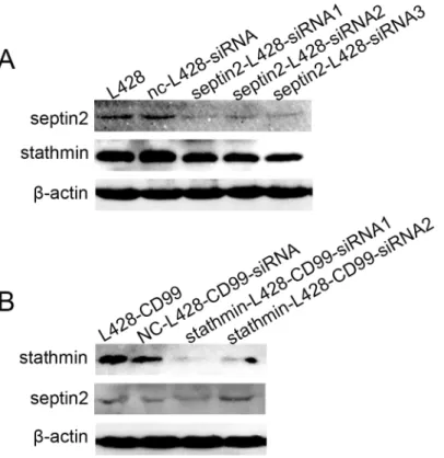
SEPTIN2
silencing leads to downregulation of
STATHMIN
Our results clearly showed thatSEPTIN2andSTATHMINwere closely associated withCD99
expression, suggesting a potential relationship betweenSEPTIN2andSTATHMIN. Therefore, we examined the expression of bothSEPTIN2andSTATHMINin L428 cells after downregula-tion ofSEPTIN2with siRNA and in L428-CD99cells following downregulation ofSTATHMIN
with siRNA, respectively. Surprisingly,STATHMINexpression was reduced when the expres-sion ofSEPTIN2was decreased in L428 cells (Fig 6A). In contrast, almost no change in SEP-TIN2levels was observed in the L428-CD99cells followingSTATHMINdownregulation (Fig 6B). These results indicate thatSEPTIN2regulatesSTATHMIN. The mechanism remains to be determined.
Fig 6. Correlation betweenSEPTIN2andSTATHMINprotein expression.(A) Protein expression levels of SEPTIN2andSTATHMINin L428 cells transfected withSEPTIN2-siRNAs. (B) Protein expression levels of SEPTIN2andSTATHMINin L428-CD99cells transfected withSTATHMIN-siRNAs.
Discussion
In this study, we elucidated the molecular mechanisms ofCD99-mediated H/RS cellular differ-entiation, using 2D-DIGE and MALDI-TOF MS analyses and identified proteins that were dif-ferentially expressed in L428-CD99vs L428-CTR cells and A20-mCD99-L2- vs A20-CTR cells.
SEPTIN2andSTATHMIN, which are involved in cytoskeleton organization, were found to be significantly expressed (S6andS8Tables), and were selected for further validation and functional analysis.
SEPTIN2is a member of the septin family that is involved in many cellular functions, such as cell polarity, cell cortex compartmentalization, vesicle transport, and regulation of the actin and tubulin cytoskeleton [27]. Septins are highly expressed in many tumors [28]. Moreover, septins are also associated with a filament-forming cytoskeletal GTPase, which is required for normal organization of the actin cytoskeleton [29]. Actin cytoskeleton not only plays a pivotal role in the regulation of the morphology and apoptosis of B-cells [30], but also mediates the BCR signal transmission during B-cell differentiation [31]. Depolymerization of the actin cyto-skeleton inhibits surface BCR clustering [32] and induces a signaling cascade in the absence of antigen [33]. In addition, evidence suggests thatSEPTINSplay an important role in cytoskeletal dynamics.SEPTIN-F-actinlinkage may contribute to higher-order organization of the cortical cytoskeleton [34,35]. The relationship betweenSEPTINfilaments and actin has been well es-tablished, however, very little is known about its role in HL and H/RS cell differentiation. In this study, we found thatSEPTIN2expression was negatively correlated withCD99and SEP-TIN2downregulation triggered the reduction ofF-actinand pseudopodia apophysis in L428 cells. These results were consistent with the previous finding that upregulation ofCD99altered cytoskeleton in L428 cells [13]. These data indicate thatSEPTIN2plays an important role in maintaining H/RS cell shape. Thus,SEPTIN2-mediated cytoskeleton reorganization may be one of the induction mechanisms of H/RS cellular differentiation byCD99overexpression. Nevertheless, the relationship betweenSEPTIN2and BCR is unknown and needs
further investigation.
STATHMINis another cytoskeletal protein, which is overexpressed in several malignancies with a significant role in cell differentiation [23]. Reducing the level ofSTATHMINreverses many phenotypes associated with transformation [36]. Moreover,STATHMINis a highly con-served cytosolic phosphoprotein implicated in regulating microtubule dynamics [37,38], cell proliferation and differentiation [39–42]. Nylander et al. reported thatSTATHMINexhibited variable expression in malignant lymphomas and proposed that it may be involved in B-cell differentiation [25]. Our results obtained from IHC support this idea. However, whether
STATHMINparticipates inCD99-mediated H/RS cell differentiation toward terminal B-cells is unclear.
Interestingly, the present study demonstrated that downregulation ofSTATHMINin L428 cells resulted in reduction of CD15, a characteristic marker of H/RS cells [43], and expression of plasma-cell markers CD38 and CD138. CD38 expression is tightly regulated during B-cell ontogenesis and is present at high levels in terminally differentiated plasma cells [44]. Further-more,PRDM1is a necessary and crucial factor in the regulation of B-cell differentiation toward plasma cells [26]. The results showed that silencing ofSTATHMINincreased the expression of
PRDM1in L428 cells. Our results suggested that downregulation ofSTATHMINmay stimulate L428 cellular differentiation toward plasmablasts or plasma cells (terminal B-cells). In addition, we investigated the role ofSTATHMIN-mediated differentiation inCD99cells, by silencing
STATHMINwas involved in the regulation ofCD99-mediated differentiation of H/RS cells to-ward terminal B-cells.
In addition, our previous study demonstrated that ectopic overexpression ofCD99resulted in cell growth inhibition in L428 cells, but the underling mechanism was unclear [13]. It was re-ported thatSTATHMINregulates dynamics of the mitotic spindle by promoting microtubule depolymerization [45]. When cells enter mitosis, phosphorylation-dependent inactivation of
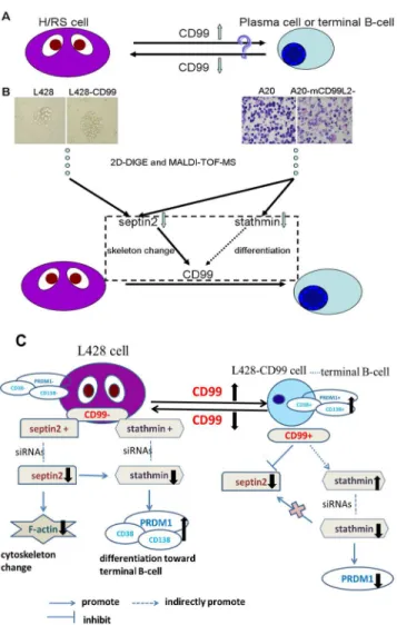
Fig 7. Summary figure.(A)CD99downregulation leads to the transformation of murine B lymphoma cells (A20) into cells with a H/RS phenotype, whereasCD99upregulation induces differentiation of classical Hodgkin’s lymphoma (cHL) cells (L428) into terminal B-cells. (B) 2D-DIGE and MALDI-TOF MS identified differentially expressed proteins respectively inCD99upregulation (L428-CD99)vs mock L428 cells, and in mouseCD99antigen-like 2 (mCD99L2) downregulation (A20-mCD99L2-) vs mock A20 cells.SEPTIN2and STATHMINwere chosen for further study. We found thatSEPTIN2induced the cellular cytoskeleton reorganization in L428 cells and downregulation ofSTATHMINinduced L428 cells differentiation toward terminal B-cells, which partially explained the observation that upregulation ofCD99induced H/RS cells to differentiate toward terminal B-cell. (C) Schematic model of the regulation of genes expression in L428 and L428-CD99cells. Low expression ofCD99and high expression ofSEPTIN2andSTATHMINwere present in L428 cells. Downregulation ofSEPTIN2with siRNAs in L428 cells induced change ofF-actinexpression. Downregulation ofSTATHMINwith siRNAs in L428 cells increased the expression ofPRDM1, CD38 and CD138, which suggests the treated cell were differentiated toward terminal B-cell. Upregulation ofCD99in L428 cells decreasedSEPTIN2while increasedSTATHMIN. Downregulation ofSTATHMINin L428-CD99 cells with siRNAs reduced the expression ofPRDM1.
STATHMINallows microtubules to polymerize and assemble into a mitotic spindle. The mi-crotubule-depolymerizing activity ofSTATHMINmust be restored in the later phases of mito-sis to allow mitotic spindle disassembly and proper exit from mitomito-sis [37]. Both overexpression and downregulation ofSTATHMINresults in mitotic spindle abnormalities and accumulation of cells in G2/M phases of the cell cycle [46]. Therefore, we speculate that downregulation of
STATHMINin L428-CD99cells may contribute to its growth inhibition.
To the best of our knowledge, we are the first to report the expression ofSEPTIN2in cHL tissues. The staining intensity ofSEPTIN2in the cytoplasm of H/RS cells was weaker than in other inflammatory cells; whereas in RH tissues,SEPTIN2was expressed at higher levels in the mantle zone than in the GC. These results suggest that the patterns ofSEPTIN2expression in RH and cHL were contrary toSTATHMINexpression, for unknown reasons.
Furthermore, several proteins known to be involved in cHL likeARHGDIB,MSH2or
PRDX2were found differentially expressed in L428 cells as well as in mCD99L2 downregulated A20 cells (S5andS7Tables). Evidence shows that the guanosine triphosphatase (GTPase) in-hibitorARHGDIBis downregulated in H/RS cells and the absence ofARHGDIBmight contrib-ute to the apoptotic resistance of H/RS [47].PRDX2is also downregulated in H/RS cells and epigenetic silencing of this gene may contribute to the loss of B-cell identity and survival of H/ RS cells [48].MSH2transcript is present in most B-cell lymphoma with the exception of plas-ma cell lymphoplas-ma and deregulation ofMSH2in B-cell lymphoma types is characterized by ag-gressive biologic behavior [49]. In this study, proteomic analysis showed upregulation ofCD99
in L428 cells led to high expression ofARHGDIBandPRDX2, while low expression ofMSH2. These results suggest thatARHGDIB,PRDX2andMSH2may play a role inCD99-induced transformation of H/RS cells toward B-cell.
In summary, we characterized the expression pattern ofSEPTIN2in cHL and provided evi-dence thatSEPTIN2-mediated cytoskeleton reorganization plays an important role in H/RS cell differentiation. Furthermore, we found thatCD99induced the transformation of H/RS cells toward B-cell by regulating the expression ofSEPTIN2andSTATHMIN(Fig 7). The pres-ent study provides novel insights into the mechanisms underlyingCD99-mediated H/RS cell differentiation toward B-cells.
Supporting Information
S1 Fig. Representative DIGE fluorescence images.(A-D) Representative DIGE images of L428-CD99and L428-CTR cells. (A) Cy3-labeled images of L428-CD99cells. (B) Cy5-labeled images of L428-CTR cells. (C) Internal images labeled with Cy2. (D) Merged images of the Cydye-labeled images. (E-H) Representative DIGE images of A20-mCD99L2- and A20-CTR cells. (E) Cy3-labeled images of A20-mCD99L2- cells. (F) Cy5-labeled images of A20-CTR cells. (G) Internal images labeled by Cy2. (H) Merged images of the Cydye-labeled images. (TIF)
S2 Fig. Gene ontology analysis.(A) Biological process annotation. (B) Cellular component an-notation. (C) Molecular function anan-notation.
(TIF)
S3 Fig. Relative expression level ofSTATHMINwhen L428 or L428-CD99cells were treated withSTATHMIN-siRNA for 72h.Left panel: relative expression level ofSTATHMINin L428
cells transfected withSTATHMIN-siRNA for 72h by qRT-PCR. Right panel: relative expression levels ofSTATHMINin L428-CD99cells transfected withSTATHMIN-siRNA for 72h by qRT-PCR.
S1 Table. SiRNA sequences used in transfection experiments.
(XLSX)
S2 Table. Primers used in qRT-PCR analysis.
(XLSX)
S3 Table. Antibodies used for IHC, ICC, western blot, immunofluorescence analysis.
(XLSX)
S4 Table. Antibodies used for flow cytometry analysis.
(XLSX)
S5 Table. MALDI-TOF MS identification of 38 characteristic proteins with differential ex-pression in L428-CD99vs L428-CTR cells.
(XLSX)
S6 Table. Closely related proteins involved in theCD99regulation of cell transformation.
(XLSX)
S7 Table. MALDI-TOF MS identification of 41 characteristic proteins with differential ex-pression levels in A20-mCD99L2- vs A20-CTR cells.
(XLSX)
S8 Table. Closely related proteins involved in the mCD99L2 regulation of cell transforma-tion.
(XLSX)
S9 Table. IHC analyses ofSEPTIN2andSTATHMINin cHL and RH.
(XLSX)
S10 Table. IHC analyses ofSTATHMINin lymphoma subtypes.
(XLSX)
S1 Text. Relationship betweenCD99andSTATHMINwas tested in the L428-CD99cells and BJAB cells.
(DOC)
Acknowledgments
We thank the Nanfang Hospital Affiliated to Southern Medical University for providing paraf-fin-embedded specimens for this study. We also thank Dr. Chan (the Nebraska Medical Cen-ter, Omaha, NE, USA) for generously providing cell lines L428 and A20.
Author Contributions
Conceived and designed the experiments: XHZ TZ. Performed the experiments: LZ. Analyzed the data: JW YT. Contributed reagents/materials/analysis tools: BQ ZQW JHY. Wrote the paper: WJJ XHZ TZ. Performed the supplemental experiments: WJJ LZ.
References
1. Kuppers R. The biology of Hodgkin's lymphoma. Nature reviews Cancer. 2009; 9(1):15–27. doi:10. 1038/nrc2542PubMed PMID:19078975.
2. Farrell K, Jarrett RF. The molecular pathogenesis of Hodgkin lymphoma. Histopathology. 2011; 58 (1):15–25. doi:10.1111/j.1365-2559.2010.03705.xPubMed PMID:21261680.
4. van den Berg A, Visser L, Poppema S. High expression of the CC chemokine TARC in Reed-Sternberg cells. A possible explanation for the characteristic T-cell infiltratein Hodgkin's lymphoma. The American journal of pathology. 1999; 154(6):1685–91. doi:10.1016/S0002-9440(10)65424-7PubMed PMID: 10362793; PubMed Central PMCID: PMC1876772.
5. Re D, Muschen M, Ahmadi T, Wickenhauser C, Staratschek-Jox A, Holtick U, et al. Oct-2 and Bob-1 deficiency in Hodgkin and Reed Sternberg cells. Cancer research. 2001; 61(5):2080–4. PubMed PMID:11280769.
6. Melamed I, Downey GP, Aktories K, Roifman CM. Microfilament assembly is required for antigen-re-ceptor-mediated activation of human B lymphocytes. Journal of immunology. 1991; 147(4):1139–46. PubMed PMID:1907989.
7. Buettner M, Greiner A, Avramidou A, Jack HM, Niedobitek G. Evidence of abortive plasma cell differen-tiation in Hodgkin and Reed-Sternberg cells of classical Hodgkin lymphoma. Hematological oncology. 2005; 23(3–4):127–32. doi:10.1002/hon.764PubMed PMID:16342298.
8. Dworzak MN, Fritsch G, Buchinger P, Fleischer C, Printz D, Zellner A, et al. Flow cytometric assess-ment of human MIC2 expression in bone marrow, thymus, and peripheral blood. Blood. 1994; 83 (2):415–25. PubMed PMID:7506950.
9. Lin O, Filippa DA, Teruya-Feldstein J. Immunohistochemical evaluation of FLI-1 in acute lymphoblastic lymphoma (ALL): a potential diagnostic pitfall. Applied immunohistochemistry & molecular morphology: AIMM / official publication of the Society for Applied Immunohistochemistry. 2009; 17(5):409–12. doi: 10.1097/PAI.0b013e3181972b6dPubMed PMID:19349856.
10. Isobe K, Tamaru J, Uno T, Yasuda S, Aruga T, Itoyama S, et al. Immunoglobulin heavy chain variable region (VH) genes of B cell chronic lymphocytic leukemia cells from lymph nodes show somatic muta-tions and intraclonal diversity irrespective of follicular dendritic cell network. Leukemia & lymphoma. 2001; 42(3):499–506. doi:10.3109/10428190109064607PubMed PMID:11699415.
11. Lee IS, Kim SH, Song HG, Park SH. The molecular basis for the generation of Hodgkin and Reed-Sternberg cells in Hodgkin's lymphoma. International journal of hematology. 2003; 77(4):330–5. PubMed PMID:12774919.
12. Kim SH, Choi EY, Shin YK, Kim TJ, Chung DH, Chang SI, et al. Generation of cells with Hodgkin's and Reed-Sternberg phenotype through downregulation of CD99 (Mic2). Blood. 1998; 92(11):4287–95. PubMed PMID:9834235.
13. Huang X, Zhou X, Wang Z, Li F, Liu F, Zhong L, et al. CD99 triggers upregulation of miR-9-modulated PRDM1/BLIMP1 in Hodgkin/Reed-Sternberg cells and induces redifferentiation. International journal of cancer Journal international du cancer. 2012; 131(4):E382–94. doi:10.1002/ijc.26503PubMed PMID: 22020966.
14. Liu F, Zhang G, Liu F, Zhou X, Chen X, Han X, et al. Effect of shRNA targeting mouse CD99L2 gene in a murine B cell lymphoma in vitro and in vivo. Oncology reports. 2013; 29(4):1405–14. doi:10.3892/or. 2013.2244PubMed PMID:23338758.
15. Suh YH, Shin YK, Kook MC, Oh KI, Park WS, Kim SH, et al. Cloning, genomic organization, alternative transcripts and expression analysis of CD99L2, a novel paralog of human CD99, and identification of evolutionary conserved motifs. Gene. 2003; 307:63–76. PubMed PMID:12706889.
16. Kim KJ, Kanellopoulos-Langevin C, Merwin RM, Sachs DH, Asofsky R. Establishment and characteri-zation of BALB/c lymphoma lines with B cell properties. Journal of immunology. 1979; 122(2):549–54. PubMed PMID:310843.
17. Passineau MJ, Siegal GP, Everts M, Pereboev A, Jhala D, Wang M, et al. The natural history of a novel, systemic, disseminated model of syngeneic mouse B-cell lymphoma. Leukemia & lymphoma. 2005; 46(11):1627–38. PubMed PMID:16334907.
18. Montes-Moreno S, Roncador G, Maestre L, Martinez N, Sanchez-Verde L, Camacho FI, et al. Gcet1 (centerin), a highly restricted marker for a subset of germinal center-derived lymphomas. Blood. 2008; 111(1):351–8. doi:10.1182/blood-2007-06-094151PubMed PMID:17898315.
19. Rui L, Emre NC, Kruhlak MJ, Chung HJ, Steidl C, Slack G, et al. Cooperative epigenetic modulation by cancer amplicon genes. Cancer cell. 2010; 18(6):590–605. doi:10.1016/j.ccr.2010.11.013PubMed PMID:21156283; PubMed Central PMCID: PMC3049192.
20. Liu F, Zhang Y, Tang H, Zhou X, Wu Z, Tang D, et al. CXC chemokine ligand 16, inversely correlated with CD99 expression in Hodgkin Reed-Sternberg cells, is widely expressed in diverse types of lym-phomas. Oncology reports. 2013; 30(2):783–92. doi:10.3892/or.2013.2522PubMed PMID:23743627. 21. Iancu-Rubin C, Nasrallah CA, Atweh GF. Stathmin prevents the transition from a normal to an
endomi-totic cell cycle during megakaryocytic differentiation. Cell cycle. 2005; 4(12):1774–82. PubMed PMID: 16258287.
(17):4580–9. doi:10.1182/blood-2010-09-305540PubMed PMID:21364187; PubMed Central PMCID: PMC3099574.
23. Doye V, Kellermann O, Buc-Caron MH, Sobel A. High expression of stathmin in multipotential teratocar-cinoma and normal embryonic cells versus their early differentiated derivatives. Differentiation; re-search in biological diversity. 1992; 50(2):89–96. PubMed PMID:1323493.
24. Gratiot-Deans J, Keim D, Strahler JR, Turka LA, Hanash S. Differential expression of Op18 phospho-protein during human thymocyte maturation. The Journal of clinical investigation. 1992; 90(4):1576–81. doi:10.1172/JCI116026PubMed PMID:1401087; PubMed Central PMCID: PMC443205.
25. Nylander K, Marklund U, Brattsand G, Gullberg M, Roos G. Immunohistochemical detection of onco-protein 18 (Op18) in malignant lymphomas. The Histochemical journal. 1995; 27(2):155–60. PubMed PMID:7775200.
26. Calame K. Activation-dependent induction of Blimp-1. Current opinion in immunology. 2008; 20 (3):259–64. doi:10.1016/j.coi.2008.04.010PubMed PMID:18554885.
27. Hall PA, Russell SE. The pathobiology of the septin gene family. The Journal of pathology. 2004; 204 (4):489–505. doi:10.1002/path.1654PubMed PMID:15495264.
28. Liu M, Shen S, Chen F, Yu W, Yu L. Linking the septin expression with carcinogenesis. Molecular biolo-gy reports. 2010; 37(7):3601–8. doi:10.1007/s11033-010-0009-2PubMed PMID:20195767.
29. Thavarajah R, Vidya K, Joshua E, Rao UK, Ranganathan K. Potential role of septins in oral carcinogen-esis: An update and avenues for future research. Journal of oral and maxillofacial pathology: JOMFP. 2012; 16(1):73–8. doi:10.4103/0973-029X.92977PubMed PMID:22438646; PubMed Central PMCID: PMC3303527.
30. Melamed I, Gelfand EW. Microfilament assembly is involved in B-cell apoptosis. Cellular immunology. 1999; 194(2):136–42. doi:10.1006/cimm.1999.1506PubMed PMID:10383816.
31. Maus M, Medgyesi D, Kiss E, Schneider AE, Enyedi A, Szilagyi N, et al. B cell receptor-induced Ca2+ mobilization mediates F-actin rearrangements and is indispensable for adhesion and spreading of B lymphocytes. Journal of leukocyte biology. 2013; 93(4):537–47. doi:10.1189/jlb.0312169PubMed PMID:23362305.
32. Fleire SJ, Goldman JP, Carrasco YR, Weber M, Bray D, Batista FD. B cell ligand discrimination through a spreading and contraction response. Science. 2006; 312(5774):738–41. doi:10.1126/science. 1123940PubMed PMID:16675699.
33. Treanor B, Batista FD. Organisation and dynamics of antigen receptors: implications for lymphocyte signalling. Current opinion in immunology. 2010; 22(3):299–307. doi:10.1016/j.coi.2010.03.009 PubMed PMID:20434893.
34. Weirich CS, Erzberger JP, Barral Y. The septin family of GTPases: architecture and dynamics. Nature reviews Molecular cell biology. 2008; 9(6):478–89. doi:10.1038/nrm2407PubMed PMID:18478031. 35. Piekny AJ, Maddox AS. The myriad roles of Anillin during cytokinesis. Seminars in cell & developmental
biology. 2010; 21(9):881–91. doi:10.1016/j.semcdb.2010.08.002PubMed PMID:20732437.
36. Jeha S, Luo XN, Beran M, Kantarjian H, Atweh GF. Antisense RNA inhibition of phosphoprotein p18 ex-pression abrogates the transformed phenotype of leukemic cells. Cancer research. 1996; 56(6):1445– 50. PubMed PMID:8640838.
37. Gavet O, Ozon S, Manceau V, Lawler S, Curmi P, Sobel A. The stathmin phosphoprotein family: intra-cellular localization and effects on the microtubule network. Journal of cell science. 1998; 111 (Pt 22):3333–46. PubMed PMID:9788875.
38. Charbaut E, Curmi PA, Ozon S, Lachkar S, Redeker V, Sobel A. Stathmin family proteins display spe-cific molecular and tubulin binding properties. The Journal of biological chemistry. 2001; 276 (19):16146–54. doi:10.1074/jbc.M010637200PubMed PMID:11278715.
39. Sobel A, Boutterin MC, Beretta L, Chneiweiss H, Doye V, Peyro-Saint-Paul H. Intracellular substrates for extracellular signaling. Characterization of a ubiquitous, neuron-enriched phosphoprotein (stath-min). The Journal of biological chemistry. 1989; 264(7):3765–72. PubMed PMID:2917975.
40. Holmfeldt P, Larsson N, Segerman B, Howell B, Morabito J, Cassimeris L, et al. The catastrophe-pro-moting activity of ectopic Op18/stathmin is required for disruption of mitotic spindles but not interphase microtubules. Molecular biology of the cell. 2001; 12(1):73–83. PubMed PMID:11160824; PubMed Central PMCID: PMC30569.
41. Alli E, Bash-Babula J, Yang JM, Hait WN. Effect of stathmin on the sensitivity to antimicrotubule drugs in human breast cancer. Cancer research. 2002; 62(23):6864–9. PubMed PMID:12460900. 42. Baldassarre G, Belletti B, Nicoloso MS, Schiappacassi M, Vecchione A, Spessotto P, et al.
43. Hertel CB, Zhou XG, Hamilton-Dutoit SJ, Junker S. Loss of B cell identity correlates with loss of B cell-specific transcription factors in Hodgkin/Reed-Sternberg cells of classical Hodgkin lymphoma. Onco-gene. 2002; 21(32):4908–20. doi:10.1038/sj.onc.1205629PubMed PMID:12118370.
44. Malavasi F, Deaglio S, Funaro A, Ferrero E, Horenstein AL, Ortolan E, et al. Evolution and function of the ADP ribosyl cyclase/CD38 gene family in physiology and pathology. Physiological reviews. 2008; 88(3):841–86. doi:10.1152/physrev.00035.2007PubMed PMID:18626062.
45. Rubin CI, Atweh GF. The role of stathmin in the regulation of the cell cycle. Journal of cellular biochem-istry. 2004; 93(2):242–50. doi:10.1002/jcb.20187PubMed PMID:15368352.
46. Iancu C, Mistry SJ, Arkin S, Wallenstein S, Atweh GF. Effects of stathmin inhibition on the mitotic spin-dle. Journal of cell science. 2001; 114(Pt 5):909–16. PubMed PMID:11181174.
47. Ma L, Xu G, Sotnikova A, Szczepanowski M, Giefing M, Krause K, et al. Loss of expression of LyGDI (ARHGDIB), a rho GDP-dissociation inhibitor, in Hodgkin lymphoma. British journal of haematology. 2007; 139(2):217–23. doi:10.1111/j.1365-2141.2007.06782.xPubMed PMID:17897297.
48. Schneider M, Szaumkessel M, Richter J, Ammerpohl O, Hansmann ML, Kuppers R, et al. The PRDX2 gene is transcriptionally silenced and de novo methylated in Hodgkin and Reed-Sternberg cells of clas-sical Hodgkin lymphoma. Blood. 2014; 123(23):3672–4. doi:10.1182/blood-2014-02-553263PubMed PMID:24904103.