Functional Impact of Corticotropin-Releasing
Factor Exposure on Tau Phosphorylation and
Axon Transport
Michelle H. Le1, April M. Weissmiller1, Louise Monte1, Po Han Lin1, Tia C. Hexom1, Orlangie Natera1, Chengbiao Wu1, Robert A. Rissman1,2*
1Department of Neurosciences, University of California San Diego, La Jolla, CA 92093, United States of America,2Veterans Affairs San Diego Healthcare System, San Diego, CA 92161, United States of America
Abstract
Stress exposure or increased levels of corticotropin-releasing factor (CRF) induce hippo-campal tau phosphorylation (tau-P) in rodent models, a process that is dependent on the type-1 CRF receptor (CRFR1). Although these preclinical studies on stress-induced tau-P provide mechanistic insight for epidemiological work that identifies stress as a risk factor for Alzheimer’s disease (AD), the actual impact of stress-induced tau-P on neuronal function remains unclear. To determine the functional consequences of stress-induced tau-P, we developed a novel mouse neuronal cell culture system to explore the impact of acute (0.5hr) and chronic (2hr) CRF treatment on tau-P and integral cell processes such as axon transport. Consistent with in vivo reports, we found that chronic CRF treatment increased tau-P levels and caused globular accumulations of phosphorylated tau in dendritic and axo-nal processes. Furthermore, while both acute and chronic CRF treatment led to significant reduction in CREB activation and axon transport of brain-derived neurotrophic factor (BDNF), this was not the case with mitochondrial transport. Acute CRF treatment caused increased mitochondrial velocity and distance traveled in neurons, while chronic CRF treat-ment modestly decreased mitochondrial velocity and greatly increased distance traveled. These results suggest that transport of cellular energetics may take priority over growth fac-tors during stress. Tau-P was required for these changes, as co-treatment of CRF with a GSK kinase inhibitor prevented CRF-induced tau-P and all axon transport changes. Collec-tively, our results provide mechanistic insight into the consequences of stress peptide-induced tau-P and provide an explanation for how chronic stress via CRF may lead to neu-ronal vulnerability in AD.
Introduction
Alzheimer’s disease (AD) is dementia disorder characterized by extensive synaptic and neuro-nal loss, extracellular amyloid beta (Aβ) plaques and intracellular neurofibrillary tangles
OPEN ACCESS
Citation:Le MH, Weissmiller AM, Monte L, Lin PH, Hexom TC, Natera O, et al. (2016) Functional Impact of Corticotropin-Releasing Factor Exposure on Tau Phosphorylation and Axon Transport. PLoS ONE 11 (1): e0147250. doi:10.1371/journal.pone.0147250
Editor:Andrey E Ryabinin, Oregon Health and Science University, UNITED STATES
Received:October 27, 2015
Accepted:January 3, 2016
Published:January 20, 2016
Copyright:© 2016 Le et al. This is an open access article distributed under the terms of theCreative Commons Attribution License, which permits unrestricted use, distribution, and reproduction in any medium, provided the original author and source are credited.
Data Availability Statement:All relevant data are within the paper and its supporting information files.
(NFTs). The accumulation of Aβplaques is hypothesized to precede dementia in AD (for review, [1]), while synaptic loss and NFT accumulation are thought to occur later and have been found to directly correlate with worsening cognitive impairment [2–4].
NFTs are composed of hyperphosphorylated and aggregated forms of the microtubule asso-ciated protein, tau. Tau is a soluble phospho-protein that exists in multiple isoforms and plays an important role in stabilizing microtubules and in the maintenance of neuronal structure, polarity and axon transport [5–12]. Although some information is known about hyperpho-sphorylated tau in AD NFTs [9,13–15], the precise mechanistic role that tau phosphorylation (tau-P) plays in neuronal compromise has been difficult to pinpoint. One hypothesis in the field suggests that excessive or sustained levels of phosphorylation reduce the ability of tau to bind and stabilize microtubules, leading to tau aggregation, impaired axonal transport and eventually NFTs [16–20].
AD is predominantly a sporadic disease, with less than 2% of cases linked to specific genetic mutations. An extensive literature implicates chronic stress in the development of sporadic AD [21–25] which supports a body of epidemiological work demonstrating that individuals prone to experience psychological distress or anxiety have accelerated rates of cognitive decline and are three times more likely to be diagnosed with AD [26–29]. Furthermore, stress exposure in humans affects learning, memory, hippocampal function and morphology [26,27].
Although corticotropin-releasing factor (CRF) is best recognized as the hypothalamic neu-ropeptide that governs the endocrine stress response [30,31], its distribution and actions in the CNS are similar to that of a neuromodulator/neurotransmitter [32–34]. The CRF family of peptides exert their biological effects via two G protein-coupled receptors (CRFRs) that are positively coupled to adenylate cyclase. CRF binds CRFR1 with high affinity, and in the pitui-tary gland this interaction mediates the neuroendocrine stress response [35]. CRFR1 is widely expressed in the brain, including AD-relevant areas such as the neocortex and hippocampus [36]. CRFR2 is a structurally related receptor but displays very limited CNS distribution [37]. The low affinity of CRF for CRFR2 led to the identification of three additional members of the CRF family, the urocortin (UCN) peptides. UCN 2 and 3 serve as high affinity CRFR2-selective ligands, and UCN 1 binds both receptors with comparably high affinities [38–40].
Supporting the hypothesis of broad central activity of CRF, studies have found prominent changes in the CRF signaling system in brain areas that are vulnerable to AD neuropathology and cell loss [41–43]. In addition to the CRF peptide itself, considerable attention has been focused on stress steroids (e.g. cortisol, corticosterone), effectors of the stress cascade, as media-tors of neuronal vulnerability in AD. Increased circulating levels of cortisol in aged individuals has been linked to brain atrophy and a range of adverse effects in the hippocampus [21,44–
46]. Furthermore, results from clinical trials suggest that corticosteroid treatment worsens behavioral symptoms in AD patients [47]. Taken together, these human studies suggest that alternations occur in CRF/stress circuitry in AD and that these changes may directly impact AD-symptomology and neuropathology.
Work in AD models also supports a role for stress in disease pathogenesis. For example, many AD mouse models display increased anxiety behavior and perturbations in central stress signaling [48–51]. With regard to AD tau neuropathology, an abundance of rodent studies demonstrate that exposure to physiological and emotional stressors induces rapid hippocampal and cortical tau-P (for review, [42]). Accordingly, animal models of chronic stress employing overexpression of CRF and/or exposure to stressors are characterized by changes in synaptic structure, brain atrophy, neurodegeneration, hippocampal-dependent cognitive deficits, tau-P and detergent-soluble pools of phosphorylated tau with definable structure [52–59]. Mechanis-tic studies suggest that stress-induced tau-P can occur in the absence of an increase in stress steroids, but is dependent on CRF signaling through CRFR1 [55–58,60].
Although the human and rodent in vivo data reviewed above suggest important mechanistic roles for CRF, CRFR1 and stress exposure in AD neuronal vulnerability and pathology, the actual cellular changes that lead to these reported detrimental effects have not been elucidated. Here we developed an in vitro stress platform to explore the effects of acute and chronic CRF exposure and tau-P on vital cellular processes such as axonal transport of growth factors and energetics.
Materials and Methods
Neuronal Cultures
Primary hippocampal neuronal cultures were generated from embryos at day 18.5 (E18.5) from timed-pregnant C57BL/6J mice (Charles River Labs) using established methods [61,62]. Dissociated neurons were plated in poly-D-lysine coated 12-well culture plates at a density of 1–1.5 million cells per well for mass cultures, and 0.2 million cells per coverslip for immunocy-tochemistry. Plating density of cultures for all experiments were based on previous reports [63] and our internal testing/validation (data not shown). Plating media was replaced with mainte-nance media (Neurobasal, 1% Glutamax and 2% B27) 2 hours after plating. To maintain maxi-mum viability, cells were maintained in culture for 7 days before experiments and two-thirds of the medium was replaced every other day. All experimental protocols involving the use of animals were approved by University of California, San Diego Institutional Animal Care and Use Committee.
In vitro stress treatment
To establish a time course of CRF-induced tau phosphorylation, cells were treated with 1μM
(low) and 10μM (high) CRF for 0, 0.5, 2, 4, 6, 8 and 24 hours (hr) (n = 3 each), and processed
for Western blot as described below. CRF (Bachem) was reconstituted in 10% ethanol. Dosages of CRF treatment were based on previous reports [63] and our internal testing/validation (data not shown). Based on the results of the time course, 10μM CRF for 0.5hr (acute stress) or 2hr
(chronic stress) was selected for immunocytochemistry and axon transport experiments. To isolate the impact of CRF-induced tau-P on axonal transport, GSK activity was blocked by pre-treating cultures with 25mM Lithium Chloride (Sigma Aldrich) for 30 minutes prior to adding CRF. Vehicle controls were run in parallel for all conditions.
Axonal transport studies
To define the impact of CRF-induced tau-P on axon transport, we elected to examine axonal transport of two types of cargoes: 1) mitochondria that displays both anterograde and retro-grade movement within axons [64] and 2) BDNF signaling in endosomes are initiated from axonal terminals and are retrogradely transported to the soma to deliver trophic support [61,
62].
To separate axons from cell bodies, 450 nm microfluidic chambers (Xona Microfluidics) were prepared using established methods [61,62]. Axons were imaged using an inverted epi-fluorescent microscope equipped with an environmental chamber (37°C, 5% CO2) and a 100x
For mitochondria studies, cells were treated with 10μM CRF and 2.5μM Mitotracker (Life
Technologies) for 30 minutes. Video image series of mitochondria in the distal axons were col-lected at 1 frame/sec for a total 60 sec [62]. For BDNF studies, maintenance media was replaced with Neurobasal media for one hour prior to treatment in order to deplete cells and axons of BDNF. 45 minutes prior to live imaging, 0.2 nM BDNF conjugated to QD655 (Life Technolo-gies) was added to the distal axons.
Western blot analysis
Hippocampal neurons plated in 12-well plates (as described above) were washed with cold phosphate buffered saline (PBS), collected and centrifuged at 1000 rcf for 5 minutes at 4°C. Resultant pellets were lysed in detergent-containing radioimmunoprecipitation assay (RIPA) buffer containing phosphatase inhibitors (1μM okadaic acid, 1 mM Na3VO4) and a protease
inhibitor cocktail (Thermo Scientific) as described [56,57]. Western blot data was analyzed using NIH ImageJ software as described [56,57].
Immunocytochemistry
Hippocampal neurons were plated on poly-L-lysine coated coverslips. After treatment with CRF (10μM for 2hr), cultures were washed with cold PBS, fixed with 4% paraformaldehyde for
10 min, quenched with 0.1 M ammonium chloride, and permeabilized with 0.1% PBS-triton for 10 minutes. Coverslips were blocked with 1% bovine or goat serum albumin in PBS for 30 minutes and then incubated in primary antibodies overnight at 4°C. Secondary antibody (Alexa 488, Life Technologies) was applied the next morning for 1hr at room temperature, fol-lowed by DAPI or Hoechst stain for 10 minutes, and then mounted for imaging. Images were acquired using a Leica DMI6000B inverted microscope with 20x objective lens by scanning the cells directly adjacent to the microgrooves. The threshold for all images was set equally using NIH ImageJ software and the“analyze particles”function was used to automatically count pos-itive nuclei (Hoechst and pCREB experiments).
Antibodies. The well-characterized phospho-specific antibody, PHF-1, was used to detect
tau-P at S396/404(gift of Dr. P. Davies, Albert Einstein College of Medicine). PHF-1 was selected as a representative marker of stress-induced tau phosphorylation based on extensive character-ization in our prior work [56–58,60]. We previously validated PHF-1 for use in mouse tissue treated with alkaline phosphatase, which eliminated detectable PHF-1 labeling [58]. Changes in the GSK, implicated in the phosphorylation of tau at S396/404was also assessed. Activated GSK-3βwas detected using anti-GSK-pY216(BD Bioscience), and inactive GSK-3βwas assessed using anti-GSK-pS9(Cell signaling) [65]. Antibodies targeting the C-terminus ofβ -actin (Sigma Aldrich) were used as a control for protein loading on Western blots. An antibody to pCREB antibody was used to target phosphorylated CREB at S133(Cell Signaling).
Statistical Analysis
Optical density readings from Western blots and data from axon transport experiments were analyzed by one or two way ANOVA using Prism 6 software (GraphPad, San Diego, CA). Data are expressed as mean ± SEM, normalized toβ-actin loading.
Results
Timecourse of CRF-Induced Tau-P
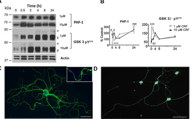
system. Western blot analysis was used to examine levels of tau-P at the AD-relevant C-termi-nal site (PHF-1) in extracts from hippocampal neuroC-termi-nal cultures after 0, 0.5, 2, 4, 8 or 24hr of 1μm (low) or 10μm (high) CRF treatment (Fig 1A and 1B). Compared to vehicle control (V),
neurons treated with either 1μm or 10μm CRF exhibited a significant increase in tau-P
imme-diately after treatment (0hr, p = 0.02), 2hr (p = 0.01) and through 24hr of treatment (p<
0.001). In terms of upstream kinase mediators, we observed parallel changes in activated GSK-3β(pY216), in concordance with the PHF-1 data. A significant increase was observed in active GSK-3βat the 2hr (p<0.001), 4hr (p = 0.01), 8hr and 24hr (each, p<0.001) timepoints with
10μm CRF treatment. No change was observed in the inactive form of GSK-3β(pS9) for any
timepoint after 1μm or 10μm CRF treatment (all p>0.05 compared to vehicle, data not
shown). Because elevation in tau-P was stable overtime, the 10μm CRF treatment and the 2hr
timepoint were selected for immunohistochemistry, and both the 0.5hr and 2hr timepoints were selected to simulate acute and chronic stress, respectively, in axon transport experiments.
Cellular Localization of Tau-P
Immunocytochemical methods were used for qualitative analysis of tau-P as a function of CRF treatment. Consistent with our biochemical data, vehicle-treated cultures showed low basal lev-els of PHF-1 labeling, which was prominently increased with CRF treatment. In vehicle treated cultures, we observed a relatively linear distribution of PHF-1 along neuron projections, consis-tent with basal levels of tau-P in neurons (Fig 1D). However, in CRF-treated cultures, we observed greatly increased intensity of PHF-1 staining and the presence of globular accumula-tions localized in neurites (Fig 1C, inset). These qualitative results suggest that CRF induces
Fig 1. Stress-induced tau-P and kinase activation. (A)Western blot of PHF-1 and GSK-3 pY216in cultured mouse hippocampal cells exposed to low or
high concentrations of stress hormone CRF (1μM or 10μM) over a period of 0, 0.5, 2, 4, 8, or 24 hours withβ-actin as a loading control.(B)Quantitative analysis of western blots (n = 3). Treatments differ significantly from controls; PHF-1†(p = 0.01),†††(p<0.001),***(p<0.001); Active GSK 3βpY216††
(p = 0.006),†††(p<0.001),***(p<0.001).(C)Immunostaining with PHF-1 (green) of neuronal treated with 10μM CRF and(D)vehicle control cell nuclei with DAPI staining (blue).
tau-P in vivo, which accumulates in dendrites and axons, and may cause blockages that are consistent with impaired axon transport.
Impact of CRF-Induced Tau-P on BDNF Axonal Transport
Microtubule-based axonal transport of organelles and other cargoes is a vital process for main-tenance of neuronal structure and function, and synaptic plasticity [66]. Impairment or disrup-tion of axonal transport has been proposed to be an early pathological feature of
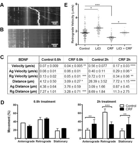
neurodegenerative diseases such as AD [67,68]. Because BDNF plays a crucial role in neuronal development and neuronal plasticity, we examined the effects of CRF-induced tau-P on BDNF transport (Fig 2). Live imaging of Quantum-dot labeled BDNF was performed using our previ-ously published protocols [61,62] and kymographs were generated from time-lapse videos recorded from both vehicle and 10μm CRF-treated neurons (Fig 2A and 2B). Measurements
Fig 2. Impact of stress-induced tau-P on BDNF transport.Overall velocity of QD-BDNF was reduced in 0.5hr CRF cultures (overall velocity**p = 0.03, retrograde***p = 0.0002) and 2hr CRF treatment (****p = 0.008); 2hr CRF treated cultures also exhibited reduced anterograde and retrograde velocity (*p = 0.03,**0.0002, respectively); Distance travelled was reduced in 0.5hr CRF treated cultures (overall *p = 0.04, retrograde***p = 0.01) and 2hr CRF treatment (****p = 0.0001). All compared to vehicle treated controls;(D)Analysis of percent mitochondrial movement at 0.5hr and 2hr revealed no changes with 0.5hr CRF treatment (all p>0.05), though 2hr CRF treatment induced dramatic changes in anterograde (p = 0.001),
retrograde (p = 0.0001) and stationary (p = 0.01) movement;(E)As seen in D, CRF treatment reduced retrograde velocity (***p = 0.02); 25mM LiCl treatment increased basal levels of retrograde velocity in vehicle control cultures (*p = 0.03) and prevented CRF-induced reductions in retrograde velocity (p>0.05, vehicle control compared to CRF+LiCl; CRF vs LiCl,*p = 0.03).
of distance travelled, velocity and directionality of movement relative to the cell body (antero-grade, retro(antero-grade, or stationary). With acute CRF treatment (0.5hr timepoint), CRF-treated neurons exhibited significantly reduced overall and retrograde distance travelled of QD-BDNF compared to vehicle treated controls (p = 0.04 and p = 0.01, respectively) (Fig 2C). We also observed a significant reduction in overall and retrograde velocity (p = 0.03 and p = 0.0002, respectively) (Fig 2C). Conversely, no significant change in percent movement was observed with acute CRF treatment (all, p>0.05,Fig 2D). Similar to that seen with acute CRF treatment,
chronic CRF treatment caused significant reductions in overall (p = 0.008), anterograde (p = 0.03) and retrograde (p = 0.0002) velocity and distance travelled (p = 0.0001) of QD-BDNF (Fig 2C,S1 MovievsS2 Movie). Unlike that seen with acute CRF treatment, chronic CRF treatment led to large changes in transport direction relative to the cell body, with increased anterograde transport (p = 0.001), decreased retrograde transport (p = 0.0001), and increased stationary (p = 0.01) QD-BDNF (Fig 2D) compared to vehicle treated controls.
To directly isolate the mechanistic involvement of tau-P in observed axon transport deficits, cultures were incubated with 25mM lithium chloride (LiCl) alone and in combination with acute CRF treatment, and the movement of QD-BDNF was studied. Compared to vehicle con-trol treated cells the LiCl treated cells showed a significant increase in retrograde velocity (p = 0.03) while CRF treatment significantly decreased retrograde velocity (p<0.0001) (Fig 2C
and 2E). Co-treatment of CRF and LiCl prevented CRF-induced decrease in retrograde trans-port of QD-BDNF (p = 0.03) compared to cells treated with only CRF (Fig 2E). The retrograde velocity of QD-BDNF in cells treated with CRF and LiCl were not significantly different from vehicle control-treated cells (p>0.05). Collectively these results demonstrate that CRF-induced
alterations in BDNF transport is dependent on tau-P.
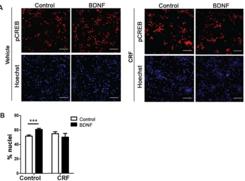
CRF treatment reduced pCREB activation in neuronal soma
One principal effector of retrograde axonal transport and signaling of BDNF is the activation of CREB (cAMP response element-binding protein) through its phosphorylation [62]. We pre-dicted that impairment of BDNF trafficking, as elicited by CRF treatment, could also disrupt neuronal function by decreasing the activation state of CREB following axonal stimulation with BDNF. Our results demonstrate that vehicle-treated cells exhibit a significant increase (p = 0.0005) in the percent of nuclei positive for phosphorylated CREB in response to BDNF, as expected (Fig 3). However, neurons treated with CRF failed to show a response to axonal BDNF application (Fig 3). These results suggest that altered BDNF transport as a result of CRF-induced tau-P involves changes in pathways involved in activation/phosphorylation of CREB.
Impact of CRF-Induced Tau-P on axonal movement of mitochondria
(Fig 4D). These results suggest that, unlike that seen for BDNF (Fig 2), mitochondrial transport may be sensitive to the effects of acute vs chronic stress. Acute exposure to CRF induces increased mitochondrial transport velocity, while chronic exposure lead to a reduction in mito-chondrial velocity but an increase in distance travelled.
Fig 3. Activation of CREB Pathways.Neurons that were cultured in microfluidic chambers were treated with BDNF (50 ng/ml) for 0.5hr. Neurons were fixed and stained for pCREB and the nuclei were stained with Hoechst dye.(A)Representative images are shown (scale bar = 50μM).(B)Quantitative analysis of the percentage of nuclei that were pCREB-positive (p = 0.005, n = 10 images).
doi:10.1371/journal.pone.0147250.g003
Fig 4. Impact of stress-induced tau-P on mitochondria transport.Kymographs showing axons of cultured mouse hippocampal cells after 2hr treatment;(A)Vehicle control.(B)CRF.(C)Table of quantitative analysis of fluorescent live images acquired from axons of cultured mouse hippocampal cells treated with 10μM CRF compared to controls; Average velocity of mitochondria 0.5hr (***p = 0.0002), 2hr (*p = 0.02); Distance travelled 0.5hr (*p = 0.04), 2h (*p = 0.01); Density of mitochondria after 0.5hr and 2hr.(D)Percent of mitochondrial movement at 0.5hr and 2hr.
As performed in BDNF experiments, parallel cultures where incubated with LiCl during acute CRF treatment. As seen for BDNF, the mitochondrial velocity was significantly increased in LiCl treated cells (p = 0.03) compared to vehicle control cells. Accordingly, combined treat-ment of CRF and LiCl resulted in a significant increase in velocity and distance travelled of mitochondria (p = 0.03, each) compared cells treated with only CRF (data not shown).
Discussion
In this study, we developed an in vitro model system to explore the functional consequences of acute and chronic stress-induced tau-P on neuronal function. We find that CRF treatment induces tau-P in a manner similar to that reported with stress models in vivo, and demonstrate that the axon transport deficits observed in our model involves CREB phosphorylation and is reliant on tau-P via GSK activation. Collectively, our data underscore the importance of CRF as a neuromodulator/neurotransmitter and provides a mechanistic underpinning for stress-induced tau-P and work that implicates CRF as a target for therapeutic intervention in AD.
Axon transport of Mitochondria and BDNF
In our in vivo work, we observed phospho-tau positive beading in varicosities along axons in stressed mice (RA Rissman, unpublished observations) similar to dystrophic neurite pathology in AD, suggesting structural alterations (i.e. blockages) and transport dysfunction [19,20,69]. A major hypothesis in the AD field suggests that defects in transport of important vesicles con-tributes to neurodegeneration [67,68]. Phosphorylated tau accumulations have been reported to block transport of organelles, mitochondria, and other vesicles in neurons [70,71], consis-tent with our current results. To probe the functional relevance of CRF-induced tau-P, we monitored changes in axon transport of two critical processes, energetics (i.e. mitochondria) and a major neuronal growth factor, BDNF, as a function of CRF treatment.
Mitochondria are concentrated in areas of high ATP demand, such as pre and post synaptic terminals, and neuronal growth cones. With the dynamic nature of neurons, mitochondria must be able to rapidly redistribute to different areas in order to meet increased energy demands to support mobilization of synaptic vesicles, actin assembly and disassembly, and generation of membrane potentials. Mitochondria are transported to activated synapses in response to ATP levels: velocity increases in areas of high ATP and decrease in areas of depleted ATP [70]. As observed in our study, short-term (i.e. 0.5hr) CRF exposure increased velocity of mitochondria, consistent with neurons eliciting an adaptive response of mobilizing energy resources to address to the challenge. We hypothesize that mitochondrial movement increased due to higher energy demands in the cell body and synaptic terminal during stress. Conversely, we observed a reduction in mitochondrial velocity with chronic CRF treatment, which we hypothesize may be due to increased accumulation of tau-Pand axonal blockage [19]. Accordingly, models of tau overexpression are characterized by blockages of microtubule tracks and reduction of transport of vesicles and cell organelles [19]. Blockage of vesicles and cell organelles can lead to depletion of crucial supplies required for cellular processes. Although not examined in this study, tau-P has also shown to inhibit movement of motor proteins [72,
73], which may also have a substantial impact on changes in mitochondrial movement. We hypothesize that the physical blockage of axons by tau-P leads to dysfunction of motor proteins and therefore a decrease in mitochondrial velocity, depletion of local ATP, oxidative stress, and interruption of neuronal function.
demonstrated in many animal models of AD [75]. Chronic stress exposure in vivo decreases BDNF expression levels in the dentate gyrus and hippocampus [24,76,77]. Tau pathology is a common component of chronic stress and AD, but it is unclear what effects these have on BDNF transport. Efficient retrograde axonal trafficking of BDNF is essential to properly initi-ate and sustain signaling both locally in the axon and at the soma. Previous studies suggest that treatment of neurons with other AD-related proteins, such as Aβ, cause deficits in BDNF traf-ficking [78]. We therefore investigated the effects of CRF-induced tau-P on BDNF transport. Using the same parameters as the mitochondria study, changes in mitochondrial movement were apparent with both acute and chronic CRF treatment. However, unlike that seen in our mitochondria experiments, we did not observe acute stress-like increases in transport of BDNF with CRF treatment—both acute and chronic treatment caused reduction in velocity and dis-tance travelled of BDNF, suggesting that cellular energetics may be a priority to cells undergo-ing stress as compared to growth factor transport. Although activation of the stress axis is a crucial response to challenge, our findings suggest that under chronic/repeated stress, neurons may be starved of growth factors. This response may be restricted to stressful stimuli, as studies of“good stressors”such voluntary exercise demonstrate increases in BDNF bioavailability [79,
80].
Activation of the transcription factor, cAMP response element-binding protein (CREB) has been observed in the nuclei of neurons after axonal treatment with BDNF [81]. CREB activa-tion is responsible for the expression of several genes including those involved in maintenance of neuronal morphology and dendritic development [82]. Importantly, CREB is downstream of the mitogen-activated protein kinase (MAPK) pathway, one of the signaling pathways induced by BDNF stimulation. Activation of CREB by BDNF and the ensuing retrograde axo-nal transmission of CREB to the soma is an integral part of the sigaxo-naling pathways of BDNF, which provides critical trophic support to neurons. Blockade or inhibition of these processes often results in axonal degeneration and neuronal atrophy [61,62]. Our current studies reveal that stress hormones such as CRF, through induction of tau-P, may exert a similar adversary effect on axonal and neuronal function.
Conclusions
Our study demonstrates that CRF induces tau-P in a manner consistent with our in vivo stud-ies [56–58]. In these in vivo studies, we found that chronic stress or CRF overexpression caused sequestration of tau aggregates into detergent-soluble cellular fractions [56,57]. The data pre-sented here provide functional information regarding the consequences that CRF and tau-P may have on neuronal function. CRF-induced tau-P interfered with axon transport of mito-chondria and BDNF, which we hypothesize can lead to impairments in transportation of car-goes, depleted trophic factor supply, and oxidative stress that may contribute to AD
pathogenesis. Continued exploration of the circuits and mechanism of stress-induced tau-P is central to further uncover the relationship between stress and AD tau pathology.
Supporting Information
S1 Movie. BDNF movement under control/vehicle conditions.Time lapsed video of
QD-BDNF movement in neuronal axons under vehicle conditions. (MP4)
S2 Movie. BDNF movement with CRF treatment.Time lapsed video demonstrating altered
S3 Movie. Mitochondrial movement under control/vehicle conditions.Time lapsed video of
mitochondrial movement in neuronal axons under vehicle conditions. (MP4)
S4 Movie. Mitochondrial movement with CRF treatment.Time lapsed video demonstrating
altered movement of mitochondria in neuronal axons after 2hr CRF treatment. (MP4)
Author Contributions
Conceived and designed the experiments: RAR CW. Performed the experiments: MHL AW PHL TCH ON. Analyzed the data: MHL AW PHL CW RAR. Wrote the paper: MHL LM CW RAR.
References
1. Selkoe DJ. Alzheimer's disease: genes, proteins, and therapy. Physiol Rev. 2001; 81(2):741–66. Epub 2001/03/29. PMID:11274343.
2. Terry RD, Masliah E, Salmon DP, Butters N, DeTeresa R, Hill R, et al. Physical basis of cognitive alter-ations in Alzheimer's disease: synapse loss is the major correlate of cognitive impairment. Ann Neurol. 1991; 30(4):572–80. PMID:1789684.
3. Gomez-Isla T, Hollister R, West H, Mui S, Growdon JH, Petersen RC, et al. Neuronal loss correlates with but exceeds neurofibrillary tangles in Alzheimer's disease. Ann Neurol. 1997; 41(1):17–24. PMID: 9005861.
4. Guillozet AL, Weintraub S, Mash DC, Mesulam MM. Neurofibrillary tangles, amyloid, and memory in aging and mild cognitive impairment. Arch Neurol. 2003; 60(5):729–36. doi:10.1001/archneur.60.5. 729PMID:12756137.
5. Kosik KS. Tau protein and Alzheimer's disease. Current opinion in cell biology. 1990; 2(1):101–4. PMID:2109621.
6. Kosik KS. Tau protein and neurodegeneration. Molecular neurobiology. 1990; 4(3–4):171–9. doi:10. 1007/BF02780339PMID:2135393.
7. Caceres A, Kosik KS. Inhibition of neurite polarity by tau antisense oligonucleotides in primary cerebel-lar neurons. Nature. 1990; 343(6257):461–3. doi:10.1038/343461a0PMID:2105469.
8. Dickson DW, Ksiezak-Reding H, Liu WK, Davies P, Crowe A, Yen SH. Immunocytochemistry of neuro-fibrillary tangles with antibodies to subregions of tau protein: identification of hidden and cleaved tau epitopes and a new phosphorylation site. Acta Neuropathol. 1992; 84(6):596–605. PMID:1281953.
9. Kopke E, Tung YC, Shaikh S, Alonso AC, Iqbal K, Grundke-Iqbal I. Microtubule-associated protein tau. Abnormal phosphorylation of a non-paired helical filament pool in Alzheimer disease. J Biol Chem. 1993; 268(32):24374–84. PMID:8226987.
10. Goedert M, Spillantini MG, Jakes R, Rutherford D, Crowther RA. Multiple isoforms of human microtu-bule-associated protein tau: sequences and localization in neurofibrillary tangles of Alzheimer's dis-ease. Neuron. 1989; 3(4):519–26. PMID:2484340.
11. Goedert M, Spillantini MG, Cairns NJ, Crowther RA. Tau proteins of Alzheimer paired helical filaments: abnormal phosphorylation of all six brain isoforms. Neuron. 1992; 8(1):159–68. Epub 1992/01/01. 0896-6273(92)90117-V [pii]. PMID:1530909.
12. Sengupta A, Kabat J, Novak M, Wu Q, Grundke-Iqbal I, Iqbal K. Phosphorylation of tau at both Thr 231 and Ser 262 is required for maximal inhibition of its binding to microtubules. Archives of biochemistry and biophysics. 1998; 357(2):299–309. doi:10.1006/abbi.1998.0813PMID:9735171.
13. Kenessey A, Yen SH. The extent of phosphorylation of fetal tau is comparable to that of PHF-tau from Alzheimer paired helical filaments. Brain Res. 1993; 629(1):40–6. PMID:8287279.
14. Anderton BH, Betts J, Blackstock WP, Brion JP, Chapman S, Connell J, et al. Sites of phosphorylation in tau and factors affecting their regulation. Biochem Soc Symp. 2001;( 67):73–80. PMID:11447841.
16. Gustke N, Steiner B, Mandelkow EM, Biernat J, Meyer HE, Goedert M, et al. The Alzheimer-like phos-phorylation of tau protein reduces microtubule binding and involves Ser-Pro and Thr-Pro motifs. FEBS Lett. 1992; 307(2):199–205. PMID:1644173.
17. Bramblett GT, Goedert M, Jakes R, Merrick SE, Trojanowski JQ, Lee VM. Abnormal tau phosphoryla-tion at Ser396 in Alzheimer's disease recapitulates development and contributes to reduced microtu-bule binding. Neuron. 1993; 10(6):1089–99. PMID:8318230.
18. Alonso AC, Grundke-Iqbal I, Iqbal K. Alzheimer's disease hyperphosphorylated tau sequesters normal tau into tangles of filaments and disassembles microtubules. Nat Med. 1996; 2(7):783–7. PMID: 8673924.
19. Mandelkow EM, Stamer K, Vogel R, Thies E, Mandelkow E. Clogging of axons by tau, inhibition of axo-nal traffic and starvation of synapses. Neurobiol Aging. 2003; 24(8):1079–85. Epub 2003/12/04. S0197458003001787 [pii]. PMID:14643379.
20. Cuchillo-Ibanez I, Seereeram A, Byers HL, Leung KY, Ward MA, Anderton BH, et al. Phosphorylation of tau regulates its axonal transport by controlling its binding to kinesin. Faseb J. 2008; 22(9):3186–95. Epub 2008/05/31. fj.08-109181 [pii] doi:10.1096/fj.08-109181PMID:18511549.
21. Sapolsky RM, Krey LC, McEwen BS. The neuroendocrinology of stress and aging: the glucocorticoid cascade hypothesis. Endocr Rev. 1986; 7(3):284–301. PMID:3527687.
22. Sapolsky RM. Why stress is bad for your brain. Science (New York, NY. 1996; 273(5276):749–50. PMID:8701325.
23. McEwen BS, Sapolsky RM. Stress and cognitive function. Current opinion in neurobiology. 1995; 5 (2):205–16. PMID:7620309.
24. Smith MA, Makino S, Kvetnansky R, Post RM. Stress and glucocorticoids affect the expression of brain-derived neurotrophic factor and neurotrophin-3 mRNAs in the hippocampus. J Neurosci. 1995; 15 (3 Pt 1):1768–77. PMID:7891134.
25. Swaab DF, Bao AM, Lucassen PJ. The stress system in the human brain in depression and neurode-generation. Ageing Res Rev. 2005; 4(2):141–94. PMID:15996533.
26. Wilson RS, Evans DA, Bienias JL, Mendes de Leon CF, Schneider JA, Bennett DA. Proneness to psy-chological distress is associated with risk of Alzheimer's disease. Neurology. 2003; 61(11):1479–85. PMID:14663028.
27. Wilson RS, Bennett DA, Mendes de Leon CF, Bienias JL, Morris MC, Evans DA. Distress proneness and cognitive decline in a population of older persons. Psychoneuroendocrinology. 2005; 30(1):11–7. PMID:15358438.
28. Wilson RS, Arnold SE, Schneider JA, Kelly JF, Tang Y, Bennett DA. Chronic psychological distress and risk of Alzheimer's disease in old age. Neuroepidemiology. 2006; 27(3):143–53. PMID:16974109.
29. Wilson RS, Schneider JA, Boyle PA, Arnold SE, Tang Y, Bennett DA. Chronic distress and incidence of mild cognitive impairment. Neurology. 2007; 68(24):2085–92. PMID:17562829.
30. Vale W, Spiess J, Rivier C, Rivier J. Characterization of a 41-residue ovine hypothalamic peptide that stimulates secretion of corticotropin and beta-endorphin. Science (New York, NY. 1981; 213 (4514):1394–7. PMID:6267699.
31. Sawchenko PE, Yuan ZF, Laplante F, Rissman RA, Bittencourt JC. Corticotropin-Releasing Factor: Integration of Adaptive Responses to Stress. New Encyclopedia of Neuroscience ( London: Elsevier). 2008;in press.
32. Chadwick D, Marsh J, Ackrill K. Corticotropin-releasing factor. Chichester; New York: Wiley; 1993. x, 357 p. p.
33. Bale TL, Vale WW. CRF and CRF receptors: role in stress responsivity and other behaviors. Annu Rev Pharmacol Toxicol. 2004; 44:525–57. PMID:14744257.
34. Turnbull AV, Rivier C. Corticotropin-releasing factor (CRF) and endocrine responses to stress: CRF receptors, binding protein, and related peptides. Proceedings of the Society for Experimental Biology and Medicine Society for Experimental Biology and Medicine. 1997; 215(1):1–10. PMID:9142133.
35. Chen R, Lewis KA, Perrin MH, Vale WW. Expression cloning of a human corticotropin-releasing-factor receptor. Proc Natl Acad Sci USA. 1993; 90:8967–71. PMID:7692441
36. Van Pett K, Viau V, Bittencourt JC, Chan RKW, Li H-Y, Arias C, et al. Distribution of mRNAs encoding CRF receptors in brain and pituitary of rat and mouse. J Comp Neurol. 2000; 428:191–212. PMID: 11064361
37. Lovenberg TW, Chalmers DT, Liu C, De Souza EB. CRF2aand CRF2breceptor mRNAs are
38. Vaughan J, Donaldson C, Bittencourt J, Perrin MH, Lewis K, Sutton S, et al. Urocortin, a mammalian neuropeptide related to fish urotensin I and to corticotropin-releasing factor. Nature. 1995; 378:287–92. PMID:7477349
39. Reyes TM, Lewis K, Perrin MH, Kunitake KS, Vaughan J, Araias CA, et al. Urocortin II: A member of the corticotropin-releasing factor (CRF) neuropeptide family that is selectively bound by type 2 CRF recep-tors. Prog Natl Acad Sci. 2001; 98:2843–8.
40. Lewis K, Li C, Perrin MH, Blount A, Kunitake K, Donaldson C, et al. Identification of urocortin III, an addi-tional member of the corticotropin-releasing factor (CRF) family with high affinity for the CRF2 receptor. Proc Natl Acad Sci, USA. 2001; 98:7570–5. PMID:11416224
41. Rehman HU. Role of CRH in the pathogenesis of dementia of Alzheimer's type and other dementias. Curr Opin Investig Drugs. 2002; 3(11):1637–42. PMID:12476966.
42. Rissman RA. Stress-induced tau phosphorylation: functional neuroplasticity or neuronal vulnerability? J Alzheimers Dis. 2009; 18(2):453–7. Epub 2009/07/09. T23W75R9KQ126534 [pii] doi: 10.3233/JAD-2009-1153PMID:19584431.
43. Davis KL, Mohs RC, Marin DB, Purohit DP, Perl DP, Lantz M, et al. Neuropeptide abnormalities in patients with early Alzheimer disease. Arch Gen Psychiatry. 1999; 56(11):981–7. PMID:10565496.
44. Sapolsky RM, Krey LC, McEwen BS. Prolonged glucocorticoid exposure reduces hippocampal neuron number: implications for aging. J Neurosci. 1985; 5(5):1222–7. PMID:3998818.
45. McEwen BS. Possible mechanisms for atrophy of the human hippocampus. Mol Psychiatry. 1997; 2 (3):255–62. Epub 1997/05/01. PMID:9152991.
46. Bremner JD. Stress and brain atrophy. CNS Neurol Disord Drug Targets. 2006; 5(5):503–12. Epub 2006/11/01. PMID:17073653.
47. Aisen PS, Davis KL, Berg JD, Schafer K, Campbell K, Thomas RG, et al. A randomized controlled trial of prednisone in Alzheimer's disease. Alzheimer's Disease Cooperative Study. Neurology. 2000; 54 (3):588–93. PMID:10680787.
48. Touma C, Ambree O, Gortz N, Keyvani K, Lewejohann L, Palme R, et al. Age- and sex-dependent development of adrenocortical hyperactivity in a transgenic mouse model of Alzheimer's disease. Neu-robiol Aging. 2004; 25(7):893–904. doi:10.1016/j.neurobiolaging.2003.09.004PMID:15212843.
49. Sterniczuk R, Antle MC, Laferla FM, Dyck RH. Characterization of the 3xTg-AD mouse model of Alzhei-mer's disease: part 2. Behavioral and cognitive changes. Brain Res. 2010; 1348:149–55. doi:10.1016/ j.brainres.2010.06.011PMID:20558146.
50. Guo Q, Zheng H, Justice NJ. Central CRF system perturbation in an Alzheimer's disease knockin mouse model. Neurobiol Aging. 2012; 33(11):2678–91. doi:10.1016/j.neurobiolaging.2012.01.002 PMID:22336193; PubMed Central PMCID: PMC3361634.
51. Dong H, Wang S, Zeng Z, Li F, Montalvo-Ortiz J, Tucker C, et al. Effects of corticotrophin-releasing fac-tor recepfac-tor 1 antagonists on amyloid-beta and behavior in Tg2576 mice. Psychopharmacology (Berl). 2014. doi:10.1007/s00213-014-3629-8PMID:24862368.
52. Heinrichs SC, Stenzel-Poore MP, Gold LH, Battenberg E, Bloom FE, Koob GF, et al. Learning impairment in transgenic mice with central overexpression of corticotropin-releasing factor. Neurosci-ence. 1996; 74(2):303–11. Epub 1996/09/01. 0306452296001406 [pii]. PMID:8865183.
53. Magarinos AM, Verdugo JM, McEwen BS. Chronic stress alters synaptic terminal structure in hippo-campus. Proceedings of the National Academy of Sciences of the United States of America. 1997; 94 (25):14002–8. PMID:9391142.
54. Goebel M, Fleming SM, Million M, Stengel A, Tache Y, Wang L. Mice overexpressing corticotropin-releasing factor show brain atrophy and motor dysfunctions. Neurosci Lett. 2010; 473(1):11–5. Epub 2010/02/06. S0304-3940(10)00121-7 [pii] doi:10.1016/j.neulet.2010.01.068PMID:20132869; PubMed Central PMCID: PMC2848985.
55. Carroll JC, Iba M, Bangasser DA, Valentino RJ, James MJ, Brunden KR, et al. Chronic stress exacer-bates tau pathology, neurodegeneration, and cognitive performance through a corticotropin-releasing factor receptor-dependent mechanism in a transgenic mouse model of tauopathy. J Neurosci. 2011; 31 (40):14436–49. doi:10.1523/JNEUROSCI.3836-11.2011PMID:21976528; PubMed Central PMCID: PMC3230070.
56. Rissman RA, Staup MA, Lee AR, Justice NJ, Rice KC, Vale W, et al. Corticotropin-releasing factor receptor-dependent effects of repeated stress on tau phosphorylation, solubility, and aggregation. Pro-ceedings of the National Academy of Sciences of the United States of America. 2012; 109(16):6277– 82. doi:10.1073/pnas.1203140109PMID:22451915; PubMed Central PMCID: PMC3341026.
58. Rissman RA, Lee KF, Vale W, Sawchenko PE. Corticotropin-releasing factor receptors differentially regulate stress-induced tau phosphorylation. J Neurosci. 2007; 27(24):6552–62. PMID:17567816.
59. Dirks A, Groenink L, Bouwknecht JA, Hijzen TH, Van Der Gugten J, Ronken E, et al. Overexpression of corticotropin-releasing hormone in transgenic mice and chronic stress-like autonomic and physiological alterations. Eur J Neurosci. 2002; 16(9):1751–60. Epub 2002/11/15. 2245 [pii]. PMID:12431228.
60. Roe AD, Staup MA, Serrats J, Sawchenko PE, Rissman RA. Lipopolysaccharide-induced tau phos-phorylation and kinase activity—modulation, but not mediation, by corticotropin-releasing factor recep-tors. Eur J Neurosci. 2011; 34(3):448–56. doi:10.1111/j.1460-9568.2011.07764.xPMID:21722209; PubMed Central PMCID: PMC3148267.
61. Zhao X, Weissmiller AM, Pearn ML, Mobley WC, Wu C. Real-time imaging of axonal transport of quan-tum dot-labeled BDNF in primary neurons. J Visualized Exp. 2014;In press.
62. Weissmiller AM, Natera-Naranjo O, Reyna SM, Pearn ML, Zhao X, Nguyen P, et al. A gamma-secre-tase inhibitor, but not a gamma-secregamma-secre-tase modulator, induced defects in BDNF axonal trafficking and signaling: evidence for a role for APP. PloS one. 2015; 10(2):e0118379. doi:10.1371/journal.pone. 0118379PMID:25710492; PubMed Central PMCID: PMC4339551.
63. Choi JS, Pham TT, Jang YJ, Bui BC, Lee BH, Joo KM, et al. Corticotropin-releasing factor (CRF) and urocortin promote the survival of cultured cerebellar GABAergic neurons through the type 1 CRF recep-tor. J Korean Med Sci. 2006; 21(3):518–26. Epub 2006/06/17. 200606518 [pii]. PMID:16778399; PubMed Central PMCID: PMC2729961.
64. Frederick RL, Shaw JM. Moving mitochondria: establishing distribution of an essential organelle. Traf-fic. 2007; 8(12):1668–75. doi:10.1111/j.1600-0854.2007.00644.xPMID:17944806; PubMed Central PMCID: PMC3739988.
65. Liu SJ, Zhang AH, Li HL, Wang Q, Deng HM, Netzer WJ, et al. Overactivation of glycogen synthase kinase-3 by inhibition of phosphoinositol-3 kinase and protein kinase C leads to hyperphosphorylation of tau and impairment of spatial memory. J Neurochem. 2003; 87(6):1333–44. Epub 2004/01/10. 2070 [pii]. PMID:14713290.
66. Millecamps S, Julien JP. Axonal transport deficits and neurodegenerative diseases. Nature reviews Neuroscience. 2013; 14(3):161–76. doi:10.1038/nrn3380PMID:23361386.
67. Stokin GB, Goldstein LS. Axonal transport and Alzheimer's disease. Annual review of biochemistry. 2006; 75:607–27. doi:10.1146/annurev.biochem.75.103004.142637PMID:16756504.
68. Stokin GB, Goldstein LS. Linking molecular motors to Alzheimer's disease. Journal of physiology, Paris. 2006; 99(2–3):193–200. doi:10.1016/j.jphysparis.2005.12.085PMID:16459060.
69. Morfini GA, Burns M, Binder LI, Kanaan NM, LaPointe N, Bosco DA, et al. Axonal transport defects in neurodegenerative diseases. J Neurosci. 2009; 29(41):12776–86. Epub 2009/10/16. 29/41/12776 [pii] doi:10.1523/JNEUROSCI.3463-09.2009PMID:19828789.
70. Sheng ZH, Cai Q. Mitochondrial transport in neurons: impact on synaptic homeostasis and neurode-generation. Nature reviews Neuroscience. 2012; 13(2):77–93. doi:10.1038/nrn3156PMID:22218207.
71. Stamer K, Vogel R, Thies E, Mandelkow E, Mandelkow EM. Tau blocks traffic of organelles, neurofila-ments, and APP vesicles in neurons and enhances oxidative stress. J Cell Biol. 2002; 156(6):1051–63. Epub 2002/03/20. [pii]. PMID:11901170; PubMed Central PMCID: PMC2173473.
72. Ebneth A, Godemann R, Stamer K, Illenberger S, Trinczek B, Mandelkow E. Overexpression of tau pro-tein inhibits kinesin-dependent trafficking of vesicles, mitochondria, and endoplasmic reticulum: impli-cations for Alzheimer's disease. J Cell Biol. 1998; 143(3):777–94. Epub 1998/11/13. PMID:9813097; PubMed Central PMCID: PMC2148132.
73. Trinczek B, Ebneth A, Mandelkow EM, Mandelkow E. Tau regulates the attachment/detachment but not the speed of motors in microtubule-dependent transport of single vesicles and organelles. Journal of cell science. 1999; 112 (Pt 14):2355–67. PMID:10381391.
74. Dugich-Djordjevic MM, Peterson C, Isono F, Ohsawa F, Widmer HR, Denton TL, et al. Immunohisto-chemical visualization of brain-derived neurotrophic factor in the rat brain. Eur J Neurosci. 1995; 7 (9):1831–9. PMID:8528456.
75. Nagahara AH, Merrill DA, Coppola G, Tsukada S, Schroeder BE, Shaked GM, et al. Neuroprotective effects of brain-derived neurotrophic factor in rodent and primate models of Alzheimer's disease. Nat Med. 2009; 15(3):331–7. doi:10.1038/nm.1912PMID:19198615; PubMed Central PMCID: PMC2838375.
76. Smith MA, Makino S, Kim SY, Kvetnansky R. Stress increases brain-derived neurotropic factor mes-senger ribonucleic acid in the hypothalamus and pituitary. Endocrinology. 1995; 136(9):3743–50. doi: 10.1210/endo.136.9.7649080PMID:7649080.
78. Poon WW, Blurton-Jones M, Tu CH, Feinberg LM, Chabrier MA, Harris JW, et al. beta-Amyloid impairs axonal BDNF retrograde trafficking. Neurobiol Aging. 2011; 32(5):821–33. doi:10.1016/j.
neurobiolaging.2009.05.012PMID:19540623; PubMed Central PMCID: PMC3038182.
79. Adlard PA, Perreau VM, Cotman CW. The exercise-induced expression of BDNF within the hippocam-pus varies across life-span. Neurobiol Aging. 2005; 26(4):511–20. doi:10.1016/j.neurobiolaging.2004. 05.006PMID:15653179.
80. Oliff HS, Berchtold NC, Isackson P, Cotman CW. Exercise-induced regulation of brain-derived neuro-trophic factor (BDNF) transcripts in the rat hippocampus. Brain Res Mol Brain Res. 1998; 61(1–2):147– 53. PMID:9795193.
81. Zhou B, Cai Q, Xie Y, Sheng ZH. Snapin recruits dynein to BDNF-TrkB signaling endosomes for retro-grade axonal transport and is essential for dendrite growth of cortical neurons. Cell Rep. 2012; 2(1):42– 51. doi:10.1016/j.celrep.2012.06.010PMID:22840395; PubMed Central PMCID: PMC3408618.