Drug Development in Conformational
Diseases: A Novel Family of Chemical
Chaperones that Bind and Stabilise Several
Polymorphic Amyloid Structures
Marquiza Sablón-Carrazana1,2☯, Isaac Fernández2,3☯, Alberto Bencomo1,2, Reyna
Lara-Martínez4, Suchitil Rivera-Marrero1, Guadalupe Domínguez5, Rafaela Pérez-Perera1, Luis Felipe Jiménez-García4, Nelly F. Altamirano-Bustamante6, Massiel Diaz-Delgado1,
Fernand Vedrenne2, Lina Rivillas-Acevedo2, Karina Pasten-Hidalgo6,7, María de Lourdes Segura-Valdez4, Sergio Islas-Andrade2, Eulalia Garrido-Magaña8,
Alejandro Perera-Pintado9, Anaís Prats-Capote9, Chryslaine Rodríguez-Tanty1*, Myriam M. Altamirano-Bustamante2
*
1Dpto. Neurodiagnóstico, Centro de Neurociencias de Cuba, Cubanacán, Playa, La Habana, Cuba, 2Unidad de Investigación Médica en Enfermedades Metabólicas, Hospital de Cardiología, Centro Médico Nacional Siglo XXI, Instituto Mexicano del Seguro Social, México D.F., México,3Departamento de Microbiología, Escuela Nacional de Ciencias Biológicas, Instituto Politécnico Nacional, México D.F., México, 4Laboratorio de Nanobiología Celular, Departamento de Biología Celular, Facultad de Ciencias, UNAM, México D.F., México,5Instituto de Fisiología Celular, UNAM, México D.F., México,6Servicio de Endocrinología, Instituto Nacional de Pediatría, SS, México D.F., México,7Cátedra Conacyt, México D.F., México,8Servicio de Endocrinología, Hospital de Pediatría, CMNXXI, IMSS, México D. F., México, 9Centro de Investigaciones Clínicas, La Habana, Cuba
☯These authors contributed equally to this work.
*[email protected](CR-T);[email protected](MMA-B)
Abstract
The increasing prevalence of conformational diseases, including Alzheimer's disease, type 2 Diabetes Mellitus and Cancer, poses a global challenge at many different levels. It has devastating effects on the sufferers as well as a tremendous economic impact on families and the health system. In this work, we apply a cross-functional approach that combines ideas, concepts and technologies from several disciplines in order to study,in silicoandin vitro, the role of a novel chemical chaperones family (NCHCHF) in processes of protein aggregation in conformational diseases. Given that Serum Albumin (SA) is the most abun-dant protein in the blood of mammals, and Bovine Serum Albumin (BSA) is an off-the-shelf protein available in most labs around the world, we compared the ligandability of BSA: NCHCHF with the interaction sites in the Human Islet Amyloid Polypeptide (hIAPP): NCHCHF, and in the amyloid pharmacophore fragments (Aβ17–42 and Aβ16–21): NCHCHF. We posit that the merging of this interaction sites is a meta-structure of pharma-cophore which allows the development of chaperones that can prevent protein aggregation at various states from: stabilizing the native state to destabilizing oligomeric state and proto-filament. Furthermore to stabilize fibrillar structures, thus decreasing the amount of toxic oligomers in solution, as is the case with the NCHCHF. The paper demonstrates how a set of NCHCHF can be used for studying and potentially treating the various physiopathological
OPEN ACCESS
Citation:Sablón-Carrazana M, Fernández I, Bencomo A, Lara-Martínez R, Rivera-Marrero S, Domínguez G, et al. (2015) Drug Development in Conformational Diseases: A Novel Family of Chemical Chaperones that Bind and Stabilise Several Polymorphic Amyloid Structures. PLoS ONE 10(9): e0135292. doi:10.1371/journal.pone.0135292
Editor:Jie Zheng, University of Akron, UNITED STATES
Received:May 1, 2015
Accepted:July 20, 2015
Published:September 1, 2015
Copyright:© 2015 Sablón-Carrazana et al. This is an open access article distributed under the terms of theCreative Commons Attribution License, which permits unrestricted use, distribution, and reproduction in any medium, provided the original author and source are credited.
Data Availability Statement:All relevant data are within the paper and its Supporting Information file.
stages of a conformational disease. For instance, when dealing with an acute phase of cyto-toxicity, what is needed is the recruitment of cytotoxic oligomers, thus chaperone F, which accelerates fiber formation, would be very useful; whereas in a chronic stage it is better to have chaperonesA,B,C, andD, which stabilize the native and fibril structures halting self-catalysis and the creation of cytotoxic oligomers as a consequence of fiber formation. Fur-thermore, all the chaperones are able to protect and recondition the cerebellar granule cells (CGC) from the cytotoxicity produced by the hIAPP20–29fragment or by a low potassium
medium, regardless of their capacity for accelerating or inhibitingin vitroformation of fibers.
In vivoanimal experiments are required to study the impact of chemical chaperones in cog-nitive and metabolic syndromes.
Introduction
Developing innovative and cost-effective strategies for diagnosing and treating conformational diseases (CDs) is a major challenge of modern biomedical science. Misfolded proteins (more than 40 different kinds) play a central role in the pathophysiology of CDs such as Alzheimer's disease (AD), Huntington's disease (HD), Parkinson's disease (PD), type 2 Diabetes Mellitus (DM2), and Cancer among others. Molecular mechanisms by which an initially innocuous protein adopts an amyloidogenic conformation capable of forming fibers and toxic oligomers, contributing to the onset of CDs is an unanswered question as of yet [1–4]. It is known that sequence-independent amyloid states are formed by elongated fibers with spines built out ofβ -sheets strands.β-sheets interact with each other by means of a dense hydrogen-bond network, forming a closed dry surface. These structures, known as“steric zippers”, are repeated along the fiber [2,5,6]. In order to reach a minimum in their total free energy, most misfolded pro-teins, regardless of their structure, can form amyloid aggregates as alternative packing [7–9]. Finding ways to disrupt the formation of amyloid structures, therefore, could lay the founda-tions for developing novel drugs to prevent, delay the onset, and treat these diseases [10].
Take as example theβamyloid protein (Aβ) whose polymorphic aggregates (toxic Aβ oligo-mers, protofilaments and fibers) and its circulation or deposition in brain, are the pathological hallmarks of AD. Similarly, recent studies suggest that the aggregation of Human Islet Amyloid Polypeptide (hIAPP), a 37-residue peptide produced in pancreatic beta cells, is a key factor in the development of diabetes [6]. Its fibrillating core, the decapeptide SNFFGAILSS (hIAPP20– 29), is thus a key point of study. These peptides, however, are costly, and thus inaccessible for
large-scale use in some research facilities. In this article we show that Bovine Serum Albumin (BSA), a familiar,‘off-the-shelf’protein shares a common meta-structure with IAPP and the amyloid pharmacophore fragments (Aβ17–42and Aβ16–21). Systematic comparison suggests
that BSA has remarkable potential as a model molecule for further study in the field of pharma-ceutics in conformational diseases.
BSA, a protein highly abundant in blood, is composed by 585 amino acids. Under normal conditions, 67% of the BSA’s tertiary structure is in anα-helix conformation [11]. This protein has a globular and rigid structure that can suffer conformational changes depending on the medium’s condition from their nativeα-helix structure to aβ-sheet structure. Although there have been numerous studies investigating the mechanisms of BSA aggregation, the conditions which elicit such conformational changes are still unknown [12]. Battacharya, et al.[12] have found that pH and ionic strength significantly influence the kinetics of aggregation of BSA, while Holm, et al.[13] point out that high temperatures also promote the formation of fibers,
Novel Potential Building Blocks for Treating Conformational Diseases?
indicating that BSA first needs to go into a denatured state in order to aggregate. It has also been proven that various conditions can elicit the formation of structurally different oligomers [7,9,13,14].
As part of the quality control system of proteins, there is a group of molecules known as chaperones. These are proteins that interact with, stabilize, or help other proteins acquire their functionally active conformation, without affecting their final native structure [15]. Several dif-ferent compounds are classified as chaperones due to their ability to interact with protein [16]. Chaperones have been typified as molecular, chemical, and pharmacological [17–19]. They res-cue the functionality of other proteins and promote their correct folding by temporarily bind-ing to partially unfolded intermediaries to prevent their aggregation and interactions [16,20]. It is of great interest, therefore, to study and develop compounds that can bind to a wide variety of polymorphic aggregates, marking them and inhibiting their development. Various studies have proved that there are several proteins, lipids, and small chemical compounds that can inhibit protein aggregation into amyloid structures, acting like chaperones. Worth of mention among these, are Non-Steroid Anti-inflammatory Drugs (NSAIDs), curcumin, heparin, small heat shock proteins (sHsp), inositol derivatives, and many other polycyclic, polyphenolic, and aromatic substances [2,21–25]. Other substances, such as Gemini surfactants, have been stud-ied because of their property of refolding. Some naphthalenes derivatives, such as 2-dialkyla-mino-6-acylmalononitrile (DDNP,J), substituted naphthalenes, and Orange G (I) [2], have been likewise successfully bound to amyloid fibers by forming aromatic interactions within the steric zippers’dry surface, thus disrupting their structure.
In this work we apply a cross-functional approach that combines ideas, concepts and tech-nologies from several disciplines to study,in silicoandin vitro, a Novel Chemical Chaperones Family (NCHCHF) The NCHCHF is composed by: N-(2-aminoethyl)-N'-1-naphthylsuccina-mideA; methyl (2-{[4-(1-naphthylamino)-4-oxobutanoyl]amino}ethyl) dithiocarbamateB; N-[4-(1-naphthylamino)-4-oxobutanoyl]-β-alanineD; 6-{[4-(1-naphthylamino)-4-oxobutanoyl] amino} hexanoic acidE; N3,N3'-ethane-1,2-dyilbis(N1-1-naphthylsuccinamide)Fand N-(4-aminobutyl)-N'-1-naphthylsuccinamideG. Also, the (2R)-2-(6-methoxy-2-naphthyl)pro-panoic acid (Naproxen)C, was used as control, because its anti-amyloid activity reported [26] (Fig 1) [27,28] and their role in protein aggregation in conformational diseases; using BSA and the hIAPP fragment 20–29 (hIAPP20–29) considered among the most amyloidogenic of their
class. We reveal that some members of NCHCHF have the ability to interact directly with native and/or aggregation-prone species in solution and/or fiber structures to decrease the amount of toxic oligomers in solution. Furthermore they are able to protect and recondition the cerebellar granules cells (CGC) from the citotoxicity produced by hIAPP20–29.
Results
Molecular Frameworks of Action of Chemical Chaperones
In order to understand the ligand–protein interactions, we used the molecular docking tech-nique to design and test a novel chemical chaperones family using BSA, hIAPP1–37, and the
amyloid pharmacophore fragments (Aβ17–42and Aβ16–21). as target molecules. All these
struc-tures were downloaded directly from the PDB: 3VO3, 2L86, 2BEG and 1IYT codes, respec-tively. Six novel compounds and four references were docked into the 3D structure of BSA using AutoDockVina. The compounds were clustered into four sets, according to the terminal functional group of the amidoalkylic chain and the logP values (calculated by ACD program.
docking simulation protocol points out the most probable binding sites, poses and the energies between the chemical chaperones and the protein.
BSA Molecular Docking
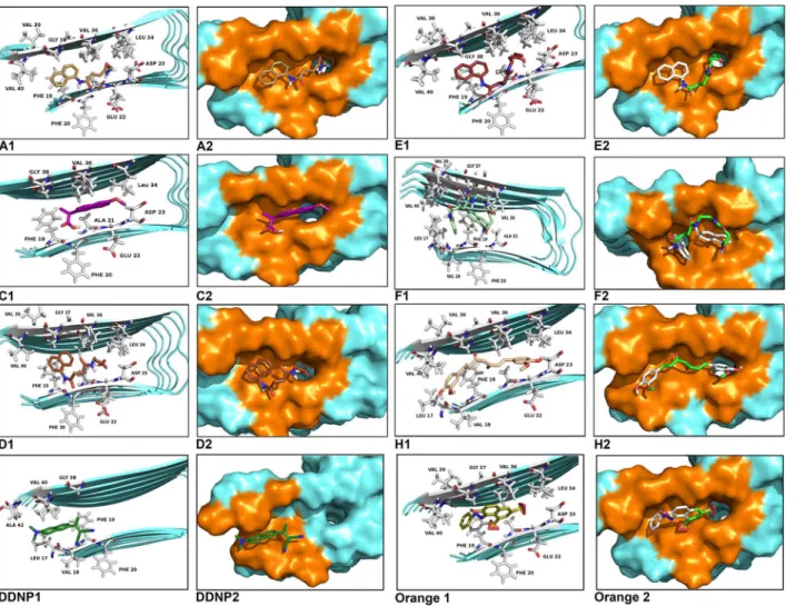
Observations of the 3D structure of crystalline albumin have shown that BSA is constituted by three homologous domains (I, II, III): I (residues 1–183), II (184–276) and III (377–583), each one containing two subdomains (A or B). Two hydrophobic cavities are located within the sub-domains IIA and IIIA, which are considered drug-binding sites. Its secondary structure is dominated by anα-helix. The binding sites of all the chaperones tested with BSA were in the similar vicinity. The main regions of ligand binding to BSA were located in hydrophobic cavi-ties of the subdomains IIA and IIIA, known as drug’s pocket. This site is a large hydrophobic cavity, which accommodates chaperone molecules. Examples of the location of these sites in the 3D structure of BSA are shown inFig 2.
The conformer with the lowest binding energy was calculated from 10 different conforma-tions for each docking simulation, at 5 Å distance between chaperones and BSA (S1 Table). Finally, it was found that the chaperone-BSA binding site was located into the domain IIA and IIIA of the protein, which corresponds to the lowest free energy values calculated. According to the analysis of the calculated free energy, the binding process of BSA with chaperones is spon-taneous (ΔG<0). The common amino acid residues lining this binding site were: His145, Glu186, Leu189, Thr190, Ala193, Arg196, Glu424, Ser428, Lys431, Val432, Arg435, Tyr451, Ile455, Arg458 and Asp107, Ser109, Leu189, Thr190, Ala193, Arg196, Glu424, Ser428 Lys431, Val432, Arg435, Tyr451, Ile455, Arg458, for the 1G and 2G groups respectively. The
Fig 1. Structures and LogP of the chemical chaperones.N-(2-aminoethyl)-N'-1-naphthylsuccinamideA; methyl (2-{[4-(1-naphthylamino)-4-oxobutanoyl] amino}ethyl) dithiocarbamateB; (2R)-2-(6-methoxy-2-naphthyl)propanoic acid (Naproxen)C;N-[4-(1-naphthylamino)-4-oxobutanoyl]-β-alanineD; 6-{[4-(1-naphthylamino)-4-oxobutanoyl]amino} hexanoic acidE;N3,N3'-ethane-1,2-dyilbis(N1-1-naphthylsuccinamide)F;N-(4-aminobutyl)-N
'-1-naphthylsuccinamideG; (1E,6E)-1,8-bis(4-hydroxy-3-methoxyphenyl)octa-1,6-diene-3,5-dione (Curcumine)H; 7-hydroxy-8-[(Z)-phenyldiazenyl] naphthalene-1,3-disulfonic acid (Orange G)I; (2Z)-3-[6-(dimethylamino)-2-naphthyl]-2-isocyanobut-2-enenitrile (DDNP)J.*The logP was calculated using
ACD/Log P software (ACD/1-Lab Service,Toronto,Ontario,Canada).**Valueestimatedfor uncharged compound.
doi:10.1371/journal.pone.0135292.g001
compounds of the 3G group interact with the following common amino acid residues: Asp108, Glu186, Leu189, Thr190, Ser192, Ala193, Arg196, Glu424, Ser428, Lys431, Val432, Arg435, Tyr451, Leu454, Ile455, and Arg458. Similarly, the binding amino acid residues forCand cur-cumin (H) are Glu186, Leu189, Thr190, Ser192, Ala193, Arg196, Glu424, Ser428, Lys431, Val432, Arg435, Tyr451, Ile455, and Arg458. In 85% of calculations, the interaction zone was comprised between amino acids 145 to 458, specifically with the residues His145, Glu186, Leu189, Thr190, Ser192, Ala193, Arg196, Glu424, Ser428, Lys431, Val432, Arg435, Tyr451, Ile455, and Arg458. In general, these molecules bind to the sites via hydrophobic and Van der Waals forces. In some cases, hydrogen-bonding interactions have been observed. (Fig 3).
Taking chaperonesA,B,DandHas representative examples of the different groups (1G, 2G, 3G and 4G), the docking results reveal that the chaperones bind to the sites IIA and IIIA of BSA, mainly via hydrophobic interactions with the Leu189, Ala193, Val432, and Ile455 resi-dues, as well as by Van der Waals forces. The interactionsππwere located between the
naphthyl ring of the chaperones and the phenyl ring of the Tyr451. In the case of the
Fig 2. Molecular docked model of chemical chaperones (stick representation), located within the hydrophobic pocket of BSA (cartoon and sphere views).At 5Ådistance, the amino acid residues surrounding chaperones (A,B,C,D,E,F,IandH) are represented in orange. The H-bonding interaction between chaperones and amino acid residues of BSA, are shown as a dotted line.
chaperonesAandDthere was hydrogen bonding with two residues of Ser (198 and 428) of the BSA. In the case of chaperoneB, this interaction was found with Leu189. OtherwiseHdid not expose the hydrogen bonding interaction (Fig 3). The binding process of BSA with chaperones
Fig 3. Visualization of binding site of chaperones (A, B, C, D, E, F, G and H) into IIA and IIIA regions of BSA, within 5Ådistance.
doi:10.1371/journal.pone.0135292.g003
is spontaneous (ΔG<0). The interaction forces are mainly hydrophobic and hydrogen bonding.
Amyloid Pharmacophore Molecular Docking
Eisenberg et al.[2] demonstrated that amyloid pharmacophores, offering cavities alongβ -sheets, are similar to those observed binding of amyloid markers toβ-sheets in non-fibrillar structures and the oligomers and fibers that are inhibited by similar compounds, includingH. The amyloid model is used as target for docking simulations in order not only to understand the aggregation process, but also to find compounds that could prevent the formation ofβ -structure. Also, three crystal structures of small molecules bound to amyloid-like segments of
tauand amyloid-β(I,HandJ) were reported. In this work, they described the molecular framework of small-molecule binding, within cylindrical cavities running along the spines of the fibers. The segment selected for Eisenberg (KLVFFA, residues 16–21) corresponding to Aβ1–42has also been reported by other authors as an amyloidogenic region [29–31]. This
pep-tide segment contains two Phe residues and a charged amino acid (Lys), involved in the forma-tion of aromatic interacforma-tions and salt bridges, which allow the formaforma-tion of theα/β-structure.
Pharmacophore-based virtual screening is a widely used strategy for drug design based on the identification of novel hits. As a part of on-going effort to identify novel chemical chaper-ones for potential drug development, we used the pharmacophoric features (the atomic struc-tures of the adhesive segments of amyloid fibers, termed steric zippers) of Aβ17–42in its fibril
model, for docking studies that reveal the interaction between them and our set of chemical chaperones.Fig 4shows the interaction sites between the chaperones and the amino acids of the Aβ17–42segment, represented as a 3D structure. All the simulations demonstrate that the
chaperones interact with two regions, mainly with the amino acid residues: Leu17, Phe19, Phe20, Ala21 and Met36, Val37, Gly38, Gly39, Val40. These amino acids are involved in the so called steric zipper of the Aβ17–42segment, which several studies report as key in the formation
of fibrils [2],[29–31]. The docking results reveal that the chaperones bind to the Aβ17–42region,
essentially via hydrophobic interactions and by Van der Waals forces, mainly with the above-mentioned residues. The interactionsππwere located between the naphthyl ring of the
chap-erones and the phenyl ring of the Phe19 and Phe20. Also, the weak interactions by salt bridge between Asp23 and the chaperoneF,H,IandJwere observed. Our docking results are in good agreement with the ones reported by Eisenberg et al [2].
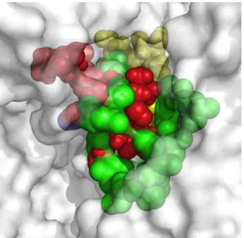
Protein-ligand docking is the most intuitive and powerful tool for structure-based drug dis-covery. Using protein-structure similarity clustering, as described by Waldmann and co-work-ers [32–36], we identified the conservation of structural motifs in the binding region of four target proteins (BSA, Aβ17–42, hIAPP1–37and Aβ16–21, the two last data no shown) with the
that is to say, from stabilizing the native state to destabilizing the oligomeric state and protofila-ment formation, which is the case of interest to this paper.
Influence of Potential Drugs on Fibril Formation Monitored by Thioflavin
T Fluorescence
The Thioflavin T (ThT) fluorescence is widely used to study the kinetics of amyloid fibril for-mation in different proteins [13]. According to the studies consulted, the fibril formation mechanism of BSA is distinguished from others on account of its mechanism of aggregation, which is composed of two states: a logarithmic phase of rapid growth, corresponding to fibril elongation, and a plateau phase of stationary growth, during which there may be continuing morphological changes that can be fitted into a single exponential decay without any lag phase. The ThT fluorescent assay was used to monitor the fibril formation of BSA in the presence or absence of the novel family of chemical chaperones. NCHCHF were added to the ThT assay, in a molar ratio of 1:1 BSA: chaperone.Fig 6Ashowsex situmeasurements of ThT emission intensity with and without chaperones. Triplicate aggregation kinetics were developed at differ-ent times at 65°C. The samples were cooled in order to stop the aggregation process as
Fig 4. Molecular docked model of chemical chaperones (stick representation), located in the amino acids of Aβ17–42segment (cartoon and sphere
views).At 5Ådistance, the amino acid residues surrounding chaperonins (A,B,C,D,E,F,H,IandJ) are represented in orange. The hydrogen bonding interaction between chaperones and amino acid residues of Aβ17–42segment are shown as a dotted line.
doi:10.1371/journal.pone.0135292.g004
described by Vetry et al. [9] BSA aggregation was significantly inhibited with increasing doses of chemical chaperonesA,C,DandE, with A IC50 of 6.34μmol/L, 9.21μmol/L, 6.37μmol/L
and 5.38μmol/L, respectively. Although chaperoneF(40μmol/L) showed the highest IC50
value, this compound delayed the BSA aggregation and also diminished the total amount of fiber.
At a concentration of 100μmol/L (BSA: chaperone ratio of 1:1) chaperonesA,C, D, Band
Ehad a strong inhibitory effect on BSA fibrillation and showed a decrease in ThT fluorescence values between 41% and 14% with respect to BSA alone. At the same concentrations, the chap-eronesFandGincreased BSA fibrillation and had ThT fluorescence values between 22% and 9% greater than BSA alone (Fig 6B).
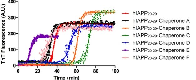
On the other hand, experimental studies have shown that hIAPP20–29fragment is capable of
forming fibrilsin vitro. The ThT fluorescence kinetics of hIAPP20–29fragment with or without
chemical chaperones are presented inFig 7. The experiments were performed at 25°C in 100 mmol/L phosphate buffer, at pH 7.4, containing 100 mmol/L NaCl with continuous stirring. The results indicate that chaperonesB,CandDcan bind to native hIAPP20–29fragment and
delay fibril formation (tlagof 48.2 ± 9.4, 69.9 ± 0.9 and 47.8 ± 1.6, respectively) with a tlaghigher
and significantly different (p<0.05) than the control (tlagof 31.9 ± 3.2). However, chaperones
EandFaccelerate the fibril formation (tlagof 9.7 ± 0.2 and 25.6 ± 0.6 respectively), whereasA
does not present any modulating effect with respect to control (tlagof 33.6 ± 1.3) (Fig 7).
The Effects of Potential Drugs on Aggregation Monitored by Circular
Dichroism Spectroscopy
The conformational differences of the amyloid-like structure of BSA at 70°C in absence and presence of a NCHCHF were investigated by Circular Dichroism (CD) spectroscopy. The
far-Fig 5. Meta-structure of pharmacophore.A structural alignment using Pymol, whereby the interaction zones betweenβ-amiloyd17–42(Aβ17–42), IAPP and Eisenberg’s pharmacophore molecules and the
chaperons were set in position with the interaction zone between BSA and the chaperons. BSA is shown in white at 70% transparency, the interaction zone between the Aβ17–42and the chaperons is shown in red, the
one corresponding to the Eisenberg pharmacophore in blue, with IAPP in yellow and with BSA in green.
Fig 6. A. ThT fluorescence kinetics during amyloid fibrillation of BSA.Experiments were carried out at 65°C in glycine buffer (pH = 3; 0.05M, NaCl 100 mM), in presence or absence of the selected chaperones at molar relation 1:1 of BSA: Chap. (ThT: 24μM). Time dependent changes in ThT intensity was fitted by exponential function (solid line). In all cases, the adjusted R-square values were greater than 0.9. The experiments were carried out in triplicate. The control BSA alone (black square) and BSA plus DMSO (white triangles). All the chaperones (empty squares) were compared with BSA plus DMSO (black squares). B: Normalized maximum values of the ThT fluorescence intensity of BSA fibrilations in presence or absence of chaperones (A,B,C,D,E,FandG).
doi:10.1371/journal.pone.0135292.g006
UV CD spectra of BSA incubated at room temperature shows minima at 222 and 209 nm, characteristic of a helical structure [12]. After incubating the sample at 70°C for 240 minutes, CD spectra showed changes in protein structure composition obtained from CD analysis.
Table 1shows the secondary structure variation on BSA samples at the end (240 min) of incu-bation at 70°C in absence and presence ofA,B,C,D,EandF. We observed the differences of secondary structure composition (expressed in %) at zero and 240 minutes of incubation. The negative signs stand for a decrease in the corresponding structure, as described by Juárez [7,14]. Within the NCHCHF analyzed here, the content ofα-helical structure is negative and larger for BSA alone in comparison to chaperones (F>D>B>C>A>E) (seeTable 1). There was a loss inα-helix structure and the formation of bothβ-sheet and unordered conformation. The amyloid-like fibrils were found to increaseβ-sheet content in the BSA alone more than in the presence of chaperones (F>D>E>B>A>C) (Table 1). It is important to note that without chaperones, the BSA loses its helical shape, which suggests that the former stabilize theα-helix structure and diminish aggregation.
Fig 7. ThT fluorescence kinetics during amyloid fibrillation of IAPP20–29(100μmol/L).Experiments were carried out at 25°C in PBS buffer (pH 7.4; 10
mmol/L; NaCl 100 mmol/L), in presence or absence of the selected chaperones at molar relation 1:1 of IAPP20–29: Chap. (ThT: 24μM). Time dependent
changes in ThT intensity was fitted by sigmoidal function (solid line). The experiments were carried out in triplicate. tlagof fibril formation of the tested
chaperones*p<0,05 show significant differences with regard to control.
doi:10.1371/journal.pone.0135292.g007
Table 1. Values of molar ellipticity differences of the BSA fibrilogenesis assays at 70°C, in absence and presence of chaperones A, D, E, B, F and C, calculated from the CD spectra.
Δ(Θ240min-Θ0min) (%)*
Δ α-helix Δ β-sheet Δothers
BSA -24.2 15.3 7.0
BSA:A(1:1) -20.5 12.9 6.6
BSA:B(1:1) -22.2 13.7 6.2
BSA:C(1:1) -16.5 8.0 5.2
BSA:D(1:1) -19.8 9.0 5.4
BSA:E(1:1) -17.5 7.2 5.3
BSA:F(1:1) -15.1 9.2 5.0
*Differences of molar ellipticity at 240 and zero min. of incubation time.
Electron and Atomic Force Microscopy Revealed the Effects of Potential
Drugs in Fibril Formation
The morphology of the BSA and hIAPP20–29aggregates formed with and without chemical
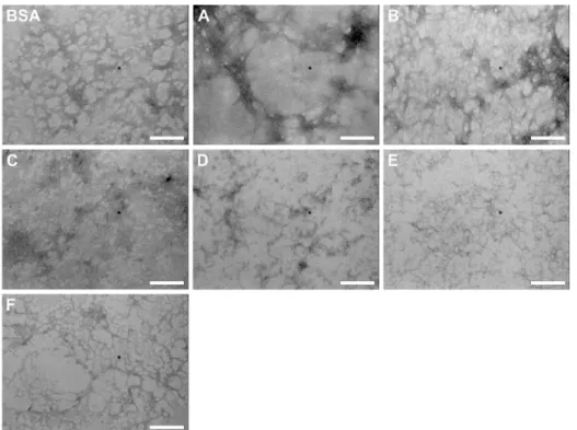
chaperones was examined under transmission electron and atomic force microscopies (TEM and AFM, respectively), in order to check the evolution of fibril formation (Fig 8).Fig 8A
shows images of BSA fibrillation process at the end of the incubation time (240 min at 70°C). In the first micrograph of BSA alone, the presence of long and curly fibers is observed. In con-trast, at room temperature neither fibrils nor other types of aggregates are detected (data not shown), in agreement with previous reported work [12,13]. When the incubation of BSA was carried out in the presence of chaperones it was not possible to distinguish fibers but several amorphous structures were detected. This result was also confirmed by AFM, which provided micrographs at the end of the incubation time of BSA aggregation that show non-fibrillar structures in presence of chaperoneB, as can be observed inFig 8C(BSA alone and BSA:B
(1:1)).
A similar modulating effect on the fibril formation of hIAPP20–29was observed in presence
of chaperonesA,BandC, at a molar ratio of 1:1, at 25°C (Fig 8B). According to TEM micro-graphs, fibers are evidently formed in hIAPP20–29alone, but in presence of chaperonesA,B
andCthese fibrillar structures were not observed. When BSA was pre-aggregated for 24 h at
Fig 8. A. Transmission Electron Microscopy (TEM) micrographs of BSA fibrilogenesis process (75μmol/L), with and without N-(2-aminoethyl)-N'-1-naphthylsuccinamideA; methyl (2-{[4-(1-naphthylamino)-4-oxobutanoyl]amino}ethyl) dithiocarbamate B; (2R)-2-(6-methoxy-2-naphthyl)propanoic acid (Naproxen) C; N-[4-(1-naphthylamino)-4-oxobutanoyl]-β-alanine D and 6-{[4-(1-naphthylamino)-4-oxobutanoyl]amino} hexanoic acid E, at 70°C. B: Transmission Electron Microscopy (TEM) micrographs of hIAPP20–29fibrilogenesis process with and without N-(2-aminoethyl)-N'-1-naphthylsuccinamide A;
methyl (2-{[4-(1-naphthylamino)-4-oxobutanoyl] amino} ethyl) dithiocarbamate B and (2R)-2-(6-methoxy-2-naphthyl)propanoic acid (Naproxen) C; at 25°C. Molar ratio hIAPP20–29: chaperones was 1:1. C: Atomic Force Microscopy (AFM) micrographs of BSA fibrilogenesis process (75μmol/L), with and without B,
at 70°C. Molar ratio BSA:chaperones was 1:1.
doi:10.1371/journal.pone.0135292.g008
75°C before incubation with chemical chaperonesA,B,C,D, andEat 1:1 molar ratio, all of them were capable of disaggregating mature fibrils of BSA (Fig 9).
Cytoprotective Effect of the Chaperones Family on Cerebellar Granule
Cells
Cytoprotective properties of chaperonesBandDwere evaluated in cerebellar granule cells (CGC) after exposal to hIAPP20–29, a toxic protein (in monomeric and aggregated form). This
was done by means of viability and apoptosis cellular assays. In all assays,Hwas used as refer-ence due to its reported cytoprotective effect [37]. InFig 10, the estimation results of cell viabil-ity by means of MTT assay is observed. The data was analyzed with statistical test-way
ANOVA, followed by a least significant difference (LSD) posteriori test. The results indicate that there are significant differences between the cytotoxic effect of monomeric and aggregated hIAPP20–29forms (p<0.05). This protein fragment in its monomeric form is nearly three times more cytotoxic to the CGC than in its aggregated form.
Moreover, when cells were exposed to hIAPP20–29in monomeric form and to the
chaper-ones of interest, the viability results were twice higher for the chaperoneBin the two molar ratios tested, being significantly different from the rest of the other compounds tested (p<0.05). Also, the CGC viability in presence of chaperoneDwas almost twice as high for a molar ratio of 1:0.3 (p<0.05). In contrast,H, used as reference, did not show any cytoprotec-tive effect.
In the case of the aggregated form of hIAPP20–29in presence of chaperoneD, the CGC
via-bility did not differ significantly from cells exposed to hIAPP20–29(p>0.05). As expected,H showed a marked cytoprotective effect that differed statistically from chaperonesBandD
Fig 9. Transmission Electron Microscope (TEM) images of the BSA fibril formed with or without the chaperones (A, B, C, D, E, F and G) at a molar relation of BSA: chaperones 1:1, in buffer Tris (pH = 7.4, 20 mM) and 75°C.Scale bar 200 nm.
(p<0.05).Fig 11shows the results of the cell apoptosis assays, in which the level of caspase-3 was measured by an immunofluorescence assay. The caspase-3 levels of CGC exposed to monomeric or aggregated hIAPP20–29were not significantly different between them (p>0.05) and they were the highest apoptotic levels observed. Contrastingly, in presence of chaperones, the caspase-3 levels of CGC exposed to hIAPP20–29diminished (p<0.05).
When the cells were exposed to the monomeric form of hIAPP20–29in presence of
chaper-oneB(molar ratio hIAPP20–29:B, 1: 0.01 and 1: 0.3), the caspase-3 levels were similar to those
obtained with the protein in its monomeric form (p>0.05). However, in the case of chaperone
D, the caspase-3 levels were lower, but without reaching the levels obtained forHin a molar ratio of 1:0.3. Lower caspase-3 levels were achieved for a molar ratio hIAPP20–29: chaperone 1:1
Fig 10. Cell Viability of CGC exposed to hIAPP20–29, in monomeric and aggregated form, through MTT assay, with and without
N-[4-(1-naphthylamino)-4-oxobutanoyl]-β-alanine D; methyl (2-{[4-(1-naphthylamino)-4-oxobutanoyl]amino}ethyl) dithiocarbamate B and H used as reference; at different molar ratio hIAPP20–29:chaperones.All the experiments have the same concentration of hIAPP20–29
doi:10.1371/journal.pone.0135292.g010
Fig 11. Cell Apoptosis of CGC exposed to hIAPP20–29, in monomeric and aggregated form, with and without
N-[4-(1-naphthylamino)-4-oxobutanoyl]-β-alanine D; methyl (2-{[4-(1-naphthylamino)-4-oxobutanoyl]amino}ethyl) dithiocarbamate B and H used as reference; at different molar ratio hIAPP20–29:chaperones.In all assays caspase-3 levels were measured by immunofluorescence.
doi:10.1371/journal.pone.0135292.g011
for both chaperones (p<0.05). Thus, a six to seven times higher survival average of CGC was observed in the presence of chaperonesBas forD, with regards to exposition to hIAPP20–29
alone.
Reconditioning Activity of Chaperones B, C and D on CGC
In this study the effect of chaperonesB,CandDon the survival of CGC, exposed to a pro-apo-ptotic stimulus, in this case the reduction of potassium concentration (from 25 mmol/L to 5 mmol/L) was evaluated. In this experiment the CGC were exposed individually to these chap-erones at different times: at initial time (Group I) and after 4 h of stimulus induction (Group II). Two controls were also used at 25 mmol/L (K25) and 5 mmol/L (K5).
Group I (Fig 12), treated with chaperonesB,CandD, showed a considerable reduction in the caspase-3 levels, with regard to control K5. This reduction was of 92%, 80% and 78% for the samples treated withB,CandD, respectively. With regard to control K25, it was observed that chaperoneBreduced the caspase-3 level in 84%, whileCandDdid so in a 57% and a 52%, respectively.
In group II (Fig 13), treated with chaperonesB,CandD, the caspase-3 levels were lower in a 56%, 66% and 26%, respectively, relative to the control group K5, which is associated with increased cell survival. The CGC withBandCshow values that are very close to those of con-trol test K25, whereas the cells exposed toDhad an activity that is 94% higher. These results indicate that the chaperones tested were able to reverse the apoptosis induced in CGC through a low potassium environment.
Discussion
In this article, we show how a novel chemical chaperones family (NCHCHF) can regulate fibrillogenesis of target proteins, as they interact with both the native and fibrillar structures. ChaperonesA,B,CandDarrest the aggregation of BSA and hIAPP20–29and prevent the
Fig 12. Cell Apoptosis of CGC exposed to pro-apoptotic stimulus as low potassium concentration (K5) in the presence of N-[4-(1-naphthylamino)-4-oxobutanoyl]-β-alanine D; methyl (2-{[4-(1-naphthylamino)-4-oxobutanoyl] amino}ethyl) dithiocarbamate B and C used as reference.In all assays caspase-3 levels were measured by inmunofluorescence.
formation of potentially cytotoxic species. Conversely, chaperonesE,FandGcaused the t1/2to
be prolonged in the BSA aggregation. It is worth noting that in the case of hIAPP20–29,
chaper-onesEandFaccelerate the process of fibril formation. One possible explanation for this
phe-nomenon is related to the fact that chaperones stabilize the structure of the fiber, shifting the equilibrium towards fiber formation and to halt self-catalysis and the creation of cytotoxic olig-omers as a consequence of fiber formation. The differential effects of chemical chaperones on the fibril formation of BSA and hIAPP20–29are similar to the results reported for Blancas y
coworkers on the t50 value of fibril formation kinetics of AL proteins [38].
That the ThT intensity decreases in the presence of chaperones, indicates a decrease inβ -rich amyloid-like aggregates, which is corroborated when exposed to far-UV CD (Fig 6and
Table 1). The TEM experiments show that without chaperones BSA can form long and curled fibers, but not thick ones. This is consistent with a number of studies [7,9,13]. When BSA at 70°C is treated with chaperonesA,B,C,D,E,FandG, smaller fibers, amorphous aggregates,
and ring shaped oligomers are formed. Holm, et al.[13] have found that the structure of the fibrillation intermediaries is highly dependent on the protein-solvent interactions, and the con-ditions of the medium. Therefore, we assume that the structural variation found in the treated samples is due to a change in the type of interactions that the proteins establish with the solvent and chaperones. ChaperonesA,B,CandDproved to be more effective than chaperonesE,F
andGin the disaggregation processes of BSA [22,39].
The molecular simulation indicates that the interaction between BSA and the chaperones is mainly of two kinds: hydrophobic and hydrogen bonding. A similar behaviour has been previ-ously reported for other small molecules [22,40–45]. These interactions, furthermore, play a crucial role in the binding process of chaperones with BSA, which is consistent with our experi-mental results. The bioinformatic results and the kinetics experiexperi-mental results are correlated (Figs3and6) since chaperones decrease the fibril concentration and its formation rate. This process evidently occurs on account of the interaction of these compounds with protein thus modulating the transformation fromα-helix toβ-amyloids-like structure. In accordance to experimental results, we demonstrated that this set of chemical chaperones can influence pro-tein aggregation and fibril formation, as well as protecting cerebellar granule cells from apopto-sis upon exposure to hIAPP20–29. A possible explanation of these results, based on virtual
screening studies, is that these chaperones interact with the amino acids of the so-called
Fig 13. Cell Apoptosis of CGC exposed to pro-apoptotic stimulus as low potassium concentration (K5) during 4 h and later, the chaperones N-[4-(1-naphthylamino)-4-oxobutanoyl]-β-alanine D; methyl (2-{[4-(1-naphthylamino)-4-oxobutanoyl]amino}ethyl) dithiocarbamate B and C (reference), were inoculated.In all assays caspase-3 levels were measured by inmunofluorescence.
doi:10.1371/journal.pone.0135292.g013
amyloid-like steric zipper zone (through Van der Waals forces, interactionsππand H-bond),
which are the binding sites of the proteins superposed in the structure, defined as meta-pharmacophore by us (Fig 5). Although the steric zippers cannot have all the features of the full amyloid target proteins, they share many properties with them, thus shedding light upon the functional attributes of specific and broad-spectrum amyloid binders. These findings agree with and build on the work of Eisenberg et al.[2] by arguing that there might be a universal pharmacophore in proteins, which forms a common meta-structure in which chemical chaper-ones can be bound even if it have different effects. From this follows that, being akin to hIAPP in important respects, BSA can serve as an affordable and accessible model protein that can be used when evaluating new therapeutic agents for conformational diseases.
The development of new compounds with anti-amyloid activity is of great interest for neu-roscientists and diabetes researchers. While all of the chaperones tested showed a modulating effect of BSA and hIAPP20–29aggregation, the mechanisms by which these bind to the protein,
either to arrest, accelerate the fibrillation process, or to disaggregate fibers is shown in Figs6,8
and9. Further research usingin vivostudies could be of great use for determining the mecha-nism of action of these chaperones, thus laying a stronger foundation from whence to develop other compounds.
Mechanistic Insights
While several studies focus on finding novel therapeutic agents to fight conformational dis-eases, they are still only moderately successful [22–24]. Recent work on the atomic structure of a segment of the steric zipper of the amyloid-beta (Aβ) pointed out at new compounds with potential as therapeutic agents, which diminish the cytotoxicity of the Aβbut do not reduce the formation of fibers [46].
In this article, we show how by selecting compounds based on the two states of the amyloid protein (native and amyloid pharmacophore structure) we managed to obtain chaperones that bind to the native state, to the cytotoxic oligomers and to the amyloid fibrils (Figs6,9and14
andTable 1). The novel chemical chaperones can regulate fibril formation processes by binding to the native state, minimizing the formation of amorphous aggregates, as well as cytotoxic oligomers. It is worth mentioning that these compounds also adhere to the fibrils and stabilize them. ChaperoneFin particular was found to accelerate the formation of hIAPP20–29fibrils by
binding to cytotoxic oligomers (Fig 7). In this article we demonstrate the physiopathological conditions in which a set of chaperones can be used. We observed that in acute processes, with abundance of cytotoxic oligomers, chaperoneFis the most suitable for arresting the former, forming fibers and thus preventing apoptosis caused by membrane pores. For chronic pro-cesses, chaperonesA,B,CandDwere found to be apposite for stabilizing the monomer and reducing the formation of fibres (Table 1, Figs5–14). This is extremely relevant as it allows for a close examination of the fiber-formation phenomena and, potentially, to control cytotoxicity. If taken to the next stage—assaying the dynamics of these chaperones in living organisms in order to learn their effects within cognitive and metabolic processes—this could lead to the development of therapeutic agents derived from this chaperone family that could help control the devastating effects of conformational diseases. Finally, the findings reported in this article are useful for encouraging research in reverse engineering, as well as building increasingly effective chemical libraries for the treatment of conformational diseases.
Materials and Methods
MilliQ water (18.2 MOresistance at 25°C) for the preparation of all working solutions is used.
sulphoxide (DMSO) and 3- (4,5-dimethyl-2-thiazoyl) -2,5-diphenyltetrazolium bromide (MTT) from Sigma-Aldrich were used. All buffers were freshly prepared before every aggrega-tion experiment. Thioflavin (ThT) was purchased from Sigma-Aldrich. Bovine serum albumin (BSA, fraction V) was purchased from Roche and stored at 4°C. Stock solution of BSA
(700μmol/L) was freshly prepared by dissolving it in glycine buffer (50 mmol/L, pH 3; NaCl
100 mmol/L), and then passed through acrodisc syringe filters with 0.45μm pores (Sigma
Aldrich). Accurate protein concentration was determined by using BSA molar extinction coef-ficient at 280 nm (43 824 Lmol−1 cm−1).[12] hIAPP fragment 20–29 (hIAPP20–29) was
synthe-sized according to the procedures described in the literature.[47–49] This peptide was purified by semi preparative reverse high performance liquid chromatography (SP-HPLC) and its purity was greater than 95%. The expected molecular mass (MM: 1029 Da) was confirmed by mass spectrometry (ESI-MS). Chemical chaperones (A, B, D, E, F and G) were gently provided from Neurosciences Center of Cuba [27,28]. Solutions of each chaperone (25 mmol/L) were prepared in DMSO and stored at -20°C. Naproxen and curcumin, used as reference substances, were dissolved in DMSO and ethanol absolute (25 mmol/L), and then stored at -20°C y a -70°C, respectively. Cerebellar granule cells (CGC) cultures (8 DIV), which were prepared as previously described [44–46], were donated by Dr. J. Moran Institute of Cellular Physiology, UNAM, Mexico. Kinetic assays were monitored by fluorescence spectroscopy and microscopy techniques. Fluorescence measurements were performed on Cary Eclipse fluorescence
Fig 14. Proposal mechanism of chaperone on protein aggregation/disaggregation.Novel chemical chaperones family can regulate fiber formation processes by binding to the native state, minimizing the formation of amorphous aggregates, as well as cytotoxic oligomers.
doi:10.1371/journal.pone.0135292.g014
spectrophotometer (methacrylate cell) and Infinite M1000, TECAN (96-well plates, polysty-rene, Costar 3615, Special Optics Plate), equipped with temperature control. Transmission electron microscopy (TEM, JEM-1010, JEOL, 80 kV, magnifications between 1 000 and 120 000) and atomic force microscopy (AFM, Veeco Bioscope, Digital Instruments, mounted on an inverted microscope) were performed.
Experimental Procedure
Kinetic assays of fiber formation. Fluorescence measurements. Aggregation kinetics
were followed at 65°C at different times.
BSA. Stock solution of BSA (700μmol/L) was diluted using glycine buffer (50 mmol/L,
pH 3; NaCl 100 mmol/L) to yield a final BSA concentration of 100μmol/L as described by
Bhattacharya, et al.[12]. Aliquots of 250μL of mixtures of BSA:chemical chaperones (A,B,C,
D,E,FandG) (1:1) were prepared and mixed with 1μL of ThT (final concentration 20μmol/
L) and incubated during different times at 65°C. The samples were cooled in order to stop the aggregation process as described by Vetry et al [9].These aliquots were deposited into the wells of a 96-well plate, by sextuplicate. For detecting the fiber formation, ThT fluorescence intensity was measured in an Infinite M1000 spectrophotometer, at excitation and emission wavelengths of 450 and 482 nm, respectively. Signals were corrected with the background signal (ThT blank).
Fiber Disaggregation Experiments. BSA was pre-aggregated for 24 h at 75°C before
incu-bation with chemical chaperonesA,B,C,D, andEat 1:1 molar ratio, in buffer Tris (pH = 7.4, 20 mmol/L). The samples were applied (6μL) to 400-mesh copper grids coated with carbon/
collodion film, and left for 3 minutes. Excess solution on the grids was absorbed with filter paper, grids were stained with 3μL of 1% filtered uranyl acetate for 5 min, and staining solution
was absorbed with filter paper again. After air drying, grids were examined with an electron microscope JEM-1010, JEOL, 80 kV, magnifications between 1 000 and 120 000.
hIAPP20–29. Stock solution of hIAPP20–29(5.8 mmol/L) was diluted using PBS buffer (100
mmol/L, pH 7,4 with sodium chloride 100 mmol/L) to prepare experiments at a final hIAPP20–29
concentration of 100μmol/L. Experiments were carried out in the methacrylate cells (4.5 mL,
10 mm path length, Sigma Aldrich) in the presence of 1μL of Th-T (20 mmol/L). Aliquots of
1.5 mL of mixtures of hIAPP20–29:chemical chaperones (25 mmol/L,A,B,C,D,E,FandG) (1:1)
were incubated at 25°C with stirring. IF values were recorded continuously toλemis = 482 nm, exciting at 450 nm with a bandwidth of 5 nm in a Cary Eclipse spectrofluorometer. Signals were corrected with the background signal (ThT blank). The tlag was obtsined by fitting the data to a sigmoidal function (Botzmann function), using Origin Software.
IC50. Stock solution of BSA (700μmol/L) was mixed with chaperonesA,B,C,DandE
solutions (25 mmol/L) to prepare mixtures of BSA (300μmol/L): Chaperones (0, 15, 150, 300,
750 and 1500μmol/L) and ThT (24μmol/L) to reach a final volume of 250μL into the wells of
a 96-well plate. The plate was read in an Infinite M1000 spectrofluorometer every 20 min at 42°C. All the ThT fluorescence experiments were replicated six times. For the calculation of IC50, the maximum velocities were computed and plotted against the Log Chaperone concentrations.
Microscopy measurements. Transmission Electron Microscopy (TEM). The samples
BSA:chaperones A, B, C, D, E, F, and G (1:1) and BSA from kinetic assays at 65°C were diluted 1/10 or 1/20 in glycine buffer (50 mmol/L, pH 3; NaCl 100 mmol/L) and applied (6μL) to
400-mesh copper grids coated with carbon/collodion film, and left for 3 minutes. Excess solu-tion on the grids was absorbed with filter paper, grids were stained with 3μL of 1% filtered
drying, grids were examined with an electron microscope JEM-1010, JEOL, 80 kV, magnifica-tions between 1 000 and 120 000.
Atomic force microscopy (AFM). The samples BSA:chaperonesA,B,C,D,E,F, andG
(1:1) and BSA from kinetic assays at 65°C were diluted with milli-Q water in a 1:9 (v/v). 100μL
of the diluted sample were deposited on a mica and dried under vacuum over moisture-free sil-ica gel. After drying msil-icas, 500μL of milli-Q water was added and allowed to stand for 5 min
before removing the excess of water using filter paper. The micas were dried again for 24 hours and deposited over glass slides used in light microscopy. The samples were observed with an atomic force microscope (Bioscope, Digital Instruments, Santa Barbara CA, USA, equipped with a Nanoscope IIIa controller) working in contact mode with a silicon nitride probe (DNP, Veeco, USA). Microscope works on an inverted Diaphot 200 (Nikon) microscope with a 100 microns scanner and silicon nitride tips with 50 nm radius of curvature (DNP–Veeco). Sweep speeds used between 1969–1285 Hz, 10 nN force and 0.5 arbitrary units of gain.
Cell viability and apoptosis of cerebellar granule cells (CGC). Exposure of CGC cultures
to hIAPP20–29, as cytotoxic agent. Solutions of hIAPP20–29in PBS buffer (pH 7.4, 0.1 mol/L,
0.1 mol/L NaCl) were prepared. Monomeric and aggregated peptide concentration were 5.8 mmol/L and 32.6 mmol/L, in DMSO, respectively. Viability tests were performed according to the procedures reported by Moran et al. [50]. In particular, in this experiment, the viability of CGC cultures (8 DIV) exposed to hIAPP20–29, in its monomeric and aggregated form, was
eval-uated. CGC cultures were exposed or not to chaperonesBandDat different molar ratios hIAPP20–29: chaperones (1:0.01, 1:0.3 or 1:1) andHas reference. In brief, in all samples of
CGC, half the volume of medium is removed to add, independently, hIAPP20–29(monomeric
or aggregated): chaperone at different molar ratios. Next, the enriched cell culture was reintro-duced and again incubated at 37°C. After 4 h, the apoptosis was estimated by measuring cas-pase-3 activity (fluorogenic method according to Moran et al.) or after 20 h, the viability levels were assessed by the MTT test.[51]
Exposure of CGC cultures to low potassium medium. CGC cultures (8DIV) containing
KCl (25 mmol/L, K25) were transferred to the same medium containing KCl (5 mmol/L, K5). The chaperonesB,Dand curcumin as reference (25μmol/L) were independently added to
cul-tures, at the initial time (Group I) or after 4 h (Group II) of the induction of the pro-apoptotic stimulus (K5) and incubated 4 h additional. Also, negative (K5) and positive (K25) controls were carried out. Apoptosis was estimated by measuring caspase-3 activity (fluorogenic method according to Moran et al.) [51].
Molecular docking
The crystallographic structure of the BSA was downloaded from Protein Data Bank (PDB) (3V03 code), with a resolution of 2.7 Å. The3D fibril structure of the Aβ17-42amyloid peptide,
as a NMR image, was downloaded from the PDB (2BEG code) with 10 configurations [31].The structures of the chemical chaperones were refined usingAvogadrosoftware and converted into pdbqt format withAutodock Tools, considering all ligand bonds as flexible. These com-pounds were docked with both proteins usingAutoDock Vina, an interactive molecular graph-ics program used to understand the drug-protein interaction.Vinauses a scoring function, which can be seen as an attempt to approximate the standard chemical potentials of the system, in order to search for the optimum binding site of small molecules into the protein. For these simulations all amino acids of tested protein were considered in rigid position. Folders with the data for the calculation were compiled each file of which contained the BSA and Aβ17-42
config-urations (pdbqt) and the corresponding ligand configuration (pdbqt). During docking, a maxi-mum of 10 conformers was considered for each molecule. To recognize the binding sites in
BSA or Aβ17-42a blind docking was carried out. The grid size set to 108, 100, and 100 along X,
Y and Z axes with 1.00 Å grid spacing. Thus, each ligand can move freely across the surface of the BSA or Aβ17-42. The center of the grid was set to 38.00, 25.00, and 36.00 Å. The
visualiza-tion and determinavisualiza-tion of the interacvisualiza-tion zones between the amino acids and all tested com-pounds were carried out using the molecular graphic program (Chimera). All calculations were performed on a cluster of 10 computers (30 CPU) with Linux as operating system.
Statistical analysis. STATISTICA software, version 8, was used and the significance level
was set at 0.05. Experimental data from kinetic assays were processed using OriginPro 8.5 program.
Supporting Information
S1 Table. Energy of the binding complex (chaperones-BSA) and interaction sites of BSA-chaperonines obtained from molecular docking using AutoDockingVina.
(DOC)
Acknowledgments
We are indebted to Dinorah Gamero, Denise Bixler and Annia Hernández Reyes for helping with the initial Tht assays. The authors would like to acknowledge the experimental support of Dr. Julio Morán and Dr. Liliana Quintanar. We also wish to thank Dr. Fabio Salamanca, Dr. Ramón Paniagua and Dr. Javier Torres for their support. The contributions of Anaclara Cas-tro-Santana for the proofreading of the manuscript and Rosa Angélica Castillo-Rodríguez for her help in the artwork and for her participation in fruitful discussions are greatly appreciated.
Author Contributions
Conceived and designed the experiments: MS-C CR-T MMA-B. Performed the experiments: MS-C IF AB RL-M SR-M GD RP-P LFJ-G MD-D FV LR-A KP-H MLS-V AP-P AP-C CR-T MMA-B. Analyzed the data: MS-C IF AB RL-M GD KP-H MLS-V CR-T MMA-B. Contrib-uted reagents/materials/analysis tools: MS-C EG-M NFA-B SR-M LFJ-G CR-T SI-A MMA-B. Wrote the paper: MS-C CR-T MMA-B. Contributed with helpful discussions: MS-C IF RL-M GD EG-M NFA-B LFJ-G CR-T KP-H SI-A MMA-B.
References
1. Gidalevitz T, Ben-Zvi A, Ho KH, Brignull HR, Morimoto RI (2006) Progressive disruption of cellular pro-tein folding in models of polyglutamine diseases. Science 311: 1471–1474. 1124514 [pii];doi:10.1126/ science.1124514PMID:16469881
2. Landau M, Sawaya MR, Faull KF, Laganowsky A, Jiang L, Sievers SA, et al. (2011) Towards a pharma-cophore for amyloid. PLOS Biol 9: e1001080. doi:10.1371/journal.pbio.1001080 ;PBIOLOGY-D-11-00136 [pii]. PMID:21695112
3. Welch WJ (2004) Role of quality control pathways in human diseases involving protein misfolding. Semin Cell Dev Biol 15: 31–38. doi:10.1016/j.semcdb.2003.12.011;S1084952103001162 [pii]. PMID:
15036204
4. Westermark P, Benson MD, Buxbaum JN, Cohen AS, Frangione B, Ikeda S, et al. (2007) A primer of amyloid nomenclature. Amyloid 14: 179–183. 781269331 [pii];doi:10.1080/13506120701460923
PMID:17701465
5. Herczenik E, Gebbink MF (2008) Molecular and cellular aspects of protein misfolding and disease. FASEB J 22: 2115–2133. fj.07-099671 [pii];doi:10.1096/fj.07-099671PMID:18303094
7. Juarez J, Taboada P, Mosquera V (2009) Existence of different structural intermediates on the fibrilla-tion pathway of human serum albumin. Biophys J 96: 2353–2370. S0006-3495(09)00322-1 [pii];doi:
10.1016/j.bpj.2008.12.3901PMID:19289061
8. Khurana R, Ionescu-Zanetti C, Pope M, Li J, Nielson L, Ramirez-Alvarado M, et al. (2003) A general model for amyloid fibril assembly based on morphological studies using atomic force microscopy. Bio-phys J 85: 1135–1144. S0006-3495(03)74550-0 [pii];doi:10.1016/S0006-3495(03)74550-0PMID:
12885658
9. Vetri V, D'Amico M, Fodera V, Leone M, Ponzoni A, Sberveglieri G, et al. (2011) Bovine Serum Albumin protofibril-like aggregates formation: solo but not simple mechanism. Arch Biochem Biophys 508: 13–
24. S0003-9861(11)00048-8 [pii];doi:10.1016/j.abb.2011.01.024PMID:21303653
10. Eisenberg D, Nelson R, Sawaya MR, Balbirnie M, Sambashivan S, Ivanova MI, et al. (2006) The struc-tural biology of protein aggregation diseases: Fundamental questions and some answers. Acc Chem Res 39: 568–575. doi:10.1021/ar0500618PMID:16981672
11. Babcock JJ, Brancaleon L (2013) Bovine serum albumin oligomers in the E- and B-forms at low protein concentration and ionic strength. Int J Biol Macromol 53: 42–53. S0141-8130(12)00427-8 [pii];doi:10. 1016/j.ijbiomac.2012.10.030PMID:23148944
12. Bhattacharya M, Jain N, Mukhopadhyay S (2011) Insights into the mechanism of aggregation and fibril formation from bovine serum albumin. J Phys Chem B 115: 4195–4205. doi:10.1021/jp111528c
PMID:21417250
13. Holm NK, Jespersen SK, Thomassen LV, Wolff TY, Sehgal P, Thomsen LA, et al. (2007) Aggregation and fibrillation of bovine serum albumin. Biochim Biophys Acta 1774: 1128–1138. S1570-9639(07) 00150-1 [pii];doi:10.1016/j.bbapap.2007.06.008PMID:17689306
14. Juarez J, Lopez SG, Cambon A, Taboada P, Mosquera V (2009) Influence of electrostatic interactions on the fibrillation process of human serum albumin. J Phys Chem B 113: 10521–10529. doi:10.1021/ jp902224dPMID:19572666
15. Hartl FU, Bracher A, Hayer-Hartl M (2011) Molecular chaperones in protein folding and proteostasis. Nature 475: 324–332. nature10317 [pii];doi:10.1038/nature10317PMID:21776078
16. Papp E, Csermely P (2006) Chemical chaperones: mechanisms of action and potential use. Handb Exp Pharmacol (172: ): 405–416. PMID:16610368
17. Mogk A, Tomoyasu T, Goloubinoff P, Rudiger S, Roder D, Langen H, et al. (1999) Identification of ther-molabile Escherichia coli proteins: prevention and reversion of aggregation by DnaK and ClpB. EMBO J 18: 6934–6949. doi:10.1093/emboj/18.24.6934PMID:10601016
18. Seong IS, Oh JY, Lee JW, Tanaka K, Chung CH (2000) The HslU ATPase acts as a molecular chaper-one in prevention of aggregation of SulA, an inhibitor of cell division in Escherichia coli. FEBS Lett 477: 224–229. S0014-5793(00)01808-1 [pii]. PMID:10908725
19. Zahn R, Perrett S, Fersht AR (1996) Conformational states bound by the molecular chaperones GroEL and secB: a hidden unfolding (annealing) activity. J Mol Biol 261: 43–61. S0022-2836(96)90440-4 [pii]; doi:10.1006/jmbi.1996.0440PMID:8760501
20. Scheibel T, Buchner J (2006) Protein aggregation as a cause for disease. Handb Exp Pharmacol 199–
219. PMID:16610361
21. Tomoyasu T, Tabata A, Ishikawa Y, Whiley RA, Nagamune H (2013) Small heat shock protein AgsA: an effective stabilizer of enzyme activities. J Biosci Bioeng 115: 15–19. S1389-1723(12)00323-4 [pii]; doi:10.1016/j.jbiosc.2012.08.001PMID:22929984
22. Hard T, Lendel C (2012) Inhibition of amyloid formation. J Mol Biol 421: 441–465. S0022-2836(12) 00006-X [pii];doi:10.1016/j.jmb.2011.12.062PMID:22244855
23. Sacchettini JC, Kelly JW (2002) Therapeutic strategies for human amyloid diseases. Nat Rev Drug Dis-cov 1: 267–275. doi:10.1038/nrd769PMID:12120278
24. Bartolini M, Andrisano V (2010) Strategies for the inhibition of protein aggregation in human diseases. Chembiochem 11: 1018–1035. doi:10.1002/cbic.200900666PMID:20401887
25. Hard T, Lendel C (2012) Inhibition of amyloid formation. J Mol Biol 421: 441–465. S0022-2836(12) 00006-X [pii];doi:10.1016/j.jmb.2011.12.062PMID:22244855
26. Breitner JC, Welsh KA, Helms MJ, Gaskell PC, Gau BA, Roses AD, et al. (1995) Delayed onset of Alz-heimer's disease with nonsteroidal anti-inflammatory and histamine H2 blocking drugs. Neurobiol Aging 16: 523–530. 019745809500049K [pii]. PMID:8544901
27. Sablón CM, Rodriguez-Tanty C, Perera PA, Rivera MS, Perez PR, López BRM, et al. (2010) Procedi-miento de obtención de nuevos derivados de naftaleno para el diagnóstico in vivo de la enfermedad de alzheimer. Google Patents.EP2436666A2, EP2436666A4, EP2436666B1, EP2860169A2,
US20120321560, WO2010118706A2, WO2010118706A3, WO2010118706A4
28. Sablón CM, Rodriguez-Tanty C, Altamirano-Bustamante MM, Rivillas ALA, López BRM, Lara MR, et al. (2014) Chemical chaperonins as novel molecular modulators of beta protein aggregation present in conformational diseases. Google Patents. WO20141313
29. Tjernberg LO, Callaway DJ, Tjernberg A, Hahne S, Lilliehook C, Terenius L, et al. (1999) A molecular model of Alzheimer amyloid beta-peptide fibril formation. J Biol Chem 274: 12619–12625. PMID:
10212241
30. Tjernberg LO, Naslund J, Lindqvist F, Johansson J, Karlstrom AR, Thyberg J, et al. (1996) Arrest of beta-amyloid fibril formation by a pentapeptide ligand. J Biol Chem 271: 8545–8548. PMID:8621479
31. Luhrs T, Ritter C, Adrian M, Riek-Loher D, Bohrmann B, Dobeli H, et al. (2005) 3D structure of Alzhei-mer's amyloid-beta(1–42) fibrils. Proc Natl Acad Sci U S A 102: 17342–17347. 0506723102 [pii];doi:
10.1073/pnas.0506723102PMID:16293696
32. Breinbauer R, Vetter IR, Waldmann H (2002) From protein domains to drug candidates-natural prod-ucts as guiding principles in the design and synthesis of compound libraries. Angew Chem Int Ed Engl 41: 2879–2890. doi:10.1002/1521-3773(20020816)41:16<2878::AID-ANIE2878>3.0.CO;2-BPMID:
12203413
33. Koch MA, Breinbauer R, Waldmann H (2003) Protein structure similarity as guiding principle for combi-natorial library design. Biol Chem 384: 1265–1272. doi:10.1515/BC.2003.140PMID:14515987
34. Koch MA, Waldmann H (2005) Protein structure similarity clustering and natural product structure as guiding principles in drug discovery. Drug Discov Today 10: 471–483. S1359644605034197 [pii];doi:
10.1016/S1359-6446(05)03419-7PMID:15809193
35. Konrat R (2009) The protein meta-structure: a novel concept for chemical and molecular biology. Cell Mol Life Sci 66: 3625–3639. doi:10.1007/s00018-009-0117-0PMID:19690801
36. Naranjo Y, Pons M, Konrat R (2012) Meta-structure correlation in protein space unveils different selec-tion rules for folded and intrinsically disordered proteins. Mol Biosyst 8: 411–416. doi:10.1039/ c1mb05367aPMID:22108787
37. Yang F, Lim GP, Begum AN, Ubeda OJ, Simmons MR, Ambegaokar SS, et al. (2005) Curcumin inhibits formation of amyloid beta oligomers and fibrils, binds plaques, and reduces amyloid in vivo. J Biol Chem 280: 5892–5901. M404751200 [pii];doi:10.1074/jbc.M404751200PMID:15590663
38. Blancas-Mejia LM, Hammernik J, Marin-Argany M, Ramirez-Alvarado M (2015) Differential effects on light chain amyloid formation depend on mutations and type of glycosaminoglycans. J Biol Chem 290: 4953–4965. M114.615401 [pii];doi:10.1074/jbc.M114.615401PMID:25538238
39. Lendel C, Bertoncini CW, Cremades N, Waudby CA, Vendruscolo M, Dobson CM, et al. (2009) On the mechanism of nonspecific inhibitors of protein aggregation: dissecting the interactions of alpha-synu-clein with Congo red and lacmoid. Biochemistry 48: 8322–8334. doi:10.1021/bi901285xPMID:
19645507
40. Bulic B, Pickhardt M, Khlistunova I, Biernat J, Mandelkow EM, Mandelkow E, et al. (2007) Rhodanine-based tau aggregation inhibitors in cell models of tauopathy. Angew Chem Int Ed Engl 46: 9215–9219. doi:10.1002/anie.200704051PMID:17985339
41. Bulic B, Pickhardt M, Schmidt B, Mandelkow EM, Waldmann H, Mandelkow E (2009) Development of tau aggregation inhibitors for Alzheimer's disease. Angew Chem Int Ed Engl 48: 1740–1752. doi:10. 1002/anie.200802621PMID:19189357
42. Larbig G, Pickhardt M, Lloyd DG, Schmidt B, Mandelkow E (2007) Screening for inhibitors of tau protein aggregation into Alzheimer paired helical filaments: a ligand based approach results in successful scaf-fold hopping. Curr Alzheimer Res 4: 315–323. PMID:17627489
43. Pickhardt M, Gazova Z, von Bergen M, Khlistunova I, Wang Y, Hascher A, et al. (2005) Anthraquinones inhibit tau aggregation and dissolve Alzheimer's paired helical filaments in vitro and in cells. J Biol Chem 280: 3628–3635. M410984200 [pii];doi:10.1074/jbc.M410984200PMID:15525637
44. Pickhardt M, Larbig G, Khlistunova I, Coksezen A, Meyer B, Mandelkow EM, et al. (2007) Phenylthiazo-lyl-hydrazide and its derivatives are potent inhibitors of tau aggregation and toxicity in vitro and in cells. Biochemistry 46: 10016–10023. doi:10.1021/bi700878gPMID:17685560
45. Pickhardt M, Biernat J, Khlistunova I, Wang YP, Gazova Z, Mandelkow EM, et al. (2007) N-phenyla-mine derivatives as aggregation inhibitors in cell models of tauopathy. Curr Alzheimer Res 4: 397–402. PMID:17908042
46. Jiang L, Liu C, Leibly D, Landau M, Zhao M, Hughes MP, et al. (2013) Structure-based discovery of fiber-binding compounds that reduce the cytotoxicity of amyloid beta. Elife 2: e00857. doi:10.7554/ eLife.00857;00857 [pii]. PMID:23878726
glycol-polystyrene (PEG-PS) graft supports, with 1,8-diazobicyclo [5.4.0]-undec-7-ene (DBU) deprotec-tion. Pept Res 9: 106–113. PMID:8875589
48. Kates SA, McGuinness BF, Blackburn C, Griffin GW, Sole NA, Barany G, et al. (1998) "High-load" poly-ethylene glycol-polystyrene (PEG-PS) graft supports for solid-phase synthesis. Biopolymers 47: 365–
380. doi:10.1002/(SICI)1097-0282(1998)47:5<365::AID-BIP4>3.0.CO;2–8 [pii];10.1002/(SICI)1097-0282(1998)47:5<365::AID-BIP4>3.0.CO;2–8. PMID:9973867
49. Hood CA, Fuentes G, Patel H, Page K, Menakuru M, Park JH (2008) Fast conventional Fmoc solid-phase peptide synthesis with HCTU. J Pept Sci 14: 97–101. doi:10.1002/psc.921PMID:17890639
50. Blancas S, Moran J (2011) Role for apoptosis-inducing factor in the physiological death of cerebellar neurons. Neurochem Int 58: 934–942. S0197-0186(11)00110-0 [pii];doi:10.1016/j.neuint.2011.03.017
PMID:21447364
51. Moran J, Itoh T, Reddy UR, Chen M, Alnemri ES, Pleasure D (1999) Caspase-3 expression by cerebel-lar granule neurons is regulated by calcium and cyclic AMP. J Neurochem 73: 568–577. PMID:
10428052
![Fig 1. Structures and LogP of the chemical chaperones. N-(2-aminoethyl)-N'-1-naphthylsuccinamide A; methyl (2-{[4-(1-naphthylamino)-4-oxobutanoyl]](https://thumb-eu.123doks.com/thumbv2/123dok_br/17054139.234404/4.918.63.717.114.486/structures-chemical-chaperones-aminoethyl-naphthylsuccinamide-methyl-naphthylamino-oxobutanoyl.webp)


![Fig 8. A. Transmission Electron Microscopy (TEM) micrographs of BSA fibrilogenesis process (75 μmol/L), with and without N-(2-aminoethyl)-N'- N-(2-aminoethyl)-N'-1-naphthylsuccinamide A; methyl (2-{[4-(1-naphthylamino)-4-oxobutanoyl]amino}ethyl) dithiocarb](https://thumb-eu.123doks.com/thumbv2/123dok_br/17054139.234404/12.918.69.760.515.917/transmission-microscopy-micrographs-fibrilogenesis-naphthylsuccinamide-naphthylamino-oxobutanoyl-dithiocarb.webp)