Supercompetitive Behavior of Hippo Pathway Mutant
Cells
Marcello Ziosi1., Luis Alberto Baena-Lo´pez2., Daniela Grifoni1,3
*, Francesca Froldi1, Andrea Pession4, Flavio Garoia5, Vincenzo Trotta3, Paola Bellosta6, Sandro Cavicchi3, Annalisa Pession1
1Dipartimento di Patologia Sperimentale, Alma Mater Studiorum, Bologna, Italy,2National Institute for Medical Research, London, United Kingdom,3Dipartimento di Biologia Evoluzionistica Sperimentale, Alma Mater Studiorum, Bologna, Italy,4Dipartimento di Ginecologia, Ostetricia e Pediatria, Alma Mater Studiorum, Bologna, Italy, 5NGB Genetics s.r.l, University of Ferrara, Ferrara, Italy,6Department of Biology, City College of the City University of New York, New York, New York, United States of America
Abstract
Genetic analyses inDrosophilaepithelia have suggested that the phenomenon of ‘‘cell competition’’ could participate in organ homeostasis. It has been speculated that competition between different cell populations within a growing organ might play a role as either tumor promoter or tumor suppressor, depending on the cellular context. The evolutionarily conserved Hippo (Hpo) signaling pathway regulates organ size and prevents hyperplastic disease from flies to humans by restricting the activity of the transcriptional cofactor Yorkie (yki). Recent data indicate also that mutations in several Hpo pathway members provide cells with a competitive advantage by unknown mechanisms. Here we provide insight into the mechanism by which the Hpo pathway is linked to cell competition, by identifying dMyc as a target gene of the Hpo pathway, transcriptionally upregulated by the activity of Yki with different binding partners. We show that the cell-autonomous upregulation of dMyc is required for the supercompetitive behavior of Yki-expressing cells and Hpo pathway mutant cells, whereas the relative levels of dMyc between Hpo pathway mutant cells and wild-type neighboring cells are critical for determining whether cell competition promotes a tumor-suppressing or tumor-inducing behavior. All together, these data provide a paradigmatic example of cooperation between tumor suppressor genes and oncogenes in tumorigenesis and suggest a dual role for cell competition during tumor progression depending on the output of the genetic interactions occurring between confronted cells.
Citation:Ziosi M, Baena-Lo´pez LA, Grifoni D, Froldi F, Pession A, et al. (2010) dMyc Functions Downstream of Yorkie to Promote the Supercompetitive Behavior of Hippo Pathway Mutant Cells. PLoS Genet 6(9): e1001140. doi:10.1371/journal.pgen.1001140
Editor:Gregory S. Barsh, Stanford University, United States of America
ReceivedDecember 11, 2009;AcceptedAugust 24, 2010;PublishedSeptember 23, 2010
Copyright:ß2010 Ziosi et al. This is an open-access article distributed under the terms of the Creative Commons Attribution License, which permits unrestricted use, distribution, and reproduction in any medium, provided the original author and source are credited.
Funding:This work was supported by grants from ‘‘Fondazione Cassa di Risparmio in Bologna’’ to Annalisa Pession and Sandro Cavicchi, from the Italian AIRC (RG 6238) to Andrea Pession, and from NIH (SC1DK085047) to Paola Bellosta. Marcello Ziosi was a Fellow of the PhD Program in ‘‘Biodiversity and Evolution,’’ Universita` di Bologna, in the first part of the work and currently is supported by the ‘‘Fondazione Cassa di Risparmio in Bologna’’. Luis Alberto Baena-Lo´pez is supported by the Wellcome Trust Foundation, grant nr. 082694/Z/07/Z; Daniela Grifoni is supported by a Senior Research Fellowship, Universita` di Bologna, and Francesca Froldi is a Fellow of the PhD Program in ‘‘Cellular Biology and Physiology,’’ Universita` di Bologna. The funders had no role in study design, data collection and analysis, decision to publish, or preparation of the manuscript.
Competing Interests:The authors have declared that no competing interests exist.
* E-mail: [email protected]
.These authors contributed equally to this work.
Introduction
Growth regulation requires the fine tuning between the rate of cell death and cell proliferation in developing organs. Studies in
Drosophila have revealed that somatic cells within a growing epithelium compete with one another for contribution to the adult organ and this phenomenon, known as ‘‘cell competition’’ [1], is possibly conserved among organisms, for a review [2]. Cell competition was discovered several decades ago comparing the clonal growth parameters of Drosophilawild type cells (+/+) and slow-dividing Minute/+ cells [1]. From those analyses and recent data [3], it has been concluded that the contact between wild type and slow-growing cells, in genetic mosaics, favors the positive selection and clonal expansion of faster cells (winners) at the expense of slow-dividing ones (losers), although eventually the final
number of cells in the organs is unaffected [3]. The biological function of cell competition remains unclear but it is thought to contribute to tissue homeostasis by coordinating the rate of cell proliferation and cell death [4,5]. One of the best examples illustrating cell competition was obtained from the analysis of
surrounded by wild type cells [5]. By contrast,dmycoverexpressing cells become ‘‘supercompetitors’’ able to kill wild type surrounding cells [4,5]. Remarkably, dMyc upregulation is related with many types of human cancers [13] and it favors the clonal expansion of cells carrying additional oncogenic mutations [14,15].
During the last years, the Hippo (Hpo) tumor suppressor pathway has emerged as a safeguard system restricting organ growth and preventing hyperplastic disease in metazoans [16,17]. Mutations in several members of this pathway have been associated with tumor formation both inDrosophilaand in humans [18]. It has also been reported that mutations in many members of the Hpo pathway can rescue the viability of heterozygous M/+
cells in genetic mosaics [19], suggesting that these mutant cells behave as ‘‘supercompetitors’’. Therefore the detailed analysis of Hpo pathway members appears to be an attractive model in which to evaluate the relationship between cell competition and tumor growth, as well as the molecular mechanisms required for this crosstalk. Hpo, Salvador (Sav) and Warts (Wts) constitute the core of the Hpo pathway that regulates by phosphorylation the downstream transcriptional co-activator Yorkie (Yki) [18,20]. The hyperphosphorylated form of Yki is retained in the cytoplasm [21,22], thereby preventing the expression of several target genes involved in cell proliferation control (Cyclin E, E2F1, bantam
miRNA) [16,23–25], cell death (dIAP1) [16] and cell signaling regulation (dallyanddally-like) [26]. It has been demonstrated that Yki regulates its target genes by binding to Scalloped (Sd), a TEAD/TEF family transcription factor [27–30]. In addition, recent data indicate that Yki is also able to bind to the homeoprotein Homothorax (Hth) forming a complex which regulates the transcription of bantam in the eye disc [31]. The atypical cadherins Fat (Ft) [26,32–37] and Dachsous (Ds) [20,26,33,38], as well as the FERM-domain proteins Expanded (Ex) and Merlin (Mer) [39], have also been implicated in the pathway as upstream components. Although their biochemical functions are still uncertain, it is assumed that they converge on Wts to regulate Yki activity [40,41].
Here we provide a detailed analysis of the autonomous and non-autonomous effects on growth ofyki-expressing cells and mutations of members of the Hpo pathway. In addition we show thatdmycis a transcriptional target of Yki, able to confer competitive properties to the Hpo pathway mutant cells in the Drosophila
wing. Furthermore, dmyc upregulation is essential to sustain the high rate of cell proliferation of Hpo mutant cells and to protect
them from being eliminated in a competitive background. Finally, we show that the relative levels of dMyc protein between neighboring cells are critical in order to define the role of cell competition during tumor progression.
Results
Hpo pathway mutant cells display supercompetitive properties
In order to analyze the competitive properties of Hpo pathway mutant cells, we used mosaic analysis to compare the size ofyki
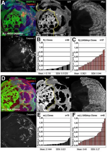
overexpressing clones (hereafter referred to as ykiover) with their wild type twins. While clones and twins showed a comparable size in the wild type control (Figure 1A, 1F, and 1G, and Figure S1A, S1C, S1D),ykioverclones were notably larger than their wild type twins in wing discs dissected either 60h (Figure 1D, 1H, and 1I) or 48h (Figure S1B, S1E, S1F) after heat-shock induction. Further-more,ykioverwild type twins were almost disappeared from the epithelium at 120h after egg laying (AEL) (Figure 1B and 1C). These differences in size were also prominent when discs were dissected at 96h AEL (Figure 1D and 1E). Interestingly, the clonal expansion ofykiovercells was also correlated with non-autonomous apoptosis, as revealed by active Caspase 3 immunoreactivity of a subset of surrounding wild type cells (Figure 1B–1E). The size advantage ofykioverclones and the induction of apoptosis in wild type cells is consistent with the broadly assumed definition of cell competition, which implies that the clonal expansion of the winner cells occurs at the expense of the juxtaposed losers, that are eliminated by apoptotic death [2,42,43]. The pattern of cell death in wild type andykiovercells (Figure 1B–1E) was not confined to the interface between the two cell types; as can be seen in Figure 1E, cell death extends several cell diameters away and wild type cells tend to die massively when enclosed between nearby mutant clones (Figure 1E, yellow arrowhead). A similar pattern of non-autonomous cell death was observed in wild type cells nearby mutant clones for other members of the Hpo pathway, such asft
andex(Figure S2A, S2B, S2C). Strikingly,ykiover
clones and wts
mutant clones grown for a longer period presented autonomous cell death (Figure 1C, see active Caspase 3 staining, and Figure S2D), despite the upregulation of anti-apoptotic molecules such as dIAP1 [16]; this might be possibly due to either developmental constraints compensating for excessive proliferation of the entire organ or toxicity caused by high and constant levels of Yki. Altogether, these results confirm the previously suggested supercompetitive properties of the Hpo pathway mutant clones [19] by revealing their ability to overgrow and eliminate surrounding wild type cells.
dmycis a Hpo pathway target gene regulated by the activity of Yki
It is well documented that the confrontation of different levels of dMyc protein between two populations of cells either in vivo
[4,5] or in cell culture [44] can trigger cell competition, however the molecular mechanism by which this occurs is unknown. In addition,myc family oncogenes are frequently overexpressed in human cancers and it contributes to tumor progression ofYAP -expressing cells (mammalian orthologue of yki) [17]. We have previously shown that a transcriptional activation ofdmycoccurs in ft mutant tissues and that ft clones fail to grow in a dmyc
hypomorphic background [45], indicating a possible regulation of this oncogene by the Hpo pathway. Moreover, the expression pattern of dMyc is complementary to that of Ds in the wing imaginal disc (Figure 2A), suggesting a possible functional interaction. To validate this hypothesis, we analyzed dMyc Author Summary
expression in mutant clones for several members of the Hpo pathway and inykiovercells by immunofluorescence. Noticeably, we found that dMyc was upregulated in a cell-autonomous manner inykioverclones throughout the wing disc (Figure 2B and Figure S3), with the weakest activation in the lateral regions, and in a subset of clones mutant for several Hpo pathway members (Figure 2C–2F). These differences in dMyc activation between
ykioverclones and clones mutant for other members of the Hpo signaling pathway might be due to additional levels of regulation of the Hpo cascade operating on upstream members. According to our previous observations, we would predict a repression of dMyc upon Hpo pathway hyperactivation. To investigate this hypothesis, we expressed Hpo in the spaltexpression domain of the developing wing disc. Since Hpo overexpressing cells die
massively by apoptosis during development [25], we coexpressed the anti-apoptotic factor p35. As expected, cells coexpressing Hpo and p35 show reduced levels of dMyc with respect to the control (Figure S4A) in both late (Figure S4B) and early (Figure S4C) wing discs. Thus dMyc levels can be regulated by the Hpo pathway activity.
dmycis transcriptionally regulated by Yki
dmyc was observed upregulated in RT-PCRs performed on ft
mutant imaginal discs [45], suggesting that it could be a transcriptional target of the Hpo pathway. In order to investigate this, we first performed anin situhybridization inDrosophilawing discs expressing yki under the control of the decapentaplegic (dpp) promoter. As expected, dmyc transcript is detectable in the dpp
Figure 1.ykioverexpression confers cells a supercompetitive behavior.(A)yw, hs-Flp,tub-Gal4, UAS-GFP; FRT42D,tub-Gal80/FRT42D,
Ubi-GFPclones induced at 48–72 h AEL. Twin clones are marked by the lack of GFP (0xGFP); imaginal discs were dissected at 120h AEL. (B–E)yw,
hs-Flp,tub-Gal4, UAS-GFP; FRT42D,tub-Gal80/FRT42D, Ubi-GFP; UAS-yki/+clones (2xGFP = 2XUbi-GFP+tub-GFP) sorrounded by wild type cells. (B,D) Clones were induced at 24–48 h AEL and dissected at 120 h AEL (B) or 96h AEL (D). C and E are magnifications of B and D respectively. Cell death assayed by Caspase 3 staining is shown in red (A–E). Yellow arrowhead indicates a wild type twin in C (0xGFP), whereas the red arrowhead indicates wild type cells dying far fromykioverclones. Note that wild type cells preferentially die when enclosed byykiovercells (yellow arrowhead in E). (F–I) Histograms showing the surface area of wild type andykioverclones and respective twins induced at 48–72 h AEL and allowed to grow until 120h AEL. (F, G) Wild type clones (F) and their twins (G) display the same size profile. (H) The size profile ofykioverclones indicates that they are larger either than wild type controls (F, G) or than their wild type twins (I). Note also that wild type twins (I) ofykioverclones (H) are smaller than wild type clones induced at the same stage of development (F, G). SEM = Standard Error of the Mean.P,0.0001.
domain both inykiand controldmyc-expressing discs (Figure 3A). No signal within thedppdomain was detected indpp.GFPcontrol discs (not shown). We were able to reproduce these data using a
dmyc.lacZline [46] which recapitulates accurately thedmycpattern throughout the wing disc during development [7,47]. As can be seen in Figure 3B, the ßGal expression is increased in the dpp
domain upon yki expression, indicating that Yki acts upondmyc
transcription. This result was supported using clonal analysis, both inykiovercells, as shown in Figure 3C, and in cells mutant forft
(Figure S5). Altogether, these data demonstrate the ability of the Hpo pathway to regulatedmyctranscription in the imaginal wing disc.
Yki transcriptional activity depends on the formation of tissue-specific complexes with different partners such as Scalloped and Homothorax [27–31]. In order to study the contribution of Sd to
dmyc upregulation by Yki in the wing disc, we generated ykiover
clones coexpressing either a UAS-sdor a UAS-sd-RNAi construct (see Figure S6A for validation). As can be seen in Figure 3D,sdover;
ykiover clones overgrew relative to ykiover clones (compare with Figure 2B, 68% increase on average, n = 27,P,0,005) confirming previous data [29], but we were not able to detect significant differences in dMyc protein levels compared to ykiover clones (n = 22, P= 0,43). As expected, control sdover clones did not overgrow and did not deregulate dMyc (Figure 3E), demonstrating that Yki is required for dMyc upregulation. We were not able to recover sd-RNAi; ykiover
clones in the wing pouch region, but clones generated in other territories of the wing disc, although large, did not upregulate dMyc (Figure 3F), nor showed the same degree of hyperplasia as Yki expression alone (Figure 1B–1E).sd -RNAi control clones were very small and did not deregulate dMyc
(not shown). These data indicate a key role for Sd in vivo in upregulating dMyc inykiover clones, and in contributing to the
ykiovertumorous phenotype.
Interestingly, examination of dmyc locusrevealed the existence of several CATTCCA repeats in non-coding regions of the gene, which perfectly match the mammalian [48,49] and Drosophila
[28,29] TEAD/TEF family transcription factor consensus binding motifs (mammaliam orthologues of Scalloped). In ad-dition, these putative binding motifs for Yki/Sd complexes are evolutionarily conserved inD. simulans(Figure 3G) and relatively close to the insertion point of P elements that recapitulate the endogenous expression of the gene (dmPL35 LacZ [50,51] and
dmBG02383Gal4 insertions - http://flybase.org/reports/FBti0018138. html).
To test the significance of these sequences indmycregulation, we generated admyc-fireflyreporter containing the putative responsive elements for Yki/Sd complexes (Figure 3H) and performed a transient dual luciferase assay in S2 cells. As can be seen in Figure 3I, the reporter was specifically activated upon Sd and Yki cotransfection but, unexpectedly, the transfection of Yki alone was able to activate the reporter as efficiently as the cotransfection Yki/Sd (Figure 3I). This result suggests that in presence of high levels of Yki alone, additional partners such as Hth [31] could bind it and co-regulatedmycexpression.
Indeed, complementarily to Yki/Sd complexes, Yki/Hth complexes seemed to play the same role in the presumptive thoracic region of the wing disc. Supporting this conclusion,hth -RNAi; ykiover clones down-regulated dMyc in the notum (30% reduction on average, n = 15,P,0,05, Figure S6B, yellow arrows) and did not grow as tumors in that region. By contrast, they were
Figure 2.dmyconcogene is regulated by the Hpo pathway.(A) Double staining with anti-dMyc (red) and anti-Ds (green) reveals that dMyc and Ds proteins show a complementary pattern of expression in the wing disc; dMyc is highly expressed in the wing pouch and less in the notum, while Ds localizes mainly in the hinge and pleura, where dMyc expression is the lowest. (B) dMyc staining ofyw, hs-Flp;actFRTy+FRTGal4, UAS-GFP/
UAS-ykiFlp-Out clones (GFP+) show increased dMyc protein level. (C–F) dMyc protein levels are increased indsd36,ftG-rv,exE1andwtsX1LOF clones (arrows). Clones were induced by the Flp/FRT system and are marked by the lack of GFP (0xGFP). Clones were induced at 42–54 h AEL (C–F) or at 54– 66 h AEL (B).
undistinguishable from ykiover clones in the wing pouch region (Figure S6B, white arrowhead), where Hth expression is almost undetectable (Figure S6C). Altogether, these latter results indicate that Sd and Hth play a role in Yki-induced tumorigenesis by regulatingdmycexpression in the wing disc, with Sd playing a more critical role in the pouch and Hth acting in the presumptive thorax.
dMyc upregulation enhances cell proliferation of the Hpo pathway mutant cells in an autonomous manner
With the aim to investigate the cell-autonomous contribution of dMyc overexpression toykioverphenotypes, we first compared the size ofykioverclones with that ofykiover;dmyc-RNAi clones (Figure 4,
see also Figure S7A, S7A9, and [7] for RNAi construct validation ). As expected,dmyc-RNAi clones showed a reduced number of cells with respect to that observed in wild type clones (21% reduction on average, compare Figure 4B and 4B9 with Figure 4A and 4A9,
P,0.01). The reduction in cell number displayed by the ykiover;
dmyc-RNAi clones with respect to theykioverclones was even more evident (43% reduction on average, compare Figure 4D and 4D9 with Figure 4C and 4C9,P,0.01), and this percentage raised up to 65% (n = 87,P,0,001) when these clones were induced earlier in development (42–54h AEL), indicating a strong cell-autonomous requirement of dMyc protein for the expansion ofykioverclones. We also observed that the non-autonomous apoptosis induced byyki
overexpression was reduced upon dmyc deprivation (32% on
Figure 3. Yki regulatesdmyctranscription.(A)In situ hybridization on imaginal wing discs with a full-lengthdmyc RNA probe. Thedmyc
transcript is robustly upregulated in thedpp.ykidisc on the left. Adpp.dmycdisc is shown on the right as a control. (B) ßGal staining (red) of
dmyc.lacZG0354/+;dpp.Gal4, UAS-GFP/UAS-ykiimaginal wing discs.dmycexpression is upregulated in thedppdomain (GFP+). (C) ßGal staining (red)
ofdmyc.lacZG0354/hs-Flp;actFRTy+FRTGal4, UAS-GFP/UAS-ykiimaginal wing discs.ykiover
clones (GFP+) were induced at 42–54h AEL. As can be
observed, a robust activation ofdmycregulatory sequences is visible within the mutant clones. (D,E) dMyc staining ofyw, hs-Flp/+; UAS-sd/+;
actFRTy+FRTGal4, UAS-GFP/UAS-ykiandyw, hs-Flp/
+; UAS-sd/+;actFRTy+FRTGal4, UAS-GFP/
+imaginal wing discs respectively. Clones (GFP+) were
induced at 42–54 h AEL. As can be observed in E, dMyc levels are comparable inside and outside the mutant clones. (F) dMyc staining ofyw, hs-Flp/+; UAS-sd-RNAi/+;actFRTy+FRTGal4, UAS-GFP/UAS-ykiimaginal wing discs. Clones (GFP+) were induced at 42–54 h AEL. No clones in the wing pouch
region were recovered and those outside the wing pouch never took the tumorous shape typical ofykioverclones. All larvae were dissected at 120h AEL. (G)dIAP1Hpo Responsive Elements (HRE) for Yki/Sd complexes are indicated in red [28]. The same consensus sites are found twice in the second intron ofdmycboth inD. melanogasterandD. simulans, indicating a possible functional conservation. (H) Scheme of thedmyc locusshowing the exons (E) and introns (I). The red bar indicates the region of the second intron containing the putative Yki/Sd consensus sites. (I) Luciferase assay on S2Drosophilacells. NT: cells transfected withdmyc-fireflyalone.Pvalue is significative for Sd/Yki (P,0,05) and Yki (P,0,01). Error bars represent standard deviation (triplicate wells).
average, n = 28, P,0,01, Figure S7B). These data suggest that dMyc upregulation promotes cell proliferation ofykioverclones in an autonomous manner, and also promotes their competitive behavior. To further characterize this proliferation-promoting effect of dMyc, we compared the clonal behavior of various mutations in members of the Hpo pathway grown in two different genetic backgrounds: a wild type context and a genetic background overexpressingdmycunder the control of ahedgehogpromoter in the posterior (P) compartment of the wing disc. We found thatft,ex
and dsmutant clones were consistently larger in those territories expressing uniform levels of dMyc than in the wild-type background (Figure 5 and Figure S8). It is however described that the overexpression of dMyc is able to autonomously increase apoptosis [8–11]. In fact, the wild type tissue expressing high amounts of dMyc tends to die and does not overgrow (see active Caspase 3 stainings in Figure 5A and 5D). Noticeably, the apoptosis mediated by dMyc overexpression seems to be extremely reduced insideftandexclones (Figure 5A and 5D) with respect to the wild type surrounding territories, likely due to the upregulation of antiapoptotic genes such asdIAP1, a target of the Hpo pthway [20]. In addition, the dying cells in this genetic background might induce morphogens to promote compensatory proliferation [52] that may contribute to the extra-growth of ft- or ex-UAS-dmyc
expressing clones. To circumvent this problem, we repeated the same experiment coexpressingdmycanddIAP1. As can be seen in Figure S9, bothft(Figure S9A) andex(Figure S9D) mutant clones grown in the P compartment were still consistently larger than those originated in the A compartment, thus confirming a specific cooperation of dmyc and Hpo pathway mutants in clonal expansion.
Hpo mutant cells therefore seem to show the ability to take advantage of the cell mass accumulation boosted by dMyc overexpression to proliferate faster.
dMyc upregulation prevents the Hpo pathway mutant clones from being restrained in a competitive
background
To address the non-autonomous relevance ofdmycupregulation in providing ykiover cells with a supercompetitive behavior, we compared the size of ykiover clones generated in a wild type
background to that of ykiover
clones generated in a background ubiquitously overexpressingdmycunder the control of atubulin(tub) promoter (cell competition assay, [4,5]). In this assay cells express the endogenousdmyc gene plus an extra copy of the gene under the control of atubpromoter that ensures two-to-threefold increase of
dmyc transcript [5]. This extra copy of dmyc is located in a removable cassette between thetubpromoter and a Gal4 cDNA. Upon dmyc cassette excision, the tub promoter drives Gal4 expression in the clones and, as a result, those cells express lower levels ofdmycrelative to the background and are rapidly eliminated from the tissue by cell competition. Only few genes have so far been found whose overexpression rescues cell viability in this context [5]. The relative difference in dMyc levels between yki -expressing cells and the surroundingtub.dmyccells was minimized in a competitive background compared to a wild type context (compare Figure S7C and S7C9 with Figure 2B). In this competitive background,ykiover
clones showed a diminished ability to overgrow compared to a wild type background (44% reduction on average, compare Figure 6C and 6C9and Figure 6B and 6B9;
P,0,01). Besides the reduction in size,ykioverclones showed an important reduction in clone number both in discs (Figure 6C) and adult wings (compare Figure S7E to Figure S7D). Moreover,ykiover
clones induced earlier in development (42–54h AEL) were never recovered at the end of larval development (not shown). These data indicate that the competitive properties of ykiover cells are extremely reduced when they are surrounded by cells expressing very high amounts of dMyc.
We then performed the same competition assay as before while reducingdmycactivity inside the clones. We used the pupal lethal
dmycPL35allele [49] and, taking advantage ofdmyc locusassociation to chromosome X, we were able to analyze both female (heterozygous condition, the expression of dmyc is halved) and male (hemizygous condition, the expression ofdmycis completely removed) larvae. IndmycPL35
/+; tub.dmyc females,ykiover clones were smaller than those described in the previous assay (28% reduction on average, compare Figure 6D and 6D9to Figure 6C and 6C9,P,0,05), whereas they were completely outcompeted by 48h after the heat shock in males (not shown). Since it has been observed that admycPL35heterozygous condition does not impair cell growth or proliferation rate [49], our results reveal an
Figure 4. dMyc boosts the proliferative abilities ofykiovercells.Four types of clones were simultaneously induced through the Flp-Out
system at 48–72h AEL and allowed to grow for a comparable period (until 120h AEL). Clones are GFP+, nuclei are counterstained with DAPI. (A–A9)
act.GFP control clones. (B–B9)act.dmyc-RNAi clones. (C–C9)act.ykioverclones proliferate faster than wild type clones. (D–D
9)act.dmyc-RNAi;
ykioverclones: proliferation is reduced relative toact.ykioverclones. (A9–D9) Histograms showing the number of cells/clone of the genotypes indicated in A–D. SEM = Standard Error of the Mean.P,0.01.
important role fordmyc-induced cell competition in controlling the clonal expansion ofykiovercells, which may occur via their non-autonomous capabilities to compete with neighboring wild type cells.
dMyc expression alone is not sufficient to prevent the elimination ofykimutant cells
ykiLOF clones generated in a wild type background are not able to grow [16,25] and the ectopic expression of the antiapoptotic proteins dIAP1 [25] or p35 (Figure S10A) poorly rescues their viability, whereas aMinutebackground [53] orbantam overexpres-sion within yki clones has been shown to partially rescue their growth [25]. Since our results have indicated thatdmycparticipates in tumor growth of the Hpo pathway mutant cells, we therefore analyzed if the expression of dMyc was sufficient to prevent the death of yki mutant cells. The overexpression of dMyc failed to rescue the viability ofyki2/2cells (Figure S10B). Sinceykimutant
cells express low levels of the apoptosis inhibitor dIAP1 (not shown), this result is not surprising, considering the autonomous cell death described for cells overexpressing dMyc [11]. However,
yki mutant cells coexpressing dMyc and p35 also failed to grow (Figure S10C). The lack of expression of additional antiapoptotic genes and cell cycle regulators [18] possibly impedes the clonal growth of yki mutant cells even though they overexpress dMyc. This result suggests that dmyc expression is able to enhance the ability of Hpo pathway mutant cells to grow, but it is not sufficient to rescue tissue growth ofyki2/2clones.
Discussion
Cells within a tissue coordinate and execute complex genetic programs in order to succeed in completing a variety of processes during development. In this context, the phenomenon of cell competition may be part of the developmental plan that ensures removal and replacement of defective cells in growing organs, thus keeping their size invariant. In this work, we have evaluated in details the relationships between the phenomenon of cell competition and the clonal expansion of tumorous cells, using for that purpose mutants in components of the evolutionarily conserved Hpo pathway. From our studies we reveal that the Hpo pathway regulates dMyc expression, and show that this is critical for the tissue growth and competitive behavior of Hpo pathway mutant clones.
dMyc is a Hpo pathway transcriptional target
dmyc upregulation has been demonstrated in many studies to provide cells with supercompetitive properties [4,5,7]. The model explaining how dMyc can confer competitive properties to cells is based on the relative levels of this protein in neighboring cell populations, transforming those cells expressing higher levels of dMyc into supercompetitors [4,5].dmyc overexpression is never-theless insufficient to drive tumorous growth;dmycoverclones fail to overproliferate and show strong autonomous apoptosis [9]. Interestingly, we found that dMyc protein is overexpressed in Hpo pathway mutant clones, indicating an involvement for this cascade indmycregulation (Figure 2). Furthermore, the upregula-tion of dMyc in Yki-expressing cells correlates with an increase in the amount of mRNA, observed byin situhybridization (Figure 3A) and using admyc.lacZline (Figure 3B and 3C). Finally, we have identified a regulatory region in the second intron ofdmycthat is sensitive to Yki abundance; importantly, this regulatory region includes predicted consensus-binding motifs for Sd (Figure 3H). Clonal experiments in the wing disc indicate that Sd is necessary for Yki functionin vivo, since upon Sd downregulation Yki is no longer able to induce tumorous growth and does not upregulate dMyc (Figure 3F). All these findings support the notion that there is a transcriptional regulation of dMyc mediated by Yki/Sd complexes in the wing pouch. Importantly, similar results were observed for dMyc regulation in the notum by Yki/Hth complexes, suggesting that tumor growth and dmyc regulation are tissue-specific.
What is the contribution of dMyc to the Hpo pathway mutant phenotypes?
We found that dMyc upregulation is a common feature of Hpo pathway mutant cells. Sincedmyc has been repeatedly associated with tumor progression and cell competition, we analyzed its role in the clonal expansion of Hpo pathway mutant cells. We observed that the reduction of dMyc expression restricts the ability of Hpo pathway mutant cells to proliferate (Figure 4), whereas its uniform overexpression strongly promotes their proliferation (Figure 5).
Figure 5. dMyc overexpression enhances the proliferation of Hpo pathway mutant cells.ft(A–C) andex(D–F) LOF clones (0xGFP) generated in a background where posterior (P) cells ectopically express
dmyc under the control of hh-Gal4 (A and P compartments are separated by a yellow line in A and D; P is on the right). dMyc overexpression strongly enhances the proliferative activity offt(A–C) andex(D–F) mutant cells; mutant clones are larger in dMyc-expressing territories (P compartment in histograms C and F) than in a wild type background (A compartment in histograms B and E). SEM = Standard Error of the Mean.P,0.001. High levels of active Caspase 3 signal are evident in dMyc-expressing cells outsideftandexmutant clones in the posterior compartment.
Furhermore, while dMyc-expressing wild type cells surrounding mutant clones are rapidly eliminated by autonomous apoptosis, Hpo pathway mutant cells are able to take advantage of dMyc role in protein biosynthesis and cellular growth to divide rapidly. This is a clear example of functional cooperation between different genes in order to favor tumor progression, but it also indicates a specific role of dMyc in promoting the clonal expansion of Hpo pathway mutant cells. According to these data, we conclude that dMyc behaves as a growth-promoting factor which sustains the hyperplastic phenotype of Hpo pathway mutant cells. Importantly, this specific cooperation might be evolutionarily conserved, since
c-myc appears to be upregulated in a murine model of YAP-induced carcinoma [17].
Relative levels of dMyc in neighboring cells restrict/ promote clonal expansion of hyperplastic cells, likely through cell competition
It has been suggested that cell competition may be a mechanism potentially restricting the clonal expansion of tumorous cells [7], but it might also help faster proliferation of transformed cells. Our data indicate that Hpo pathway mutant cells are able to use high levels of dMyc to proliferate rapidly (Figure 5), but in a competitive context, where neighboring cells express high levels of dMyc, clonal expansion of ykiovercells is restrained (Figure 6), therefore suggesting a tumor suppressor role for cell competition. Con-versely, dMyc upregulation inykioverclones grown in a wild type background favors their clonal expansion promoting cell auton-omous proliferation and also conferring the ability to outcompete sourrounding cells in a non-autonomous manner. These findings suggest that the phenomenon of cell competition may play a dual role in tumor progression depending on the output of the genetic interactions occurring between adjacent cells.
In summary, we have shown a tumor-braking gene network in
Drosophila epithelia which tightly controls cell proliferation, apoptosis and cell competitionviathe Hpo pathway and dMyc expression. Importantly, YAP deregulation has been reported in
several types of human cancers [54–56], therefore the mecha-nism of clonal expansion of Hpo pathway mutant cells in
Drosophilamight be relevant to understand tumor progression in mammals.
Materials and Methods
Genotypes and clonal analysis
The fly strains used in the present work were obtained by the Bloomington Stock Center and are described at http://flybase.bio. indiana.edu. The following strains were instead obtained by: w;
UAS-yki (D Pan); yw, tubFRTdmycFRTGal4 and yw, dmycPL35,
actFRTy+FRTGal4(P Gallant);w,hs-FLP;actFRTy+FRTGal4,UAS
-GFP(B Edgar);w;FRT40A,dsD36(I Rodrı´guez). The UAS-RNAi constructs fordmyc,sd and hthwere obtained from the VDRC.
All experiments were carried out at 25uC unless otherwise indicated.
MARCM UAS-yki twin-spot clones were induced at different stages of development by a 35-minutes heat shock at 37uC and larvae of the following genotype were dissected at either 84-100h AEL or 120h AEL:yw, hs-Flp,tub-Gal4, UAS-GFP; FRT42D, tub-Gal80/FRT42D, Ubi-GFP; UAS-yki/+. Clones of the same genotype were induced 54–66 h AEL and dissected 48h after a 20-minutes heat shock (Figure S1). For FRT-Flp twin analysis, the following hypomorphic or null alleles were used:dsD36,ftG-rv,exE1,
wtsX1, ykiB5. Loss-of-function clones of ds,ft,exand wtsin either wild-type or mutant backgrounds overexpressing different trans-genes in the posterior compartment were induced at 48–72h AEL by 1 hour heat shock at 37uC. Larvae of the following genotype were dissected at 120h AEL:
yw, hs-Flp; FRT40A, Ubi-GFP/FRT40A,dsD36orftG-rvorexE1 yw, hs-Flp; FRT82B, Ubi-GFP/FRT82B,wtsX1
yw, hs-Flp; FRT40A, Ubi-GFP/FRT40A,dsD36orftG-rvorexE1;
hh-Gal4/UAS-dmyc
yw, hs-Flp; FRT40A, Ubi-GFP/FRT40A,ftG-rvorexE1;hh-Gal4/ UAS-dmyc, UAS-dIAP1
Figure 6.ykioverclonal expansion is restrained bydmyc-induced cell competition.(A–D
9) Cell competition assay shows that, while wild type clones are outcompeted in this genetic background [5], the clonal expansion ofykiovercells is partially restrained. Clones were induced at 60–84 h AEL and allowed to grow until 120h AEL. Clones are GFP+, nuclei are counterstained with DAPI. (A–A9) wild type control clones generated through a
tubulin(tub) Flp-OUT system. (B–B9)ykioverclones generated with the same system as the wild type control. (C–C9)ykioverclones in atub.dmyc
background are smaller than in a wild type background.P,0.01. (D–D9)dmycPL35/
+;tub.ykioverclones in atub.dmycbackground are smaller than in C (P,0.01), confirming that the relative dMyc levels outsidevsinside the clones affect the competitive ability ofykiovercells. (A9–D9) Histograms showing the number of cells/clone of the genotypes indicated in A–D. In C and D discs are larger due to the overall increase in body size of
The size of non-confluent clones was measured drawing each Z-stack of the confocal images using ImageJ software (http://rsbweb. nih.gov/ij). Afterwards the area of the clones was normalized dividing by the area of the wing pouch, considered as the territory encircled by the first outer folding of the wing. In Figure S1, the narrower window of clonal induction allowed us to compare clonal size without size normalization respect to the wing pouch. Statistical analysis was performed with Microsoft Excel and R (www.r-project.org). Statistical significance was determined by two tailed Student’s t test and reported as the associated probability value (P).
Flp-Out clones were induced at 60h AEL by a 8-minutes heat shock at 37uC; imaginal discs of the following genotype were dissected at 120h AEL:
yw, hs-Flp;actFRTy+FRTGal4, UAS-GFP
yw, hs-Flp; UAS-dmycRNAi/+;actFRTy+FRTGal4, UAS-GFP/ +
yw, hs-Flp;actFRTy+FRTGal4, UAS-GFP/UAS-yki
yw, hs-Flp; UAS-dmycRNAi/+; actFRTy+FRTGal4, UAS-GFP/
UAS-yki.
yw, hs-Flp/w, dmyc.lacZG0354; actFRTy+FRTGal4, UAS-GFP/
UAS-yki.
Cell competition assays were performed at 72h AEL inducing a 40-minutes heat shock at 36uC. Larvae of the following genotype were dissected at 120h AEL:
yw,tubFRTy+FRTGal4/hs-Flp; UAS-GFP/ +
yw,tubFRTy+FRTGal4/hs-Flp; UAS-GFP/
+; UAS-yki/+
yw,tubFRTdmycFRTGal4/hs-Flp; UAS-GFP/+; UAS-yki/+
yw, dmycPL35
, hs-Flp, tubFRTdmycFRTGal4/+-Y; UAS-GFP/+; UAS-yki/+.
MARCMyki clones overexpressing p35, dMyc or both were generated at 48–72h AEL by a 45-minutes heat shock at 37uC and larvae were dissected 48h later.
Immunofluorescence
Immunostainings were performed using standard protocols. The following primary antibodies were used: mouse anti-dMyc (1:5, P Gallant), mouse anti-En (1:50, DSHB), rabbit anti-active Caspase 3 (1:100, Cell Signaling Technology), rabbit anti-p35 (1:1000, Stratagene), rabbit Ds (1:100, D Strutt), rabbit anti-Hth (1:400, A Salzberg, [57]), mouse anti-dIAP1 (1:100, B Hay) and rabbit ßGal (1:400, F Graziani). Anti-mouse and anti-rabbit Alexa Fluor 555 (1:200) (Molecular Probes) and anti-mouse Cy5 (1:200) (Jackson Laboratories) against corresponding primary antibodies were used as secondary antibodies. Imaginal discs were mounted in Vectashield (Vector Laboratories) for confocal imaging. Single Z stacks were acquired with Leica SP2 and SP5 confocal microscopes. Images for Figure 4 and Figure 6 were captured with an epifluorescence Nikon 90i microscope. Entire images were elaborated with Photoshop CS2 (Adobe) and the projections along the Z axis were rebuilt starting from 35–55 Z stacks using the ImageJ public software (NIH). For measurements of dMyc abundance, fluorescence intensity was calculated using the ImageJ public software (NIH) as the average gray value within selectioned portions of confocal Z stacks. For measurement of active Caspase 3 signal outside UAS-dmyc-RNAi; UAS-yki and UAS-ykiclones, staged wing discs were chosen containing as few clones as possible and single cells positive to active Caspase 3 observed at a maximum distance of five nuclei (counterstained with DAPI) from the border of the clone were counted on confocal Z stacks.In situhybridization was performed with a full lengthdmyc
probe [9] on wing imaginal discs of L3 larvae expressing
UAS-GFP, UAS-dmycor UAS-ykiunder the control ofdpp-Gal4. RNAin situhybridization was carried out using digoxigenin-labeled RNA probes [58].
Luciferase transient expression assays
Drosophila S2 cells were grown at 25uC in Schneider medium (GIBCO) supplemented with 10% heat-inactivated FCS and 100 units of penicillin.
1189 base pairs located in the second intron of the dmyc
sequence (Figure 3H) were subcloned into a pGL3-firefly vector (Promega) and co-transfected with Sd and/or Yki-expressing pAc5.1/V5-HisB plasmids [28] using Effectene Qiagen Transfec-tion Kit. The primers used for that purpose were:
59CAGCGGTACCAGTTTGCTGTCCTCTGC 39
59GCACTCTAGAGCCATGCGGAATTGTGCG 39.
The PCR product was first cloned in pCR 2.1 TOPO-TA (Sigma) and then subcloned in KpnI/XhoI sites of pGL3 Promoter vector. For luciferase transient expression assays, 26104 cells were plated in 96-well dishes. Cells were harvested at 48 hours after transfection and luciferase activity was measured using the Luciferase reporter assay system (Promega). Dual-Luciferase measurements were performed using a FLUOstar Optima luminometer (BMG Labtech) and normalized to the
Renilla luciferase activity using pAct5C-seapansy as an internal control. All transient expression data reported in this paper represent the means from three parallel experiments, each performed in triplicate. Average relative luciferase activity was graphed and statistically analyzed by the Student’st-test. Supporting Information
Figure S1 ykiovercells supercompetitive behavior is indeed visible at 48h after induction. (A,B) yw, hs-Flp, tub-Gal4, UAS-GFP; FRT42D, tub-Gal80/FRT42D, Ubi-GFP (A) and yw, hs- Flp,tub-Gal4, UAS-GFP; FRT42D, tub-Gal80/FRT42D, Ubi-GFP;
UAS-yki/+(B) clones induced at 54–66h AEL and dissected 48h after the heat-shock. Wild type andykioverclones are GFP2+and twin
clones are marked by the lack of GFP. Cell death is assayed by active Caspase 3 inmunoreactivity in red. Note that cell death is almost absent in the wild type experiment (A0) and marks wild type cells in theykioverexperiment (B0). (C–F) Histograms showing the surface area of wild type andykiover
clones and respective twins. (C,F) Wild type clones (C) and their twins (D) display the same size profile. (E) The size profile indicates thatykioverclones are larger than wild type controls (C) as than their wild type twins (F) after only 48h of growth in the wing. SEM = Standard Error of the Mean.P,0.0001.
Found at: doi:10.1371/journal.pgen.1001140.s001 (1.60 MB TIF)
Figure S2 Hpo pathway LOFs induce cell competition. (A,B) Activated Caspase 3 staining of yw, hs-Flp/+; ftG-rv, FRT40A/
Ubi.GFPnls, FRT40A discs in which mutant clones (0xGFP) were grown for 48 hours (48–96 in A and 72–120 in B); apoptotic cell death occurs mainly in wild type cells surrounding the mutant clones (arrowheads). (C,D) Activated Caspase 3 staining ofyw,
hs-Flp/+; exE1, FRT40A/Ubi.GFPnls, FRT40A (C) and hs-Flp/+;
wtsX1, FRT82B/Ubi.GFPnls, FRT82B (D) discs in which mutant clones (0xGFP) were grown for a longer period (48–108 and 48– 120 hours respectively); apoptotic death is visible in both wild type (D, arrowheads) and mutant (D, arrows) cells.
Found at: doi:10.1371/journal.pgen.1001140.s002 (2.75 MB TIF)
Figure S3 dMyc upregulation in ykiover
clones is cell-autono-mous. dMyc staining in yw, hs-Flp/+; actFTRy+FRTGal4,
UAS-GFP/UAS-ykiimaginal wing discs.ykioverclones (GFP+, in green)
express high levels of dMyc (in red) compared to the endogenous background. Z-section indicates that dMyc (in red) up-regulation is confined toyki-expressing cells (in green).
Figure S4 Hpo overexpression reduces dMyc protein levels. (A) dMyc staining inw;sal.Gal4/+; UAS-p35/+imaginal wing discs. (B–C) dMyc staining of late (B) and early (C)w;sal.Gal4/+;
UAS-Hpo/+; UAS-p35/+imaginal wing discs. p35 is shown in the green channel and dMyc in red. As can be observed in the Z-sections dMyc abundance is lower inside thesaldomain. The position of Z-section is indicated by white bars in the surface view of the wing discs.
Found at: doi:10.1371/journal.pgen.1001140.s004 (2.76 MB TIF)
Figure S5 dmyc is transcriptionally upregulated in ft mutant clones. ßGal staining (red) of dmyc.lacZG0354/hs-Flp; ftG-rv, FRT40/UbiGFPnls, imaginal wing discs. As can be observed, a robust activation ofdmycregulatory sequences is visible within the mutant clones (arrows). Larvae were dissected at 120h AEL. Found at: doi:10.1371/journal.pgen.1001140.s005 (0.93 MB TIF)
Figure S6 Hth is necessary for Yki-induced dMyc overexpres-sion in the presumptive thoracic region of the wing disc. (A) Wing from aw; UAS-sd-RNAi/en.Gal4 individual. For UAS-sd-RNAi line validation, we induced the expression of the sd-RNAi construct in the posterior compartment of the wing by means of theengrailed(en) promoter. As can be observed, the wing lacks the posterior compartment (green-colored in the insert). (B) dMyc staining in yw, hs-Flp/+; UAS-hth-RNAi/+; UAS- yki/actF-TRy+FRTGal4, UAS-GFPimaginal wing discs. Note that mutant clones (GFP+) overgrow and overexpress dMyc in the wing pouch
region (white arrowhead) and not in the notum region (yellow arrows). (C) For UAS-hth-RNAi line validation, we stained for Hth [57] yw, hs-Flp/+; UAS-hth-RNAi/+; actFTRy+FRTGal4,
UAS-GFP/+wing discs. Hth expression is lacking in the clone originated in the pleural region (arrow).
Found at: doi:10.1371/journal.pgen.1001140.s006 (1.49 MB TIF)
Figure S7 dmyc is involved in the competitive ability ofyki. (A) dMyc levels are strongly affected insidedmyc-RNAi;ykioverclones. (A9) A projection along the Z axis of the clone presented in Figure A is shown. The percentage of dMyc abundance reduction inside the mutant clones calculated as the abatement of fluorescence intensity (see Methods - Immunofluorescence) with respect to the neighboring tissue was 62% on average (n = 12). (B)dmyc-RNAi;
ykioverclones display a reduced non-autonomous apoptotic activity (yellow arrows, see Methods - Immunofluorescence - for calculation) compared to ykiover clones (see Figure 1). (C) ykiover
clones can compete in a highdmyc level background, where wild type clones fail to grow; clones were induced at 66–78h AEL and allowed to grow until 120h AEL. In red, staining for dMyc indicates that dMyc levels are quite similar inside theykiover
clone and in thetub.dmyc background. (C9) A projection along the Z axis of the clone presented in figure C is shown. (D–E) In adult wings,tub.ykiover
clones generated in a wild type background (D) are bigger than tub.ykiover clones generated in a tub.dmyc
background (red arrows, E) confirming the results illustrated in Figure 6. Clones were induced at 66–78h AEL and survived up to the adult stage.
Found at: doi:10.1371/journal.pgen.1001140.s007 (1.18 MB TIF)
Figure S8 dMyc overexpression boosts proliferation indsmutant cells. (A)dsLOF clones (0xGFP) generated in a background where posterior cells ectopically expressdmycunder the control of thehh
promoter (on the right). dMyc overexpression strongly enhances the proliferative activity of ds mutant cells; mutant clones are larger in dMyc-expressing territories (posterior compartment in C) than in a wild type background (anterior compartment in B). SEM = Standard Error of the Mean.P,0.001.
Found at: doi:10.1371/journal.pgen.1001140.s008 (0.67 MB TIF)
Figure S9 dMyc overexpression boosts proliferation of Hpo pathway mutant cells also when wild type cells are protected from cell death.ft(A–C) andex(D–F) LOF clones (0xGFP) generated in a background where posterior (P) cells ectopically coexpressdmyc
anddIAP1under the control ofhh-Gal4(A and P compartments are separated by a white line in A and D; P is on the right). dMyc overexpression enhances the proliferative activity offt(A–C) andex
(D–F) mutant cells; mutant clones are larger in dMyc-dIAP1 expressing territories (P compartment in histograms C and F) than in a wild type background (A compartment in histograms B and E). All panels show Caspase 3 staining in red and dIAP1 in blue. SEM = Standard Error of the Mean.P,0.001.
Found at: doi:10.1371/journal.pgen.1001140.s009 (2.59 MB TIF)
Figure S10 dmycfails to rescueykiLOF upon inhibition of cell death. Three types ofyki LOF clones were induced through the MARCM system. In (A),ykimutant clones were generated while overexpressing the antiapoptotic protein p35 (in red). (B) Overexpression of dmyc fails to rescue yki mutant cells viability and Caspase 3 activation (red arrows). (C) The overexpression of p35 anddmyctogether also fails to rescueykimutant cells viability. Found at: doi:10.1371/journal.pgen.1001140.s010 (1.47 MB TIF)
Acknowledgments
We thank A Baonza, A Garcia-Bellido, G Gargiulo, and JP Vincent for scientific support. Thanks to L Quinn for sharing information about the expression of thedmyc.lacZG0354line prior to publication that was helpful for our analysis in this paper and our previous publication [7]. We thank L Johnston for having hosted us in her laboratory to perform part of the experiments on S2 cells. We are grateful to HE Richardson, FA Martı´n, and G Perini for manuscript revision and constructive criticisms. We finally thank L Johnston, D Strutt, PJ Briant, P Gallant, FA Martı´n, G Morata, D Pan, I Rodriguez, BA Hay, F Graziani, A Salzberg, the Bloomington Stock Center, and the VDRC for flies and reagents.
Author Contributions
Conceived and designed the experiments: Marcello Ziosi, Luis Alberto Baena-Lo´pez, Daniela Grifoni, Flavio Garoia. Performed the experiments: Marcello Ziosi, Luis Alberto Baena-Lo´pez, Daniela Grifoni, Francesca Froldi. Analyzed the data: Marcello Ziosi, Luis Alberto Baena-Lo´pez, Daniela Grifoni, Francesca Froldi, Vincenzo Trotta, Sandro Cavicchi, Annalisa Pession. Contributed reagents/materials/analysis tools: Andrea Pession, Paola Bellosta. Wrote the paper: Marcello Ziosi, Luis Alberto Baena-Lo´pez, Daniela Grifoni, Andrea Pession.
References
1. Morata G, Ripoll P (1975) Minutes: mutants of Drosophila autonomously affecting cell division rate. Dev Biol 42: 211–221.
2. Moreno E (2008) Is cell competition relevant to cancer? Nat Rev Cancer 8: 141–147.
3. Martı´n FA, Herrera FC, Morata G (2009) Cell competition, growth and size control in the Drosophila wing imaginal disc. Development 136: 3747–3756.
4. de la Cova C, Abril M, Bellosta P, Gallant P, Johnston LA (2004) Drosophila myc regulates organ size by inducing cell competition. Cell 117: 107–116. 5. Moreno E, Basler K (2004) dMyc transforms cells into super-competitors. Cell
117: 117–129.
7. Froldi F, Ziosi M, Garoia F, Pession A, Grzeschik NA, et al. (2010) The lethal giant larvae tumour suppressor mutation requires dMyc oncoprotein to promote clonal malignancy. BMC Biol 8: 33.
8. Oster SK, Ho CS, Soucie EL, Penn LZ (2002) The myc oncogene: Marvelous Complex. Adv Cancer Res 84: 81–154.
9. Johnston LA, Prober DA, Edgar BA, Eisenman RN, Gallant P (1999) Drosophila myc regulates cellular growth during development. Cell 98: 779–790.
10. Meyer N, Kim SS, Penn LZ (2006) The Oscar-worthy role of Myc in apoptosis. Semin Cancer Biol 16: 275–287.
11. Montero L, Mu¨ller N, Gallant P (2008) Induction of Apoptosis by Drosophila Myc. Genesis 46: 104–111.
12. Grewal SS, Li L, Orian A, Eisenman RN, Edgar BA (2005) Myc-dependent regulation of ribosomal RNA synthesis during Drosophila development. Nat Cell Biol 7: 295–302.
13. Vita M, Henriksson M (2006) The Myc oncoprotein as a therapeutic target for human cancer. Semin Cancer Biol 16: 318–330.
14. Land H, Parada LF, Weinberg RA (1983) Cellular oncogenes and multistep carcinogenesis. Science 222: 771–778.
15. Zhan L, Rosenberg A, Bergami KC, Yu M, Xuan Z, et al. (2008) Deregulation of Scribble Promotes Mammary Tumorigenesis and Reveals a Role for Cell Polarity in Carcinoma. Cell 135: 865–878.
16. Huang J, Wu S, Barrera J, Matthews K, Pan D (2005) The Hippo signaling pathway coordinately regulates cell proliferation and apoptosis by inactivating Yorkie, the Drosophila homolog of YAP. Cell 122: 421–434.
17. Dong J, Feldmann G, Huang J, Wu S, Zhang N, et al. (2007) Elucidation of a universal size-control mechanism in Drosophila and mammals. Cell 130: 1120–1133.
18. Saucedo LJ, Edgar B (2007) Filling out the Hippo pathway. Nat Rev Mol Cell Biol 8: 613–621.
19. Tyler DM, Li W, Zhuo N, Pellock B, Baker NE (2006) Genes affecting cell competition in Drosophila. Genetics 175: 643–657.
20. Harvey K, Tapon N (2007) The Salvador-Warts-Hippo pathway - an emerging tumour-suppressor network. Nat Rev Cancer 3: 182–191.
21. Oh H, Irvine KD (2009) In vivo analysis of Yorkie phosphorilation sites. Oncogene 28: 1916–1927.
22. Ren F, Zhang L, Jiang J (2009) Hippo signaling regulates Yorkie nuclear localization and activity through 14-3-3 dependent and independent mecha-nisms. Dev Biol 337: 303–312.
23. Nicolay BN, Frolov MV (2008) Context-dependent requirement for dE2F during oncogenic proliferation. PLoS Genet 4: e1000205. doi:10.1371/journal. pgen.1000205.
24. Nolo R, Morrison CM, Tao C, Zhang X, Halder G (2006) The bantam MicroRNA is a target of the Hippo tumor-suppressor pathway. Curr Biol 16: 1895–1904.
25. Thompson BJ, Cohen SM (2006) The Hippo pathway regulates the bantam microRNA to control cell proliferation and apoptosis in Drosophila. Cell 126: 767–774.
26. Baena-Lopez LA, Rodriguez I, Baonza A (2008) The tumor suppressor genes dachsous and fat modulate different signalling pathways by regulating dally and dally-like. Proc Natl Acad Sci USA 105: 9645–9650.
27. Goulev Y, Fauny JD, Gonzalez-Marti B, Flagiello D, Silber J, et al. (2008) SCALLOPED Interacts with YORKIE, the Nuclear Effector of the Hippo Tumor-Suppressor Pathway in Drosophila. Curr Biol 18: 435–441. 28. Wu S, Liu Y, Zheng Q, Dong J, Pan D (2008) The TEAD/TEF family protein
Scalopped mediates transcriptional output of the Hippo growth-regulatory pathway. Dev Cell 14: 388–398.
29. Zhang L, Ren F, Zhang Q, Chen Y, Wang B, et al. (2008) The TEAD/TEF family of transcription factor Scalopped mediates Hippo signaling in organ size control. Dev Cell 14: 377–387.
30. Zhao B, Ye X, Yu J, Li L, Li W, et al. (2008) TEAD mediates YAP-dependent gene induction and growth control. Genes Dev 22: 1962–1971.
31. Peng HW, Slattery M, Mann RS (2009) Transcription factor choice in the Hippo signaling pathway: homothorax and yorkie regulation of the microRNA bantam, in the progenitor domain of the Drosophila eye imaginal disc. Genes Dev 1: 2307–2319.
32. Willecke M, Hamaratoglu F, Kango-Singh M, Udan R, Chen C, et al. (2006) The Fat Cadherin Acts through the Hippo Tumor-Suppressor Pathway to Regulate Tissue Size. Curr Biol 16: 1–11.
33. Silva E, Tsatskis Y, Gardano L, Tapon N, McNeill H (2006) The Tumor-Suppressor Gene fat Controls Tissue Growth Upstream of Expanded in the Hippo Signaling Pathway. Curr Biol 16: 2081–2089.
34. Cho E, Feng Y, Rauskolb C, Maitra S, Fehon R, et al. (2006) Delineation of a Fat tumor suppressor pathway. Nat Genet 38: 1142–1150.
35. Bennett FC, Harvey KF (2006) Fat Cadherin Modulates Organ Size in Drosophila via the Salvador/Warts/Hippo Signaling Pathway. Curr Biol 16: 2101–2110.
36. Cho E, Irvine KD (2004) Action of fat, four-jointed, dachsous and dachs in distal-to-proximal wing signaling. Development 131: 4489–4500.
37. Feng Y, Irvine KD (2007) Fat and expanded act in parallel to regulate growth through warts. Proc Natl Acad Sci USA 104: 20362–20367.
38. Willecke M, Hamaratoglu F, Sansores-Garcia L, Tao C, Halder G (2008) Boundaries of Dachsous Cadherin activity modulate the Hippo signaling pathway to induce cell proliferation. Proc Natl Acad Sci USA 105: 14897–14902.
39. Hamaratoglu F, Willecke M, Kango-Singh M, Nolo R, Hyun E, et al. (2006) The tumour suppressor genes NF2/Merlin and Expanded act through Hippo signalling to regulate cell proliferation and apoptosis. Nat Cell Biol 8: 27–36. 40. Wu S, Huang J, Dong J, Pan D (2003) hippo encodes a Ste-20 family protein
kinase that restricts cell proliferation and promotes apoptosis in conjunction with salvador and warts. Cell 114: 445–456.
41. Reddy BVVG, Irvine KD (2008) The Fat and warts signaling pathways: new insights into their regulation, mechanism and conservation. Development 135: 2827–2838.
42. Moreno E, Basler K, Morata G (2002) Cells compete for decapentaplegic survival factor to prevent apoptosis in Drosophila wing development. Nature 416: 755–759.
43. Li W, Baker NE (2007) Engulfment is required for cell competition. Cell 15: 1215–1225.
44. Senoo-Matsuda N, Johnston LA (2007) Soluble factors mediate competitive and cooperative interactions between cells expressing different levels of Drosophila Myc. Proc Natl Acad Sci USA 104: 18543–18548.
45. Garoia F, Grifoni D, Trotta V, Guerra D, Pezzoli MC, et al. (2005) The tumor suppressor gene fat modulates the EGFR-mediated proliferation control in the imaginal tissues of Drosophila melanogaster. Mech Dev 122: 175–187. 46. Peter A, Scho¨ttler P, Werner M, Beinert N, Dowe G, et al. (2002) Mapping and
identification of essential gene functions on the X chromosome of Drosophila. EMBO Rep 31: 34–38.
47. Cranna N, Quinn L (2009) Impact of steroid hormone signals on Drosophila cell cycle during development. Cell Div 20: 4:3.
48. Xiao JH, Davidson I, Matthes H, Garnier JM, Chambon P (1991) Cloning, expression, and transcriptional properties of the human enhancer factor TEF-1. Cell 65: 551–568.
49. Larkin SB, Farrance IK, Ordahl CP (1996) Flanking sequences modulate the cell specificity of M-CAT elements. Mol Cell Biol 16: 3742–3755.
50. Bourbon HM, Gonzy-Treboul G, Peronnet F, Alin MF, Ardourel C, et al. (2002) A P-insertion screen identifying novel X-linked essential genes in Drosophila. Mech Dev 110: 71–83.
51. Benassayag C, Montero L, Colombie N, Gallant P, Cribbs D, et al. (2005) Human c-Myc isoforms differentially regulate cell growth and apoptosis in Drosophila melanogaster. Mol Cell Biol 25: 9897–9909.
52. Martı´n FA, Pere´z-Garijo A, Morata G (2009) Apoptosis in Drosophila: compensatory proliferation and undead cells. Int J Dev Biol 53: 1341–1347. 53. Oh H, Irvine KD (2008) In vivo regulation of Yorkie phosphorylation and
localization. Development 135: 1081–1088.
54. Lam-Himlin DM, Daniels JA, Gayyed MF, Dong J, Maitra A, et al. (2006) The hippo pathway in human upper gastrointestinal dysplasia and carcinoma: a novel oncogenic pathway. Int J Gastrointest Cancer 37: 103–109.
55. Steinhardt AA, Gayyed MF, Klein AP, Dong J, Maitra A, et al. (2008) Expression of Yes-associated protein in common solid tumors. Hum Pathol 39: 1582–1589.
56. Overholtzer M, Zhang J, Smolen GA, Muir B, Li W, et al. (2006) Transforming properties of YAP, a candidate oncogene on the chromosome 11q22 amplicon. Proc Natl Acad Sci USA 103: 12405–12410.
57. Kurant E, Pai CY, Sharf R, Halachmi N, Sun YH, et al. (1998) Dorsotonals/ homothorax, the Drosophila homologue of meis1, interacts with extradenticle in patterning of the embryonic PNS. Development 125: 1037–1048.
58. Johnston LA, Edgar BA (1998) Wingless and Notch regulate cell-cycle arrest in the developing Drosophila wing. Nature 394: 82–84.
![Figure 6. yki over clonal expansion is restrained by dmyc -induced cell competition. (A–D9) Cell competition assay shows that, while wild type clones are outcompeted in this genetic background [5], the clonal expansion of yki over cells is partially restra](https://thumb-eu.123doks.com/thumbv2/123dok_br/16477084.199552/8.918.92.607.97.361/expansion-restrained-competition-competition-outcompeted-background-expansion-partially.webp)
