The Intracellular Localisation and
Phosphorylation Profile of the Human
5-Lipoxygenase
Δ
13 Isoform Differs from
That of Its Full Length Counterpart
Eric P. Allain1, Luc H. Boudreau1, Nicolas Flamand2, Marc E. Surette1*
1Département de Chimie et Biochimie, Université de Moncton, Moncton, Canada,2Centre de recherche de l’Institut universitaire de cardiologie et de pneumologie de Québec, Département de médecine et Faculté de médecine, Université Laval, Québec, Canada
Abstract
5-Lipoxygenase (5-LO) catalyzes leukotriene (LT) biosynthesis by a mechanism that involves interactions with 5-lipoxygenase activating protein (FLAP) and coactosin-like pro-tein (CLP). 5-LO splice variants were recently identified in human myeloid and lymphoid cells, including the catalytically inactiveΔ13 isoform (5-LOΔ13) whose transcript lacks exon 13. 5-LOΔ13 inhibits 5-LO product biosynthesis when co-expressed with active full length 5-LO (5-LO1). The objective of this study was to investigate potential mechanisms by which 5-LOΔ13 interferes with 5-LO product biosynthesis in transfected HEK293 cells. When co-expressed with 5-LO1, 5-LOΔ13 inhibited LT but not 5-hydroxyeicosatetraenoic acid (5-HETE) biosynthesis. This inhibition was independent of 5-LOΔ13—FLAP interactions since it occurred in cells expressing FLAP or not. In cell-free assays CLP enhances 5-LO activity through interactions with tryptophan-102 of 5-LO. In the current study, the require-ment for W102 was extended to whole cells, as cells expressing the 5-LO1-W102A mutant produced little 5-LO products. W102A mutants of 5-LOΔ13 inhibited 5-LO product biosyn-thesis as effectively as 5-LOΔ13 suggesting that inhibition is independent of interactions with CLP. Confocal microscopy showed that 5-LO1 was primarily in the nucleoplasm whereas W102A mutants showed a diffuse cellular expression. Despite the retention of known nuclear localisation sequences, 5-LOΔ13 was cytosolic and concentrated in ER-rich perinuclear regions where its effect on LT biosynthesis may occur. W102A mutants of 5-LOΔ13 showed the same pattern. Consistent with subcellular distribution patterns, 5-LOΔ13 was hyper-phosphorylated on S523 and S273 compared to 5-LO1. Together, these results reveal a role for W102 in nuclear targeting of 5-LO1 suggesting that interactions with CLP are required for nuclear localization of 5-LO1, and are an initial characterisation of the 5-LOΔ13 isoform whose inhibition of LT biosynthesis appears independent of interactions with CLP and FLAP. Better knowledge of the regulation and properties of alternative 5-LO isoforms will con-tribute to understanding the complex regulation of LT biosynthesis.
OPEN ACCESS
Citation:Allain EP, Boudreau LH, Flamand N, Surette ME (2015) The Intracellular Localisation and Phosphorylation Profile of the Human
5-LipoxygenaseΔ13 Isoform Differs from That of Its Full Length Counterpart. PLoS ONE 10(7): e0132607. doi:10.1371/journal.pone.0132607
Editor:Rajeev Samant, University of Alabama at Birmingham, UNITED STATES
Received:March 6, 2015
Accepted:June 16, 2015
Published:July 14, 2015
Copyright:© 2015 Allain et al. This is an open access article distributed under the terms of the
Creative Commons Attribution License, which permits unrestricted use, distribution, and reproduction in any medium, provided the original author and source are credited.
Data Availability Statement:All relevant data are within the paper.
Introduction
5-Lipoxygenase (5-LO) catalyses the initial steps of the conversion of arachidonic acid (AA) to leukotrienes (LTs), lipid mediators that play a crucial role in the inflammatory response [1]. While LTs are active participants in host defence, excessive levels of these bioactive lipids have long been linked to diseases with an inflammatory component such as asthma, atherosclerosis and inflammatory arthritis [2–7]. A better understanding of the mechanisms of control of 5-LO activation and of LT biosynthesis could therefore uncover new therapeutic approaches to the treatment of these diseases.
5-LO is mainly expressed by leukocytes. The enzyme is localized in the cytoplasm and/or the nucleoplasm of resting cells, and translocates to peri-nuclear membranes upon cell stimula-tion [8]. For instance, 5-LO is intra-nuclear in alveolar macrophages [9], rat basophilic leuke-mia cells [10] and bone-marrow derived mast cells [11], while human neutrophils have mostly cytosolic 5-LO [12]. Numerous factors are involved in the translocation and activation of 5-LO, notably arachidonic acid [13], ATP [14] andFFfffff, calcium ions [15,16]. In addition, 5-LO also interacts with coactosin-like protein (CLP) [17], which also participates in 5-LO translocation [18]. In cell-free experiments, the tryptophan residue 102 (W102) in the N-termi-nal domain of 5-LO was shown to be responsible for the interaction of 5-LO with CLP, and for the CLP-induced increase in 5-LO activity in cell-free assays [19]. CLP also interacts with F-actin [20] suggesting that the cytoskeleton has a role to play in 5-LO translocation. Upon cell stimulation and subsequent binding to CLP, 5-LO translocates to the nuclear envelope where it interacts with the five-lipoxygenase-activating protein (FLAP). This interaction has yet to be fully characterized but is important for LT biosynthesis and the stable translocation to the nuclear membrane [13,18,21,22] where 5-LO dimerization may also be associated with its activation [23,24].
The gene that codes for 5-LO, ALOX5, was suggested to be part of a multitranscript family in a study on human brain tumors where malignancy was positively correlated with 5-LO tran-script abundance and multiple trantran-scripts were observed [25]. More recently, we and others described the presence of alternatively spliced variants of 5-LO in several human cell lines and showed that at least one of these splice variants, theΔ13 isoform, is expressed in both
B-lym-phocyte derived cell lines and in human neutrophils [26,27]. TheΔ13 isoform protein is
cata-lytically inactive due to the excision of exon 13 which encodes an important section of the catalytic domain [28] (Fig 1). Although the known alternative isoforms are without catalytic activity, some splice variants, including theΔ13 isoform, interfere with LT biosynthesis when
co-expressed with the active 5-LO in HEK293 cells [26]. The mechanism by which alternative 5-LO protein isoforms affect LTs biosynthesis is unknown, however, a better understanding of the mechanisms by which they interfere with LT biosynthesis may provide new clues regarding the control of 5-LO activation in both healthy and diseased states.
Given the recent identification of several alternatively spliced isoforms of 5-LO, a nomencla-ture has been adopted for clarity. The full length catalytically active protein coded by all 14 exons of the ALOX5 gene will be designated 5-LO1 (Fig 1). The alternative isoforms will be named according to the alternative splicing event. For example theΔ13 isoform that lacks exon
13 is termed 5-LOΔ13, while theα10 isoform where intron 10 is retained is termed 5-LOα10.
An analysis of the protein sequence of the 5-LOΔ13 shows that all known regulatory factors
usually housed within the active 5-LO1 are retained despite the lack of amino acids coded by exon 13 (Fig 1). The N-terminal domain is retained which is essential for calcium binding, the interaction with CLP via W102, and translocation to the nucleus upon cell stimulation. LT bio-synthesis is also regulated by phosphorylation of the 5-LO enzyme. Multiple phosphorylation sites have been identified some of which are housed within classic nuclear import/export
recherche du Québec-Santé. The funders had no role in study design, data collection and analysis, decision to publish, or preparation of the manuscript.
sequences that regulate 5-LO activation and localization [29]. Phosphorylation on serine 271 is associated with nuclear import and increased LT production [30], while phosphorylation on serine 523 is associated with reduced LT biosynthesis and exclusion from the nucleus [31]. Accordingly, the regions identified as basic region (BR)518, BR112and BR158[32] that regulate nuclear import of 5-LO1, as well as the nuclear export region associated with amino acids 270 to 272 [30] are also retained in 5-LOΔ13 (Fig 1). These nuclear import/export sites are highly
relevant since Ser-523 lies within the BR518import region and Ser-271 lies within the nuclear export region.
In this study, we further characterize 5-LOΔ13 by demonstrating that it is regulated
differ-ently than the 5-LO1 enzyme. We show here for the first time that the two proteins reside in different sub-cellular compartments, are differentially phosphorylated and possess different capacities to translocate following cell stimulation. Importantly, the inhibitory effect of 5-LOΔ13 on LT biosynthesis is independent of interactions with FLAP or with CLP.
Methods
Plasmids and site-directed mutagenesis
pcDNA3.1 expression vectors for 5-LO1 and 5-LOΔ13 and a pBUDCE4.1 vector expressing
FLAP-hemagglutinin (FLAP-HA) were prepared as previously described [26]. W102A mutants were generated by directed mutagenesis of 5-LO1 and 5-LOΔ13 constructs using the
Quick-Change Lightning Site-Directed mutagenesis kit according to the manufacturer’s protocol (Agilent Technologies). New constructs were then transformed into MAX efficiency DH5α
competent cells (Life Technologies), cells were plated and colonies were selected and grown overnight in 3 ml of LB broth containing 100μg/ml of ampicillin. Alkaline lysis preparations
for each vector were done the next day. Tubes were centrifuged at 600×g for 10 min and pellets
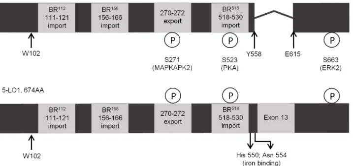
Fig 1. Known regulatory factors are retained in the protein sequence of 5-LOΔ13.chematic of the protein sequences of the 617 amino acid 5-LOΔ13 isoform (top) and of the 674 amino acid 5-LO1 isoform (bottom). All known phosphorylation sites, import and export sequences are shown to be present maintained in 5-LOΔ13 despite the lack of exon 13. The kinases (MAPKAPK2, PKA and ERK) responsible for phosphorylation of the different sites are indicated as is residue W102 that is responsible for the interaction of 5-LO1 with CLP, and for the CLP-induced increase in 5-LO1 activity in cell-free assays.
were resuspended in 100μL of a buffer containing 25 mM Tris, 10mM EDTA, 50mM glucose
and 20μg/mL RNase A. Lysozyme (20μL) was then added at a concentration of 10mg/mL and
tubes were incubated for 2 min before adding 200μL of a second buffer containing 1% SDS
and 200 mM NaOH. Samples were then put on ice for 5 min before adding a third buffer con-taining 3 M potassium (KOAc) and 5 M acetate (HAc). Tubes were then mixed by inverting, placed on ice 5 min and centrifuged at 21,000×g. Supernatants were conserved and 400μL of
phenol:chloroform was added. The upper phase was transferred to a new tube and 1 mL 99% ethanol was added before incubating for 2 min. Samples were then centrifuged for 5 min in a cold centrifuge and pellets were left to dry completely before resuspending in TE buffer. DNA constructs were then sent to the Plateforme de séquencage et génotypage du CHUL (Québec, QC) for sequencing to confirm base pair change. Colonies expressing proper sequences were incubated overnight in 200 mL LB broth containing 100μg/mL ampicilin. Purification was
then carried out using the PureLink HiPure Plasmid FP Maxiprep kit according to the manu-facturer’s protocol.
Transfections of HEK293 cells
Transfections were completed by detaching and resuspending HEK293 cells (ATCC) at a con-centration of 1.5×10⁷cells/mL. Cells were then transferred to a 400μL electroporation cuvette
(Bio-Rad), the indicated plasmids (37.5μgDNA/ml) were added and the solutions were
incu-bated at room temperature for 10 minutes. Cells were then shocked (250 volts, 950μF) using a
Gene Pulser Xcell (Bio-Rad) and left to sediment for 10 minutes and were then transferred to pre-warmed culture flasks containing DMEM medium supplemented with 10% foetal bovine serum (FBS) at 37°C. Experiments with transient transfections were carried out within 24–48 hours. Stable transfectants were obtained by culturing cells in DMEM medium supplemented with 10% FBS at 37°C in a humidified 5% CO2environment in the presence of 400 ng/mL
geneticin (Life Technologies) for pcDNA3.1 vectors or 200 ng/mL zeocin (Invivogen) for pBUDCE4.1 vectors.
Immunofluorescence microscopy
Glass cover slides were washed with 70% ethanol and placed at the bottom of six-well plates (CellStar). Cells were centrifuged and re-suspended in DMEM containing 10% FBS at a con-centration of 3×10⁵cells/mL. One mL was then added to each six-well plate and incubated overnight. Wells were then washed with HBSS and stimulation was initiated by adding 1 ml of HBSS solution containing 1.6 mM CaCl2, 1μM calcium ionophore A23187 (Sigma-Aldrich)
and 10μM arachidonic acid (AA) (Nu-Check Prep). After 10 minutes at 37°C, the glass slides
were rinsed with PBS and cells were fixed with 4% paraformaldehyde (Alfa Aesar). The plates were incubated at room temperature for 20 min, rinsed with PBS and permeabilized with 0.25% Triton X-100 (Sigma-Aldrich). Following permeabilisation, the slides were rinsed with PBS containing 10% FBS for 15 minutes. Afterwards, 80μL of rabbit anti-5-LO (1:50 in PBS
60X oil objective. Images are 200μm cuts from samples taken by scanning at 450 nm or 488
nm. Data were acquired and exported using Fluoview 10-ASW software.
Cell stimulation and analysis of 5-LO products
Cells were stimulated as previously described with slight modifications [26]. Briefly, HEK293 cells were detached by trypsinization and re-suspended in HBSS containing 1.6 mM CaCl2,
1μM ionophore A23187 and 10μM AA at a concentration of 1×10⁷cells/mL and were
incu-bated at 37°C in a water bath for 30 min. Stimulations were stopped by adding 0.5 volumes of a methanol:acetonitrile (1:1) solution containing 100 ng/mL each of prostaglandin B2(PGB2;
Cayman Chemical) and 19-OH-PGB2as internal standards, and samples were then kept at
-20°C overnight for protein denaturation. Samples were then centrifuged at 12,000×g for 5 min, supernatants were collected and subjected to automated in-line solid phase extraction on Oasis HLB columns prior to reverse-phase high-performance liquid chromatography analy-sis with diode array detection [33].
SDS-PAGE and Western blots
Cells were trypsinized and centrifuged, then re-suspended in a lysis buffer (150 mM NaCl, 2 mM EDTA and 50 mM Tris-HCl, pH 7.6) containing 0.1% NP-40, complete mini-EDTA free protease inhibitor tablets (Roche) and phosphatase inhibitors (Sigma-Aldrich). Laemmli solution (5X, Sigma-Aldrich) was then added to a final concentration of 1X and were heated for 10 minutes in a boiling water bath.
SDS-PAGE was carried out on a 4–12% acrylamide gel gradient before transferring proteins onto a polyvinylidene fluoride membrane (GE Healthcare). Western blotting was done using rabbit monoclonal anti-5-LO (1:500 in TBS-Tween; Cell Signaling Technology), rabbit mono-clonal hemagglutinin (1:2000 in TBS-Tween; Cell Signaling Technology), rabbit S523 5-LO (1:500 in TBS-Tween; Cell Signalling Technology), rabbit anti-phospho-S271 5-LO (1:500 in TBS-Tween; Cell Signalling Technology), and a horseradish-conjugated mouse anti-rabbit IgG (Jackson Immunoresearch). Membranes were then developed using ECL prime western blotting detection reagent (GE Healthcare) and detection was performed using an Alpha Innotech Fluorchem imager.
Statistical analyses
Student’s t-tests and one-way ANOVA were performed to determine differences in 5-LO prod-uct biosynthesis. All values are means ± SEM. Statistics were done using Graphpad Prism ver-sion 5.
Results
5-LO
Δ
13 affects LT but not 5-hydroxyeicosatetraenoic acid (5-HETE)
biosynthesis
We previously reported that 5-LOΔ13 could inhibit the biosynthesis of 5-LO products when
co-expressed with the active 5-LO1 [26]. To further investigate this process, we performed experiments in which we modulated the expression of 5-LOΔ13 while keeping that of 5-LO1
constant. This was achieved by manipulating vector ratios in transfection experiments (Fig 2A). Increasing the amount of 5-LOΔ13 decreased 5-LO product biosynthesis by
the same co-expression experiments were performed in HeLa cells and yielded similar results (Fig 2C).
W102 is required for 5-LO product biosynthesis in intact stimulated cells
Given that interactions of 5-LO1 with CLP greatly increase the capacity to synthesize LTs, the possibility that 5-LOΔ13 inhibits 5-LO product biosynthesis by a mechanism related to CLPwas next considered. A single amino acid (W102) on 5-LO1 has been shown to be directly involved in the interaction of 5-LO1 with CLP [19]. However, the role of W102 for the stimu-lated biosynthesis of 5-LO products in intact cells had not previously been assessed. When the
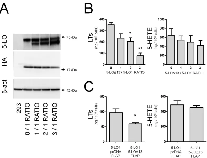
Fig 2. TheΔ-13 isoform of 5-lipoxygenase inhibits LT biosynthesis in a dose-dependant manner. (A)Immunoblots showing expression of 5-LO1 (top band) and 5-LOΔ13 (lower band) after transfections with the indicated ratios of 5-LOΔ13/5-LO1 expression vectors, as well as the presence of FLAP-HA and
β-actin as loading control in HEK293 cells.(B)HEK293 cells expressing FLAP-HA and transfected with the indicated ratios of 5-LO1 and 5-LOΔ13 expression vectors were stimulated with 1μM thapsigargin and 10μM AA for 30 minutes. 5-LO products were measured by HPLC as described in the Methods section. Leukotrienes (LTs) are the sum of LTB4and its trans isomers. 5-HETE = 5-hydroxyeicosatetraenoic acid.(C)HeLa cells transfected with a
1:1 ratio of vectors expressing 5-LO1 and 5-LOΔ13 were stimulated under the same conditions as in (B) and LTs and 5-HETE production were measured. Immunoblots are representative of 4 independent experiments. Data represent means±SEM of 4 independent experiments.*Different from control p<0.05,
and**p<0.01.
W102A mutant of 5-LO1 (5-LO1-W102A) was expressed in HEK293 cells, the stimulated bio-synthesis of 5-LO products was drastically reduced compared to that of 5-LO1 indicating that W102 is required for product biosynthesis, and suggesting that the interaction of CLP with the W102 of 5-LO1 is required for proper 5-LO1 activation and product biosynthesis in intact HEK293 cells (Fig 3, second column).
Modulation of LT biosynthesis by 5-LO
Δ
13 does not involve interactions
with CLP or FLAP
Having confirmed the importance of W102 in intact cells, a W102A mutant of the catalytically inactive 5-LOΔ13 (5-LOΔ13-W102A) was generated to investigate whether the 5-LOΔ13
protein might inhibit LT biosynthesis by trapping CLP. Co-expression experiments of 5-LOΔ13-W102A with 5-LO1 showed a similar inhibition profile for the biosynthesis of 5-LO
products to that of its non-mutant counterpart indicating that the inhibitory effect of 5-LOΔ13
does not involve the binding and sequestration of CLP (Fig 3, third and fourth columns). In addition to CLP, interactions of the 5-LO1 enzyme with FLAP are necessary for the effi-cient production of LTs in stimulated cells [34]. To evaluate the possibility that 5-LOΔ13 may
inhibit the biosynthesis of 5-LO products by interfering with 5-LO1/FLAP interactions, the inhibitory effect of 5-LOΔ13 was assessed in HEK293 cells co-transfected with a vector
express-ing recombinant FLAP or a mock vector (HEK293 cells do not naturally express FLAP [26, 35]). 5-LO1 and FLAP-HA expression were confirmed by western blotting (not shown) and, as expected, cells expressing 5-LO1 stimulated in the absence of FLAP generated significantly less 5-LO products than cells co-expressing FLAP (Fig 3, compare 5thcolumn with 1stcolumn). However, 5-LOΔ13 still effectively inhibited the biosynthesis of 5-LO products in the absence
of FLAP suggesting that the impact of 5-LOΔ13 on 5-LO product biosynthesis is independent
of the presence of FLAP (Fig 3, 5thand 6thcolumns).
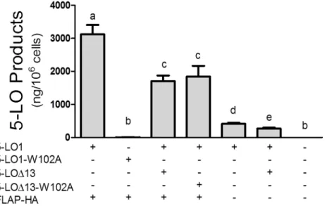
Fig 3. Impact of W102A mutations and of FLAP expression on the biosynthesis of 5-LO products.
HEK293 cells expressing FLAP-HA or not were transfected with expression vectors coding for 5-LO1, the 5-LO1-W102A mutant, 5-LOΔ13 or the 5-LOΔ13-W102A mutant as shown. Control cells (last column) were transfected with a control pcDNA3.1 vector. HEK293 cells were then stimulated with 1μM thapsigargin and 10μM AA. 5-LO products were measured by HPLC as described in the Methods section and represent the sum of LTB4, its trans isomers and 5-hydroxyeicosatetraenoic acid. Data represent means±SEM of 3 or 4
independent experiments.*Values without a common superscript are different, p<0.05.
Confocal microscopy evaluation of the subcellular distribution of 5-LO
isoforms and their W102A mutants
To further characterize the behaviour of the 5-LO isoforms, the intracellular localization of 5-LO1 and 5-LOΔ13 were evaluated by confocal microscopy. 5-LO1 was primarily located in
the nucleoplasm of resting HEK293 cells, whether the cells expressed FLAP or not (Fig 4). Upon cell stimulation, 5-LO1 translocation was observed only in cells co-expressing FLAP as shown by the strong peri-nuclear rings, confirming the importance of FLAP in 5-LO1 translo-cation to the nuclear envelope in these cells (Fig 4). In addition to the confocal microscopy
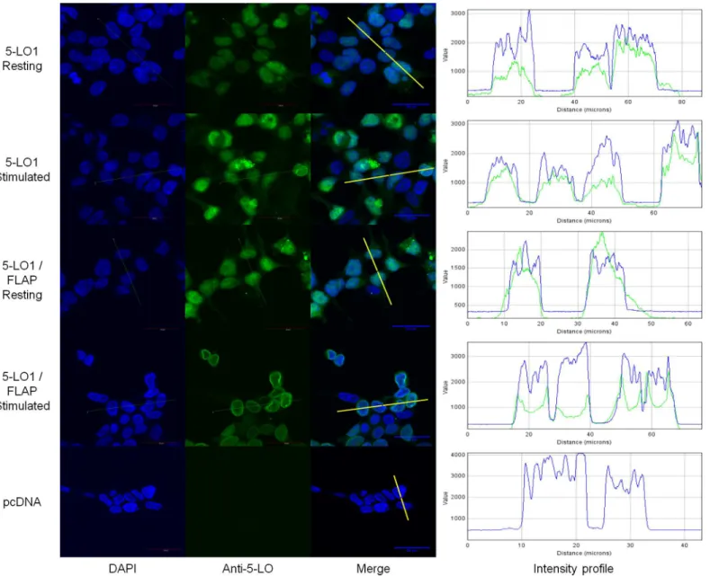
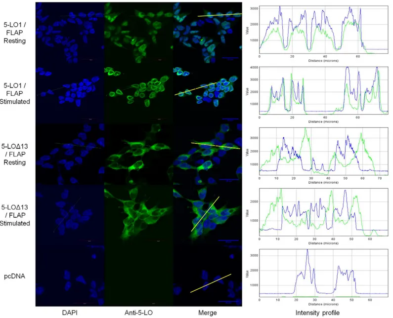
Fig 4. Sub-cellular localization of wild-type (WT) 5-lipoxygenase in cells expressing FLAP or not.HEK293 cells expressing FLAP or not were transfected with a vector expressing 5-LO1 or with a control vector (pcDNA). Cells were stimulated with 1μM A23187 and 10μM AA or were incubated with their diluent (resting) for 10 minutes. Cells were then fixed and permeabilized and then incubated with rabbit anti-5-LO. Slides were then incubated with an Alexa488-conjugated secondary anti rabbit antibody (green) and with DAPI (blue) to visualize nuclei, and slides were then mounted. Samples were analysed by confocal microscopy and images are presented on the left panels. The intensity of the signals could be visualized by intensity profiles (right panels) of the regions indicated by the white line on the merge images. Images are representative of three independent experiments. Scale bar represents 30μm.
images, the intracellular location of 5-LO can also be visualized graphically with intensity pro-files of cross sections of cells (Fig 4, right panels). The intensity propro-files show that 5-LO stain-ing overlaps primarily with that of nuclear DNA stainstain-ing (DAPI) in the nuclear compartment of resting cells, and spikes of 5-LO intensity appear at the periphery of DNA staining in stimu-lated FLAP-expressing cells, consistent with translocation to the nuclear membrane.
Unlike the 5-LO1, 5-LOΔ13 was primarily located in the cytoplasm of resting cells (Fig 5).
Interestingly, no translocation of 5-LOΔ13 was observed following cell stimulation in the
pres-ence of FLAP (Fig 5) or in its abspres-ence (not shown). Closer observation of cells using the
Fig 5. Sub-cellular localization of 5-LO1 and 5-LOΔ13 in resting and stimulated cells.HEK293 cells expressing FLAP which were transfected to express either 5-LO1 or 5-LOΔ13, or which were transfected with a control vector (pcDNA) were stimulated with 1μM A23187 and 10μM AA or were incubated with their diluent (resting) for 10 minutes. Cells were then fixed and permeabilized and then incubated with rabbit anti-5-LO. Slides were then incubated with an Alexa488-conjugated secondary anti-rabbit antibody (green) and with DAPI (blue) to visualize nuclei, and slides were then mounted. Samples were analysed by confocal microscopy and images are presented on the left panels. The intensity of the signals could be visualized by intensity profiles (right panels) of the regions indicated by the white line on the merge images. Images are representative of three independent experiments. Scale bar represents 30μm.
intensity profiles of cross sections of cells revealed that the maximum of the intensity profile signal for 5-LOΔ13 was at close proximity to the nuclear envelope, and that this signal tends to
fade in more distal cytoplasmic regions (Fig 5, right panels). However, this small perinuclear accumulation is not as pronounced or sharp as that observed for translocation of 5-LO1 to nuclear membranes in stimulated cells.
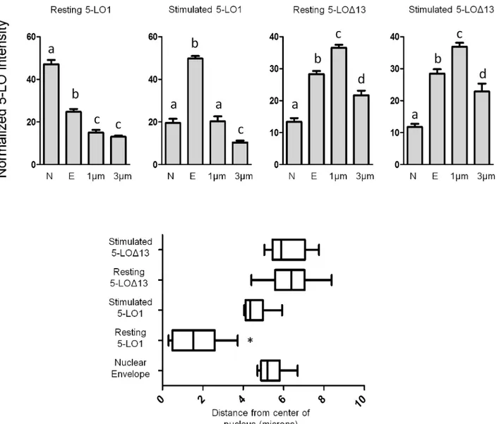
The ability to measure intensity profiles of cross-sections of cells was utilized to quantify the cellular distribution of 5-LO1 and 5-LOΔ13 in resting and stimulated cells. The intensity of
anti-5-LO staining was measured at the centre of the nucleus, at the nuclear envelope as deter-mined by the edge of DAPI staining, 1μm from the edge of the nucleus to represent perinuclear
staining and at 3μm from the nucleus to represent cytosolic staining (Fig 6, top panels). 5-LO1
was significantly more abundant in the nucleus in resting cells compared to other areas, while resting 5-LOΔ13 staining was significantly greater in the perinuclear and nuclear envelope
regions. Upon cell stimulation 5-LO1 became significantly more intense at the nuclear enve-lope region whereas the pattern of 5-LOΔ13 staining was unchanged. When site of maximum
staining intensity was determined on cell cross sections, resting 5-LO1 was significantly closer to the centre of the nucleus than stimulated 5-LO1 or 5-LOΔ13 (resting or stimulated) whose
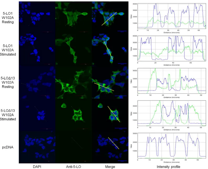
maximum intensities were not significantly different from one another (Fig 6, bottom panel). Although 5-LO1 and CLP have been shown to co-associate in nuclear fractions of stimu-lated cells [17,19] and 5-LO1 translocation is greatly diminished in the absence of CLP [18], the subcellular distribution of 5-LO1-W102A mutants had not been previously investigated. Fig 7shows that the 5-LO1-W102A has a uniform cellular distribution in resting cells suggest-ing that W102 is required for the nuclear location of WT 5-LO. Stimulation of cells expresssuggest-ing 5-LO1-W102A did not induce 5-LO translocation to the nuclear envelope, consistent with its inability to generate 5-LO products. 5-LOΔ13-W102A mutants displayed a cellular distribution
profile similar to that of 5-LOΔ13 with the increased staining in the perinuclear region and no
difference between stimulated and non-stimulated cells (Fig 7).
5-LO isoforms have distinct phosphorylation states
Given the distinct sub-cellular distribution of 5-LO1 and 5-LOΔ13, the phosphorylation
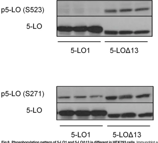
pat-terns of these two isoforms were investigated. Western blotting was performed on extracts from stable transfectants using specific antibodies against phosphorylated S-523 and S-271. Despite the fact that both protein isoforms are identical other than the absence of amino acids 559–615 in 5-LOΔ13, 5-LOΔ13 was phosphorylated on S523 while 5-LO1 was not (Fig 8).
Sim-ilarly, S271 was heavily phosphorylated on 5-LOΔ13 while 5-LO1 was only moderately
phosphorylated.
Discussion
5-LO is subjected to a number of regulatory mechanisms that impact on the capacity to pro-duce leukotrienes. Recently a potentially new mechanism of 5-LO regulation was revealed when several catalytically inactive splice variants of human 5-LO were identified [26,27,36] and shown to negatively impact on the biosynthesis of 5-LO products when co-expressed with the active 5-LO1. Human polymorphonuclear leukocytes (PMN) and several human myeloid and lymphoid cell lines were shown to express alternatively-spliced 5-LO transcripts, and west-ern blots from human PMN and Raji lymphoma cells showed immunoreactive bands that co-migrated with 5-LOΔ13 [26].
In the current study, further investigation of the 5-LOΔ13 protein isoform provides new
increased expression of 5-LOΔ13 resulted in a greater inhibition of 5-LO product biosynthesis
(Fig 2). Surprisingly, in both HEK293 and HeLa cells 5-LOΔ13 mainly inhibited cellular LT
biosynthesis with a less pronounced impact on 5-HETE production. This was akin to the effect of CLP on recombinant 5-LO1 activity where CLP enhances LT synthesisin vitrobut has little influence on 5-HETE synthesis [17,19]. Likewise, FLAP silencing in MM6 cells was also recently associated with a decrease in LT biosynthesis but not that of 5-HETE [18]. Therefore, this inhibition profile was suggestive of an antagonist-like effect of 5-LOΔ13 where this isoform
may interfere with the ability of 5-LO1 to interact with CLP and/or FLAP.
Fig 6. Intensity profiles of of 5-LO1 and 5-LOΔ13 staining in resting and stimulated cells.HEK293 cells expressing FLAP which were transfected to express either 5-LO1 or 5-LOΔ13 were stimulated with 1μM A23187 and 10μM AA or were incubated with their diluent (resting) for 10 minutes. Cells were then fixed and permeabilized and then incubated with rabbit anti-5-LO. Slides were then incubated with an Alexa488-conjugated secondary anti-rabbit antibody and with DAPI to visualize nuclei, and slides were then mounted. Samples were analysed by confocal microscopy and the intensity of the signals of cross-sections of cells were measured as inFig 5.Top panels:The intensity of each cross section was measured at the centre of the nucleus (N), at the nuclear envelope (E) identified by the edge of DAPI staining, at 1μm and at 3μm from the nuclear envelope (cytoplasmic side). Values are means±SEM, n = 9. Values in the same figure without a common superscript are different, p<0.05.Bottom panel:Box and whisker plots showing the distance from the centre of the nucleus of the most intense anti-5-LO staining of cross sections. The box represents the two middle quartiles of data, the vertical line in the box is the median and the whiskers represent the minimum and maximum of all the data.*Median value different from others, p<0.05. Values in all panels are
from intensity cross sections of 9 cells per condition from three different fields of view, each field representing a separate experiment.
This observation prompted the investigation of the potential implication of CLP or FLAP in the inhibitory effect of 5-LOΔ13. Previous reports had shown that CLP enhances recombinant
5-LO1 activity in cell-free systems, while CLP silencing experiments recently confirmed a role for CLP in 5-LO1 activation in intact cells [18]. W102 is the key 5-LO1 residue required for the activity-enhancing interaction of 5-LO1 with CLP in cell free assays [17,19], however the importance of W102 on 5-LO1 activation in stimulated whole cells had never been evaluated. The current study shows that the stimulation of HEK293 cells expressing the W102A mutant of 5-LO1 resulted in very little biosynthesis of 5-LO products (Fig 3). Having confirmed the
Fig 7. Sub-cellular localization of the W102A mutants of 5-LO1 and 5-LOΔ13 in resting and stimulated cells.HEK293 cells expressing FLAP and expressing either the 5-LO1-W102A mutant or the 5-LOΔ13-W102A mutant, or which were transfected with a control vector (pcDNA) were stimulated with 1μM A23187 and 10μM AA or were incubated with their diluent (resting) for 10 minutes. Cells were then fixed and permeabilized and then incubated with rabbit anti-5-LO. Slides were then incubated with an Alexa488-conjugated secondary anti-rabbit antibody (green) and with DAPI (blue) to visualize nuclei, and slides were then mounted. Samples were analysed by confocal microscopy and images are presented on the left panels. The intensity of the signals could be visualized by intensity profiles (right panels) of the regions indicated by the white line on the merge images. Images are representative of three independent experiments. Scale bar represents 30μm.
requirement of W102 in intact cells, the interaction of 5-LOΔ13 with CLP was investigated
using 5-LOΔ13-W102A mutants that should also be unable to interact with CLP. However, the
5-LOΔ13-W102A mutants showed the same inhibitory capacity as the non-mutated form of
the protein (Fig 3) suggesting that the mechanism by which the 5-LOΔ13 inhibits 5-LO1 does
not involve an interaction or competition with CLP. Similarly, although the absence of FLAP decreased the cellular capacity to synthesize 5-LO products, 5-LOΔ13 was fully capable of
inhibiting 5-LO product biosynthesis in stimulated HEK293 cells in the absence of FLAP indi-cating that the inhibitory effect of 5-LOΔ13 does not involve FLAP.
The control of the subcellular location of 5-LO1 and its ability to translocate to nuclear membranes following cell stimulation are also mechanisms involved in the regulation of 5-LO product biosynthesis. Surprisingly, the location of 5-LO1, 5-LOΔ13 and their W102A mutants
varied greatly in resting HEK293 cells (Figs5and6). Similar to previous studies using HEK293 cells as well as human alveolar macrophages, mast cells and adherent neutrophils and eosino-phils [9,11,37,38], 5-LO1 was primarily located in the nucleoplasm of resting HEK293 cells although some cytoplasmic 5-LO was also evident. 5-LO1 protein also translocated to nuclear membranes following cell stimulation in a FLAP-dependent manner, as previously shown in other cell types [13,18]. Unexpectedly, expression of 5-LO1-W102A mutants revealed a signif-icant shift in the protein’s localization in resting cells showing a diffuse cellular staining as well as a complete loss of the ability to translocate following stimulation (Fig 7), consistent with the
Fig 8. Phosphorylation pattern of 5-LO1 and 5-LOΔ13 is different in HEK293 cells.Immunoblot analysis of resting HEK293 cells expressing either 5-LOΔ13 or 5-LOΔ13. All cells also expressed FLAP and CLP. After separation of proteins by SDS-PAGE, membranes were subject to western blots using anti phospho-serine 523 (S523) or anti-phospho-phospho-serine 271 (S271). Expression of 5-LO isoforms was verified by blotting using anti-5-LO (5-LO). The immunoblots show the results of 3 independent experiments.
inability to activate this mutant following cell stimulation. It was recently shown that CLP silencing results in a loss in the ability of 5-LO to translocate to the nucleus in stimulated MM6 cells. However, in the present study the loss of nuclear localization in resting HEK293 cells indicates that W102 is important for nuclear localisation of 5-LO1 and suggests that interaction with CLP may be required for nuclear targeting. Thus the role of CLP may be more complex than previously thought.
The cytosolic location of 5-LOΔ13 differed considerably from that of the 5-LO1 protein.
The intensity profiles obtained from microscopy images show that 5-LOΔ13 is primarily
non-nuclear and appears to be concentrated in the ER-rich peri-non-nuclear region. However, this sub-cellular location was not altered by cell stimulation or by expression of the 5-LOΔ13-W102A
mutant (Figs5–7). This peri-nuclear position of 5-LOΔ13 is the location in HEK293 cells
where both proteins isoforms are in proximity to one another following cell stimulation and suggests that this is where 5-LOΔ13 may interact with 5-LO1 and exert its inhibitory effect.
Since 5-LO forms dimers that might be required for its activation [22–24], this inhibitory effect could possibly occur through the formation of inactive heterodimers, or indirectly through another unknown mechanism. Additionally, the altered location of 5-LOΔ13 compared to
5-LO1 suggests that the presence of amino acids 559 through 615 of 5-LO1 that are coded by exon 13 are required for both nuclear import and stimulus-induced translocation. Accordingly, it was previously suggested that mutations that eliminate enzymatic activity also prevent nuclear localization of 5-LO by a yet-to-be-described mechanism implying that enzymatic activity is required for nuclear import [39]. Interestingly, splice variants of the human 15-lipox-ygenase 2, the most abundant AA-metabolizing enzyme in prostate cancer, are also catalytically inactive, cytosolic proteins as opposed to their nuclear, enzymatically-active counterpart [40]. Therefore, the existence of alternatively spliced lipoxygenase isoforms that do not co-localize with their enzymatically active counterparts is not limited to 5-LO.
The known nuclear localisation sequences and phosphorylation sites associated with the control of the subcellular location of 5-LO are all retained in theΔ-13 isoform (Fig 1). However
5-LO1 and 5-LOΔ13 show strikingly dissimilar phosphorylation patterns on S523 and S271
where 5-LOΔ13 is hyper-phosphorylated (Fig 8). This phosphorylation pattern may explain
the differential localization of these proteins since phospho-S523, that lies within the BR518 import region and is targeted by protein kinase A, prevents nuclear import [31] thus prevent-ing 5-LOΔ13 entry into the nucleus. On the other hand, 5-LO1 that enters the nucleus is
retained due to the phosphorylation of Ser-271, which prevents nuclear export of 5-LO1 [30]. Therefore, the differential location of the 5-LO protein isoforms may be the consequence of dif-ferent susceptibilities of the isoforms to protein kinases or phosphatases.
Overall this study provides new information regarding inhibition of LT biosynthesis by 5-LOΔ13, as well as the role of the sequence coded by exon 13, the W102 residue of 5-LO1, and
possibly that of CLP, on nuclear targeting of 5-LO. Given that alternatively spliced isoforms of 5-LO have been identified in human leukocytes, a better understanding of the regulation of their expression and of the mechanisms by which they modulate LT biosynthesis will contrib-ute to our understanding of the complex regulation of LT biosynthesis and may possibly lead to new approaches to the treatment of diseases like asthma and atherosclerosis in which 5-LO plays an active role.
Acknowledgments
from the Fonds de recherche du Québec-Santé. E.A was supported by a graduate student schol-arship from the New Brunswick Health Research Foundation. L.H.B was the recipient of a fel-lowship from the Fonds de recherche du Québec-Santé.
Author Contributions
Conceived and designed the experiments: EPA MES LHB NF. Performed the experiments: EPA LHB. Analyzed the data: EPA MES NF. Contributed reagents/materials/analysis tools: MES NF. Wrote the paper: MES EPA LHB NF.
References
1. Radmark O, Samuelsson B. Regulation of the activity of 5-lipoxygenase, a key enzyme in leukotriene biosynthesis. Biochem Biophys Res Commun. 2010; 396: 105–110. doi:10.1016/j.bbrc.2010.02.173
PMID:20494120
2. Peters-Golden M, Henderson WR Jr. Leukotrienes. N Engl J Med. 2007; 357: 1841–1854. PMID:
17978293
3. Haeggstrom JZ, Funk CD. Lipoxygenase and leukotriene pathways: biochemistry, biology, and roles in disease. Chem Rev. 2011; 111: 5866–5898. doi:10.1021/cr200246dPMID:21936577
4. Bishayee K, Khuda-Bukhsh AR. 5-lipoxygenase antagonist therapy: a new approach towards targeted cancer chemotherapy. Acta Biochim Biophys Sin (Shanghai). 2013; 45: 709–719.
5. Fanning LB, Boyce JA. Lipid mediators and allergic diseases. Ann Allergy Asthma Immunol. 2013; 111: 155–162. doi:10.1016/j.anai.2013.06.031PMID:23987187
6. Chu J, Li JG, Pratico D. Zileuton improves memory deficits, amyloid and tau pathology in a mouse model of Alzheimer's disease with plaques and tangles. PLoS One. 2013; 8: e70991. doi:10.1371/ journal.pone.0070991PMID:23951061
7. Chen M, Lam BK, Kanaoka Y, Nigrovic PA, Audoly LP, Austen KF, et al. Neutrophil-derived leukotriene B4 is required for inflammatory arthritis. J Exp Med. 2006; 203: 837–842. PMID:16567388
8. Brock TG, McNish RW, Peters-Golden M. Translocation and leukotriene synthetic capacity of nuclear 5-lipoxygenase in rat basophilic leukemia cells and alveolar macrophages. J Biol Chem. 1995; 270: 21652–21658. PMID:7665580
9. Woods JW, Coffey MJ, Brock TG, Singer II, Peters-Golden M. 5-Lipoxygenase is located in the euchro-matin of the nucleus in resting human alveolar macrophages and translocates to the nuclear envelope upon cell activation. J Clin Invest. 1995; 95: 2035–2046. PMID:7738170
10. Brock TG, Peters-Golden M. Localization of 5-lipoxygenase to the nucleus of resting rat basophilic leu-kemia cells. Adv Prostaglandin Thromboxane Leukot Res. 1995; 23: 151–153. PMID:7732822 11. Chen XS, Naumann TA, Kurre U, Jenkins NA, Copeland NG, Funk CD. cDNA cloning, expression,
mutagenesis, intracellular localization, and gene chromosomal assignment of mouse 5-lipoxygenase. J Biol Chem. 1995; 270: 17993–17999. PMID:7629107
12. Woods JW, Evans JF, Ethier D, Scott S, Vickers PJ, Hearn L, et al. 5-lipoxygenase and 5-lipoxygen-ase-activating protein are localized in the nuclear envelope of activated human leukocytes. J Exp Med. 1993; 178: 1935–1946. PMID:8245774
13. Flamand N, Lefebvre J, Surette ME, Picard S, Borgeat P. Arachidonic acid regulates the translocation of 5-lipoxygenase to the nuclear membranes in human neutrophils. J Biol Chem. 2006; 281: 129–136. PMID:16275640
14. Ochi K, Yoshimoto T, Yamamoto S, Taniguchi K, Miyamoto T. Arachidonate 5-lipoxygenase of guinea pig peritoneal polymorphonuclear leukocytes. Activation by adenosine 5'-triphosphate. J Biol Chem. 1983; 258: 5754–5758. PMID:6406506
15. Borgeat P, Samuelsson B. Arachidonic acid metabolism in polymorphonuclear leukocytes: effects of ionophore A23187. Proc Natl Acad Sci U S A. 1979; 76: 2148–2152. PMID:377292
16. Hammarberg T, Provost P, Persson B, Radmark O. The N-terminal domain of 5-lipoxygenase binds calcium and mediates calcium stimulation of enzyme activity. J Biol Chem. 2000; 275: 38787–38793. PMID:10984486
18. Basavarajappa D, Wan M, Lukic A, Steinhilber D, Samuelsson B, Radmark O. Roles of coactosin-like protein (CLP) and 5-lipoxygenase-activating protein (FLAP) in cellular leukotriene biosynthesis. Proc Natl Acad Sci U S A. 2014; 111: 11371–11376. doi:10.1073/pnas.1410983111PMID:25034252 19. Esser J, Rakonjac M, Hofmann B, Fischer L, Provost P, Schneider G, et al. Coactosin-like protein
func-tions as a stabilizing chaperone for 5-lipoxygenase: role of tryptophan 102. Biochem J. 2010; 425: 265– 274.
20. Provost P, Doucet J, Stock A, Gerisch G, Samuelsson B, Radmark O. Coactosin-like protein, a human F-actin-binding protein: critical role of lysine-75. Biochem J. 2001; 359: 255–263. PMID:11583571 21. Abramovitz M, Wong E, Cox ME, Richardson CD, Li C, Vickers PJ. 5-lipoxygenase-activating protein
stimulates the utilization of arachidonic acid by 5-lipoxygenase. Eur J Biochem. 1993; 215: 105–111. PMID:8344271
22. Plante H, Picard S, Mancini J, Borgeat P. 5-Lipoxygenase-activating protein homodimer in human neu-trophils: evidence for a role in leukotriene biosynthesis. Biochem J. 2006; 393: 211–218. PMID:
16144515
23. Zhang YY, Hammarberg T, Radmark O, Samuelsson B, Ng CF, Funk CD, et al. Analysis of a nucleo-tide-binding site of 5-lipoxygenase by affinity labelling: binding characteristics and amino acid sequences. Biochem J. 2000; 351 Pt 3: 697–707. PMID:11042125
24. Hafner AK, Cernescu M, Hofmann B, Ermisch M, Hornig M, Metzner J, et al. Dimerization of human 5-lipoxygenase. Biol Chem. 2011; 392: 1097–1111. doi:10.1515/BC.2011.200PMID:22050225 25. Boado RJ, Pardridge WM, Vinters HV, Black KL. Differential expression of arachidonate
5-lipoxygen-ase transcripts in human brain tumors: evidence for the expression of a multitranscript family. Proc Natl Acad Sci U S A. 1992; 89: 9044–9048. PMID:1357659
26. Boudreau LH, Bertin J, Robichaud PP, Laflamme M, Ouellette RJ, Flamand N, et al. Novel 5-lipoxygen-ase isoforms affect the biosynthesis of 5-lipoxygen5-lipoxygen-ase products. FASEB J. 2011; 25: 1097–1105. doi:
10.1096/fj.10-173856PMID:21098726
27. Ochs MJ, Suess B, Steinhilber D. 5-lipoxygenase mRNA and protein isoforms. Basic Clin Pharmacol Toxicol. 2014; 114: 78–82. doi:10.1111/bcpt.12115PMID:24020397
28. Hammarberg T, Kuprin S, Radmark O, Holmgren A. EPR investigation of the active site of recombinant human 5-lipoxygenase: inhibition by selenide. Biochemistry. 2001; 40: 6371–6378. PMID:11371199 29. Jones SM, Luo M, Healy AM, Peters-Golden M, Brock TG. Structural and functional criteria reveal a
new nuclear import sequence on the 5-lipoxygenase protein. J Biol Chem. 2002; 277: 38550–38556. PMID:12140292
30. Flamand N, Luo M, Peters-Golden M, Brock TG. Phosphorylation of serine 271 on 5-lipoxygenase and its role in nuclear export. J Biol Chem. 2009; 284: 306–313. doi:10.1074/jbc.M805593200PMID:
18978352
31. Luo M, Jones SM, Flamand N, Aronoff DM, Peters-Golden M, Brock TG. Phosphorylation by protein kinase a inhibits nuclear import of 5-lipoxygenase. J Biol Chem. 2005; 280: 40609–40616. PMID:
16230355
32. Jones SM, Luo M, Peters-Golden M, Brock TG. Identification of two novel nuclear import sequences on the 5-lipoxygenase protein. J Biol Chem. 2003; 278: 10257–10263. PMID:12525477
33. Borgeat P, Picard S, Vallerand P, Bourgoin S, Odeimat A, Sirois P, et al. Automated on-line extraction and profiling of lipoxygenase products of arachidonic acid by high-performance liquid chromatography. Methods Enzymol. 1990; 187: 98–116. PMID:2122189
34. Dixon RA, Diehl RE, Opas E, Rands E, Vickers PJ, Evans JF, et al. Requirement of a 5-lipoxygenase-activating protein for leukotriene synthesis. Nature. 1990; 343: 282–284. PMID:2300173
35. Gerstmeier J, Weinigel C, Barz D, Werz O, Garscha U. An experimental cell-based model for studying the cell biology and molecular pharmacology of 5-lipoxygenase-activating protein in leukotriene biosyn-thesis. Biochim Biophys Acta. 2014; 1840: 2961–2969. doi:10.1016/j.bbagen.2014.05.016PMID:
24905297
36. Ochs MJ, Sorg BL, Pufahl L, Grez M, Suess B, Steinhilber D. Post-transcriptional regulation of 5-lipoxy-genase mRNA expression via alternative splicing and nonsense-mediated mRNA decay. PLoS One. 2012; 7: e31363. doi:10.1371/journal.pone.0031363PMID:22363630
37. Brock TG, Anderson JA, Fries FP, Peters-Golden M, Sporn PH. Decreased leukotriene C4 synthesis accompanies adherence-dependent nuclear import of 5-lipoxygenase in human blood eosinophils. J Immunol. 1999; 162: 1669–1676. PMID:9973428
39. Chen XS, Zhang YY, Funk CD. Determinants of 5-lipoxygenase nuclear localization using green fluo-rescent protein/5-lipoxygenase fusion proteins. J Biol Chem. 1998; 273: 31237–31244. PMID:
9813031