11 -
ORIGINAL ARTICLE
Hypertonic glucose solution 10% - 25% on the mesenterium and peritoneum of
the rat: macroscopic and microscopic study
1Solução glicosada hipertônica no mesentério e no peritônio de ratos: estudo
macroscópico e microscópico
José Cícero Ferreira de Carvalho2, Antenor Teixeira Leal3, Luis Ferreira de Sousa4, Benedito Herani Filho5
1. Paper from the Surgery Gastroenterology Discipline Graduate Program of the Federal University of São Paulo (UNIFESP) and edical Doctor School of São Paulo (EPM).
2. Assistant Professor at the Surgery Techniques Department of UNCISAL. 3. Chairman Professor of Pathology of UNCISAL.
4. Associate Professor of Morphology Department and Human Anatomy of UFAL.
5. Associate Professor of Gastroenterology Surgery Discipline at the Surgery Department of UNIFESP/EPM.
ABSTRACT
Purpose: The objective of the experimental study is to detect the macroscopic and microscopic alterations of the mesenterium and parietal peritoneum when hypertonic glucose aqueous solution 10%-25% is administrated into the peritoneal cavity of the rat. Methods: 90 Wistar females young rats adults were used weighin between 180-250 g, numbered 1 to 90, establishing unique group and divided in three groups (A, B, C) of 30 animals chosen aleatory manner. 0,9% saline solution was used called control group, or group A, 10% glucose solution named group B, and in the others 30 was used 25% glucose solution named group C, differing in the observation period, (06h, 24h and 48h), but with the same procedure. A midline abdominal wall laparotomy was made and in the animals of the control group was injected 2 ml of a 0,9% saline solution into the peritoneal cavity. After, we made a suture in mass without to include the peritoneum. For the others groups (B, C) the rats received 10% glucose solution and 25% glucose solution injected into the peritoneal cavity respectively. All groups were kept under observation and the results were submitted to statistical analysis by a longitudinal and transversal comparative study. Results: A new surgery was done in 6h, 24h and 48h, and we observed in macroscopic evaluation, the presence of fluid, serous uniforme and rosy all over the cavity. Vascular congestion was present. We dried out 90 fragments of mesenterium and 90 fragments of parietal peritonium bilateral. In the microscopic study, necrosis was not present. For the mesenterium histological study we observed 16 cases (17,8%) unspecific chronic inflammation, 30 cases (33,4%) hiperplasic linfonod, 10 cases (11,1%) high vascular congestion, 6 cases (6,6%) reaction fibrosis and 28 cases (31,1%) no alteration. For the parietal peritonium histological study we observed 6 cases (3,3%) reaction fibrosis and 174 cases (96,7%) no alteration. Giant cell was not present. In the statistical analisys statistic there is no significance between the groups (p>0,05). Conclusion: Hypertonic glucose solution and NaCl 0,9% on the mesenterium and parietal peritonium do not produce tissue necrosis in a rat and the inflammation process has the same intensity.
Key words: Glucose Solution, Hypertonic. Laparotomy. Microscopy. Rats.
RESUMO
Objetivo: Investigar as alterações macroscópicas e microscópicas do mesentério e do peritônio parietal quando se administra a solução aquosa de glicose hipertônica a 10% e a 25% na cavidade peritoneal de rato. Métodos: 90 ratos fêmeas (n=90), adultos, “Wistar”, jovens, com peso variando de 180 a 250 gramas foram divididos em 3 sub-grupos (A, B e C) contendo cada um 30 animais com procedimentos idênticos, diferindo apenas no período de observação. Os números de 1 a 30 constituem o grupo A ou grupo-controle (NaCl 0,9%), os números de 31 a 60 constituem o grupo B ou grupo-glicose a 10% e os números de 61 a 90 constituem o grupo C ou grupo- glicose a 25%. Realizando-se posteriormente laparotomia com incisão mediana longitudinal de pele a 2 cm abaixo do processo Xiphoideus sterni, estendendo-se por 3 cm caudalmente na linha média ventral. A escolha do procedimento a ser realizado para introdução na cavidade peritoneal de 2 ml de uma solução de cloreto de sódio 0,9% (controle), de glicose hipertônica a 10% e de glicose hipertônica a 25%. Em períodos correspondentes às 6h, 24h e 48h de pós-operatório, os animais de cada grupo foram reoperados, sendo realizada avaliação macroscópica e microscópica além dos registros das alterações histológicas do mesentério e peritônio parietal. Resultados: Na microscopia do mesentério observou-se que 30 animais (33,4%) apresentaram linfonodos hiperplásicos; 6 animais (6,6%) com fibrose reacional; 10 animais (11,1%) com intensa congestão vascular; 16 animais (17,8%) com inflamação crônica inespecífica; 28 casos (31,1%) sem alteração. A microscopia do peritônio revelou 6 casos com fibrose reacional (3,3%) 174 casos (96,7%) sem alteração histológica. Conclusão: As soluções de glicose a 10% e a 25% não causam necrose tecidual quando introduzidas na cavidade peritoneal. O processo reacional inflamatório é de igual intensidade tecidual comparando-se ao uso da solução de NaCl a 0,9%.
Introduction
The peritoneal socket is vulnerable to the chemical, bacterial and traumatic aggressions; fragile, therefore, the tissue integrity of the mesenterium and the peritoneum. It enters the illnesses of the peritoneum, the peritonitis is most frequent. The peritoneum plays important role in the protection of the abdominal socket. In the execution of this function, it makes possible the absorption, exudation and capacity to form tacks, when it is headquarters of aggressions for traumatic agent or infectious 1. The job of
sugar in the treatment of infected wounds has been used for centuries. Egyptian surgeons used honey and other boiled sugary syrups in the treatment of wounds and burns2.
The use of the glycoside hypertonic solution 10% in the laundering of the peritoneal socket, in experimental study, the validity of a peritoniti for injury of colon, determine a faster intestinal healing and resistant comparatively to the use of the 0.9% saline solution 3.The use of solution of
dextrose diluted 10% in water distilled in the same rat for the removal of the mucinous content of the tumor in the intra-peritonei irrigation, as mucolytic agent, in the surgical treatment of pseudomyxoma, prevents the intestinal blockages4. This study was made for observing the use of
the glycoside hypertonic solution 10% and 25% in the peritoneal socket and if the crystals contained in these chemical solutions would cause injury to the mesenterium and parietal peritoneum. Thus being, valid to point out that in consulted literature it has registered on the use of these hypertonic glycoside solutions into the peritoneal socket, however, had not been evidence references of works that detected the morphologic alterations (macroscopic and microscopic) of the mesenterim and the parietal peritoneum when submitted to the laundering with watery hypertonic glucose solution, without mentioning stories of tissue injury to the celomatic content. These facts in had motivated them to the present research.
Methods
90 young rats “Wistar”, adults had been selected, with weight varying of 180-250g, originating the breeding Lab Surgery of the Center of Experimental Surgery of the University Foundation of Sciences of the Health of Alagoas (UNCISAL), and the surgical procedures had obeyed the criteria, norms techniques and international laws of research in animals of the Committee of Ethics by Colégio Brasileiro de Experimentação Animal (COBEA) with approval of the Advice of Ethics of the same Institution ( The Brazilian Association for Animal Studies.
Experiment design
The sample was numbered 1 to 90, constituting on groups randomly distributed in 3 sub-groups (A, B and C) each one contains 30 animals with identical procedures, differing only in the period of observation, receiving water and ration ad libitum. The numbers 1 through 30 constitute of group A or group-control (NaCl 0.9%),numbers 31 through 60 constitute group B or group of glycoside hypertonic solution 10%; and numbers 61 through 90
constitute group C or group of glycoside hypertonic solution 25%. After weighing each animal was anesthetically induced, enclose a cotton ball with fixed weight (3,0 g), impregnated with 7 ml of diethyl ether, saturated previously, for period of 1 the 4 minutes. The induction was carried through until reaching the anesthesia level (without movements of head, extremities and body; with disappearance of the interdigital reflexes, normal breath and rosy extremities).5 Continuously maintaining the anesthetic
level with anesthetic induction tube of 10 cm of length for 2,5 cm of diameter in semi closed system. The anesthetized animal was carried to the operation table with thichtomy of the coats of the abdominal area, using an arched shears (with the purpose to prevent injuries of the skin); setting of the legs in the extremities of the plate in position dorsal decubitus, and antisepsis of the skin, the same region with Iodine solution 10%. The position of the fenestrated cloth, sterilized, delimiting the area to be operated.
Dissection of the abdominal region
Acute and chronic inflammatory reaction
The removed material was placed for setting during 36h in formaldehyde 10%, for the dehydration, had been used four alcohols in changeable degrees of dehydration: 1º 70%; 2º 90% and 2 in absolute alcohol. This material was dyed by the Hematoxilina and Eosina, in 30 second (HE). One verified presence and intensity of the reactions inflammatory and written down predominant the cellular elements: hyperplasc of linfonods, reaction fibrosis, vascular congestion and unspecific chronic inflammation.
Results
The operations proposals had been carried through adequately, not having occurred difficulty technique, as well as had not occurred trans-operative complications. The animals had presented good healing, without serous or purulent secretion elimination for the operated wounds. Microscopical findings for the mesenterium for group: Group A- The hyperplasic linfonods had appeared in 12 cases with 40% percentage; reaction fibrosis, 4 cases with 13,3% percentage; vascular congestion, 2 cases with 6,7% percentage; unspecific chronic inflammation, 4 cases with 13,3% percentage; 8 cases without alteration, with 26,7% percentage. There were a total of 30 cases with 100% percentage. Group B - The hyperplasic linfonods had appeared in 8 cases with 26,7% percentage; reaction fibrosis, 2 cases with 6,7% percentage; vascular congestion, 6 cases with 20,0% percentage; unspecific chronic inflammation, 4 cases with percentage of 13,3% and 10 cases without Postoperative care
The animals, warmed in artificial light during the aesthetical recovery, had remained in double river steamers under the same conditions of the daily pay-operator, in the Laboratory of Experimental Surgery. All the animals had received the daily clinical evaluation in postoperative during the evolution from the experiment, being the findings registered in individual records. The clinical evolution of these animals was characterized as good in the absence of complications; to regulate when suppuration occurred and partial or total dehiscence of the skin; harm when the outcome was the death of the animal.
Variable studies
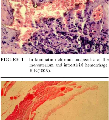
Macroscopic - After anesthesia with diethyl ether, magnifying the previous incision of the abdominal wall, in the direction skull-volume, of the Xiphoideus sterni process to the pubis, enough ample for the comments of the structures to be studied: mesenterium and parietal peritoneum. In the peritoneal socket, we observe the presence of liquid with uniformity of the serosal and the coloration was rosy in all extension of the area and studied place. We also noticed an abundant presence of vascular congestion. These fabric blocks had been struck laterally, allowing to the comment and photographic documentation. Later the section of these structures was continued. In the mesenterium, removed it of a total of 90 fragments was measuring greater 2,5 X 2,0 X 1,8 cm and the minor 1,7 X 1,2 X 1,0 cm, irregular form or oval, yellowish external surface with white traces. It was completed removed it of the surgical parts of the parietal peritoneum, bilateral, totalizing 180 fragments measuring the greater 2,3 X 1,3 X 1,0 cm and the minor 1,2 X 0,8 X 0,6 cm, all of irregular form and external surface of clear coloration chestnut and firm consistency. The parts had been analyzed following previous protocol; the animals had been submitted to euthanasia for inhalation of lethal dose of diethyl ether. Microscopy of the mesenterium - the cuts of the fragments of fabric of this region had shown to infiltrations of lynfocites and some plasmocites. One associated, presence of arterioles and capillaries presenting ecstasis and vascular congestion, characterizing histological picture of chronic inflammation process unspecific. Presence of linfonods was proven, presenting proliferation of lynfocites elements and reticular cells, designating histological picture of linfo-reticular hyperplasia. It was developed, diffuse fibroblastic proliferation, without inflammation, resulting only in fibrosis reacional. The excessively fragments ones had been presented without histological alteration (Figure 1). Microscopy of the parietal peritoneum - In the pathology of the material of the parietal peritoneum, in the cuts of the fragments of fabric, only fibroblastic proliferation with formation of collagen excess was observed constituting reacional typical histological picture of fibrosis, beyond fragments without histological alteration (Figure 2).
FIGURE 1 - Inflammation chronic unspecific of the mesenterium and intresticial hemorrhage. H-E (100X).
alteration, with percentage of 33,3%. There were a total of 30 cases with 100% percentage. Group C - The hyperplasic linfonods had appeared in 10 cases with 33,3% percentage; without reaction register for fibrosis; vascular congestion, 2 cases with 6,7% percentage; unspecific chronic inflammation, 8 cases with 26,7% percentage; without alteration, 10 cases with 33,3% percentage. There were a total of 30 cases with 100% percentage. In all groups p > 0,05, no significance. Still we can affirm that the histological alterations of the mesenterium had been distributed also by re-operation time, being, thus, informed: Group - The hyperplasic linfonods, in 6h, had appeared in 6 cases, 24h, 2 cases, and 48h, 4 cases; reaction fibrosis, in 6h appeared in 1 in case that, 24h, in 3 cases, without register in 48h; vascular congestion, in 6h, appears in number of 2 cases, without register in 24h and 48h; unspecific chronic inflammation, in 6h, appears in 1 in case that, 24h, 3 cases and without register in 48h; without alteration, in 6h, without
TABLE 1 - Distribution of histological alterations of fragments of the mesenterium for group.
Histological Alterations Group A Group B Group C
n % n % n %
Hyperplasic linfonods 12 40,0 8 26,7 10 33,3 Reaction fibrosis 4 13,3 2 6,7 - -Vascular congestion 2 6,7 6 20,0 2 6,7 Chronic unspecific inflamation 4 13,3 4 3,3 8 26,7 No alteration 8 26,7 10 33,3 10 33,3 T O T A L 30 100,0 30 100,0 30 100,0
p>0,05
Legend: n= number
register, in 24h it appears in 2 cases and 48h, in 6 cases. There were a total of 30 cases. Group B - The hyperplasic linfonods, in 6h, appear in 1 in case that, 24h, 3 cases and 48h, 4 cases; reaction fibrosis, in 6h, appears in 2 cases, without register for 24h and 48h; the vascular congestion, in 6h and 24h, appears in 1 in case that e, 48h, 2 cases; unspecific chronic inflammation, in 6h and 24h, 1 in case that and 48h, 2 cases; without alteration, in 6h and 24h, it appears in 5 cases, without register in 48h. There were a total of 30 cases. Group C - The hyperplasic linfonods, in 6h, had appeared in 2 cases, 24h and 48h, 4 cases; reaction fibrosis, in 6h, appeared in 2 cases, without register in 24h and 48h; vascular congestion, without register in 6h, 24h and 48h; unspecific chronic inflammation, in 6h and 24h, appears in 2 cases and 48h, in 4 cases; without alteration, in 6h and 24h, it appears in 4 cases and 48h in and 2 cases. Totalizing 30 cases and p 0,05, that is, without significance (Tables 1 and 2, Figures 3 and 4).
TABLE 2 - Distribution of the histological alterations of the mesenterium for group and time of reoperation. Group A - Control Gruop B - Glucose 10% Gruop C - Glucose 25% Histological Alterations 6 h 24 h 48 h 6 h 24 h 48 h 6 h 24 h 48 h Hyperplasic linfonods 6 2 4 1 3 4 2 4 4
Reaction fibrosis 1 3 - 2 - - 2 -
-Vascular congestion 2 - - 1 1 4 - - -Chronic Unspecific
Infection 1 3 - 1 1 2 2 2 4
No alteration - 2 6 5 5 - 4 4 2
Fisher’s test accurate p>0,05
Legend: n= number
FIGURE 3 - Distribution of histological alterations of fragments of the mesenterium for group.
To the microscopy of the peritoneum it disclosed to 6 cases with fibrosis (3,3%) and 174 cases (96,7%) without histological alteration reaction (Table 3, Figure 5).
Discussion
In the present work, we detected the macroscopic and microscopic alteration of the mesenterium and the parietal peritoneum when we manage the watery glucose solution 10% and 25% in the peritoneal socket of rat “Wistar”, and statistical study, where a sample of 90 animals, subdivided in 3 groups of 30 animals and the comparison of the histological reactios of the mesenterium and the parietal peritoneum was analyzed. One chose adult young and females, without taking in consideration possible differences in the healing process or factor of increase6.
the animals had been submitted the anesthesia with diethyl ether under inhalation, did not observe aesthetical complications. As chosen anatomical region, longitudinal incision of 3 cm in the regio abdominis was measured with tecidual resection until the parietal peritoneum7. All these
animals had been re-operated in 6h, 24h and 48h, for being an animal that possess a capacity of fast tecidual regeneration8. Practiced synthesis in.mass, by simple suture,
abstaining the parietal peritoneum “ipsi-lateral” with the purpose not to allow that the wire used for the suture intervened with the process of peritoneal healing. What it was confirmed in analyzes histological, as also to prevent TABLE 3 - Global distribution of histological alterations of
fragments of parietal peritoneum. Histological Alterations n % Reaction fibrosis 6 3,3 No Alteration 174 96,7 Total 180 100,0
p > 0,05
Legend: nº = number
FIGURE 5 - Percentage of the global distribution of histological alterations of fragments of parietal peritoneum.
extravasation of the introduced intraperitoneal. It did not have risk of the animals to cause traumatism to the proper one abdomen or to remove the wire of suture of the operated wound, in virtue of previous comment of these rodents, to the odor of the plolivinilpirrolidona-iodine solution 10%, being applied to the wound, daily, when necessary. In literature, references of diluted watery works had been evidenced using itself solutions of hypertonic glucose in water distilled in the same ratio, for intraperitoneal laundering as mucolitic agents in the removal of the mucinous content of pseudomyxoma, thus preventing the intestinal blockages subsequently.9 10 11 In relation to the type of boarded
glycoside hypertonic solution in the research, we choose the glucose solutions 10%, due to the works of evidence in the consulted literature, praising the use of the glucose solution 10% in the laundering of the peritoneal socket in experimental study, in the validity of a peritonit for injury of colon, promoting faster and resistant intestinal healing, comparatively the use of the salty solution 0.9%,12 we
Conclusion
In the global analysis, the reaction inflammation process is of equal tecidual intensity as of the use of the glycoside hypertonic solution in the concentrations 10% and 25% or of saline solution 0.9% on the mesenterium and parietal peritoneum.
References
1. Robbins SL, Cotran RS, Kumar V, Path JFRC, Collins T. Pathologic basis of disease. 6.ed. Philadelphia: Saunders; 1999.
2. Forrest RD. Early history of wound treatment. J R Soc Med. 1982;75(3):198-5.
3. Aguilar-Nascimento JE, Soares YT, Silveira BRP, Silva MRN, Caporossi C. Influência da lavagem da cavidade peritoneal com glicose hipertônica 10 % na cicatrizaçäo por segunda intençäo do colo em vigência de peritonite fecal: estudo experimental em ratos. Acta Cir Bras. 1992;7(1):17-20.
4. Roy WJ, Thomas BL, Horowitz IR. Acute hyperglycemia following intraperitoneal irrigation with 10% dextrose in a patient with pseudomyxoma peritonei. Gynecol Oncol. 1997;65(2):360-2.
5. White PF, Johnston RR, Eger EI. Determination of
anesthetic requirement in rats. Anesthesiology. 1974;40(1):52-7.
6. Nial M, Ryan GB, O’Brien BM. The effect of epidermal growth factor on wound healing in mice. J Surg Res. 1982;33(2):164-9.
7. Stewart DJ, Matheson NA. Peritoneal lavage in fecal peritonaites in the rat. Br J Surg. 1978;65:57-9.
8. Rhoden EL, Silva UL e,Torres OM, Blankenburg E, Castro MAA de, Pereira-Lima L. Efeito do dextran sobre a intensidade das aderências peritoneais intra-abdominais pós-operatórias: estudo experimental em ratos. Rev Col Bras Cir. 2000;27(23):23-5.
9. Barbosa TV, Peixoto M, Rapoporte A, Barbosa PPP. Metodologia experimental para a determinação de drogas potencialmente cicatrizantes na cavidade oral através das fases do processo cicatricial por segunda intenção obtida na língua do rato. Rev Pós Grad Fac Odontol Univ São Paulo. 2000;71(supl 32):R128
10. Green,N, Gancedo, H, Smuth, R, Bernett, G. Pseudomyxoma peritonei-nonoperative management and biochemical findings: a case report. Câncer. 1975;36:1834-7.
11. Silva AL. Lavagem e irrigação peritoneais. In: Silva AL. Cirurgia de urgência. Rio de Janeiro: Medsi; 1985. p.447-50. 12. Piver MS, Shashikant BL, Patsner B. Pseudomixoma peritoneii: possible prevention of mucinous ascites by peritoneal lavage. Obstet Gynecol. 1984;64(suppl 3):95S-6.
Correspondence:
José Cícero Ferreira de Carvalho Rua 04, Quadra A/115
57046-040 Maceió - AL Brazil
How to cite this article:
Carvalho JCF, Leal AT, Sousa LF, Herani Filho B. Hypertonic glucose solution 10% - 25% on the mesenterium and peritoneum of the rat: macroscopic and microscopic study. Acta Cir Bras. [serial on the Internet] 2005 Nov-Dec;20(6). Available from URL: http://www.scielo.br/acb
*Color figures available from www.scielo.br/acb
Conflict of interest: none Financial source: none
Received: May 3, 2005 Review: June 12, 2005 Accepted: July 18, 2005