Arq Neuropsiquiatr 2007;65(4-A):988-991
988
BASAL ENCEPHALOCELE ASSOCIATED
WITH MORNING GLORY SYNDROME
Case report
Ivanete Minotto
1, Nitamar Abdala
2, Adriana Aparecida Siviero Miachon
3,
Angela Maria Spinola e Castro
4, Paulo Imamura
5, Roberto Gomes Nogueira
6ABSTRACT - The basal encephaloceles refer to rare entities and they correspond to herniation of brain tissue through defects of skull along the cribiform plate or the sphenoid bone. A rare morning glory syndrome, with characteristic retinal defect has been reported in association with basal encephaloceles. Hypophysis hormonal deficiencies may occur. We accounted for a pituitary dwarfism with delayed diagnosed transs-phenoidal encephalocele associated with morning glory syndrome, showing the alterations found in retin-ography, computed tomography and magnetic resonance imaging.
KEY WORDS: basal encephalocele, morning glory syndrome, computed tomography, magnetic resonance imaging, pituitary dwarfism.
Encefalocele basal associada a síndrome “morning glory”: relato de caso
RESUMO - As encefaloceles basais são entidades raras e correspondem a herniações do tecido cerebral atra-vés de um defeito do crânio, ao longo da lâmina crivosa etmoidal ou do osso esfenoidal. A rara síndrome morning glory, com alterações de fundo de olho características pode apresentar-se associada à encefaloce-le basal. Deficiências hormonais hipofisárias podem ocorrer. Relatamos caso de nanismo hipofisário com en-cefalocele transesfenoidal de diagnóstico tardio associada à síndrome de morning glory, mostrando as alte-rações na retinografia, tomografia computadorizada e ressonância magnética.
PALAVRAS-CHAVE: encefalocele basal, síndrome de “morning glory”, tomografia computadorizada, resso-nância magnética, nanismo hipofisário.
Neuroradiology Ward, Image Diagnoses Department, Federal University of São Paulo, São Paulo SP, Brazil (UNIFESP), Endocrinology Ward at Pediatrics Department, UNIFESP: 1Undermasters graduation at the Neuroradiology Department (DDI/UNIFESP); 2 Affi liated
Professor, DDI/UNIFESP; 3Physician at the Pediatrics Endocrinology Department, UNIFESP; 4 Assistant Professor, Pediatrics
Endocri-nology Department, UNIFESP; 5Professor of Neurophtalmology, Ophthalmology Department, UNIFESP; 6Professor-Assistant at the
Neuroradiology Department – DDI/UNIFESP Received 27 April 2007. Accepted 17 August 2007.
Dr. Roberto G. Nogueira - Rua Pará 126 / 52 - 01243-020 São Paulo SP - Brasil.
The basal cephaloceles refer to rare entities of dif-fi cult diagnosis and correspond to herniation of the brain tissue through a birth or acquired defect in the skull along the cribiform plate or through the sphe-noid bone. It may be associated to hormonal distur-bances or ocular malformation and, amongst, the rare morning glory syndrome1-3, which name is given due
to the retinal aspect similar to the tropical fl ower of same name (Fig 1A). It is believed that such anoma-lies result from a succession of events for the medium line conclusion during the gestation period3. The
com-puted tomography (CT) and magnetic resonance im-age (MRI) exams play a very important role for these anomalies since they evaluate the whole skull and present structures in the hernial content1,2.
We present a case of defect on the medium line in which the TC and MRI images are fundamental to clarify the diagnosis.
CASE
Arq Neuropsiquiatr 2007;65(4-A)
989 Basal encephalocele: morning glory syndrome Minotto et al.
characteristic alterations of the morning glory syn-drome in the left eye, observing an optical disk with an enlarged choanoid and cupped aspect, with a pink pigmentation and a central white mass which hid the way of the vessels at the bottom of the disk. The disk was surrounded by a little elevated grey ring, with irregular borders and mixed with colored areas. The vessels were multiple, thin and radiated (Fig 1B).
At the age of 20, he was submitted to imaging ex-ams of the sellar region. On the CT of the sella, with volumetric acquisition and three-dimensional recon-struction, a defect at the main area of the sellar fl oor
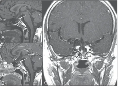
was observed (Fig 2). On the MRI (Phillips Gyroscan 1,5T), sagittal and coronal images from the sellar re-gion were obtained on the T1 weighted spin echo (T1WSE) sequence before and after the paramagnetic contrast medium intravenous administered, and in T2 weighted spin echo sequence. A sellar content consti-tuted by the pituitary stalk, optical chiasm, adenohy-pophysis and neurohyadenohy-pophysis occurring on the right side was observed. It was observed the extension of the anterior portion of the third ventricle into the in-terior of the sella, and the hernial content with tissue characteristics not defi ned on the inner side of the sellar fl oor and of the sphenoidal sinus (Fig 3).
Fig 2. (A) A 3D reconstructed sella turcica CT image showing the main damage of the fl oor (arrow). (B) CT image reconstruction in the sagittal plane, in soft window showing the dam-age on the sellar fl oor and the encephalocele (arrow). (C) Coronal CT image in bone window showing the main fl aw on the sellar fl oor (arrow).
Arq Neuropsiquiatr 2007;65(4-A)
990
Basal encephalocele: morning glory syndrome Minotto et al.
DISCUSSION
Encephalocele is a congenital defect of the skull bone and of the dura-mater with extracranial hernia-tion of any intracranial structure. It is found with a geographic variation and with different occurrences when related to sex and race, and in different asso-ciation to the neural tube malformations, suggesting that some of them may present genetic origin, and that the several types of encephalocele may corre-spond to distinct genetic defects4. It can be divided
into four groups. The cephalic meningoceles, which are constituted by the leptomeninges and the cere-brospinal fl uid (CSF), and the glioceles, which are glial cells cysts containing CSF. The meningoencephaloce-les, which consist of leptomeninges, CSF and brain pa-renchyma, recognizes the meningoencephaloventric-ulocele when parts of the ventricles and of portions of the choroid plexus participate on the herniation. And the atresic changes on the encephalocele, char-acterized by tuberous lesions situated on the medium line of the scalp, either in the vertex (parietal form) or in the occipital protuberance (occipital form)4.
The basal encephalocele is the rarest and of the most diffi cult diagnostic and it corresponds to 1% to 10% of all of them1,2. The basal encephalocele
fre-quently escapes the diagnosis and may be detected at adult age2,4,5. On the transsphenoidal
encephalo-cele, the bone defect occur as a result of the chondri-fi cation of the intersphenoidal synchondrosis defect on the sphenoid body, causing the persistence of the craniopharyngeal canal, which normally closes itself by the fi ftieth gestation day. Its persistence allows
the passage of several portions of the intracranial structure such as the hypophysis, the anterior por-tion of the third ventricle fl oor, optic chiasm and op-tic nerves1,2,4. The symptoms may be developed at the
neonatal period or at the fi rst childhood, and they consist of expansive processes of the epipharynx and pituitary dwarfi sm3,6,7, and the hypertelorism is almost
constant. When these symptoms do not manifest themselves, visual disturbances and hypothalamohy-pophysial dysfunction1 may lead to the diagnostic, as
occurred in our case.
The basal encephaloceles associate themselves to optic malformation and retinal defect1, observing an
increase on the prevailing (67.7%) of the morning glory syndrome3,7-10, which is an unusual congenital
anomaly on the optic nerve. It was described by Reis and by Handman3. Kindler called it morning glory3,11.
Its frequency on the population is unknown5 and it
is transmitted hereditarily with an autossomal domi-nant pattern. Most cases are unilateral3,4 but there
are rare cases of bilaterality6,7 with twice as much
fre-quency on females3. It is featured by the increase and
copping of the optic nerve on the optic disk region with the persistence of a glial tissue with a yellow color in the middle constituted by hyaloid remains. The vessels follow a radial pattern to the periphery. The coloboma is surrounded by a lifted ring of reti-nal pigmentation, which resembles the morning glory fl ower. Clinically, there is a decrease of the unilateral visual accuracy frequently associated to the displace-ment of the retina, which occurs in 30% to 38% of the cases12-14.
Arq Neuropsiquiatr 2007;65(4-A)
991 Basal encephalocele: morning glory syndrome Minotto et al.
Some theories have been proposed to explain the malformation. Itakura and coworkers8 described a
suc-cession of events which culminated in the bottom of the eye anomaly. According to these authors, the scir-rhous palate would have embryological origin on the fi rst brachial arch, the palatine process, which would originate itself from the maxillary process, melting completely with the septonasal, which in turn derives from a frontal saliency around the sixtieth gestational day. In this stage, no internal layers of the retina and of the optic nerve are well differentiated. During the seventh gestational week, the axons of the ganglionic cells of the retina start to form the optic nerve reach-ing its full development around the twenty-seventh gestational week. If a transsphenoidal encephalocele blocks the palate fusion, and as this phenomenon precedes the optic nerve formation, there could be an abnormal development of the nerve with a white glial tissue formation in its centre8,13,15. These
abnor-malities may present themselves in many combina-tions and in different degrees of severity and, there-fore, the possibility of association cannot be discard even on the absence of exuberating clinical evidence, but being always indicated to a detailed evaluation through imaging exams.
The hypothalamic structures involvement may be associated to endochrinal alterations, mainly the growth hormone defi ciency, though it must be con-sidered the occurrence of the defi ciency of multiple adenohypophysial hormones, which will appear later on the evolution process1. In this case, it was only
ob-served a growth hormone defi ciency and it has not yet developed any other hormonal defi ciency.
The imaging exams have an important role on the basal encephaloceles diagnosis. Machado Jr and coworkers described the case of a patient with no previous neurological and/or endochrinal symptoms subjected to a CT exam because of a mild cranial trau-ma. A right parasellar lesion was observed, requir-ing a MRI exam to identify the incidental sphenoidal meningoencephaloventriculocele16. The encephalocele
was into the sphenoidal sinus through the side wall with a discrete sellar deformation, although without hypothalamic or hypophyseal compromising.
MRI is the imaging diagnostic procedure of choice since it allows to precisely identifying the presence of meninges, brain parenchyma and blood vessels inside the bone defect1. Besides, it provides broad
enceph-alic anatomic evaluation which facilitates the identifi -cation of other anomalies. The considered T1WSE im-aging presents an anatomic resolution and it must be used to trying to identify cerebral structures which are
normally deformed inside the herniation. The usage of intravenous paramagnetic contrast media helps to identify vascular structures and it might be important to evaluate surgical risks17. The images must also be
obtained in larger areas for a complete evaluation of the brain parenchyma to disclose malformations. In our case, the structures in the interior of the sphenoi-dal sinus were not characterized as brain tissue and so being classifi ed as gliocele5. The CT exam can better
show the bone defect and it must be considered as a complementary method of imaging diagnostic, and the 3-D reconstruction facilitates this process.
The skull image study is fundamental in the di-agnostic process of the ocular congenital alteration related to the retarded growth due to a possible as-sociation to the basal encephalocele. The chosen pro-cedure is MRI using CT as a complementary method. The association of basal encephaloceles to endochri-nal disorders and visual alteration suggests that a brain imaging study must be performed and com-pleted with an exam directed to the hypothalamo-hypophysial region.
REFERENCE
1. Yokoda A, Matsukado Y, Fuwa I, Moroki K, Nagahiro S. Anterior basal encephalocele of neonatal and infantile period. Neurosurgery 1986;19: 468-478.
2. Kollias SS, Ball WS Jr. Congenital malformation of the brain. In Ball WS Jr (Ed). Pediatric neuroradiology. Philadelphia: Lippincot-Raven, 1977: 93-102. 3. Eutis S H, Sanders M R, Zimmerman T. Morning glory syndrome in chil-dren: association with endocrine and central nervous system anomalies. Arch Ophthalmol 1994;112:204-207.
4. Naidich TP, Altman NR, Braff mann BH, McLone DG, Zimmerman RA. Cephaloceles and related malformations. AJNR 1992;13:655-689. 5. Chen CS, David D, Hanieh A. Morning glory syndrome and basal
en-cephalocele. Childs Nerv Syst 2004:20:87-90.
6. Murphy BL, Griffi n JF. Optic nerve coloboma (morning glory syndrome): CT fi ndings. Radiology 1994;191:59-61.
7. Kindler P. Morning glory syndrome: unusual congenital optic disk. anomaly. Am J Ophthalmol 1970;69:376-384.
8. Itakura T, Miyamoto K, Uematsu Y, Hayashi S, Komai N. Bilateral morn-ing glory syndrome associated with sphenoid encephalocele: case re-port. J Neurosurg 1992;77:949-951.
9. De Laey JJ, Ryckaert S, Leys A. The morning glory syndrome. Ophthal-mol Paediatr Genet 1985;5:117-124.
10. Nawratzki I, Schwartzenberg T, Zambermann H.: Bilateral morning glo-ry syndrome with mid-line brain lesion in an autistic child. Metab Pedi-atr Sist Ophthalmol 1985;8:35-36.
11. Steinkuller PG. The morning glory disc anomaly: a case report and lit-erature review. J Pediatr Ophthalmol Strabismus 1980;17:81-87. 12. Lees MM, Hodgkins P. Frontonasal dysplasia with optic disc anomalies
and other midline craniofacial defects: a report of six cases. Clin Dys-morphol 1998;7:157-162.
13. Matsumoto H, Enaida H, Hisatomi T, et al. Retinal detachment in morn-ing glory syndrome treated by triamcinolone acetonide-assisted pars plana vitrectomy. Retina 2003;23:569-572.
14. Azuma N, Yamaguchi Y, Handa H, Asaka K, Kawase E, Yamada M, Mu-tations of the PAX6 gene detected in patients with optic-nerve malfor-mations. Am J Hum Genet 2003;72:1565-1570.
15. Hodgkins P, Lees M, Lawson J, et al. Optic disc anomalies and fronto-nasal dysplasia. Br J Ophthalmol 1998;82:290-293.
16. Machado MAC Jr., Barbosa VAO, Pires MCM, et al. Meningoencefalo ventriculocele transesfenoidal assintomática em adulto: relato de um caso. Arq Neuropsiquiatr 2001;59:280-282.