1. Department of Orthopaedic Surgery, Seikeikai Hospital 2. Department of Orthopaedic Surgery, Osaka City General Hospital 3. Department of Orthopaedic Surgery, Osaka City University Graduate School of Medicine
4. Department of Orthopaedic Surgery, Osaka Ekisaikai Hospital
cAse report
A 67-year-old Japanese woman (right-handed) visited our department with swelling and discharge from a fis-tula of the right wrist. Her job involved managing a bar. She had no relevant past medical conditions and no fa-mily history of infectious disease and denied a history of trauma or overuse.
She noticed mild right wrist pain about ten years ago and the wrist swelling about three months before visi-ting our hospital. She visited a local clinic and was trea-ted for rheumatoid arthritis. After conservative treat-ment, including various drug regimens (unknown), she visited another hospital because her condition did not improve. At the hospital, her wrist was punctured for examination. However, the puncture hole did not clo-se and the discharge from the fistula continued.
Physical examination revealed swelling of the dorsal aspect of the right wrist and discharge from the fistula on the ulnar side of the wrist (Figure 1a). There was no evidence of a tendon rupture or a peripheral nerve di-sorder. In addition, other joints were not swollen or painful. The grip strength for the right and left hands, measured with a Jamar digital dynamometer (Takei Scientific Instrument Co, Ltd., Niigata, Japan), were 15.6 Kg and 23.5 Kg, respectively. The respective ran-ge of motion for the right and left extremities, measu-red with a standard goniometer, was as follows: wrist dorsiflexion, 25° and 45°; wrist palmar flexion, 30° and 75°; forearm pronation, 70° and 90°; and forearm su-pination, 60° and 90°. Plain radiographs of the right wrist revealed narrowing of the radiocarpal joint (RCJ), cystic change in the distal radius and ulna and widening of the distal radioulnar joint (DRUJ) (Figure 1b). The irregularity of the cortex of ulnar side of metaphyseal ulna which was near to the fistula was also found, and this finding guided us suspicion of infection.
As the fistula had not closed in a few months, we
Osteomyelitis and arthritis of the wrist caused by
Mycobacterium intracellulare in an immunocompetent
patient: a case report and literature review
Yano K1, Kazuki K2, Ikeda M3, Yoneda M4
AbstrAct
Mycobacterium intracellulare causes infection in
hu-mans. Involvement of joint and bone, however, is ex-tremely rare. We present the case of an immunocom-petent 67-year-old female with chronic swelling of the wrist joint diagnosed as rheumatoid arthritis by her pre-vious physician. Examination revealed an unclosed fis-tula associated with a puncture, and bone and joint des-truction on radiographs. She was diagnosed with os-teomyelitis and arthritis due to M. intracellulare on his-tological and microbiological examinations. She was successfully treated with radical surgical debridement and anti-tuberculous drugs for 1 year and there was no recurrence at 3 years postoperatively.
Keywords: Mycobacterium intracellulare;
Osteomyeli-tis; ArthriOsteomyeli-tis; Wrist
IntroductIon
Non-tuberculous mycobacterium (NTM) is widely dis-tributed in nature and some of its species have been re-ported to cause infection in human beings1. NTM in-fection of the upper extremity is rare, and commonly involves the tenosynovium2-4. Involvement of a joint and bone is extremely rare; there have been a few reports of this type of involvement in immunocompetent cases2,5. We report the case of an immunocompetent patient with wrist joint and bone infection caused by Mycobacterium
intracellulare and reviewed the literature.
performed further examinations under suspicion of an infection. The patient’s blood examination revealed a white blood cell count of 6230 cells/mm3, erythrocy-te sedimentation raerythrocy-te of 21 mm/hr, CD4-positive
lymphocyte count of 1056 cells/mm3, C-reactive pro-tein level of 0.11 mg/dL; the rheumatoid factor, anti--cyclic citrullinated peptide antibody, antinuclear an-tibody, QuantiFERON TB-2G (Japan BCG Laboratory Co., Ltd, Tokyo, Japan), and human immunodeficien-cy virus antibody were negative. Gram, Ziehl-Neelsen, fluorescent and fungal staining of the discharge were also negative. Cultures of the discharge were proven to be negative for aerobic and anaerobic bacterium, fun-gus and mycobacterium in four weeks in the preopera-tive period. The polymerase chain reaction (PCR) test for
Mycobacterium tuberculosis, M avium, and M intracellulare
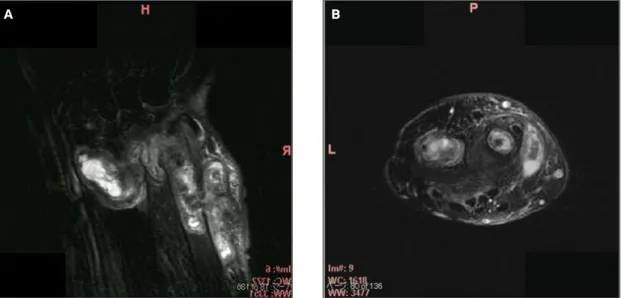
(TaqMan-PCR, Roche Diagnostics K. K., Tokyo, Japan) was negative. There was no abnormal finding on a plain radiograph of the chest. Magnetic resonance imaging (MRI) of the right wrist revealed a pathological lesion with low signal intensity in T1 weighted-image (WI) and high signal intensity in T2 WI in the bone marrow in the distal radius and ulna, RCJ, DRUJ, and subcuta-neous tissue in ulnar side of the wrist (Figure 2a and 2b). Moreover, signal intensity of distal ulna and sub-cutaneous lesion was almost the same and the subcuta-neous lesion was continuing to the ulnar fistula through the discontinuity of the cortex of ulna (Figure 2a). This discontinuity of the cortex of metaphyseal ulna made us suspicion of osteomyelitis.
To obtain a definite diagnosis, a radical resection of
FIGure 1. Preoperative appearance and plain radiograph of the right wrist. A) Swelling of the dorsal and ulnar sides; a fistula was found on the ulnar side of the right wrist. B) Plain radiograph of the right wrist showed narrowing of the radiocarpal joint, cystic change in distal radius and ulna, discontinuity of the ulnar cortex of distal ulna, and widening of the distal radioulnar joint. The irregularity of ulnar cortex of distal ulna was near to the fistula
A b
FIGure 2. Preoperative MRI of the right wrist. A) Coronal image with fat-suppression (chemical shift selective method, CHESS). The mass with increased signal intensity was found in the distal radius and ulna, distal radio-ulnar joint, and under the
subcutaneous tissue. The ulnar mass continued from the ulna to the fistula. B) Sagittal image with fat-suppression (CHESS). The mass with high signal intensity was found in the radius, ulna and ulnar subcutaneous tissue of the distal forearm, and the signal intensity of three lesions were almost same
daily), as recommended by an infectious disease spe-cialist. Bacterial and fungal cultures were all negative. At one month after the surgery, the wound had healed and the wrist swelling had decreased. Colonies were obtained from the mycobacterial culture at nine weeks after the surgery, and M. intracellulare was identified by performing DNA-DNA hybridization (Kyokuto Phar-maceutical Industrial Co., Ltd, Tokyo, Japan). At three months after the surgery, the swelling disappeared; the three anti-tuberculous drugs were continued for one year. No side effects of drugs were experienced. At the last follow-up, 3 years postoperatively, the patient felt no pain and no disability in daily activities and had no recurrence (Figure 4a). The grip strength was 14.3 Kg and 18.2 Kg for the right and left hands, respectively. The range of motion of the right extremity was as fol-lows: wrist dorsal flexion, 35 degrees; wrist palmar fle-xion, 35 degrees; forearm pronation, 90 degrees; and forearm supination, 90 degrees. Sclerotic changes were observed in the preoperative cystic lesion of the distal radius; no osteolytic changes of the distal ulna were ob-served on a wrist radiograph (Figure 4b).
dIscussIon
Mycobacterium intracellulare is one of the NTM species1. It causes lung disease, cervical lymphadenitis, and skin, bone and joint infections1. In the upper extremity, it is a rare cause of tenosynovitis, and especially of arthritis and osteomyelitis2-4. Although disseminated NTM in-the pathological lesion was performed under regional
anesthesia. A skin incision, which included the fistula, was made on the dorso-ulnar aspect of the wrist. The-re was a hole in the ulnar aspect of the metaphyseal ulna and the inflammatory synovitis continued to the fistula. The distal ulna was resected at the proximal portion from the hole. The proliferative synovium from the DRUJ and RCJ was extensively removed. There was also a hole in the dorso-ulnar aspect of the distal radius; the synovium from the radius was excised radically through the hole. A surgical specimen was examined microbiologically and histopathologically. Gram, Ziehl--Neelsen, fluorescent, and fungal staining were perfor-med. The formalin-fixed tissues were embedded in pa-raffin and processed for hematoxylin and eosin and the Ziehl-Neelsen stain. For microbiological examination, aerobic, anaerobic, bacterial, and fungal cultures were done on excised surgical tissues. For culture of the my-cobacterium, solid and broth culture systems (Kudo PD culture, Japan BCG Laboratory, Tokyo, Japan, and MGIT, Japan Becton, Dickinson and Company, Tokyo, Japan) were used under 30°C and 37°C. The PCR kit described above was also used.
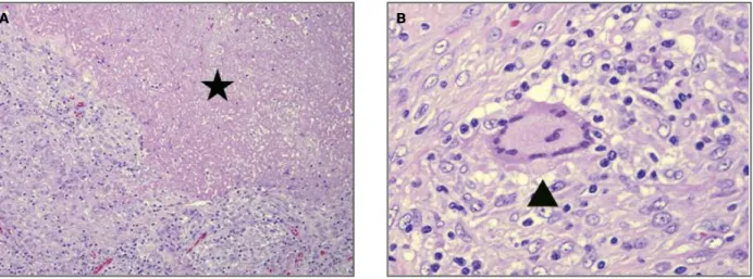
Histological examination revealed epithelioid gra-nuloma with caseous necrosis, and Ziehl-Neelsen stai-ning showed acid-fast bacilli (Figure 3a and 3b). The PCR result was positive for M. intracellulare at one week postoperatively. After the diagnosis was confirmed by the results, the patient received treatment with three anti-tuberculous drugs: rifampicin (450 mg daily), cla-rithromycin (400 mg daily), and levofloxacin (500 mg
FIGure 3. Histopathologic photograph. A,B) Histological sections stained with hematoxylin and eosin show epithelioid granuloma (arrow head) with caseous necrosis (asterisk), infiltration of inflammatory cells, and fibrosis. Langhans giant cells were found, as seen in b (magnification: a, ×100; b, ×400)
fections in immunocompromised patients have recei-ved attention recently, there also have been sporadic reports of these infections in immunocompetent pa-tients. The mechanism of musculoskeletal system in-fection due to NTM has been reported as occurring via hematogenous contamination from surgical treatment or penetrating injury, and steroid injections2,6. In this case, a hematogenous spread or a minor trauma asso-ciated with the patient’s job may be the cause as there was no history of obvious trauma.
To establish a diagnosis of mycobacterium infection, the most reliable method is an open biopsy of the pa -thological lesion. The surgical specimens should be examined histopathologically without delay. Acid-fast organisms can be confirmed by performing Ziehl-Neel-sen and fluorescent stains, and a presumptive diagno-sis can be made by observing granuloma formation. The American Thoracic Society recommends the use of two kinds of culture media, solid and broth, at tem-peratures 35°C and 28°C–32°C, for accurate detec-tion7. PCR is useful for early detection of the myco-bacterium8. A combination of histological examination and PCR results enabled an early diagnosis of M.
intra-cellulare infection; the PCR was especially helpful in the
identification of the organism and therefore, in the ini-tiation of treatment with anti-tuberculous drugs.
In this case, the patient was diagnosed with rheu-matoid arthritis by her previous physician. This mis-diagnosis may have been led by the manifestation of
the wrist swelling and radiographic findings of the wrist joint. Although radiography revealed wrist bone and joint destruction, her condition did not meet the 2010 rheumatoid arthritis classification criteria9. The pre-sentation of an unclosed fistula with discharge made us suspect infection. This case illustrates the fact that NTM infection should always be considered as a possible diagnosis in any patient with chronic swelling6.
Our treatment was successful in retaining the wrist joint by using a combination of radical surgical debri-dement and anti-tuberculous drugs. After a surgical re-duction of the infectious lesion, anti-tuberculous drugs were administrated for one year with monitoring for recurrence and side effects. As mentioned above, rheu-matoid arthritis is not a likely diagnosis in this patient presenting only a wrist monoarthritis. Literature sear-ches were made on the Medline and PubMed. And ad-ditional cross-reference checks of the bibliographies were performed. Search terms included mycobacte-rium, infection, and arthritis/ osteomyelitis. Previous reported cases with arthritis and osteomyelitis of the extremities due to Mycobacterium avium complex or
in-tracellulare in immunocompetent patients are listed in
Table I. A state without immunosuppressive therapy including steroid treatment, human immunodeficien-cy virus infection, or acquired immune deficienimmunodeficien-cy syn-drome, was defined as an immunocompetent condi-tion. Operative resection of the infected tissue appears to be necessary, even if prolonged drug therapy is admi -nistered (success rate: without surgery; 0/1, with sur-gery; 6/6). Although the period of drug therapy after surgical treatment was from 12 to 24 months, the du-ration seemed to be empirical. The successful result ob-tained in our case may be due to the good immune sta-tus of the host. A compromised immune stasta-tus has been discussed as a risk factor for recurrence and dis-semination of NTM infection. Kozin et al. reported in a series of NTM infections of the upper extremity that 13 of 15 immunocompetent patients had achieved re-solution; in contrast, only four of 10 immunocompro-mised patients achieved resolution2.
We had also planned an additional surgery, wrist arthrodesis, if the NTM infection was uncontrollable after surgical debridement and medication. Taniguchi et al. reported two cases of infected wrist arthritis trea-ted by using vascularized fibular graft10. They perfor-med two-stage procedures, involving debridement of the infected tissue and a vascularized fibular osteocu-taneous graft, and achieved resolution of infection.
In summary, excisional biopsy for histological and
FIGure 4. Final follow-up appearance and plain radiograph of the right wrist. A) Swelling of the right wrist had disappeared at the final follow-up. B) Final follow-up plain radiograph of the right wrist showed the change in distal radius from preoperative cystic lesion to sclerosis. No osteolytic change was observed at the resected ulna
microbiological analysis was essential to obtain a defini-te diagnosis for joint and bone infection due to myco-bacterium, and a successful outcome was achieved with a combination of radical surgical debridement and drug therapy. Mycobacterial infection should be included in the differential diagnosis for chronic wrist swelling.
correspondence to Koichi Yano
4-2-10 Koryonakamachi, Sakai-ku E-mail: koichiyano@hotmail.com
reFerences
1. Shinnick TM, Good RC. Mycobacterial taxonomy. Eur J Clin Microbiol Infect Dis 1994;13:884-901.
2. Kozin SH, Bishop AT. Atypical Mycobacterium infections of the upper extremity. J Hand Surg Am 1994;19:480-487. 3. Hellinger WC, Smilack JD, Greider JL, Jr., et al. Localized
soft-tissue infections with Mycobacterium avium/Mycobacterium intracellulare complex in immunocompetent patients: granu-lomatous tenosynovitis of the hand or wrist. Clin Infect Dis 1995;21:65-69.
4. Zenone T, Boibieux A, Tigaud S, et al. Non-tuberculous mycobac-terial tenosynovitis: a review. Scand J Infect Dis 1999;31:221-228.
tAble I. prevIously reported cAses oF osteomyelItIs And ArthrItIs In the extremIty cAused by nontuberculous mycobActerIA (mycobActerIum AvIum complex, IntrAcellulAre) In
Immunocompetent pAtIents
Age, Past Involved
Mycoba-Case gendera historyb sitec cteriumd Trauma Treatmentf Medicationg Durationh Outcome
1 [11] 61, M HT, DM Wrist intracellulare A INH, RFP 51 Unresolved
+ (numerous)
2 [12] 43, M - Ankle MAC + EB, A, D, F AMK 18 Resolved
(→CAM), RFP, EB, CPFX,
3 [13] 25, M gonococcal Wrist MAC - EB, F AMK, ETH, NA Resolved
urethritis CLF, RFP
4 [6] 53, F VSD Wrist, MAC Po-VSDe EB, EB, CAM, EB, 24 Resolved
MPJ A (R), RBT
EB (L)
5 [14] 33, M AS Ankle MAC - A, EB CAM, EB, NA NA
RBT
6 [15] 81, F tuberculous Knee MAC - EB RFP, SM, 12 Resolved
pleurisy INH
7 [16] 27, M - Knee, Intracellulare - A, EB, INH, ETH, NA Resolved
carpal OCTR + EB, PN,
tunnel, B, B RFP
sacroiliac joint
8 [17] 74, M NA Wrist MAC NA EB NA NA NA
9 67, F - Wrist Intracellulare - EB RFP, CAM, 12 Resolved
current LVFX
case
a. M, male; F, female
b. HT, hypertension; DM, diabetes mellitus; VSD, ventricular septal defect; AS, ankylosing spondylitis c. MPJ, metacarpophalangeal joint
d. MAC, Mycobacterium avium complex e. Po-VSD, postoperation of VSD
f. A, aspiration; EB, excisional biopsy; D, debridement; F, fusion; OCTR, open carpal tunnel release; B, biopsy
g. INH, isoniazid; CPFX, ciprofloxacin; AMK, amikacin; RFP, rifampicin; EB, ethambutol; CAM, clarithromycin; ETH, ethionamide; CLF, clofazimine; RBT, rifabutin; SM, streptomycin; PN, pyridoxine; PAS, paraaminosalicylic acid; LVFX, levofloxacin
h. Duration of anti-tuberculous drugs (month) NA, not applicable
5. Marchevsky AM, Damsker B, Green S, Tepper S. The clinico-pathological spectrum of non-tuberculous mycobacterial os-teoarticular infections. J Bone Joint Surg Am 1985;67:925-929. 6. Kanik KS, Greenwald DP. Mycobacterium avium/Mycobacte-rium intracellulare Complex-Associated Arthritis Masquerading as a Seronegative Rheumatoid Arthritis. J Clin Rheumatol 2000;6:154-157.
7. Diagnosis and treatment of disease caused by nontuberculous mycobacteria. Am Rev Respir Dis 1990;142:940-953. 8. de Charnace G, Delacourt C. Diagnostic techniques in
paedia-tric tuberculosis. Paediatr Respir Rev 2001;2:120-126. 9. Aletaha D, Neogi T, Silman AJ, et al. 2010 Rheumatoid
arthri-tis classification criteria: an American College of Rheumatolo-gy/European League Against Rheumatism collaborative initia-tive. Arthritis Rheum 62:2569-2581.
10. Taniguchi Y, Yoshida M, Doi N, Tamaki K, Tamaki T. Vasculari-zed fibular osteocutaneous grafts in the treatment of severely infected wrist arthritis. J Reconstr Microsurg 2002;18:71-75. 11. Halleran WJ, Martin NL. Nontuberculous mycobacterial
arth-ritis: report of a chronic case. J Kans Med Soc 1982;83:284--286.
12. Jones AR, Bartlett J, McCormack JG. Mycobacterium avium complex (MAC) osteomyelitis and septic arthritis in an immu-nocompetent host. J Infect 1995;30:59-62.
13. Rolfe B, Sowa DT. Mixed gonococcal and mycobacterial sepsis of the wrist. Clin Orthop Relat Res 1990;100-103.
14. Yang DH, Chang WC, Cheng MF, Lai JH, Chang DM, Chen CH. Peripheral arthritis caused by Mycobacterium avium-intracel-lulare in a patient with ankylosing spondylitis. J Clin Rheuma-tol 2009;15:323-324.
15. Tanaka M, Matsui H, Tsuji H. Atypical mycobacterium osteo-myelitis of the fibula. Int Orthop 1993;17:48-50.
16. Cheatum DE, Hudman V, Jones SR. Chronic arthritis due to My-cobacterium intracellulare. Sacroiliac, knee, and carpal tunnel involvement in a young man and response to chemotherapy. Arthritis Rheum 1976;19:777-781.
17. Whitaker MD, Jelinek JS, Kransdorf MJ, Moser RP, Jr., Brower AC. Case report 653: Arthritis of the wrist due to Mycobacte-rium avium-intracellulare. Skeletal Radiol 1991;20:291-293.