Chemical Industry & Chemical Engineering Quarterly www.ache.org.rs/CICEQ
Chem. Ind. Chem. Eng. Q. 22 (2) 145−153 (2016) CI&CEQ
DEJAN MARKOVIC1 IVANA KARADZIC2 VUKOMAN JOKANOVIC3 ANA VUKOVIC1 VESNA VUCIC2
1Department of Paediatric and Preventive Dentistry, Faculty of Dentistry, University of Belgrade, Belgrade, Serbia 2Centre of Research Excellence in Nutrition and Metabolism, Institute for Medical Research, University of Belgrade, Belgrade, Serbia 3Laboratory of Radiation Chemistry and Physics, Institute of Nuclear Sciences “Vinca”, University of Belgrade, Belgrade, Serbia
SCIENTIFIC PAPER
UDC 602.9:616:66.017/.018 DOI 10.2298/CICEQ141231028M
BIOLOGICAL ASPECTS OF APPLICATION OF
NANOMATERIALS IN TISSUE ENGINEERING*
Article Highlights
• Stem cells and scaffolds - an essential role in the production of new tissue by tissue engineering
• Nanotechnology - a field of high importance and rapid development
• Functional necessities of scaffolds – biocompatibility, biodegradability and mechanical properties
• The main challenge: transforming tissue engineering into regenerative engineering
Abstract
Millions of patients worldwide need surgery to repair or replace tissue that has been damaged through trauma or disease. To solve the problem of lost tissue, a major emphasis of tissue engineering (TE) is on tissue regeneration. Stem cells and highly porous biomaterials used as cell carriers (scaffolds) have an essential role in the production of new tissue by TE. The cellular component is important for the generation and establishment of the extracellular matrix, while a scaffold is necessary to determine the shape of the newly formed tis-sue and facilitate migration of cells into the desired location, as well as their growth and differentiation. This review describes the types, characteristics and classification of stem cells. Furthermore, it includes functional features of cell carriers – biocompatibility, biodegradability and mechanical properties of bio-materials used in developing state-of-the-art scaffolds for TE applications, as well as suitability for different tissues. Moreover, it explains the importance of nanotechnology and defines the challenges and the purpose of future research in this rapidly advancing field.
Keywords: tissue engineering, nanomaterials, scaffolds, stem cells, tissue regeneration.
Tissue engineering
Millions of patients worldwide need surgical procedures to repair or replace tissue that has been damaged through trauma or disease [1]. Today, con-ventional therapy addresses the problem of lost tissue by appropriate tissue replacement – tissue graft. The majority of defects can be healed using standard con-servative or surgical methods. However, large defects occurring after tumor surgery, cysts or multiple
Correspondence: I. Karadzic, Centre of Research Excellence in Nutrition and Metabolism, Institute for Medical Research, Uni-versity of Belgrade, Tadeusa Koscuska 1, P.O. Box 102, 11129 Belgrade, Serbia.
E-mail: ivana.colak@gmail.com Paper received: 31 December, 2014 Paper revised: 11 May, 2015 Paper accepted: 21 July, 2015
* This paper was part of a plenary lecture at the Rosov Pin Conference 2014.
tures require a more complex procedure of tissue rep-aration [2,3].
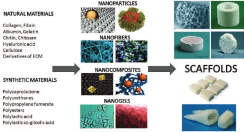
With respect to lost tissue treatment, the main emphasis of tissue engineering is on tissue regener-ation (TE) rather than tissue replacement [4–7]. Thus, stem cells and highly porous biomaterials used as scaffolds have an essential role in the production of new tissue by TE (Figure 1). The cellular component is important for the generation and establishment of extracellular matrix (ECM) in the new tissue, while a scaffold is necessary for providing mechanical stab-ility and foundation for a new three-dimensional tissue structure [6,8,9].
controlled. Further, the surface of these materials can be modified in order to enhance biocompatibility, immune compatibility and/or cell adhesion [13].
This review describes the functional necessities and types of stem cells and biomaterials used in dev-eloping state-of-the-art scaffolds for tissue engineer-ing applications. Furthermore, it defines the chal-lenges and the purpose of future research in this fast advancing field.
Stem cells and tissue engineering
Stem cells are unspecialized cells in the early stage of the development, which under normal con-ditions have the ability to differentiate into specialized mature cells and to divide in order to produce more stem cells [14–17]. Two functions define stem cells: unlimited self-renewal capacity, which makes them potentially immortal, and pluripotency [18]. Stem cells can be divided in several different ways, as shown in Table 1 [15,19–22].
Although they have less ability to differentiate, adult stem cells are far more applicable in regener-ative medicine than embryonic stem cells, primarily because of being relatively easy to isolate, lack of oncogenic potential and no ethical constraints over their application [23–26].
Architecture and nanotechnology of scaffolds
The ECM represents a biological 3D carrier for the cells and provides appropriate environment and architecture specific for each tissue [27]. Therefore, the key to a successful TE is proper design of cell carriers – scaffolds, which mimic the native ECM, combined with adequate stem cells. The role of these carriers is to determine the shape of the newly formed tissue and facilitate the migration of cells into the des-ired location, their growth and differentiation [28,29].
The key characteristic of every scaffold is that it must be biocompatible – to provide physical and mechanical functions and provoke a preferred res-ponse without causing any undesirable reactions in the host. Hence, the choice of material is a crucial point in tissue engineering [28,30]. It is desirable for the scaffold to disintegrate during the formation of new tissue, to allow the body’s own cells, over time, to eventually replace the implanted material [31]. There-fore, the biodegradability of the scaffold is also con-sidered very important in these processes.
Developing scaffolds with adequate mechanical properties is one of the greatest challenges in attempting to engineer bone or cartilage [32,33]. A balance must be achieved between the mechanical Figure 1. Basic concept of tissue engineering.
Table 1. Stem cells classification according to different criteria
Criterion Stem cells type Properties
Cell potency Totipotent stem cells Pluripotent stem cells Multipotent stem cells Unipotent stem cells
Potential to differentiate in any human cell even whole organism
Potential to differentiate in various tissue types but not whole organism
Potential to differentiate in various cell types within tissue – progenitor cell
Potential to differentiate in one cell type – precursor cell
Function Normal stem cells
Cancer stem cells
Not involved in pathologic process
Associated with most cancer disease
Sources Embryonic stem cells
Adult stem cells
Derived from inner cell mass of blastocyst
properties and a sufficiently porous architecture in order to obtain the desired scaffold [34]. Adequate porosity allows cell migration and provides a suitable microenvironment for cell proliferation and differen-tiation, with adequate vascularization, flow of nut-rients and oxygen and elimination of degradation products [35–37]. The porosity should be in the opti-mal range: sopti-mall enough to ensure mechanical integ-rity and sufficiently large to provide optimal bioactivity. For this reason, the size of the pores should be less than 300 nm [29].
Two types of materials are currently used in TE: natural and synthetic [38]. The advantage of natural materials is biological recognition regarding cell adhe-sion and function. However, the downsides are uncontrolled mechanical properties and biodegrad-ability, possible host immune reaction and the cost [39,40]. Various natural materials have been eval-uated to date. Derivatives of ECM have been inves-tigated for supporting cell growth. Proteins collagen and fibrin, as well as polysaccharides glycosam-inoglycans, have all proved appropriate regarding cell compatibility, but some potential immunogenic issues still remain [35,41,42]. Hyaluronic acid, one of the most exploited glycosaminoglycans, in combination with glutaraldehyde or water soluble carbodiimide, is considered suitable for scaffold materials [43]. Poly-saccharide chitin and chitosan based nanofibers have remarkable potential to be used as tissue engineering scaffolds, as well as drug delivery systems, wound dressing materials, antimicrobial agents and biosen-sors, due to their biocompatibility, biodegradability, antibacterial activity, low immunogenicity, wound healing capacity and cell binding capability [44]. Further, some scaffolds are tested for use in the deli-very of small molecules (drugs) to specific tissues [45]. Finally, decellularized tissue extracts in which
the remaining cellular residues or ECM act as a scaf-fold, are another form of cell carriers undergoing inv-estigation [46].
Synthetic materials, on the other hand, have the advantage of a commercial production, together with a control over mechanical properties, microstructure and degradation rate.
A commonly used synthetic material is polylactic acid (PLA). This is a polyester which degrades within the human body to form lactic acid, a chemical com-pound that plays a role in various biochemical pro-cesses and is easily removed from the body. The nanofibrous PLA mats incorporating carbon nano-tubes and rectorite, fabricated using an electrospin-ning technique, have proven suitable for biomedical applications due to their increased thermal stability and low cytotoxicity [47]. Similar to PLA are polygly-colic acid (PGA), polylactic-co-glypolygly-colic acid (PLGA) and polycaprolactone (PCL), with degradation mech-anisms similar to that of PLA but a different rate of degradation compared to PLA [48,49]. Other natural and synthetic materials have also been used in scaf-fold synthesis (Figure 2).
Due to their unique chemical, physical and bio-logical functions, nano-sized particles/fillers, of both inorganic and organic origin, have been studied in detail. They differ in their structure, composition, design and application, and can be in the form of nanofibers, nanogels, etc, as presented in Figure 2. Nanocomposite hydrogels combine the advantages of nano-fillers and hydrogel matrices and thus may result in improved mechanical and biological proper-ties and find their potential application in biomedicine as drug delivery matrices and scaffolds [50]. Rec-ently, injectable scaffolds have received attention due to their potential for avoiding the invasive surgery normally required for tissue implantation. Natural
polymers chitosan and alginate are used as coating materials to make positively and negatively charged PLGA nanoparticles, respectively. All the results demonstrate the potential use of the biodegradable colloidal gels as injectable scaffolds in tissue eng-ineering and drug release [51].
Application of scaffolds in tissue engineering
Bone disease or bone defects such as osteosar-coma, osteoporosis, and bone fractures affect millions of individuals worldwide [1]. In order to solve the prob-lem of lost bone tissue, bone tissue engineering emphasizes tissue regeneration rather than tissue replacement and is becoming a subject of growing interest. For over two decades, bioceramic material – hydroxyapatite (HAP) has been used as a substitute for bone as it has physical properties similar to the inorganic component of natural bone [52]. It is suit-able material for hard tissue replacement due to its osteoconductivity, biocompatibility and slow resorp-tion. On the other hand, the porosity of hidroxyapatite translates into poor mechanical properties [53]. In the attempt to compensate for these disadvantages, var-ious polymers have been examined, however, none of these meet all of the requirements for the ideal cell carrier in bone TE [54]. This has led to the develop-ment of composite carriers consisting of both an inorganic and organic component where the inorganic particles are embedded into the surface of a polymer matrix [36,55]. Inorganic-organic composites are designed to mimic natural bone by combining the viscoelastic properties of polymers with the strength of the inorganic part of the composite, to create bio-active materials with improved mechanical properties and ability to degrade over time [56]. Also, the basic products of decomposition of hydroxyapatite and tri-calcium-phosphate neutralize the acidity of the poly-meric compounds. Regarding higher bioactivity, inorganic nanostructured components have received more interest than equivalent microstructured ones [57]. Nanocomposites based on hydroxyapatite-col-lagen are being particularly rapidly developed and showing promising results [58,59]. Recently, negat-ively charged inorganic hydroxyapatite nanoparticles (NPs) and positively charged organic PLGA NPs were assembled to form a cohesive colloidal gel which proved to be suitable as an injectable filling for the purpose of bone tissue regeneration [60]. Further, in vivo tests revealed that a similar colloidal gel, created by mixing PLGA nanoparticles of opposite charge, capable of controlled release, has shown good results as a filler for repair of cranial bone defects [61]. Use of a composite scaffold with high porosity (low
mech-anical properties) and fast degradation kinetics has led to the production of grafts that can be used in low load sites. In the middle of the last decade, a unique composite carrier consising of a combination of bio-degradable PLGA and bioresorbable calcium phos-phate cement was created. This carrier is charact-erized by high porosity (81–91%), with macropores of 0.8–1.8 mm, and improved mechanical properties due to the polymer [62]. Another 3D scaffold suitable for bone regeneration, with porous design and mech-anical properties similar to the trabecular bone is obtained by combining calcium phosphate ceramics (low crystalline CaP) fused in biodegradable PLGA microspheres [63].
dermal reconstruction due to their nontoxic, biodeg-radable, biocompatible, antibacterial and wound heal-ing properties [69].
In contrast, in cardiac tissue engineering the aim is to produce tissue constructs that are thick and com-pact, contain physiological densities of metabolically active cardiac cells, and contract synchronously in response to electrical stimulation with sufficient force [70]. The most difficult requirements are probably related to the establishment of blood flow and to the integration and electromechanical coupling with the host tissue. Shinoka [71] presented the first results of tissue engineered heart valves implanted into the juvenile sheep model. The scaffolds used for these heart valves were created from biodegradable poly-mers which were seeded in vitro with autologous valve cells. Recently, additional studies were per-formed to examine tissue activity at the mitral site of the experimental model, allowing to assess the high-est tissue stress that can be achieved by tissue eng-ineered heart valves [72]. The next challenge is to insert these heart valves with minimally invasive tech-nique without alteration of the tissue during the imp-lantation. Finally, the field of the whole organ eng-ineering has been expanding, in which the bio-arti-ficial heart [73] could overcome the problem of organ deficiency for patients suffering from end-stage heart failure.
In the case of cartilage produced for the auricle and nose or for complex facial trauma, the emphasis is on maintaining the shape and accurate repro-duction of the intended geometry [74]. Also, it is very important to imitate the mechanical characteristics found in the native cartilage and to control the mech-anical stimulus for chondrogenesis and ECM pro-duction. Chen et al. [74] have shown that collagen in its natural form is a better surface modification mat-erial than gelatin for promoting cell adhesion, prolifer-ation and secretion of ECM components. Transplant-ation of peptide hydrogels made of nanofiber scaf-folds containing chondrocytes and growth factors, into cartilage defects in a bovine model, resulted in exten-sive synthesis of glycosaminoglycans and type-II col-lagen similar to the native cartilage [75]. In vitro exp-eriments revealed that peptide scaffolds comprising growth factors are capable of inducing chondrogenic differentiation of human mesenchymal stem cells (MSCs). These scaffolds can promote substantial reg-eneration of the articular cartilage in full thickness chondral microfractured defects in the trochlea of adult rabbits in the presence of bone marrow MSCs [76].
In nerve tissue engineering, due to the complex system, involving neural cells, the microenvironment with a variety of cell receptors, the ECM and specific chemo–physical properties, electrospun guidance channels and hydrogels are considered to be the most promising types of scaffolds [77]. The soft nat-ure of the nervous tissue potentially makes hydrogels the ideal material, considering also their biodegrad-ability, flexibility and low inflammatory potential. Ade-quate matching between the mechanical properties of different materials and specific neural environments is crucial in achieving the correct morphology, neural growth and differentiation. It has been demonstrated that neurite extension in dorsal root ganglia cells conversely correlates with the mechanical stiffness of agarose gels [78]. Many natural and synthetic poly-mers have been investigated for use as neural scaf-fold materials. For example, a biodegradable glass material was used to repair the facial or median nerve in a sheep model [79] and carbon nanostructures, including nanotubes, nanofibers and graphene have been incorporated in some experimental neural pros-theses and guides [80].
Various types of tissues and their stem cells that are commonly used and described for application in tissue engineering are shown in Figure 3.
Figure 3. Various types of adult stem cells used in tissue engineering: (a) skin, (b) ligaments and cartilage, (c) brain,
(d) muscle, (e) bone and (f) cardiovascular.
Examples of nanostructured scaffolds applied in bone TE
its proven biocompatibility and a variable degradation rate that can be regulated by modifying the propor-tions of its constituent polymers, PLA and PGA [2,5,81,82]. Moreover, the reversibility of a colloidal gel composed of oppositely charged PLGA nano-particles makes it excellent material for molding, ext-rusion or injection of tissue engineering scaffolds[83]. There are, also, newly developed materials based on PLGA/HAP composites that are interesting for TE because of their high biocompatibility and ability to mimic natural bone. These materials have become a promising tool in load-bearing bone TE, and might provide optimal cell differentiation and mineralization of the bone tissue. Cells seeded on such materials easily adhere, especially on hydroxyapatite surface, which indicates good cell proliferation and integration of the bone implants [84].
In order to obtain better characteristics of scaf-folds, new materials and new scaffold producing tech-niques are currently in development. Appropriate mor-phology of scaffold walls can be attained by using nanodesign hydroxyapatite particles inside a biomim-etic medium, where they self-assemble on a poly-mer/ceramic scaffold structure [38]. It is an improve-ment on design of scaffold obtained using polymeric foam template based in polyurethane. This biomim-etic method has shown that it stimulates the growth of “bone-like” structures on scaffold surfaces [85]. In this way, nanodesigned biomimetic apatite is very similar to biological apatite and very suitable for cell growth and proliferation [8].
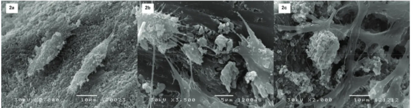
A composite scaffold PLGA/HAP registered under the name ALBO-OSS has shown good cell adhesion of cells grown in control medium. Cells were spherical with clearly visible pseudopodia or cytoplas-mic extensions (Figure 4a). After 7 days of culture in osteogenic medium, polygonal cells with very elong-ated cytoplasmic extensions were detected. The
org-anic fibrous-like structures have also been observed (Figure 4b). These structures and cell morphology are typical for stem cells undergoing osteogenic differ-entiation and indicate the beginning of the ECM form-ation. After 21 days, ECM dominated in the SEM micrographs covering the scaffold pores (Figure 4c). This highly developed ECM network demonstrated extensive differentiation and good biocompatibility between cells and materials, which is essential for use in tissue engineering [36].
Comparing the nano- and microstructured scaf-folds, it was demonstrated by a difference in the opti-cal density obtained by MTT testing and ALP activity that the larger surface area of the nanostructured scaffold allows better adhesion and provides more space for the differentiation of mesenchymal cells than in microstructured scaffolds [86].
Comparing the adhesion and quantity of human osteoblasts cultured on Bio Oss and synthetic bone substitute – Nano Bone for 7 days by SEM analysis, a significantly higher number of cells with cytoplasmic extensions was observed in the presence of Nano Bone [87].
SEM analysis showed similar results after study-ing cell morphology and adhesion of mesenchymal cells grown for 7 days on PLLA and on nano- and micro-HAP/PLLA composite scaffolds. The cells were spherical and their number was much higher in the presence of nano HAP/PLLA composite scaffold, indi-cating a superior biocompatibility of that material [88]. Hence, there are many scaffolds that exhibit good biocompatibility and could replace materials, such as golden standard Bio OSS, currently used in bone tissue engineering.
Challenges and future research
The main requirement for a successful appli-cation of the scaffold is a high control level of their
Figure 4. Scanning electron microscopy of dental pulp stem cells from deciduous teeth on Albo-Oss (porous hydroxyapatite + PLGA composite) scaffold: a) Spherical cells with cytoplasmic extensions indicate good cell adhesion after 7 days in control medium; (b) polygonal cells with elongated cytoplasmic extensions pseudopodia indicate very good cell adhesion; fibrous-like organic structures
micro- and macrostructural properties during pro-duction process. Mechanical properties of today’s composite scaffolds still do not fully satisfy the pro-perties of natural bone nor succeed in reaching their anisotropy [89].
Though a wide-range of strategies have been employed to produce the ideal scaffold that pos-sesses the optimum dimensions, porosity, topography and mechanical properties, the clinical success of such constructs remains elusive [90].
The rapid development of techniques and fab-rication tools that have arisen in the recent years have signified new beginnings in the field of TE. How-ever, there are still obstacles in achieving the success of the in vitro experiments in an in vivo system. More-over, the complexity of multiple tissues that form a functional organ poses a real challenge to tissue eng-ineers. Further developments are awaited in reaching the goal of creating a completely functional organ, thus truly transforming tissue engineering into regen-erative engineering.
Acknowledgements
This paper was supported by the Projects 41030 and 172026 financed by the Ministry of Education, Science and Technological Development of the Rep-ublic of Serbia.
REFERENCES
[1] M. Miura, S. Gronthos, M. Zhao, B. Lu, L.W. Fisher, P.G. Robey, S. Shi, Proc. Natl. Acad. Sci. U.S.A. 100 (2003) 5807-5812
[2] S.S. Kim, M. Sun Park, O. Jeon, C. Yong Choi, B.S. Kim, Biomaterials 27 (2006) 1399-1409
[3] S.C. Rizzi, D.J. Heath, A.G. Coombes, N. Bock, M. Textor, S. Downes, J. Biomed. Mater. Res. 55 (2001) 475-486
[4] E.M. Aarden, E.H. Burger, P.J. Nijweide, J. Cell. Biochem. 55 (1994) 287-299
[5] X. Miao, D.M. Tan, J. Li, Y. Xiao, R. Crawford, Acta Biomater. 4 (2008) 638-645
[6] H. Sittisak, P. Vinai, Asian Biomed. 1 (2007) 229-238
[7] L. Meinel, V. Karageorgiou, R. Fajardo, B. Snyder, V. Shinde-Patil, L. Zichner, D. Kaplan, R. Langer, G. Vunjak- -Novakovic, Ann. Biomed. Eng. 32 (2004) 112-122
[8] I. Karadzic, V. Vucic, V. Jokanovic, J. Debeljak-Martacic, D. Markovic, S. Petrovic, M. Glibetic, J. Biomed. Mater. Res., A 103 (2015) 350-357
[9] G.F. Muschler, C. Nakamoto, L.G. Griffith, J. Bone. Joint. Surg. Am. 86A (2004) 1541-1558
[10] K.E. McCracken, P.L. Tran, D.J. You, M.J. Slepian, J.Y. Yoon, J. Biol. Eng. 7 (2013) 11
[11] H. Hosseinkhani, M. Hosseinkhani, S. Hattori, R. Matsu-oka, N. Kawaguchi, J. Biomed. Mater. Res., A 94 (2010) 1-8
[12] H. Hosseinkhani, P.D. Hong, D.S. Yu, Y.R. Chen, D. Ickowicz, I.Y. Farber, A.J. Domb, Int. J. Nanomed. 7 (2012) 3035-3043
[13] H. Peng, X. Liu, R. Wang, F. Jia, L. Dong, Q. Wang, J. Mater. Chem., B 2 (2014) 6435-6461
[14] I. Singec, R. Jandial, A. Crain, G. Nikkhah, E. Y. Snyder, Annu. Rev. Med. 58 (2007) 313-328
[15] A. Vats, R.C. Bielby, N.S. Tolley, R. Nerem, J.M. Polak, Lancet. 366 (2005) 592-602
[16] E.A. de Wynter, A.J. Emmerson, N.G. Testa, Best Pract. Res., Clin. Haematol. 12 (1999) 1-17
[17] R.J. Deans, A.B. Moseley, Exp. Hematol. 28 (2000) 875- –884
[18] A. Saunders, F. Faiola, J. Wang, Stem. Cells. 31 (2013) 1227-1236
[19] I. Wilmut, A. E. Schnieke, J. McWhir, A.J. Kind, K.H. Campbell, Nature 385 (1997) 810-813
[20] H.S. Dua, A. Azuara-Blanco, Surv. Ophthalmol. 44 (2000) 415-425
[21] I. Malanchi, H. Peinado, D. Kassen, T. Hussenet, D. Metzger, P. Chambon, M. Huber, D. Hohl, A. Cano, W. Birchmeier, J. Huelsken, Nature 452 (2008) 650-653
[22] D. Brasanac, I. Boricic, V. Todorovic, N. Tomanovic, S. Radojevic, Br. J. Dermatol. 153 (2005) 1166-1175
[23] E. Fuchs, J. A. Segre, Cell 100 (2000) 143-155
[24] D. Markovic, A. Milenkovic, G. Koliakos, E. Kostidou, I. Karadzic, J. Debeljak Martacic, V. Jokanovic, T. Peric, B. Petrovic, N. Arsenijevic, T. Kanjevac, Balk. J. Stom. 14 (2010) 4-7
[25] B.C. Perry, D. Zhou, X. Wu, F.C. Yang, M.A. Byers, T.M. Chu, J.J. Hockema, E.J. Woods, W.S. Goebel, Tissue Eng., C: Methods 14 (2008) 149-156
[26] A. Graziano, R. d'Aquino, G. Laino, G. Papaccio, Stem. Cell. Rev. 4 (2008) 21-26
[27] S. Huang, D.E. Ingber, Nat. Cell. Biol. 1 (1999) E131-138
[28] C. Wang, G. Meng, L. Zhang, Z. Xiong, J. Liu, J. Biomed. Biotechnol. 2012 (2012) 579141
[29] D.W. Hutmacher, J.T. Schantz, C.X. Lam, K.C. Tan, T.C. Lim, J. Tissue. Eng. Regen. Med. 1 (2007) 245-260
[30] K.S. Chan, W. Liang, W.L. Francis, D.P. Nicolella, J. Mech. Behav. Biomed. Mater. 3 (2010) 584-593
[31] B.N. Brown, C.A. Barnes, R.T. Kasick, R. Michel, T.W. Gilbert, D. Beer-Stolz, D.G. Castner, B.D. Ratner, S.F. Badylak, Biomaterials 31 (2010) 428-437
[32] M. Kamitakahara, C. Ohtsuki, T. Miyazaki, J. Biomater. Appl. 23 (2008) 197-212
[33] F.J. O'Brien, Mater. Today. 14 (2011) 88-95.
[34] D.W. Hutmacher, Biomaterials 21 (2000) 2529-2543
[35] C.M. Murphy, M.G. Haugh, F.J. O'Brien, Biomaterials 31 (2010) 461-466
[37] E.A. Phelps, A.J. Garcia, Regen. Med. 4 (2009) 65-80
[38] M. Petrovic, B. Colovic, V. Jokanovic, D. Markovic, J. Ceram. Process. Res. 13 (2012) 398-404
[39] I. Armentano, M. Dottori, E. Fortunati, S. Mattioli, J.M. Kenny, Polym. Degrad. Stab. 95 (2010) 2126-2146
[40] O.O. Ige, L.E. Umoru, S. Aribo, ISRN Mater. Sci. 2012 (2012) 20
[41] E. Chung, J.A. Rytlewski, A.G. Merchant, K.S. Dhada, E.W. Lewis, L.J. Suggs, Acta Biomater. 17 (2015) 78-88
[42] B. Duan, L. Wu, X. Li, X. Yuan, Y. Zhang, K. Yao, J. Biomater. Sci. Polym. Ed. 18 (2007) 95-115
[43] J.Y. Lai, D.H. Ma, H.Y. Cheng, C.C. Sun, S.J. Huang, Y.T. Li, G.H. Hsiue, J. Biomater. Sci. Polym. Ed. 21 (2010) 359-376
[44] F. Ding, H. Deng, Y. Du, X. Shi, Q. Wang, Nanoscale 6 (2014) 9477-9493
[45] K.T. Shalumon, J.P. Chen, Curr. Pharm. Des. 21 (2015) 1979-1990.
[46] A.B. Daly, J.M. Wallis, Z.D. Borg, R.W. Bonvillain, B. Deng, B.A. Ballif, D.M. Jaworski, G.B. Allen, D.J. Weiss, Tissue Eng., A 18 (2012) 1-16
[47] Y. Lu, X. Li, X. Zhou, Q. Wang, X. Shi, Y. Du, H. Deng, L. Jiang, RSC Adv. 4 (2014) 33355-33361
[48] Y. Li, Z.G. Wu, X.K. Li, Z. Guo, S.H. Wu, Y.Q. Zhang, L. Shi, S.H. Teoh, Y.C. Liu, Z.Y. Zhang, Biomaterials 35 (2014) 5647-5659
[49] F. Qi, J. Wu, T. Yang, G. Ma, Z. Su, Acta Biomater. 10 (2014) 4247-4256
[50] F. Song, X. Li, Q. Wang, L. Liao, C. Zhang, J. Biomed. Nanotechnol. 11 (2015) 40-52
[51] Q. Wang, S. Jamal, M.S. Detamore, C. Berkland, J. Biomed. Mater. Res., A 96 (2011) 520-527
[52] A.M. Ambrosio, J.S. Sahota, Y. Khan, C.T. Laurencin, J. Biomed. Mater. Res. 58 (2001) 295-301
[53] M. Wang, Biomaterials 24 (2003) 2133-2151
[54] I. Hofmann, L. Müller, P. Greil, F.A. Müller, Surf. Coat. Technol. 201 (2006) 2392-2398
[55] J.B. Lee, S.I. Jeong, M.S. Bae, D.H. Yang, D.N. Heo, C.H. Kim, E. Alsberg, I.K. Kwon, Tissue Eng., A 17 (2011) 2695-2702
[56] T. Douglas, E. Pamula, D. Hauk, J. Wiltfang, S. Sivanan-than, E. Sherry, P.H. Warnke, J. Mater. Sci. Mater. Med. 20 (2009) 1909-1915
[57] S.S. Liao, F.Z. Cui, Y. Zhu, J. Bioact. Compat. Polym. 19 (2004) 117-130
[58] M.M. Pereira, J.R. Jones, R.L. Orefice, L.L. Hench, J. Mater. Sci. Mater. Med. 16 (2005) 1045-1050
[59] T. Lu, Y. Li, T. Chen, Int. J. Nanomed. 8 (2013) 337-350
[60] Q. Wang, Z. Gu, S. Jamal, M.S. Detamore, C. Berkland, Tissue Eng., A 19 (2013) 2586-2593
[61] Q. Wang, J. Wang, Q. Lu, M.S. Detamore, C. Berkland, Biomaterials 31 (2010) 4980-4986
[62] D. Lickorish, L. Guan, J.E. Davies, Biomaterials 28 (2007) 1495-1502
[63] Y.M. Khan, D.S. Katti, C.T. Laurencin, J. Biomed. Mater. Res., A 69 (2004) 728-737
[64] M. Loss, V. Wedler, W. Kunzi, C. Meuli-Simmen, V.E. Meyer, Burns 26 (2000) 644-652
[65] X. Shen, N. Nagai, M. Murata, D. Nishimura, M. Sugi, M. Munekata, J. Mater. Sci. Mater. Med. 19 (2008) 3473- –3479
[66] W.R. Gombotz, S.F. Wee, Adv. Drug. Deliv. Rev. 64 Suppl. (2012) 194-205
[67] K. Murakami, H. Aoki, S. Nakamura, M. Takikawa, M. Hanzawa, S. Kishimoto, H. Hattori, Y. Tanaka, T. Kiyo-sawa, Y. Sato, M. Ishihara, Biomaterials 31 (2010) 83-90
[68] W. Li, X. Li, Q. Wang, Y. Pan, T. Wang, H. Wang, R. Song, H. Deng, Carbohydr. Polym. 99 (2014) 218-225
[69] S. Xin, X. Li, Q. Wang, R. Huang, X. Xu, Z. Lei, H. Deng, J. Biomed. Nanotechnol. 10 (2014) 803-810
[70] G. Vunjak-Novakovic, N. Tandon, A. Godier, R. Maidhof, A. Marsano, T. P. Martens, M. Radisic, Tissue Eng., B 16 (2010) 169-187
[71] T. Shinoka, C.K. Breuer, R.E. Tanel, G. Zund, T. Miura, P.X. Ma, R. Langer, J.P. Vacanti, J.E. Mayer, Jr., Ann. Thorac. Surg. 60 (1995) S513-516
[72] J.L. Honge, J.A. Funder, H. Jensen, P.M. Dohmen, W.F. Konertz, J.M. Hasenkam, J. Heart Valve Dis. 19 (2010) 584-592
[73] F.E. Smit, P.M. Dohmen, Med. Sci. Monit. Basic Res. 20 (2014) 161-163
[74] C.H. Chen, M.Y. Lee, V.B. Shyu, Y.C. Chen, C.T. Chen, J.P. Chen, Mater. Sci. Eng., C: Mater. Biol. Appl. 40 (2014) 389-397
[75] S.A. Maher, R.L. Mauck, L. Rackwitz, R.S. Tuan, J. Tissue. Eng. Regen. Med. 4 (2010) 25-29
[76] R.N. Shah, N.A. Shah, M.M. Del Rosario Lim, C. Hsieh, G. Nuber, S. I. Stupp, Proc. Natl. Acad. Sci. U.S.A. 107 (2010) 3293-3298
[77] G.A. Saracino, D. Cigognini, D. Silva, A. Caprini, F. Gelain, Chem. Soc. Rev. 42 (2013) 225-262
[78] A.P. Balgude, X. Yu, A. Szymanski, R.V. Bellamkonda, Biomaterials 22 (2001) 1077-1084
[79] N.E. Starritt, S.A. Kettle, M.A. Glasby, Laryngoscope 121 (2011) 1614-1619
[80] F. Tavangarian, Y. Li, Ceram. Int. 38 (2012) 6075-6090
[81] S.W. Kang, H.S. Yang, S.W. Seo, D.K. Han, B.S. Kim, J. Biomed. Mater. Res., A 85 (2008) 747-756
[82] M. Ngiam, S. Liao, A.J. Patil, Z. Cheng, C.K. Chan, S. Ramakrishna, Bone 45 (2009) 4-16
[83] Q. Wang, L. Wang, M. S. Detamore, C. Berkland, Adv. Mater. 20 (2008) 236-239
[84] V. Jokanovic, Nanomedicine – the Biggest Challenge of the 21st Century, Data Status, Belgrade, 2012 (in Ser-bian)
[85] B. Colovic, V. Jokanovic, B. Markovic-Todorovic, Z. Mar-kovic, J. Optoelectron Adv. Mater. 11 (2009) 70-75
[86] M.B. Eslaminejad, F. Bagheri, M. Zandi, E. Nejati, E. Zomorodian, Cell J. 12 (2011) 469-476
[88] R.R. Recker, In: Disorders of Bone and Mineral Meta-bolism, F.L. Coe, M.J. Favus (Eds.), Raven Press, New York, 1992, pp`. 219-240
[89] M. M. Stevens, Mater. Today. 11 (2008) 18-25
[90] Y. Ikada, J. R. Soc. Interface 3 (2006) 589-601.
DEJAN MARKOVIĆ1 IVANA KARADŽIĆ2 VUKOMAN JOKANOVIĆ3 ANA VUKOVIĆ1 VESNA VUČIĆ2 1Klinika za dečju i preventivnu
stomatologiju, Stomatološki fakultet, Univerzitet u Beogradu, Dr Subotića 11, 11000 Beograd, Srbija 2
Centar izuzetnih vrednosti za ishranu i metabolizam, Institut za medicinska istraživanja, Univerzitet u Beogradu, Tadeuša Košćuška 1, 11000 Beograd, Srbija 3Laboratorija za radijacionu hemiju i fiziku, Institut za nuklearne nauke “Vinča”, University of Belgrade, Mike Petrovića Alasa 12-14, 11000 Beograd, Srbija
NAUČNI RAD
BIOLOŠKI ASPEKTI PRIMENE NANOMATERIJALA
U TKIVNOM INŽENJERSTVU
Milioni pacijenata širom sveta imaju potrebu za hirurškim procedurama radi reparacije ili nadoknade oštećenog tkiva nakon traume ili oboljenja. U cilju rešavanja problema izgub-ljenog tkiva, tkivno inžinjerstvo glavni akcenat stavlja na tkivnu regeneraciju a ne na za-menu tkiva, zbog čega postaje predmet sve većeg interesovanja. Matične ćelije i ćelijski nosači - visoko porozni biomaterijali, tzv. skafoldi, imaju esencijalnu ulogu u stvaranju novog tkiva putem tkivnog inžinjerstva. Ćelijska komponenta je neophodna zbog stvaranja i uspostavljanja ekstracelularnog matriksa, dok je skafold zadužen za da odredi oblik novo-stvorenog tkiva i olakša migraciju ćelija na željeno mesto, njihov rast i diferencijaciju. Ovaj pregledni rad opisuje vrste, karakteristike i klasifikaciju matičnih ćelija. Osim toga, uključuje i neophodne funkcionalne osobine ćelijskih nosača – biokompatibilnost, biorazgradljivost i mehanička svojstva biomaterijala u primeni tkivnog inženjerstva. Takođe, objašnjava raz-log interesovanja za praktičnu primenu nanomaterijala i nanotehnologije i definiše izazove i značaj daljih istraživanja u ovoj oblasti.