Control of Soft Rot on Potato Caused by ‘Dickeya solani’
Evelien M. Adriaenssens1,2,3, Johan Van Vaerenbergh2, Dieter Vandenheuvel1, Vincent Dunon1, Pieter-Jan Ceyssens1, Maurice De Proft3, Andrew M. Kropinski4,5, Jean-Paul Noben6, Martine Maes2, Rob Lavigne1*
1Division of Gene Technology, Katholieke Universiteit Leuven, Heverlee, Belgium,2Unit Plant – Crop Protection, Institute for Agricultural and Fisheries Research, Merelbeke, Belgium,3Division of Crop Biotechnics, Katholieke Universiteit Leuven, Heverlee, Belgium,4Laboratory of Foodborne Zoonoses, Public Health Agency of Canada, Guelph, Ontario, Canada,5Department of Molecular and Cellular Biology, University of Guelph, Guelph, Ontario, Canada,6Biomedical Research Institute and Transnational University Limburg, School of Life Sciences, Hasselt University, Diepenbeek, Belgium
Abstract
The bacterium ‘Dickeya solani’, an aggressive biovar 3 variant ofDickeya dianthicola, causes rotting and blackleg in potato. To control this pathogen using bacteriophage therapy, we isolated and characterized two closely related and specific bacteriophages, vB_DsoM_LIMEstone1 and vB_DsoM_LIMEstone2. The LIMEstone phages have a T4-related genome organization and share DNA similarity withSalmonellaphage ViI. Microbiological and molecular characterization of the phages deemed them suitable and promising for use in phage therapy. The phages reduced disease incidence and severity on potato tubers in laboratory assays. In addition, in a field trial of potato tubers, when infected with ‘Dickeya solani’, the experimental phage treatment resulted in a higher yield. These results form the basis for the development of a bacteriophage-based biocontrol of potato plants and tubers as an alternative for the use of antibiotics.
Citation:Adriaenssens EM, Van Vaerenbergh J, Vandenheuvel D, Dunon V, Ceyssens P-J, et al. (2012) T4-Related Bacteriophage LIMEstone Isolates for the Control of Soft Rot on Potato Caused by ‘Dickeya solani’. PLoS ONE 7(3): e33227. doi:10.1371/journal.pone.0033227
Editor:Eric A. Johnson, University of Wisconsin, Food Research Institute, United States of America
ReceivedNovember 14, 2011;AcceptedFebruary 6, 2012;PublishedMarch 7, 2012
Copyright:ß2012 Adriaenssens et al. This is an open-access article distributed under the terms of the Creative Commons Attribution License, which permits unrestricted use, distribution, and reproduction in any medium, provided the original author and source are credited.
Funding:EMA and the experimental work were funded through a PhD scholarship by the Katholieke Universiteit Leuven and the Institute for Agricultural and Fisheries Research Flanders (ILVO). PJC hold a post-doctoral fellowship of the Fonds voor Wetenschappelijk Onderzoek (FWO Flanders). The funders had no role in study design, data collection and analysis, decision to publish, or preparation of the manuscript.
Competing Interests:The authors have declared that no competing interests exist.
* E-mail: rob.lavigne@biw.kuleuven.be
Introduction
The plant pathogenicDickeya spp. (formerly known asErwinia chrysanthemi or Pectobacterium chrysanthemi, [1]) are Gram-negative, non-sporulating, facultative anaerobic bacteria of the family Enterobacteriaceae, which characteristically produce pectinolytic enzymes during infection. Along with other pectinolytic bacteria such asPectobacterium atrosepticumandPectobacterium carotovorumsubsp. carotovorum, they are a major cause of potato tuber soft rot during storage and blackleg disease in the field [2–4]. Samson and colleagues (2005) differentiated six species within the genusDickeya, namely D. zeae, D. dadantii, D. chrysanthemi, D. dieffenbachiae, D. dianthicolaandD. paradisiaca[1]. Of these six, onlyD. paradisiacahas not been isolated from potato [3], andD. dianthicolahas been the main species found in Europe. Recently a new, more virulent Dickeyatype, belonging to biovar 3 ofE. chrysanthemi, was described and is tentatively named ‘Dickeya solani’ [3,5]. ThisDickeyatype has become the predominant cause of blackleg of potato in certain European countries [3,5,6]. At this moment no chemical disease control measures are available forDickeyaand infected batches of potatoes are declassified or discarded, resulting in significant economic losses [3].
Traditionally, a first diagnostic tool for the identification of PectobacteriumandDickeyais a PCR analysis based on thepelYand the pelADE gene cluster, respectively [7,8]. For Dickeya spp., sequence data of both the recA gene and dnaX were used for
phylogenetic analysis of the different species in this genus and these data support the designation of the new species ‘Dickeya solani’ [5,9]. Recently, a new molecular tool was developed for the identification of this species specifically, a real-time PCR of the virulence gene fliC (Van Vaerenbergh et al., submitted manu-script).
(Bacterio)phages have been proposed as biocontrol agents for bacterial diseases in plants [10,11]. However, phage therapy has to overcome several challenges before it can be efficiently used in agriculture (summarized in [12]). In light of these challenges, Balogh and colleagues [11] argue for the application of bacteriophages in controlled and closed environments with a short window of plant susceptibility, where phages can easily access a homogenous target bacterium population and exposure to harsh environments is limited. In addition, both phage and bacterium need to be extensively characterized and efficiently purified.
investigated in phage therapy trials on calla lily tubers in the greenhouse [15]. On potato, one case of phage therapy has been reported; the application of bacteriophage øAS1 on seed potatoes infected withStreptomyces scabies which causes scab [16]. Infected seed tubers were treated with phage and produced progeny tubers with significantly reduced surface scab lesions.
Of all the phage genome sequences present in the NCBI database, less than 5% are of phages infecting plant pathogenic bacteria. ForDickeyaspp. no phage genomes are available, only for the related generaErwinia and Pectobacterium phage genomes are sequenced [17–19], illustrating the need for more genome sequence information. In this paper, we report the succesful application of a new phage species,Dickeyaphage LIMEstone in an agricultural setting, with bothin vitro andin vivo screens. Of this species, two phages were found, LIMEstone1 and LIMEstone2, which infect ‘Dickeya solani’. The microbiological characteriza-tion, as well as sequence analysis, deemed the phage isolates suitable for use in phage therapy.
Results
Phages LIMEstone1 and LIMEstone2
Isolation of bacteria and phage. Bacteria of the genus Dickeyawere isolated from diseased potato plants and tubers at the diagnostic clinic of the Institute for Agricultural and Fisheries Research (ILVO, Merelbeke, Belgium) as described by Van Vaerenbergh et al. (submitted manuscript). The isolates (Table S1) were characterized based on barcoding of thefliC amplicon and TaqMan qPCR specific for ‘Dickeya solani’ (Van Vaerenberg et al, submitted manuscript). Of the 17Dickeya isolates collected in 2008, 16 were identified as the new ‘Dickeya solani’ type and one was designated asDickeya dianthicola.
Bacteriophage isolates were made from soil samples from a potato trial field at ILVO after the harvest in September–October 2008. Out of 26 trial plots sampled, 18 contained plants infected
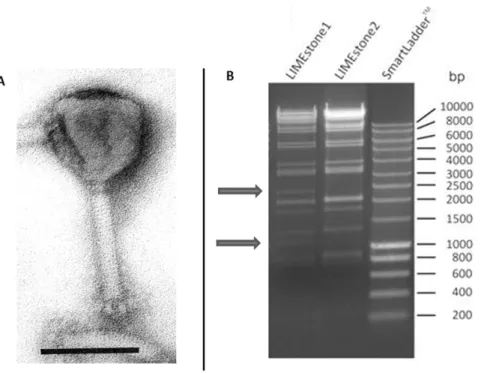
withDickeya spp. or Pectobacterium spp. Filtrates of the soil were tested for their capacity to lyse a range of Dickeya bacteria. In samples of 14 fields, of which 11 were infected and three were uninfected withDickeyaspp., phages were found. All phage isolates produced small clear plaques of 1 mm in diameter on ‘Dickeya solani’ strains and restriction digestion of the DNA of the isolates with HindII (Figure 1B) showed two closely related patterns, differing in two bands. These phages were named LIMEstone1 and LIMEstone2 (Leuven ILVO Merelbeke) belonging to the species LIMEstone (scientific names vB_DsoM_LIMEstone1 and vB_Dso_LIMEstone2 as proposed by [20]). Phage isolates belonging to the LIMEstone species were also predominant in soil samples collected from the same fields in 2009 and 2010 (data not shown), isolated according to the same protocol as in 2008. Based on the restriction patterns of the isolates, which were very similar to that of LIMEstone1 and LIMEstone2, it was decided not to further investigate these phages.
General characteristics of LIMEstone1 and LIMEstone2. LIMEstone was found to be a member of the Myoviridaeby transmission electron microscopy (Figure 1A). With an icosahedrical head of 91.4 nm and tail dimensions of 113.8617 nm, its morphology is similar to that of Salmonella
phage ViI [21]. A collar was visible (2062 nm) and several short
tail spikes of 12 nm in length. The head volume is smaller than the prolate head of phage T4 (119.5686 nm), which suggests a smaller genome size for LIMEstone1.
Adsorption and one-step-growth assays were performed for LIMEstone1 and LIMEstone2 isolates to assess the infection parameters (Figure S1). For both phages, more than 99.9999% of phages were irreversibly adsorbed to the host cell within one minute. Upon comparison of the adsorption constant k [k = (2.3/ (B*t))*log(P0/P), with B the bacterial titer at time zero and t the
time], LIMEstone2 (k at 1 min = 2.0561028ml/min) appears to
adsorb marginally faster than LIMEstone1 (k at 1 min = 9.536
1029
ml/min) and more rapid than reported for T4
Figure 1. LIMEstone isolates.A) EM picture of phage LIMEstone1. Phage negatively stained with 2% phosphotungstate. Scale bar represents 100 nm. B) HindII restriction digestion of 0.5 and 1.0mg DNA of LIMEstone1 and LIMEstone2, respectively.
(2.461029ml/min) [22]. In the one-step-growth assay, the latent
period of LIMEstone1 was determined at 60 minutes with a burst size of 160. The latent period of LIMEstone2 was 65 minutes and about 100 new particles were released. These variations are minor once more indicating the relatedness of LIMEstone1 and LIMEstone2, belonging to one proposed species, LIMEstone.
The viability of LIMEstone in a range of environmental conditions was assessed. Phage were stable at temperatures from 4uC to 37uC in phage buffer, but the titer decreased by three log10
units upon storage at 50uC for 24 hours. All viable phage were lost after freezing of the sample. LIMEstone was also stable from pH 4 to 11 for 24 hours.
Host range analysis. A collection ofDickeyastrains (Table S1) was assembled to test the host range of phages LIMEstone1 and LIMEstone2. From the reference set of Van Vaerenbergh et al (submitted manuscript PONE-D-11-23125), two strains perDickeya species were chosen, the type strain and a strain isolated from potato, except forD. paradisiacawhich has not been found on potato. This collection was supplemented with the 17 strains discussed earlier in this study and with older isolates from the culture collections of the plant clinic of ILVO (GBBC numbers) and of the diagnostic clinic in The Netherlands (PD and PRI numbers).
Only the ‘Dickeya solani’ type was found to be susceptible for infection with both LIMEstone1 and LIMEstone2, with 100% of the strains showing lysis and plaques (Table S1). The isolates of Dickeya dianthicola from Belgium and The Netherlands were not infected by either phage, but showed a clear lysis zone when a phage suspension of 109pfu or higher was spotted on a bacterial lawn. There was no lysis observed when a dilution was spotted and no plaques were formed. This is probably ‘lysis from without’ and not true infection. Of the globalDickeyacollection,D. dianthicola,D. dadantii, D. dieffenbachiae and D. chrysanthemi, all showed this lysis from without, but no phage amplification. The lastDickeyaspecies, D. zeae, was not infected with either phage and showed no lysis from without, as did a number of environmental isolates identified asPectobacteriumspp.
The genome and proteome of LIMEstone1
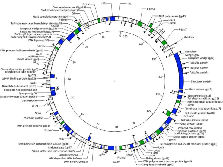
Genome organization. Genome sequencing of bacteriophage LIMEstone1 (GenBank accession number HE600015) revealed a genome of 152,427 bp and a G+C content of 49.2%, probably circularly permuted (Figure 2). A total of 201 open reading frames (ORFs) were predicted on both strands and one tRNA (Met-tRNA, anticodon CAT). Of these ORFs, 64 could be linked to bacteriophage T4 based on BLASTP similarity. To another 29 ORFs a putative function could be assigned, leaving 54% of unknown ORFs. A distribution of functional regions, typical for T4-related phages, is observed in phage LIMEstone1, where no well-defined early, middle or late region of transcription was found.
Interestingly, DNA homology has been observed between LIMEstone1 and Shigella phage phiSboM-AG3 [23], Salmonella phage ViI [21] andE. coli phage CBA120 [24]. The genome of LIMEstone1 showed a DNA homology of 69.1% with phiSboM-AG3 sharing 174 genes out of 201. Similarity with ViI and CBA120 is less with 58.7% and 59.4% respectively. The gene order is strongly conserved between the four phages, with a few insertions present, due to the other’s larger genome sizes (ViI 157,061 bp, CBA120 157,304 bp and phiSboM-AG3 158,006 bp). The overall similarity between these phages is quite remarkable, considering all four of them infect different bacterial genera.
Regulatory elements. In phage T4, transcription is mediated by three classes of promoters – early, middle and late [25]. These classes were also identified in LIMEstone1 based on sequence similarity of the235 and210 boxes with those of T4.
Five putative early promoters, five middle and 33 late promoters were located in intergenic regions on both strands (indicated in Figure 2 with arrows). Another four putative promoters were found based on the210 box ofDickeya dadantii3937 in which only five promoters have been annotated [26]. A total of 31 rho factor-independent terminators were identified throughout the genome of LIMEstone1, located on both strands (Figure 2).
Mobile elements. Homing endonucleases are mobile genetic elements, which recognize a DNA target site and generate single or double-stranded breaks in the genome to insert themselves in the target genome [27]. While their exact function is not known, Goodrich-Blair and Shub suggest they confer a selective advantage to the flanking sequences in the phage genome [28]. In phage LIMEstone1, 14 homing endonucleases were found, representing 10% of the genome (Table 1). This is comparable to the 15 homing endonucleases found in phage T4 (reviewed in [29] and [30]), but considered an oddity among the other T4-related phages. Three putative introns were identified in LIMEstone1, designated I-LimI, I-LimII and I-LimIII [31], the first two in the DNA polymerase gene, the third inuvsW. These two genes are functionally essential and strongly conserved between the T4-related phages and make thus an good target for intron homing [30]. The 11 free-standing homing endonucleases found in LIMEstone1 could be divided into three groups, the endonucleases encoding a GIY-YIG motif, the HNH-containing endonucleases and the Hef-like endonucleases (homing endonuclease-like function) [32,33] and were named F-LimI through F-LimXI according to Roberts and colleagues [31].
Structural proteome. The virion particle of LIMEstone1 consisted of at least 39 proteins, as verified by mass spectrometry (Table 2). Of these proteins, 27 had a function assigned based on sequence similarity with other phage proteins, in addition to 12 unknown structural proteins. As expected, the most abundant proteins in the sample were the major capsid protein gp23 (ORF138) and the tail sheath protein gp18 (ORF146). There is one structural region found in the genome, from ORF138 on the complementary strand (major capsid protein gp23) to ORF163 (baseplate wedge subunit gp6). In this region, the gene order is largely conserved between LIMEstone1 and T4. Four structural proteins of T4 Gp8, Gp10, Gp11 and Gp12, could not be found in phage LIMEstone1, but structural proteins were present in the corresponding locations to substitute the function of the missing T4 proteins. Two mobile elements were also located in this region; F-Lim-VIII located on the opposite strand between the tail tube and portal proteins (ORF144 and ORF146) and F-LimIX between the two subunits of the terminase complex (ORF147 and ORF149). There is another insertion of two hypothetical proteins with their own promoter and terminator between the two neck proteins, ORF151 and ORF154.
The other structural proteins were scattered throughout the entire genome on both strands, with some components of baseplate and tail tube grouping together (ORF23-27; ORF59-62). Between ORF78 and ORF85, a small group of structural proteins were clustered together, but no specific functional predictions could be made.
Table 1.Mobile elements in the genome of LIMEstone1.
ORF HEase name Phage homolog (phage name) Intron or free-standing Group Target gene or downstream genea
ORF17 F-LimI SegB (T4) Free-standing GIY-YIG ORF18
ORF12 F-LimI MobB/C/D/E Free-standing GIY-YIG ORF13 (DNA topoisomerase II)
ORF22 F-LimII SegD (133) Free-standing GIY-YIG ORF21 (Head completion protein)
ORF24 F-LimIII Hef (Acj9) Free-standing Hef-like ORF25 (Baseplate wedge subunit)
ORF36 F-LimIV MobC (phiSboM-AG3) Free-standing HNH ORF33
ORF76 F-LimV MobE (phiSboM-AG3) Free-standing HNH ORF75 (DNA primase)
ORF114 F-LimVI I-TevI Free-standing GIY-YIG ORF113
ORF123 I-LimI MobB/D Intron GIY-YIG ORF122-124 (UvsW)
ORF137 F-LimVII Hef (Acj9) Free-standing Hef-like ORF136
ORF145 F-LimVIII Hef (CP220) Free-standing Hef-like ORF144 (Tail tube protein)
ORF148 F-LimIX MobE (T4) Free-standing GIY-YIG ORF147 (Terminase large subunit)
ORF171 F-LimX Hef (Acj9) Free-standing Hef-like ORF170
ORF179 I-LimII MobE (Acj9) Intron GIY-YIG ORF178-180-182 (DNA polymerase)
ORF181 I-LimIII MobE (phiAS5) Intron HNH ORF178-180-182 (DNA polymerase)
ORF186 F-LimXI SegB (T4) Free-standing GIY-YIG ORF187
atarget gene for intron encoded homing endonucleases, downstream gene for free-standing endonucleases. doi:10.1371/journal.pone.0033227.t001
Figure 2. The genome of phage LIMEstone1 (152,427 bp).The inner ring represents ORFs on the forward strand, the outer ring the reverse strand. Proteins in italics show no sequence similarity with T4. ORFs in blue are confirmed as structural proteins, putative homing endonucleases are depicted in green. Promoters are indicated with arrows, factor-independent terminators with stem-loop structures.
Phage therapy biocontrol on potato
Virulence test on seed tubers. This test was designed to investigate whether the anti-bacterial effect of phages LIMEstone1 and LIMEstone2 on ‘Dickeya solani’ is also presentin vivo, i.e. on tubers, and to quantify this effect.
In a preliminary experiment, infection conditions for the pathogen, ‘Dickeya solani’ strain LMG 25865, were determined. A concentration of 105colony forming units (cfu) infiltrated per tuber combined with incubation at 28uC in a micro-aerophilic
environment were determined as ideal positive control conditions, since this ensured visible infection of the tubers in more than 90% of the cases.
The effect of treatment with phages on the rotting of potato tubers (cultivar (cv.) Bintje) was assessed under these micro-aerophilic conditions (Figure 3). This cultivar was chosen because it is the predominant cultivar in Belgium, with 42% of the total acreage in 2010 (National Institute for Statistics Belgium data). Phages LIMEstone1 and LIMEstone2 were added at a multiplicity Table 2.Structural proteins of LIMEstone1 as confirmed by mass spectrometry.
ORF Putative protein Size of protein (kDa) Protein coveragea
N6of unique peptides recovered
2 rIIB (T4 rIIB) 57.38 3.85% 1
6 Head outer capsid protein (T4 Hoc) 27.46 21.93% 4
23 Tail tuber associated baseplate protein (T4 gp48) 36.12 20.19% 4
26 Baseplate hub subunit (T4 gp27) 52.61 19.05% 6
27 Tail length tape measure protein 70.95 15.72% 7
32 DNA ligase (T4 gp30) 53.26 2.95% 1
50 Baseplate tail tube initiator (T4 gp54) 35.06 24.52% 7
59 Baseplate hub subunit (T4 gp26) 30.56 6.34% 1
61 Baseplate hub subunit & tail lysozyme (T4 gp5) 58.18 7.46% 2
62 Baseplate wedge subunit (T4 gp25) 14.04 19.05% 2
69 PhoH 31.47 3.93% 1
78 Unknown structural protein 13.15 28.21% 3
80 Unknown structural protein 20.28 12.17% 2
81 Unknown structural protein 40.85 55.20% 23
82 Unknown structural protein 18.76 19.63% 4
85 Unknown structural protein 22.75 16.98% 4
93 Unknown structural protein 28.32 40.78% 8
102 Unknown structural protein 17.31 35.57% 5
108 vWa containing protein 81.06 11.44% 6
119 DNA polymerase accessory protein (T4 gp44) 37.24 4.26% 1
127 Tail completion & sheath stabilizer protein (T4 gp3) 18.52 6.63% 1
129 Unknown structural protein 25.03 18.18% 2
138 Major capsid protein (T4 gp23) 48.02 73.41% 18
141 Prohead core protein 38.57 8.91% 1
143 Portal protein (T4 gp20) 63.29 38.19% 17
144 Tail tube protein (T4 gp19) 19.99 27.68% 4
146 Tail sheath protein (T4 gp18) 68.80 49.53% 27
151 Neck protein (T4 gp14) 24.97 35.19% 6
154 Neck protein (T4 gp13) 28.72 17.20% 4
157 Structural protein 177.55 24.38% 21
158 Tailspike protein 54.76 22.45% 17
159 Tailspike protein 21.59 11.76% 2
160 Tailspike protein 53.69 25.00% 8
161 Fibritin (T4 Wac) 42.69 50.62% 11
162 Baseplate wedge subunit (T4 gp7) 33.36 4.93% 1
163 Baseplate wedge subunit (T4 gp6) 64.75 18.07% 7
169 Unknown structural protein 18.45 24.42% 3
173 Unknown structural protein 17.33 11.84% 1
174 Unknown structural protein 16.62 12.50% 1
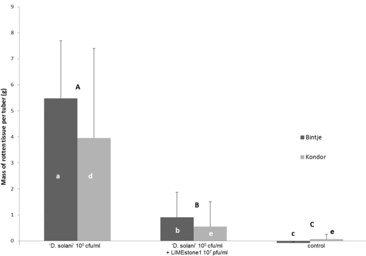
of infection (MOI) of 100 each to 20 tubers inoculated with LMG 25865. Looking at the number of rotten tubers for the positive control, 18 out of 20 tubers displayed rot. For the phage treated tubers the incidence of infection decreased significantly to 12 out of 20 for LIMEstone1 and 8 out of 20 for LIMEstone2. Moreover, a significant decrease in disease severity per tuber was observed after phage treatment. Both with LIMEstone1 and LIMEstone2, less than 10% rotten tissue per tuber was found (less than 1 g per tuber), calculated on the weight of the tuber before treatment and after the rotten tissue was scraped off, while the positive control group, which was only infected with bacteria, had an average of over 40% (5.5 g) rot per tuber (p values of 0.005073 and 0.000968, respectively). Between the two phages, no significant difference was observed in the amount of rotten tissue (p = 1.0). It can be concluded that the application of a surplus of bacteriophages can significantly reduce both the number of rotten tubers and the extent of tuber rotting caused byD. solanistrain LMG 25865. For LIMEstone1, this test was repeated on a different cultivar of potato, Kondor. At an MOI of 100 rotting of the potato tubers was significantly reduced from over 20% (4 g/tuber) to less than 5% (0.5 g) rot (p = 0.041242) (Figure 3). An MOI of 10 was also tested (data not shown) and also showed a decrease in the amount of rotten tissue, but this was not statistically significant, neither between the positive control and an MOI of 10 (p = 0.256840), nor between an MOI of 10 and an MOI of 100 (p = 0.794024). The
number of rotten Kondor tubers also showed a decrease after phage treatment with an MOI of 100.
Comparing the data of Bintje and Kondor (Figure 3) there was a variation in the percentage of tissue rot per tuber (averages of 23.5% and 42.5% respectively). This was due to the difference in size of the tubers between these two cultivars, because there was no significant difference between the two cultivars (p = 0.815129).
Field trial. The effect of phage treatment on potato tuber and plant growth was examined in a field trial. A latent infection of seed tubers with ‘D. solani’ was mimicked by vacuum infiltration of the tubers with a bacterial suspension. Next, a suspension of LIMEstone1 was nebulized over a batch of the infected tubers, to simulate a conveyor belt in a farm environment and the phage treated tubers were air dried. Three treatments were compared: an untreated control (treatment A), a positive control with only bacteria (treatment B), and co-treatment of bacteria and phage (treatment C). Tubers were kept out of direct sunlight until the moment of planting to avoid the interference of UV light in the experiment.
The emergence of the plants and disease incidence was monitored throughout the growing season. The first signs of infection, darkening and wilting of the shoot tips and young leaves, were observed 42 days after planting, for two plants of treatment B and one plant in treatment C. In the course of the next 20 days, more than 90% of the plants of treatment B showed symptoms of
Figure 3. Phage therapy assay on potato tubers cv. Bintje and Kondor.Tubers treated with ‘Dickeya solani’ strain LMG 25865 were compared with phage treated tubers and with a water-treated control. Error bars indicate standard deviation. Significant differences were tested with the Kruskal-Wallis multiple comparison tests at p,0.05 en the Mann-Whitney U test for comparison of two samples. Letters indicate significant differences, capitals between treatments, small letters within cultivars.
Dickeya infection, ranging from wilting, to leaf necrosis and stem rot (blackleg). In treatment regime C, disease incidence was a little less with 85% of plants displaying symptoms. A greater difference in disease severity was observed between treatment B and C, with none of the diseased plants of treatment C presenting stem rot, only wilting and leaf necrosis. For the control plants, no symptoms were observed throughout the growing season.
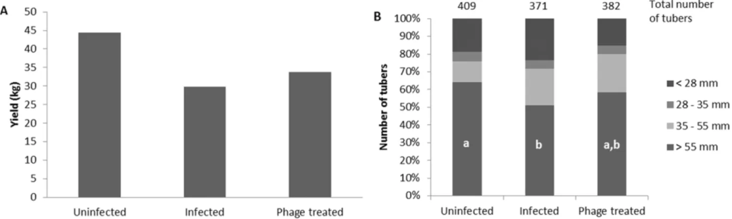
Tubers were harvested from the field 82 days after planting. The total yield for each treatment was 44.4 kg for the untreated plants (A), 29.9 kg for the ‘Dickeya solani’-treated plants (B), and 33.8 kg for the plants treated with phage LIMEstone1 (C) (Figure 4A). With a difference of 3.9 kg, phage treatment of infected potato tubers led to a 13% yield increase. This increase was mostly due to the size distribution of the tubers. The total number of tubers harvested from treatment C (382) was only 3% higher than the number of tubers from treatment B (371), both significantly less than the 409 tubers collected from group A.
The harvested tubers were divided into four groups according to their sizes, smaller than 28 mm, between 28 and 35 mm (seed tuber size), between 35 and 55 mm, and bigger than 55 mm (fry cut) (Figure 4B). This last category had the largest difference between treatments. As expected, the untreated plants had the highest number of tubers in this size range (262). For the plants inoculated with bacteria, this was significantly less, with 190 tubers (p = 0.044533). The number of tubers of the bacteria/phage treated plants was intermediate (223) and was not significantly different from either the control or the bacteria treated plants (p values of 0.820896 and 1.0, respectively) (Figure 4B).
For the control A, one rotten tuber was found, but this was not caused by ‘D. solani’, as confirmed bypelADEPCR andfliCqPCR on the rotted tissue. For treatments B and C, 9 and 6 ‘D. solani’ rotted tubers were collected respectively, a significant difference from the control treatment A. The difference between the number of rotten tubers of B and C, on the other hand was not big enough to be significant.
Discussion
Dickeyaspp. are of increasing concern in potato production in various parts of Europe [3]. It was apparent during our bacterial isolation tests that ‘D. solani’ has replacedD. dianthicolaas the most prevalent pathotype. In 2008, less than 10% of the isolatedDickeya strains belonged toD. dianthicola(only 1 out of 17 strains described in this paper). In this respect, it is logical that the phages
LIMEstone1 and LIMEstone2, isolated in the same year, specifically infect ‘D. solani’ and that no D. dianthicola phages have been isolated. These two were the onlyDickeyaphages that were isolated during the course of this study. Since they were isolated in three consecutive years (data not shown), it can be said that they are stable in this environment. They also infect 100% of the ‘Dickeya solani’ strains, which offers an explanation for the low diversity in phage types, as they might out-compete other phages. The broad host range of LIMEstone within ‘D. solani’ and its suspected abundance in the environment are characteristics of the group of T4-related phages. These are widespread around the globe and infect an array of different hosts, ranging from enterobacteria found in sewage to cyanobacteria from marine environments. Another reason for the low diversity of phages found could be the culturing method used for isolation, which seems to favor members of theCaudoviralesphage family.
The genome of phage LIMEstone1 was sequenced revealing a T4-related gene organization (Tevenvirinae), belonging to the proposed new genus of the ‘ViI-like viruses’ [23] which includes the type phage ViI, phiSboM-AG3 and CBA120. Gene order of these phages is strongly conserved in LIMEstone1. A specific feature for LIMEstone1 is the presence of a large number of homing endonucleases (not unusual for T4-related phages). As of yet, no explanation can be offered for this.
The 39 structural proteins recovered for LIMEstone1, is similar to the 41 structural proteins found for ViI [21], and all the 12 structural proteins of unknown function of LIMEstone1 have a structural counterpart in ViI. Like ViI, LIMEstone1 encodes three potential tailspike proteins. Two of them (ORF158 and 159) show significant similarity to the conserved N-terminal regions of the ViI tailspike proteins ViI_170c and ViI_171c respectively. The third one (ORF160) shows great similarity to the N-terminal domain of the putative tail fiber of ViI (ViI_173c), and the N-terminal domain of a tailspike protein of CBA120 (ORF213). Since there are no tail fibers visible on the electron micrograph and no long tail fiber genes are found, we assume ORF157 is indeed a tailspike protein. The acetyl esterase containing tailspike protein of ViI (ViI_172c) was not found in LIMEstone1. Since these acetyl esterases are thought to specifically target the Vi antigen in the capsule ofSalmonella (not present in Dickeyaspp.), the absence of this tailspike in theDickeyaphage LIMEstone1 may be explained. The extensive characterization of phages LIMEstone1 and LIMEstone2 revealed their suitability for phage therapy. They both infect all of the ‘D. solani’ strains, showed rapid adsorption
Figure 4. Field trial results.A) Total yield of the tubers in mass. B) Tuber size distribution in percentages of the total number of tubers. In the bars of fraction.55 mm, letters indicate statistical significance (p,0.05) as determined with the Kruskal-Wallis non-parametric test. Other fractions are not significantly different from each other.
and a large burst size. In addition, bio-informatic analysis of the LIMEstone1 genome showed no known toxic genes, potential allergens or integrases. Since the genome of LIMEstone2 is very similar to LIMEstone1 as determined by restriction digestion analysis, this was also assumed for LIMEstone2. No host DNA was found during sequence analysis and no host proteins during mass spectrometry, therefore no generalized transduction assays were performed. Also, T4 type phages are considered safe for administration to humans and animals because they do not cause adverse effects and are not prone to lysogenic conversion and transduction [34].
In a ‘proof-of-concept’ experiment, the effect of phage on the rotting of potato tubers was studied under conditions most favorable for disease development. The addition of an 100-fold surplus of phage compared with the bacterial inoculum in a tuber model of disease significantly decreased both the number of rotten tubers and the amount of rotted tissue in the diseased tubers. The results obtained were the same for phages LIMEstone1 and LIMEstone2, and for the different potato cultivars used (Kondor and Bintje). A decrease in the number of phage added, resulted in less suppression of rotting. This suggests that phage therapy can only work when a sufficiently large number of phage are added. Taking this into account, as well as the low bacterial titer that can lead to disease development and a phage titer that is economically feasible to produce, we chose to spray the tubers with 107pfu/ml of LIMEstone1 in the field trial.
The results of the field trial gave a first indication that phage therapy before planting of the seed tubers provides protection against a symptomless bacterial infection. The increase in yield with phage treatment was 13% when all tubers were inoculated with ‘D. solani’. Also, some of the rotten tubers found were not infected with Dickeya, but with Pectobacterium. Isolating phages against other soft rot bacteria such as P. atrosepticum and P. carotovorumsubsp.carotovorumwill undoubtedly increase the success of a therapy by using a cocktail of different phages.
The timing of phage application is also essential for a good result. One batch of tubers was sprayed with a phage suspension only minutes before planting, making sure the tubers went into the ground while still wet (data not shown). Disease development in the field was more severe for these plants and the yield was considerably less than without this phage treatment. This was probably due to the water film on the tubers creating a micro-aerobic environment, which lowers plant defenses and promotes the infection process ofDickeyaspp. It is thus important to dry the phage-treated tubers well before planting.
In conclusion, we can say that phages LIMEstone1 and LIMEstone2 belong to a group of globally abundant T4-related phages and have all the characteristics of a successful therapeutic agent in an agricultural setting. The phage therapy experiments on potato in the lab and in the field, support this statement and can be important for policymakers in the European Union (and elsewhere) to accept phage therapy as a means of biocontrol on crops.
Materials and Methods
Bacteria and growth media
Bacterial isolates were provided by the diagnostic unit of ILVO and typed as previously described by Van Vaerenbergh et al. (submitted manuscript PONE-D-11-23125). Strains were con-firmed asDickeyaspp. bypelADE PCR or asPectobacteriumspp. by pelY PCR with primers as previously described [7,8]. Further typing of theDickeyastrains was done by barcoding of thefliC gene and a TaqMan qPCR of the same gene for detection of the
‘Dickeya solani’ type (Van Vaerenbergh et al., submitted manuscript PONE-D-11-23125). Strains were grown in liquid culture in LB medium at 28–30uC or on plates of LB with 1.5% agar; LB with 0.7% agar was used for the overlays.
Bacteriophage isolation, amplification and purification
Bacteriophages LIMEstone1 and LIMEstone2 were isolated from 20 g soil samples, taken from the same potato field from which some of the bacterial strains were isolated. The soil was shaken for 30 min in sterile, demineralized water and filtered over a 0.45mm membrane (Millipore). Next, the filtrate was centrifuged for 90 min at 28,0006g (Sigma 3K30, fixed angle rotor 12156-H,
B. Braun Biotech, USA) and the pellet was resuspended in phage buffer (10 mM Tris-HCl pH 7.5; 10 mM MgSO4; 150 mM
NaCl). This suspension was spotted on a plate with a soft agar overlay of a ‘Dickeya solani’ culture. The resulting lysis zones were picked up with sterile toothpicks and three successive single plaque isolations were performed using the standard agar overlay method [35]. Phages were amplified in liquid LB medium; ‘D. solani’ strain GBBC 2072, randomly selected from the collection, was grown to an optical density at 600 nm (OD600) of 0.6 and phages
were added. The culture was left to lyse overnight. Any remaining cells were lysed with chloroform (0.5% final concentration) and kept at room temperature for at least two hours. Cell debris was removed by centrifugation for 30 min at 80006g in a Sorvall
Legend RT+ centrifuge with swing-out 4-place rotor, type 75006445 (Thermo Scientific, Waltham, MA, USA). The supernatant was filtered in a filter funnel (Nalgene) with a cellulose nitrate membrane of a pore size of 0.2mm. Phage purification was carried out with anion exchange chromatography using a CIMH
monolithic disc (QA and DEAE) (BIA Separations, Ljubljana, Slovenia) on an AKTA FPLC system (GE Healthcare, Little Chalfont, UK). Data was analyzed with UNICORNTM 5.01
software.
Electron microscopy
Phage particles were pelleted by centrifugation for 1 h at 25,0006 g and washed twice in 0.1 M ammonium acetate (pH 7.0) using a Beckman (Palo Alto, CA, USA) high-speed centrifuge and a JA-18.1 fixed angle rotor. They were then deposited on copper grids with carbon-coated Formvar films, stained with 2% (w/v) potassium phosphotungstate (pH 7.0) and examined in a Philips EM 300 electron microscope [36].
Host range and general characterization
The host range of phages LIMEstone1 and LIMEstone2 was tested by standard plaque assays and by spotting of a phage suspension on a bacterial lawn. The titer of the suspension ranged from 106pfu/ml to determine infectivity to 1010pfu/ml to assess lysis from without. TheDickeyastrains used in the host range assay are summarized in Table S1. In adsorption experiments, the host strain GBBC 2072 was grown to an OD600 of 0.4 and infected
with phages at a multiplicity of infection (MOI) of 0.001. Immediately after infection, a 100ml sample was taken and transferred into 850ml LB medium supplied with 50ml CHCl3.
Genome and proteome
DNA isolation and sequencing. DNA was isolated according to [38]. The genome was sequenced by the McGill University and Ge´nome Que´bec Innovation Centre (Montre´al, QC, Canada) using (454 technology) to 36-fold coverage. The sequence was reordered so that it was collinear with that of Salmonella phage ViI prior to annotation.
‘In silico’ analysis. The genome of LIMEstone1 was scanned for potential open reading frames (ORFs) with Kodon (Applied Math, Sint-Martens-Latem, Belgium), ORF Finder [39] and GeneMark.hmm software [40]. Shine-Dalgarno sequences were verified manually upstream from each annotated ORF. Functional bioinformatic annotation was carried out by comparing translated ORFs in a BLASTP [41] analysis against the nonredundant GenBank protein database and using the HHPred prediction software [42]. The presence of transmembrane domains was verified with TMHMM software [43], signal peptides were identified with SignalP [44] and coiled coils were found using COILS [45]. Host promoter regions were identified using the Nostradamus prediction program [46], MEME/MAST [47] and PHIRE [48] software and with Fuzznuc [49] based on the promoter consensus sequences of bacteriophage T4. Terminators were identified as palindromic repeat regions with a U-rich stretch and found with TransTerm [50] and Mfold [51]. Nucleotide similarity between phages was compared using the Stretcher algorithm [52]. The annotated genome sequence of LIMEstone1 was deposited in the EMBL GenBank database under the name vB_DsoM_LI-MEstone1 with accession number HE600015.
Proteome. Structural proteins of LIMEstone1 were identified by SDS-page gel electrophoresis, cutting out slices of the gel, subsequent trypsinization and ESI-MS/MS as previously described in [53].
Phage therapy on potato
Potato tubers (Solanum tuberosum) used for all bio-assays were prebasic or basic seed tubers, that were already tested for the presence of two quarantine bacteria,Clavibacter michiganensissubsp. sepedonicus and Ralstonia solanacearum. These tubers, from the cultivars Bintje and Kondor, were sanitized before testing with Dickeya solaniby washing them in 0.5% NaOCl household grade, for 10 min and subsequent washing with tap water. They were air dried and stored at 16uC.
Virulence test. All tubers were weighed before the experiment. Next, they were incised at the opposite side from the stolon end and a cap was removed. At this spot, 100ml of bacterial suspension (Dickeya solanistrain LMG 25865) in sterile demineralized water was pipetted or 100ml of sterile water for the negative control. The tubers were left to rest until the fluid was absorbed into the tissue, taking about 10 minutes. For the phage therapy assays, 100ml of phage suspension (LIMEstone1 or LIMEstone2) in phage buffer was added to the cut-out or 100ml of sterile phage buffer for the positive control. The tubers were again left until all fluid was absorbed and the cap was secured on the tuber with a sterile toothpick. They were placed one by one in plastic containers on a humid paper tissue and incubated at 28uC in a vacuum incubator (Memmert GmbH, Schwabach, Germany) for 70 hours. Rotten
tissue was subsequently scraped off the tubers and the weight of the remaining tuber tissue was determined.
Field trial. Sanitized potato tubers of the cultivar Kondor were submerged in a cell suspension ofD. solanistrain LMG 25865 (108cfu/l) in a vacuum incubator (50 mb, 28uC) for 30 min, and were then air dried for 30 min. A suspension of 1010pfu/l of LIMEstone1 was sprayed on the tubers and left to dry for two hours before planting (150 ml for 32 tubers). Tubers were planted on May 11th2011, in blocks of eight tubers per treatment, spaced at least 80 cm apart to minimize diffusion effects. The blocks were divided over four rows; tubers were spaced 40 cm apart and planted at a depth of 12 cm. Before emergence of the shoots, the field was treated with the herbicide RoundupH (Monsanto Company, St Louis, MO, USA) according to the manufacturer’s instructions. During the growing season, weekly applications with the fungicides TattooH C (Bayer CropScience, Monheim am Rhein, Germany) and Shirlan (Syngenta, Basel, Switzerland) were performed to prevent the emergence of the potato disease, Phytophtora infestans. Tubers were harvested by hand on August 1st, rinsed with tap water, weighed and measured.
Statistical analyses of data. Figures 3 and 4 were generated with Excel. Statistical analysis were performed with Statistica (Statsoft, Tulsa, OK, USA). Normality of data was assessed with the Shapiro-Wilk and Lilliefors tests at a significance level of 0.05. For the normally distributed data (Field trial weight data), Scheffe´’s test for multiple comparisons was used. Non-parametric tests were chosen for not normally-distributed data. Comparison of more than two groups was performed using the Kruskal-Wallis non-parametric test. For the data of the virulence test on the cultivar Kondor, the Mann-Whitney U non-parametric test for comparison of two groups was used, because the very low variance of the control group skewed the results of the Kruskal-Wallis test.
Supporting Information
Figure S1 Adsorption and one-step-growth curves of phages LIMEstone1 and LIMEstone2. A) Adsorption curves of LIME-stone1 and LIMEstone2. P/P0: ratio of free phages to original number of phage added. B) One-step-growth curves of LIME-stone1 and LIMEstone2. Burst sizes are indicated.
(TIF)
Table S1 Bacterial strains and host range of LIMEstone1 and LIMEstone2.
(DOC)
Acknowledgments
The authors would like to thank HW Ackermann for the electron microscopic analysis.
Author Contributions
Conceived and designed the experiments: EMA JVV PJC MDP MM RL. Performed the experiments: EMA DV VD AMK JPN. Analyzed the data: EMA DV VD PJC AMK JPN MDP MM RL. Contributed reagents/ materials/analysis tools: AMK JPN MDP MM RL. Wrote the paper: EMA JVV DV VD PJC MDP AMK JPN MM RL.
References
1. Samson R, Legendre JB, Christen R, Fischer-Le SM, Achouak W, et al. (2005) Transfer ofPectobacterium chrysanthemi(Burkholder et al. 1953) Brenner et al. 1973 andBrenneria paradisiacato the genusDickeyagen. nov. as Dickeya chrysanthemi comb. nov. andDickeya paradisiacacomb. nov. and delineation of four novel species,Dickeya dadantiisp. nov.,Dickeya dianthicolasp. nov.,Dickeya dieffenbachiae sp. nov. andDickeya zeaesp. nov. Int J Syst Evol Microbiol 55: 1415–1427. 2. Pe´rombelon MCM (2002) Potato diseases caused by soft rot erwinias: an
overview of pathogenesis. Plant Pathol 51: 1–12.
3. Toth IK, van der Wolf JM, Saddler G, Lojkowska E, He´lias V, et al. (2011) Dickeyaspecies: an emerging problem for potato production in Europe. Plant Pathol 60: 385–399.
4. Czajkowski R, Pe´rombelon MCM, van Veen JA, van der Wolf JM (2011) Control of blackleg and tuber soft rot of potato caused byPectobacteriumand Dickeyaspecies: a review. Plant Pathol 60: 999–1013.
Dickeyaspp. strains isolated from potato in Europe. Eur J Plant Pathol 125: 245–261.
6. Laurila J, Hannukkala A, Nykyri J, Pasanen M, He´lias V, et al. (2010) Symptoms and yield reduction caused byDickeyaspp. strains isolated from potato and river water in Finland. Eur J Plant Pathol 126: 249–262.
7. Nassar A, Bertheau Y, Dervin C, Narcy JP, Lemattre M (1994) Ribotyping of Erwinia chrysanthemiStrains in Relation to Their Pathogenic and Geographic Distribution. Appl Environ Microb 60: 3781–3789.
8. Darrasse A, Priou S, Kotoujansky A, Bertheau Y (1994) PCR and restriction fragment length polymorphism of apelgene as a tool to identifyErwinia carotovora in relation to potato diseases. Appl Environ Microb 60: 1437–1443. 9. Parkinson N, Stead D, Bew J, Heeney J, Tsror L, et al. (2009) Dickeya species
relatedness and clade structure determined by comparison of recA sequences. Int J Syst Evol Micr 59: 2388–2393.
10. Gill JJ, Abedon ST (2003) Bacteriophage Ecology and Plants. APSnet website. Available: http://www.apsnet.org/publications/apsnetfeatures/Documents/ 2003/BacteriophageEcology.pdf. Accessed February 2012.
11. Balogh B, Jones JB, Iriarte FB, Momol MT (2010) Phage therapy for plant disease control. Curr Pharm Biotechnol 11: 48–57.
12. Jones JB, Jackson LE, Balogh B, Obradovic A, Iriarte FB, et al. (2007) Bacteriophages for plant disease control. Annu Rev Phytopathol 45: 245–262. 13. Resibois A, Colet M, Faelen M, Schoonejans E, Toussaint A (1984) phiEC2, a
new generalized transducing phage of Erwinia chrysanthemi. Virology 137: 102–112.
14. Schoonejans E, Expert D, Toussaint A (1987) Characterization and virulence properties ofErwinia chrysanthemilipopolysaccharide-defective, phi EC2-resistant mutants. J Bacteriol 169: 4011–4017.
15. Ravensdale M, Blom TJ, Svircev AM, Smith RJ (2007) Bacteriophages and the control ofErwinia carotovorasubsp.carotovora. Can J Plant Pathol 29: 121–130. 16. McKenna F, El-Tarabily KA, Hardy GES, Dell B (2001) Novel in vivo use of a
polyvalentStreptomycesphage to disinfestStreptomyces scabies-infected seed potatoes. Plant Pathol 50: 666–675.
17. Gill JJ, Svircev AM, Smith R, Castle AJ (2003) Bacteriophages of Erwinia amylovora. Appl Environ Microb 69: 2133–2138.
18. Eayre CG, Bartz JA, Concelmo DE (1995) Bacteriophages ofErwinia carotovora andErwinia ananasisolated from freshwater lakes. Plant Dis 79: 801–804. 19. Muller I, Kube M, Reinhardt R, Jelkmann W, Geider K (2011) Complete
Genome Sequences of ThreeErwinia amylovoraPhages Isolated in North America and a Bacteriophage Induced from anErwinia tasmaniensisStrain. J Bacteriol 193: 795–796.
20. Kropinski AM, Prangishvili D, Lavigne R (2009) Position paper: The creation of a rational scheme for the nomenclature of viruses of Bacteria and Archaea. Environ Microbiol 11: 2775–2777.
21. Pickard D, Toribio AL, Petty NK, van TA, Yu L, et al. (2010) A conserved acetyl esterase domain targets diverse bacteriophages to the Vi capsular receptor ofSalmonella entericaserovar Typhi. J Bacteriol 192: 5746–5754.
22. Kasman LM, Kasman A, Westwater C, Dolan J, Schmidt MG, et al. (2002) Overcoming the phage replication threshold: a mathematical model with implications for phage therapy. J Virol 76: 5557–5564.
23. Anany H, Lingohr E, Villegas A, Ackermann HW, She YM, et al. (2011) A Shigella boydiibacteriophage which resemblesSalmonellaphage ViI. Virol J 8: 242. 24. Kutter EM, Skutt-Kakaria K, Blasdel B, El-Shibiny A, Castano A, et al. (2011) Characterization of a ViI-like Phage Specific toEscherichia coliO157:H7. Virol J 8: 430.
25. Miller ES, Kutter E, Mosig G, Arisaka F, Kunisawa T, et al. (2003) Bacteriophage T4 genome. Microbiol Mol Biol Rev 67: 86–156, table. 26. Glasner JD, Yang CH, Reverchon S, Hugouvieux-Cotte-Pattat N,
Condemine G, et al. (2011) Genome Sequence of the Plant-Pathogenic Bacterium Dickeya dadantii 3937. The Journal of Bacteriology 193: 2076–2077. 27. Stoddard BL (2005) Homing endonuclease structure and function. Q Rev
Biophys 38: 49–95.
28. Goodrich-Blair H, Shub DA (1996) Beyond homing: competition between intron endonucleases confers a selective advantage on flanking genetic markers. Cell 84: 211–221.
29. Belfort M (1990) Phage T4 introns: self-splicing and mobility. Annu Rev Genet 24: 363–385.
30. Edgell DR, Gibb EA, Belfort M (2010) Mobile DNA elements in T4 and related phages. Virol J 7: 290.
31. Roberts RJ, Belfort M, Bestor T, Bhagwat AS, Bickle TA, et al. (2003) A nomenclature for restriction enzymes, DNA methyltransferases, homing endonucleases and their genes. Nucleic Acids Res 31: 1805–1812.
32. Belfort M, Roberts RJ (1997) Homing endonucleases: keeping the house in order. Nucleic Acids Res 25: 3379–3388.
33. Sandegren L, Nord D, Sjoberg BM (2005) SegH and Hef: two novel homing endonucleases whose genes replace the mobC and mobE genes in several T4-related phages. Nucleic Acids Res 33: 6203–6213.
34. Denou E, Bruttin A, Barretto C, Ngom-Bru C, Brussow H, et al. (2009) T4 phages against Escherichia coli diarrhea: potential and problems. Virology 388: 21–30.
35. Adams MH (1959) Bacteriophages. New York: Interscience Publishers Inc. pp 14–15.
36. Ackermann HW (2009) Basic phage electron microscopy. Methods Mol Biol 501: 113–126.
37. Kutter E, Sulakvelidze A (2005) Bacteriophages: Biology and Applications. Boca Raton, FL, USA: CRC Press.
38. Sambrook J, Russell D (2001) Molecular Cloning: A Laboratory Manual. Cold Spring Harbor: Cold Spring Harbor Laboratory Press.
39. Tatusov T, Tatusov R (8/2011) ORF Finder. NCBI website. Available: http:// www.ncbi.nlm.nih.gov/projects/gorf/ Accessed February 2012.
40. Lukashin AV, Borodovsky M (1998) GeneMark.hmm: new solutions for gene finding. Nucleic Acids Res 26: 1107–1115.
41. Altschul SF, Wootton JC, Gertz EM, Agarwala R, Morgulis A, et al. (2005) Protein database searches using compositionally adjusted substitution matrices. FEBS J 272: 5101–5109.
42. Soding J, Biegert A, Lupas AN (2005) The HHpred interactive server for protein homology detection and structure prediction. Nucleic Acids Res 33: W244–W248.
43. Nielsen H, Engelbrecht J, Brunak S, von HG (1997) Identification of prokaryotic and eukaryotic signal peptides and prediction of their cleavage sites. Protein Eng 10: 1–6.
44. Emanuelsson O, Brunak S, von HG, Nielsen H (2007) Locating proteins in the cell using TargetP, SignalP and related tools. Nat Protoc 2: 953–971. 45. Lupas A, Van DM, Stock J (1991) Predicting coiled coils from protein sequences.
Science 252: 1162–1164.
46. Gordon L, Chervonenkis AY, Gammerman AJ, Shahmuradov IA, Solovyev VV (2003) Sequence alignment kernel for recognition of promoter regions. Bioinformatics 19: 1964–1971.
47. Bailey TL, Boden M, Buske FA, Frith M, Grant CE, et al. (2009) MEME SUITE: tools for motif discovery and searching. Nucleic Acids Res 37: W202–W208.
48. Lavigne R, Sun WD, Volckaert G (2004) PHIRE, a deterministic approach to reveal regulatory elements in bacteriophage genomes. Bioinformatics 20: 629–635.
49. Bleasby A (2/2011) Fuzznuc. EMBOSS website. Available: http://emboss. bioinformatics.nl/cgi-bin/emboss/fuzznuc Accessed February 2012. 50. Kingsford CL, Ayanbule K, Salzberg SL (2007) Rapid, accurate, computational
discovery of Rho-independent transcription terminators illuminates their relationship to DNA uptake. Genome Biol 8: R22.
51. Zuker M (2003) Mfold web server for nucleic acid folding and hybridization prediction. Nucleic Acids Res 31: 3406–3415.
52. Myers EW, Miller W (1988) Optimal alignments in linear space. Comput Appl Biosci 4: 11–17.