A REVIEW ON DISEASES
MANIFESTATION BY OCULAR
DISEASES USING COMPUTER AIDED
DIAGNOSIS (CAD)
Ridza Azri Ramlee #1, Abdul Rahman Ramli #, Hamzah Asyrani Sulaiman*
#
University Putra Malaysia, Engineering Faculty, Serdang, Selangor, 43400, Malaysia
1
Universiti Teknikal Malaysia Melaka, Faculty of Electronic and Computer Engineering,
Durian Tunggal, Malacca, 76100, Malaysia
Abstract—The use of eye for diagnosis for detecting the disease has been used long time ago. However, for conventional medical practitioners this procedure are used to detect diseases that cause vision problems. This method is widely used by practitioners of alternative medicine that uses the eyes to detect the presence of disease, such as iridology practitioners. In this paper we study the method adopted by the researchers based on conventional and alternative medical practitioners to detect the presence of disease using a computer-aided diagnosis (CAD) or automatically
Keyword-Ocular Diseases, Computer Aided Diagnosis (CAD), Automatic Diagnosis System
I. INTRODUCTION
The use of eye for biometric has been widely used as the biometric identification because the eye has a very unique and complex iris pattern. The iris pattern is not the same for every man [1], therefore it is suitable for use as a biometric identification [2]. In addition there are research studies conducted showed the potential to use eye as parameter for health monitoring such as alcohol addiction problems [3] and smoking [4], hypertension [3], diabetes [5] and obesity [3][6]. Normally analysis conducted for vision problems related cases such as retinopathy, glaucoma and cataracts. This paper will look at the use of the eye through CAD method for determining the presence of diseases that are correlated with the signs and markings on the eyes.
II. IMAGING MODALITIES
In this section we look at the types of imaging modality used to detect the disease and also the types of diseases that can be detected by ocular manifestation. Generally there are two categories of diseases discussed in this paper, the first category is related eye diseases and the second category is that the disease manifest by ocular signs. The medical imaging standard used in eye examinations such as optical coherence tomography (OCT) [7] , slit lamp [8], RetCam [9], fundus retinal and Heidelberg Retina Tomography (HRT) [10]. For the first category of diseases associated with eye problems such as age-related macular degeneration (AMD) [10], myopia [11], cataracts [12] , glaucoma [13] and retinopathy [14]. While for the second category of diseases that can be detected through the eye such as cardio heart diseases (CHD), diabetes, leukemia, cancer and tumor. Table 1 lists the type of diseases which reported able to identify the diseases through eye diagnosis.
There are various types of medical imaging device that has been developed as a diagnostic tool for detecting ocular problems related to signs of pathology. There are also cases where these signs are present have been associated with health problems such as cancer. Hence this paper see the review study conducted by researchers on the use of CAD in diagnostic to identify the diseases which associated to eye problems.
III. OCULAR DISEASES
Table 1 Types of diseases which can be diagnosed through eye.
Authors Iris Diagnose
/Ocular sign
Method Type / condition diseases Finding/Conclusion Pomerantz [15] AS Statistical analysis
CHD AS with age below 56 frequency related to coronary, statistically significant 5%.
Urbano [16] AS, CA Review paper CAD, hyperlipide mia
Patient with this sign should be screened for hyperlipidemia.
Crispin [17] OLD Comparative method
hyperlipopr oteinemia
Investigate early ocular changes in the presence of hyperlipoproteinemia. Navoyan
[18]
AS A case-control study and questionnaire
CHD Include cornea examination as part of their routine examination.
Wibawa et.al. [5]
Iris texture Pancreas’s position in iris (iridology)
pancreas, diabetes
Comparison result with the insulin normality test
Antonio Fern´andez et.al [19]
CA Review paper CHD CA represents physical evidence of early lipid deposition
Moosavi et.al.[20] Senile corneal arcus Cross sectional study
AMI CA is associated with age and hypercholesterolemia.
Forbes et.al [21]
retinal detachment
Case report AML Patients with leukemia should have an ophthalmologic examination.
Sivasankar et al. [22]
Iris texture CHT, FCM and Gray level analysis Pulmonary Diseases and Tissue imbalance
The efficiency of the proposed system support iridologist claim.
Hussein, et.al [23]
Iris texture Wavelet
analysis and ANFIS
kidney Need extensive studies to certify iridology as a valid scientific technique.
Norini et.al.[24]
Iris texture PCA and SVM-RBF
pelvis, vagina
Development of diagnostic system to monitor the human health.
Hareva et al.[25]
Iris texture Developed iridology application.
heart, lung, spleen and liver
Analyze only four type of disease, not to include all kinds of diseases
Yuan et.al [26]
Iris texture Adaptive Canny operator’s
gastrointesti nal
Detection method can extract different types of the iris intestinal loop region.
AMI=Acute myocardial infarction ANFIS=Adaptive Neuro Fuzzy Inference System
AML=Acute Myeloid Leukemia PCA=Principal Component Analysis
CHT= Circular Hough Transform SVM-RBF= Support Vector Machine with Radial Basis Function kernel
FCM=Fuzzy C-Means
A. Diabetic Retinopathy (DR)
The DR is a condition caused by severe diabetes, this condition will cause an aneurysm Micro (MAs) which will lead to complications such as blurred vision problems and blindness [27]. There are studies such as [28] using an automated method for detecting MAs using local contrast normalization (LCN) and local vessel detection (LVD) to produce a brightness image contrast to distinguish between MAs and other particles in the retina . Another study using an automatic method for detecting MAs in the retina image, as presented by [29], using the naïve Bayes (NB) as a classifier. Others studies using CAD to identify the DR such as done by [31]– [36].
B. Cataract
Shen et al. [32] have proposed the use of k-nearest classifier for the classification level of the hardness degree of cataract lens from microscope images during phacoemulsification procedures. Xinting Gao et al. [33], proposed the enhanced texture analysis (TA) method. While researchers [34] develop cataracts detector automatically based on the classification of the retina using a back propagation neural network (BPNN ). Liye Guo et al. [12] have developed an automation cataract classification and grading system based on fundus images and classify the condition to mild, moderate and severe. They had used wavelet transform for cataract classification.
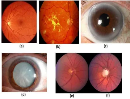
Figure 1: Various type of ocular diseases
C. Pathological Myopia (PM)
Myopia is a condition in which the eyes have vision problems resulting from retinal tissue degradation, the condition is known as pathological atrophy (PA). For this problem there are researchers [35] using an automated system to detect PA with pathological myopia detection system through peripapillary atrophy (PAMELA) in passing the analysis of gray level and texture. Authors [36] had used a minimum redundancy maximum relevancy (mRMR) as a classifier and were tested on 800 subjects. While [37] Yanwu et al., proposes a bag-of-feature and sparse learning in their automatic system for detecting PM.
D. Arcus Senilis (AS)
AS is a situation where the loop like white ring on the cornea at the limbus approximately 0.3-1mm thick [16], [38] as shown in Figure 1(c). This phenomenon is caused by abnormal lipids in human blood (hyperlipidaemia). There are research studies that support the opinion that these situations concerned with CAD, especially those aged between 30 and 60 years [39]. Among the researchers who conducted the study researchers on this situation as by [3], [4], [39], [15], [18], [19], [40]–[42] who do medical research and surveys. There are also researchers who propose an automatic method for detecting the presence of such US [43] using RBF classifier, while [63] had used Artificial Neural Network (ANN), Fuzzy classifier and Neuro-Fuzzy classifier for this purpose. Researchers [64] using iris image processing such as segmentation and normalization for the purpose of separation of parts of ROI and using Otsu method to identify the presence of the US.
IV. DISEASES MANIFESTATIONS BY OCULAR SIGNS
The eye is a precious in human body, to enable people to see. In order to ensure this eye in good condition should make regular eye examinations. Eye examination is also said to be beneficial for the early detection of health problems such as rheumatoid arthritis [44]. In this section discusses the relevant diseases manifestations by ocular signs and identify the diseases using CAD method.
A. Diabetes
accuracy of 90%. While [50] using SVM and 2-D wavelet tree for classification of diabetes, they also adopt the principle of iridology in their study.
B. Kidney
For kidney disease we have found a study done by [23], in which they use the method of wavelet analysis and neural network (NN) to classify subjects with kidney disease (168) and normal (172) subjects. The results of their studies do indicate, for normal subjects classification accuracy was 93 %, while subjects with kidney problems 82 %. Murli et al. [51], in their study suggest that ophthalmologists during screening patients undergoing treatment for eye should see if there is any abnormal condition of the eye patients because they think kidney cancer can spread to the eye.
C. Gastrointestinal or colon problem
For gastrointestinal problems and colon problems, investigations carried out in connection with ocular diagnosis by means of CAD, is like a study by Yuan et al. [26], where they perform extraction and analysis of iris texture to see the connection with intestinal and gastrointestinal problems. While [52] studied the characteristics of changes in the geometry of the iris associated with gastrointestinal problems based on the views of iridology . They use the PCA for classification of iris image for these gastrointestinal problems.
D.Pelvis and vagina
There are studies that discuss the problem of pelvic and vagina disease based on correlation with eyes as suggested by Noraini et al. [24]. Where they use the PCM and SVM for classification purposes to the problem pelvis and vaginal disease. Based on their results they have obtained the correct classification for the vagina 80% and correct classification for the pelvis is 70 %.
E. Lung
Base on ocular manifestation signs, to identify lung disease problems using CAD has been discussed by [53], where they have been reviewed regarding autonomic nervous wreath (ANW) based methods available on iridology chart for this they have used methods such as Hough transform image processing, segmentation and normalization for study the iris of the eye. While the authors [54], discuss clinical cases, 72-year-old female patient with cancer of lung squamous cell where he was found to have pain and blurred vision in his left eye, also found a number of malignant cells in the eye. Researchers [25] have developed a smart device for the purpose of health care services. For the purposes of image processing, they use canny edge detection and ANN are utilized for the purpose of classification and analysis for the detection of disease characteristics. Sivasankae et al. [22] have been proposed to identify disease-related Pulmonary they use CHT processing technique, Fuzzy C-Means clustering and gray level analysis. Where the results of the techniques they use are; 75%, sub-acute 80%, chronic 85.71% and degenerative 80%.
F. Leukaemia
Pavithran et al. [21] reported that ocular manifestations are common in patients with acute leukaemia , they have reported a case of a 65-year -old man who suffered a loss of vision in both eyes. These patients were also found to suffer from vitreous hemorrhage, glaucoma and hyphaema in the right eye. In another experimental Mateo et al. [55] have studied the relevant association with ocular manifestations of acute lymphoblastic leukaemia. They discussed the ocular manifestations that occur on the eyelids, conjunctiva, cornea, anterior chamber, retina, choroid and optic nerve. According to the authors in [21] and [55], ocular manifestations can be used as an indicator to identify the disease.
CONCLUSION
This review presents a detailed study conducted methods for detecting diseases associated with ocular diseases and diseases that can be detected through the signs contained in ocular automatically using computer aided known as CAD. The use of CAD is very useful and benefits, where it can reduce the workload of tasks to doctor or ophthalmologist in making health screening and examination of the patient. Tasks such as grading, classify and make interpretation of potential problems associated with ocular problems sometime very challenging. Where to diagnose the eye there are several steps that need to be done such as, identify eye’s anatomy, segmentation, localization and normalization on ROI. There are also processes such as extraction, classification and grading using a variety of methods and algorithms. To perform an analysis of disease associated with ocular problems most researchers using OCT images, slit lamp, RetCam, and retinal fundus. While for diseases identified by signs found on the ocular, most researchers use iris image in their analysis
to ensure that the usage of the eye as one of the parameters used in screening for health problems have firm foundation of medical terms.
ACKNOWLEDGEMENT
The authors would like to thank the staffs UPM who contribute in terms of ideas and materials to the resulting paper.
REFERENCES
[1] J. Daugman, “Iris Recognition - The colored part of the eye contains delicate patterns that vary randomly from person to person,
offering a powerful means of identification,” Am. Sci., vol. 89, no. 4, p. 326, 2001.
[2] B. J. Daugman, “Probing the Uniqueness and Randomness of IrisCodes : Results From 200 Billion Iris Pair Comparisons,” Proc.
IEEE, vol. 94, no. 11, 2006.
[3] N. Hickey, B. Maurer, and R. Mulcahy, “Arcus senilis: its relation to certain attributes and risk factors in patients with coronary heart disease.,” Br. Heart J., vol. 32, no. 4, pp. 449–52, Jul. 1970.
[4] M. Ang, W. Wong, J. Park, R. Wu, R. Lavanya, Y. Zheng, H. Cajucom-Uy, E. S. Tai, and T. Y. Wong, “Corneal arcus is a sign of
cardiovascular disease, even in low-risk persons.,” Am. J. Ophthalmol., vol. 152, no. 5, pp. 864–71.e1, Nov. 2011.
[5] A. D. Wibawa and M. H. Purnomo, “Early Detection on the Condition of Pancreas Organ as the Cause of Diabetes Mellitus by Real
Time Iris Image Processing,” APCCAS 2006 - 2006 IEEE Asia Pacific Conf. Circuits Syst., pp. 1008–1010, Dec. 2006.
[6] L. S. Chambless LE, Fuchs FD, “The Association of corneal arcus with coronary heart disease and cardiovascular disease mortality in the lipid research clinics mortality follow-up study,” Am. J. Public Health, vol. 80, no. 10, p. 1200, 1990.
[7] M. R. Lee, J. a. Izatt, E. a. Swanson, D. Huang, J. S. Schumun, C. P. Lin, C. a. Puliafito, and J. G. Fujimoto, “Optical coherence tomography for ophthalmic imaging: new technique delivers micron-scale resolution,” IEEE Eng. Med. Biol. Mag., vol. 14, 1995.
[8] R. Richa, R. Linhares, E. Comunello, A. Von Wangenheim, J. Schnitzler, B. Wassmer, C. Guillemot, G. Thuret, P. Gain, G. Hager,
and R. Taylor, “Fundus Image Mosaicking for Information Augmentation in Computer-Assisted Slit-Lamp Imaging,” vol. 33, no. 6, pp. 1304–1312, 2014.
[9] T. Aung, M. Baskaran, P. Shamira, and T. Y. Wong, “Closed Angle Glaucoma Detection in RetCam Images,” vol. 138632, pp. 4096–
4099, 2010.
[10] Y. Zheng, C. Y. Cheung, T. Y. Wong, W. Wong, S. C. Loon, and T. Aung, “Determinants of image quality of heidelberg retina
tomography II and its association with optic disc parameters in a population-based setting,” Am. J. Ophthalmol., vol. 151, no. 4, pp. 663–670, 2011.
[11] T. T. Norton and J. T. Siegwart, “Light levels, refractive development, and myopia - A speculative review,” Exp. Eye Res., vol. 114, pp. 48–57, 2013.
[12] L. Guo, J.-J. Yang, L. Peng, J. Li, and Q. Liang, “A computer-aided healthcare system for cataract classification and grading based on fundus image analysis,” Comput. Ind., 2014.
[13] U. R. Acharya, E. Y. K. Ng, L. W. J. Eugene, K. P. Noronha, L. C. Min, K. P. Nayak, and S. V. Bhandary, “Decision support system for the glaucoma using Gabor transformation,” Biomed. Signal Process. Control, vol. 15, pp. 18–26, 2015.
[14] M. R. K. Mookiah, U. R. Acharya, C. K. Chua, C. M. Lim, E. Y. K. Ng, and A. Laude, “Computer-aided diagnosis of diabetic
retinopathy: A review,” Comput. Biol. Med., vol. 43, pp. 2136–2155, 2013.
[15] H. Z. Pomerantz, “The relationship between coronary heart disease and the presence of certain physical characteristics.,” Can. Med. Assoc. J., vol. 86, pp. 57–60, Jan. 1962.
[16] F. L. Urbano, “Ocular Signs of Hyperlipidemia,” Hosp. Physician, no. November, pp. 51–54, 2001.
[17] S. Crispin, “Ocular lipid deposition and hyperlipoproteinaemia.,” Prog. Retin. Eye Res., vol. 21, no. 2, pp. 169–224, Mar. 2002.
[18] G. E. Navoyan, “A Case-Control Study of Corneal Arcus and Coronary Heart Disease in Yerevan,” Prevention, 2003.
[19] A. Fernández, A. Sorokin, and P. D. Thompson, “Corneal arcus as coronary artery disease risk factor.,” Atherosclerosis, vol. 193, no. 2, pp. 235–40, Aug. 2007.
[20] Moosavi, A. Sareshtedar, and S. Zarei-ghanavati, “Risk Factors for Senile Corneal Arcus in Patients with Acute Myocardial
Infarction,” vol. 5, no. 4, pp. 228–231, 2010.
[21] M. T. K Pavithran, BV Ajithkumar, R Aruna, “Bilateral Retinal Detachment in Acute myeloid leukaemia,” Asian J. Ophthalmol., vol. 5, no. 3, pp. 13–14, Jul. 2003.
[22] K. Sivasankar, M. Sujaritha, P. Pasupathi, and S. Muthukumar, “FCM based iris image analysis for tissue imbalance stage
identification,” 2012 Int. Conf. Emerg. Trends Sci. Eng. Technol., pp. 210–215, Dec. 2012.
[23] S. E. Hussein, O. a. Hassan, and M. H. Granat, “Assessment of the potential iridology for diagnosing kidney disease using wavelet analysis and neural networks,” Biomed. Signal Process. Control, vol. 8, no. 6, pp. 534–541, Nov. 2013.
[24] A. S. Nor’aini A.J., Rohilah S, “Classification of Iris Regions using Principal Component Analysis and Support Vector Machine,”
IEEE, pp. 134–139, 2013.
[25] D. H. Hareva, S. Lukas, and N. O. Suharta, “The smart device for healthcare service: Iris diagnosis application,” 2013 Elev. Int. Conf. ICT Knowl. Eng., pp. 1–6, Nov. 2013.
[26] W. Yuan and J. Huang, “Extraction and Analysis of Texture Information of the Iris Intestinal Loop,” pp. 328–338, 2014. [27] T. Wong and P. Mitchell, “The eye in hypertension,” Lancet, vol. 369, pp. 425–435, 2007.
[28] A. D. Fleming, S. Philip, K. a. Goatman, J. a. Olson, and P. F. Sharp, “Automated microaneurysm detection using local contrast
normalization and local vessel detection,” IEEE Trans. Med. Imaging, vol. 25, no. 9, pp. 1223–1232, 2006.
[29] I. Lazar and A. Hajdu, “Retinal microaneurysm detection through local rotating cross-section profile analysis,” IEEE Trans. Med. Imaging, vol. 32, no. 2, pp. 400–407, 2013.
[30] Z. Zhang, R. Srivastava, H. Liu, X. Chen, L. Duan, D. Wing, and K. Wong, “Open Access A survey on computer aided diagnosis for
ocular diseases,” pp. 1–29, 2014.
[31] H. Li, J. H. Lim, J. Liu, and T. Y. Wong, “Towards automatic grading of nuclear cataract,” Annu. Int. Conf. IEEE Eng. Med. Biol. - Proc., pp. 4961–4964, 2007.
[32] H. S. H. Shen, H. H. H. Hao, L. W. L. Wei, and Z. W. Z. Wang, “An Image Based Classification Method for Cataract,” 2008 Int. Symp. Comput. Sci. Comput. Technol., vol. 1, pp. 583–586, 2008.
[33] X. Gao, H. Li, J. H. Lim, and T. Y. Wong, “Computer-aided cataract detection using enhanced texture features on retro-illumination lens images,” Proc. - Int. Conf. Image Process. ICIP, pp. 1565–1568, 2011.
[35] B. Lee, D. W. K. Wong, N. M. Tan, Z. Zhang, J. H. Lim, H. Li, F. Yin, J. Liu, W. Huang, S. M. Saw, L. Tong, and T. Y. Wong, “Fusion of pixel and texture features to detect pathological myopia,” Proc. 2010 5th IEEE Conf. Ind. Electron. Appl. ICIEA 2010, pp. 2039–2042, 2010.
[36] Z. Zhang, J. Cheng, J. Liu, Y. C. M. Sheri, C. C. Kong, and S. S. Mei, “Pathological myopia detection from selective fundus image features,” Proc. 2012 7th IEEE Conf. Ind. Electron. Appl. ICIEA 2012, pp. 1742–1745, 2012.
[37] Y. Xu, J. Liu, Z. Zhang, N. M. Tan, D. W. K. Wong, S. M. Saw, and T. Y. Wong, “Learn to recognize pathological myopia in fundus
images using bag-of-feature and sparse learning approach,” Proc. - Int. Symp. Biomed. Imaging, pp. 888–891, 2013. [38] H. Lindholm, “Arcus Lipoides Corneae and Arteriosclerosis,” Acta Med. Scand., 1960.
[39] H.-T. Chen, H.-C. J. Chen, C.-H. Hsiao, D. H.-K. Ma, Y.-T. Chen, and K.-K. Lin, “Corneal arcus and cardiovascular risk factors in middle-aged subjects in Taiwan.,” Am. J. Med. Sci., vol. 338, no. 5, pp. 268–272, 2009.
[40] N. T. Cooke, “Significance of arcus senilis in Caucasians.,” J. R. Soc. Med., vol. 74, no. 3, pp. 201–4, Mar. 1981.
[41] I. Bersohn, W. M. Politzer, and D. Blumsohn, “Arcus senilis corneae--its relationship to serum lipids in the South African Bantu.,” S. Afr. Med. J., vol. 43, no. 33, pp. 1025–7, Aug. 1969.
[42] H. S. Halfon ST, Hames CG, “Corneal arcus and coronary heart disease mortality,” Br J Ophtahlmol, vol. 1, no. X, pp. 603–604, 1984. [43] R. Acharya U, L. Y. Wong, E. Y. K. Ng, and J. S. Suri, “Automatic identification of anterior segment eye abnormality,” Itbm-Rbm,
vol. 28, pp. 35–41, 2007.
[44] S. C. Reddy and U. R. Rao, “Ocular complications of adult rheumatoid arthritis.,” Rheumatol. Int., vol. 16, no. 2, pp. 49–52, Jan. 1996.
[45] G. Zahlmann, B. Kochner, I. Ugi, D. Schuhmann, B. Liesenfeld, A. Wegner, M. Obermaier, and M. Mertz, “Hybrid fuzzy image
processing for situation assessment: A Knowledge-Based system for Early Detection of Diabetic Retinopathy,” IEEE Eng. Med. Biol. Mag., vol. 19, pp. 76–83, 2000.
[46] S. Ganguly and S. Ganguly, “An Adaptive Threshold Based Algorithm for Detection of Red Lesions of Diabetic Retinopathy in a
Fundus Image,” pp. 91–94, 2014.
[47] P. P. Conde, J. De La Calleja, A. Benitez, and M. A. Medina, “Image-based classification of diabetic retinopathy using machine
learning,” Int. Conf. Intell. Syst. Des. Appl. ISDA, pp. 826–830, 2012.
[48] I. P. D. Lesmana, I. K. E. Purnama, and M. H. Purnomo, “Abnormal condition detection of pancreatic beta-cells as the cause of
diabetes mellitus based on iris image,” Proc. - Int. Conf. Instrumentation, Commun. Inf. Technol. Biomed. Eng. 2011, ICICI-BME 2011, no. November, pp. 150–155, 2011.
[49] U. M. Chaskar and M. S. Sutaone, “On a methodology for detecting diabetic presence from iris image analysis,” 2012 Int. Conf.
Power, Signals, Control. Comput., pp. 1–6, Jan. 2012.
[50] A. Bansal, R. Agarwal, and R. K. Sharma, “Determining diabetes using iris recognition system,” Int. J. Diabetes Dev. Ctries., p. 13410, 2015.
[51] M. Kurli and P. T. Finger, “The kidney, cancer, and the eye: Current concepts,” Surv. Ophthalmol., vol. 50, no. 6, pp. 507–518, 2005.
[52] L. Ma, D. Zhang, N. Li, Y. Cai, W. Zuo, K. Wang, and S. Member, “Iris-Based Medical Analysis by Geometric Deformation
Features,” vol. 17, no. 1, pp. 223–231, 2013.
[53] R. Passarella and M. Fachrurrozi, “Development of Iridology System Database for Colon Disorders Identification using Image
Processing,” vol. 2, no. June, pp. 100–103, 2013.
[54] A. Hiraki, H. Ueoka, T. Matsuo, T. Nakagawa, T. Yoshino, K. Kiura, M. Tabata, K. Sakae, Y. Ohtsuki, Y. Hiraki, and M. Harada,
“Metastasis to the iris in squamous cell lung cancer,” Int. J. Clin. Oncol., vol. 3, no. 3, pp. 186–190, Jun. 1998.
[55] J. Mateo, F. J. Ascaso, E. Núñez, C. Peiro, G. González, and J. a Cristóbal, “Ophthalmological Manifestations in Acute Lymphoblastic Leukemia,” 2010.
AUTHORPROFILE
Ridza Azri Ramlee is a Senior lecturer at the Department Electronic and Computer engineering in University Technical Malaysia Melaka (UTeM). Active in Institute of Engineer Malaysia (IEM) as cooperate member and received the status of Professional Engineer (P.Eng) from Board of Engineering Malaysia (BEM). Currently he is pursuing the PhD at University Putra Malaysia. His research interests lie in the area of image processing, industrial electronic instrumentation, security and smart house. He had achieved Master (2008) in Telecommunication and Information Engineering from University Technology MARA (UiTM); B.Eng (2000) in Electrical Engineering from University Technology MARA (UiTM), Malaysia.
Abdul Rahman Ramli is an Assistant Professor at the Department of Computer and Information Sciences in Fordham University. His research interests lie in the area of image processing and electronic imaging, multimedia system engineering, Internet computing, smart card, applications, embedded system, computer telephony integration, instrumentation, remote sensing, remote monitoring system and intelligence systems. Dr. Abdul Rahman received Ph.D., Image Processing, University of Bradford, U.K, 1995; M.SC (Eng), Information Technology System, University of Strathclyde, U.K., 1985; B.Sc., Electronics, Universiti Kebangsaan Malaysia, Bangi, 1982.
![Table 1 Types of diseases which can be diagnosed through eye. Authors Iris Diagnose /Ocular sign Method Type / condition diseases Finding/Conclusion Pomerantz [15] AS Statistical analysis](https://thumb-eu.123doks.com/thumbv2/123dok_br/16441289.196793/2.892.127.766.127.859/diseases-diagnosed-diagnose-condition-diseases-conclusion-pomerantz-statistical.webp)