http://dx.doi.org/10.1590/s2175-97902017000216127
A
r
*Correspondence: M. M. Mehanna. Industrial Pharmacy Department. Faculty of Pharmacy. Alexandria University, Egypt. E-mail: [email protected], [email protected]
Liposomes as potential carriers for ketorolac ophthalmic delivery:
formulation and stability issues
Mohammed Maher Mehanna
1,2*, Nouran Abd El-Kader
3, Magda Wadih Samaha
11Industrial Pharmacy Department, Faculty of Pharmacy, Alexandria University, Alexandria, Egypt, 2Pharmaceutical Technology Department, Faculty of Pharmacy, Beirut Arab University, Beirut, Lebanon, 3R & D Pharco B international
Company, Borg El Arab City, Alexandria, Egypt
Drug delivery to treat ocular disorders locally is a challenging endeavor. Traditional ocular dosage form - eye drops - exhibits poor availability, consequently ineicient therapeutic response. The objective of the study was to formulate and characterize a ketorolac tromethamine ocular system with a prolonged release pattern based on liposomes as a vesicular carrier and to design once daily liquid preparation realizing the thermal in situ gelation principle. Liposomes were prepared by ilm hydration method. The inluence of cholesterol concentration, pH and volume of hydration medium, and type and concentration of charging imparting agents were studied. Liposomes were characterized via, morphological examination, vesicular size, and encapsulation eiciency, and in vitro release performance, moreover its stability was assessed. The results obtained highlighted that liposomes showed a closed vesicular multi-lamellar structure. Ketorolac was successfully encapsulated within the liposomal structure in a cholesterol and charge inducing agent concentration-dependent behaviour. The dispersion of liposomes within thermosensitive Poloxamer in situ gel was able to retard the release of the drug by difusion providing a controlled prolonged delivery. The liposomal formulations were physically stable for six months. Ketorolac tromethamine in situ liposomal gel representing an eicient alternative in terms of ocular retention and patient compliance when compared with conventional eye drops.
Uniterms: Ketorolac/characterization/formulation. Liposomes/stability study. Ocular delivery. Thermosensitive gel. Prolonged release.
INTRODUCTION
Ketorolac tromethamine, Figure 1, is one of the arylacetic acid derivatives group of non-steroidal anti-inflammatory drugs which are potent cyclooxygenase
inhibitors (Ahuja et al., 2008). It has been used in the management of several ocular disorders. Ketorolac is FDA approved treatment of ocular pain following corneal refractive surgery and surface ablation and other
ocular conditions such as seasonal allergic conjunctivitis and postoperative inlammation (Robinson, Lee, 2011).
Ketorolac, free acid, is sparingly soluble in water and, therefore, it is marketed in the form of tromethamine salt, which has a higher aqueous solubility (Thakur, Kashiv, 2011). Ketorolac tromethamine is commercially available
as eye drops. Instillation of 0.5% ketorolac tromethamine aqueous solution was associated with ocular irritation, evoked as burning and stinging (Sandoval et al., 2006).
The main problem encounter eye drops as a local ocular drug delivery is its poor bioavailability due to short residence time on the eye surface as a result of several
factors among them, preparation overlow from the eye surface specially upon blinking, dilution by tears, relex
the ocular surface such as ketorolac oily solution and ophthalmic ointment preparations had been studied and
evaluated (Malhotra, Majumdar, 2006). Gelling systems
were utilized for the same purpose as ion activated in situ gel (Vodithala et al., 2010),hydrogel system based on chitosan and carbopol 940 (Zaki et al., 2011), methylcellulose and hydroxypropyl methylcellulose in
presence of sodium bicarbonate (Nanjawade, Manvi, Manjappa, 2007). Recently, Thakor et al. (2012), formulated ketorolac tromethamine in various in-situ gelling systems, which significantly improved ocular bioavailability as compared to conventional eye drops as proved through in vitro and in vivo studies through its longer precorneal residence time and ability to sustain drug release.
Phospholipid-based vesicles, i.e. liposomes, are biocompatible and biodegradable carrier system with almost no cytotoxicity. Moreover, it has the capability of enhancing drug solubility, stability and reduce drug adverse reactions (Schubert, 2002). From ophthalmic formulation point of view, liposomes are able to
intimately contact the corneal and conjunctival surfaces and modify the tears dynamics which positively relect
on its ability to prolong the drug ocular availability and thus reducing the frequency of administration, the total drug applied and both the systemic and topical side
efects which augment patient compliance to the drug
regimen. On top of these merits, liposomal preparations are still a liquid formulation that are easily installed with no discomfort and can localize and maintain drug activity at the ocular surface for a longer period, thus
allowing a controlled drug delivery (Van Der Bijl, Van
Eyk, Meyer, 2001). Several research clusters investigated
the utilization of liposomes as ocular system exempliied
by the study of Mehanna, Elmaradny and Samaha (2009) who formulated ciprofloxacin loaded-liposomes using the reverse phase evaporation technique and studied
ive diferent factors afecting the entrapment eiciency;
the results revealed that the most important factor was
the molar concentration of cholesterol, Habi, Fouad, Fathalla (2008) prepared 2 mg/mL luconazole
loaded-liposomes using reverse phases evaporation technique for the treatment of Candida keratitis in rabbits eyes and found that the liposomal formulation showed a better healing percentage over the same period of time. In 2014, Tahaa et al. (2014)studied several liposomal formulations
containing ciproloxacin and examined the inluence of diferent types of phospholipids, cholesterol incorporation,
incorporation of positively charge inducing agent, and ultrasonication on the liposomes properties and concluded that liposomal formulations showed more than three folds
of improvement in drug ocular bioavailability compared with the commercial product.
The goal of the present study is to prepare, characterize and optimize an ocular liposomal formulation
of ketorolac tromethamine as a potent anti-inlammatory
drug for treatment of ocular conditions with a prolonged release. Furthermore, formulate the prepared liposomes in a thermo-sensitive gel. The physical stability of the optimized liposomal formulation along with the liposomal
gel is evaluated upon storage at diferent temperatures.
MATERIAL AND METHODS
Material
Ketorolac tromethamine (assay<99.0%), Dicetyl phosphate, Stearylamine and Pluronic F 127(PF-127) were purchased from Sigma Aldrich (Steinheim, Switzerland). Lipoid E80, phosphatidylcholine from egg was received
as a gift from Lipoid Company (Ludwigshafen, Germany).
Cholesterol was purchased from Winlab (Leicestershire, UK). Spectrapore® 2 dialysis membrane form Spectrum
laboratories Inc. (Houston, TX, USA). All other solvents
and materials used were of analytical grade.
Preparation of Ketorolac tromethamine-loaded liposomes
Liposomes were prepared using film hydration technique, which involves dissolving the organic soluble moieties (phospholipid, cholesterol and charge inducing agent) in an organic solvent, then evaporating this solvent by means of vacuum and gentle heat, leaving an even film on the walls of the round-bottom flask.
The second major step is the ‘hydration’ that is carried
out with an aqueous phase composed of water, drug and other water-soluble ingredients. This hydration causes
immediate globulization of the phospholipid ilm where
the lipid layer tends to form liposomes incorporating the aqueous phase either inside the bilayer of the liposomal
wall and/or in the core of the lipid vesicle. The inal step
is the refrigeration of the formed liposomes to ensure intact vesicle formation.
Speciically, 100 mg phosphatidyl choline dissolved
in chloroform, along with cholesterol, and other organic
soluble ingredients. The lask is held in rotavapor under
approximately -900 mbar, 50 °C, rotating at 45 rpm
(Rotavapor, type V8000, Buchi, Switzerland) till the
solvent is completely evaporated and an even film is
formed. Hydration is achieved by addition of the aqueous
under normal pressure at 50 °C, rotating at 45 rpm, for
20 minutes till the ilm is completely hydrated and the
liposomes are formed, then stored in refrigerator for 24
hours (Ghanbarzadeh, Valizadeh, Zakeri-Milani, 2013)a 3(2.
Separation of unentraped free ketorolac tromethamine
Free ketorolac tromethamineseparation from liposomal dispersion was performed using cooling ultracentrifuge (Sigma laboratory refrigerated centrifuge,
3K-30, Germany) rotating at 17000 rpm, at a temperature
of 1°C, for 90 minutes. The supernatant representing the unentraped ketorolac tromethamine was decanted then transferred to a volumetric flask suitably diluted and drug content was determined spectrophotometrically (Shimadzu UV 1601 PC spectrophotometer, Kyoto, Japan). Liposomes was redispersed with 1 mL distilled water and stored for further characterization (Mehanna, Elmaradny, Samaha,2010).
Optimization of ketorolac liposomal entrapment efficiency
In order to develop an optimized liposomal
formulation with the highest entrapment eiciency, many variables have been explored namely; the molar ratio of cholesterol to that of the phospholipid, various pH
value of hydration medium selected based on the pka of the drug, varying the aqueous hydration phase volume,
incorporation of charge inducing agent and inally inding
out the concentration of the charge imparting agent. These experiments were assessed by comparing the entrapment
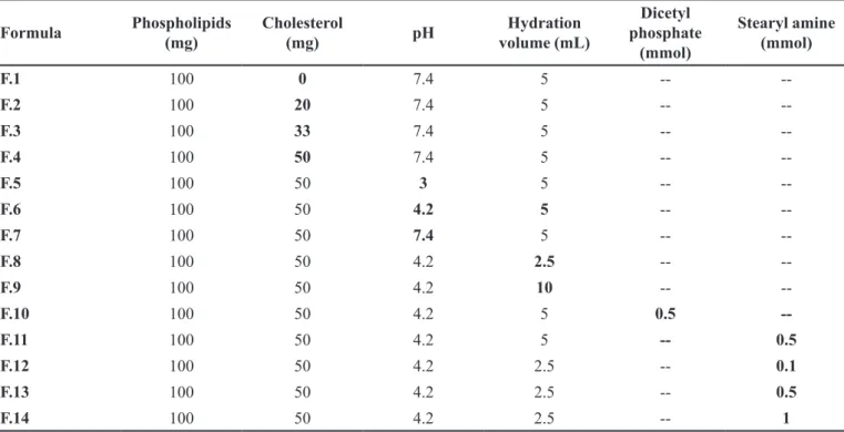
eiciency percentage. Table I illustrates the composition of diferent liposomes dispersions.
Physicochemical characterization of ketorolac-loaded liposomes
Determination of entrapment efficiency (EE%)
Entrapment efficiency was calculated from the difference between the initial drug added during preparation and the free drug determined in the supernatant
and expressed as percent entrapment eiciency, which
is defined as the percent fraction of the total input drug encapsulated in the lipid bilayers and/or aqueous compartments in the liposome structure (Mehanna, Elmaradny, Samaha, 2009). Entrapment efficiency percentage was computed from the following equation:
Morphological examination of KT-loaded liposomes Ketorolac tromethamine-loaded liposomes were morphologically inspected using optical microscope
(Euromed, Holland) in order to justify their formation and
identify their shapes. Liposome dispersions were observed
under a binocular microscope itted with a camera. A drop
of liposomes dispersion was placed on a microscope slide
(magniication power 2000), then viewed and photographed.
Transmission electron microscope examination (TEM) Ketorolac-loaded liposomal dispersion was
examined by TEM (JEM-100S; Joel, Tokyo, Japan);
negatively stained sample was prepared by applying a drop of liposomal dispersion to copper-coated grids, after 2
min, the excess was drawn of with ilter paper. A saturated
uranyl acetate aqueous solution was used as a staining agent. The excess was eliminated with distilled water and the samples were analyzed by transmission electron
microscope at magniication power 10,000 at 80 KV.
Vesicular size analysis
L i p o s o m e s s i z e w a s a s s e s s e d u s i n g l a s e r
diffractometer (CilasL100, model 1064 liquid;
Quantachrom, France), Determining the mean vesicle size and size distribution (polydispersity) by calculating the span index, assures the uniformity of the liposomal preparation, calculated using the following equation:
Span index = Dv,90 – Dv,10 / Dv,50
where D(v,10), D(v,50) and D(v,90) are the equivalent volume diameters at 10, 50 and 90% cumulative volumes, respectively (Mehanna, Elmaradny, Samaha, 2014).
In-vitro release studies
The release of ketorolac tromethamine from the different liposomal dispersions was determined using dialysis apparatus method (Ma et al., 2008). Fixed volume of each formula in a 5 cm semipermeable dialysis membrane (Molecular weight cutoff 12,000- 14,000 Da) tied from both sides and clamped to the paddle of
the dissolution instrument (DT820, Erweka, Germany)
rotating at 50 rpm, temperature kept at 34.5±1oC (simulating eye temperature), immersed in 250 ml phosphate buffer saline (PBS) as a releasing medium,
samples were withdrawn at diferent time intervals and
Release kinetics of Ketorolac from the prepared
liposomal dispersions, were examined based on the magnitude of correlation coefficients obtained after
application of zero order, irst order, and Higuchi difusion, Peppeas-Korsmeyer and Hixon Crowell models (Dash et al., 2010).
Preparation of thermosensitive liposomal gel
Pluronic F127 polymer undergoes sol-to-gel phase transition upon exposure to physiological eye temperature – was practically found to have a gelling concentration of 22%, was employed as a vehicle for liposomes. The gel preparation was performed according the cold method
(Mehanna, Elmaradny, Samaha, 2013); where PF-127 is
accurately weighed and slowly added to cold water (5 °C) with constant stirring, then refrigerated for 5 hours for complete polymer hydration and get rid of air bubbles which resulted from stirring. Liposomal PF-127 gel formula was prepared through dispersing KT-loaded liposomes in the PF-127 solutions under magnetic stirring in ice.
Physical stability of liposomes
The physical stability of KT-loaded liposomes was assessed via monitoring the liposomes size and secondly, by chemical quantitation of entrapment efficiency
percentage through diferent time intervals for six months
at room temperature 25 °C and in refrigerator at 4 °C. In addition, stability of liposomes incorporated in thermo-sensitive gel was assessed through the same time intervals
by chemical quantitation of entrapment eiciency percent.
RESULTS AND DISCUSSION
Optimization of ketorolac loaded-liposomal entrapment efficiency
Ketorolac tromethamine encapsulated within liposomes with a simple film hydration method was performed aiming to prolong the retention time of the drug within the eye hence enhancing its therapeutic
anti-inlammatory efect. In order to optimize the liposomes
capacity to entrapment KT, several variables were
studied as shown in table I. The factors investigated were;
inclusion of cholesterol and its molar concentration,
hydration at three diferent pH values, namely; pH 3, 4.2,
and 7.4, hydration volume, and the incorporation of charge inducing agents, dicetyl phosphate as a negative charge inducing agent and stearylamine as a positive charge
inducing agent. The efect of varying the concentration
of the charge-inducing agent was also addressed. The entrapment efficiency percentage was selected as the determining governing response.
TABLE I - Composition of ketorolac tromethamine –loaded liposomes dispersions*
Formula Phospholipids
(mg)
Cholesterol
(mg) pH
Hydration volume (mL)
Dicetyl phosphate
(mmol)
Stearyl amine (mmol)
F.1 100 0 7.4 5 --
--F.2 100 20 7.4 5 --
--F.3 100 33 7.4 5 --
--F.4 100 50 7.4 5 --
--F.5 100 50 3 5 --
--F.6 100 50 4.2 5 --
--F.7 100 50 7.4 5 --
--F.8 100 50 4.2 2.5 --
--F.9 100 50 4.2 10 --
--F.10 100 50 4.2 5 0.5
--F.11 100 50 4.2 5 -- 0.5
F.12 100 50 4.2 2.5 -- 0.1
F.13 100 50 4.2 2.5 -- 0.5
F.14 100 50 4.2 2.5 -- 1
Effect of molar cholesterol concentration
Ketorolac tomethamine loading level in liposomes prepared by film hydration method was expressed as
entrapment eiciency percentage (EE%), which was varied
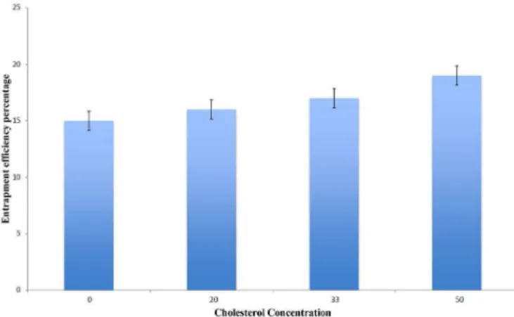
between 15 to 19%, based on the molar concentration of cholesterol included within the liposomes (F1-F4). This
low entrapment eiciency can be explained as a result of drug high water solubility (Hou et al., 1990).
The positive correlation between the molar concentration of cholesterol and EE% could be infer from Figure 2. This could be attributed to the hardening of the liposomal walls due to increase cholesterol concentration. Cholesterol molecules augment the structure integrity of liposomes bilayers membrane as it fills the spaces between the phospholipid molecules, in turns it reduces the membrane permeability and drug leakage, thus enhancing drug loading which was in accordance to many previously reported data ( Mehanna, Elmaradny, Samaha,
2010; Hosny, 2010; Alsarra et al., 2005). Correspondingly, liposomes contain 50% molar cholesterol percentage was selected for further optimization.
Effect of hydration phase pH and volume
In order to study the inluence of diferent pH values
of the aqueous phase containing KT, Formulas 5, 6 and 7
corresponding to pH 3, 4.2 and 7.4 were tested. Ketorolac tromethamine precipitated in pH 3 buffer during the
preparation F5 and thus it was not possible to use it as a hydrating solution during the early preparation stages. This precipitation can be seen as a result of poor drug solubility
in pH 3, which is below its pKa (3.5) that stopped the experiment early. Although the pH 7.4 is similar to the ocular physiological pH minimizing irritation and discomfort efect, still liposomes hydrated with ketorolac tromethamine in buffer pH 4.2 gave a higher EE% i.e.
57%. This pH is the same for the marked FDA approved
product. Similar results were observed by Mehanna, Elmaradny and Samaha (2009). Moreover, Malhotra and
Majumdar (2002)found that ketorolac tromethamine is
more permeable in goats’ corneas at acidic pH than pH 7.5.
Moreover, the volume of aqueous phase was varied 2.5-10 mL and its effect on ketorolac tromethamine entrapment was assessed, the highest EE% was that obtained with the smallest hydration volume, 2.5 mL, as shown in Figure 3. During the practical work, the least volume was observed to give a white dense colloidal liquid of high consistency, probably the reason for more
eicient entrapment. These results are supported by the
high water solubility of KT, which favor the aqueous medium leading to a reduction in EE% upon increasing the hydration volume.
Effect of incorporation and concentration of charge imparting agents
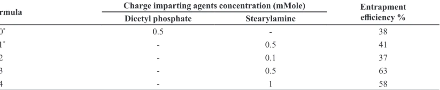
Upon comparing neutrally charged liposomal formula (F6) to negatively charge liposomal formula (F10), and to positively charge liposomal formula (F11) by incorporation of dicetyl phosphate and stearylamine, respectively, it was noted that the positively charged liposomes had the highest EE% compared to other liposomes. Meanwhile, the negatively charged and the
neutral liposomes showed lower entrapment eiciencies
(Table II). An explanation to this observation is KT is
negatively charged at pH 4.2 thus electrostatic attraction
to the positive charged stearyl amine preventing ketorolac tromethamine from leaking outside the liposome and enhance its EE% in addition to the electrostatic repulsion between the multiple bilayers of the liposomes inducing an elevation of the aqueous phase inclusion within the
liposomal bilayers and indirectly positevly inluence KT
entrapment. The higher EE% of the negatively charged FIGURE 2 - Entrapment efficiency of ketorolac–loaded
liposomes with various cholesterol molar concentration.
liposomes compared to neutral vesicles is based on solely on the repulsion between bilayers. Similar results were
obtained by Schaefer and Krohn (Schaefer, Krohn 1982). Whereas, Hosny (2010)has found that the entrapment
eiciency was best in positively charged liposomes, while
the neutral liposomes were better than the negatively charged liposomes this might be due to the zwitter ion
nature of ciproloxacin drug.
To compare the efect of varying the concentration
of positive charge imparting agent (stearylamine) on
KT liposomes entrapment eiciency, three formulations
were prepared and tested using 0.1, 0.5 and 1.0 mmole (F12, F13 and F14, respectively. Through table 2, it could be observed that increasing the molar concentration of stearylamine was linked to increase in EE% up to 0.5 mmole but further increase led to a reduction of KT entrapment. As higher stearylamine concentration may lead to disruption of liposomes structure and formation as it lacks the amphiphilic nature of phospholipids. These results are quite similar to Mehanna, Elmaradny and Samaha (2009)who reported that 0.38 molar ratio of
stearylamine was the optimal for ciproloxacin-loaded
liposomes preparation.
Morphological examination of ketorolac tromethamine-loaded liposomes
The microscopical examination using the optical
microscope -magnifying at 2000x- showed well-identiied
spherical vesicular structure with large internal space of the liposomes as illustrated in Figure 4, liposomes without cholesterol appeared as transparent bi-layer, whereas liposomes containing cholesterol showed a
fortiied bilayers, It is agreed by many researchers that
the incorporation of cholesterol increases the rigidity of the bilayer membranes (Tahaa et al., 2014; Wessman, Edwards, Mahlin, 2010). The transmission electron microscope magnifying at 10,000x showed mainly
multilamellar liposomes with some unilamellar vesicles as evident in Figure 4. Several authors reported comparable
indings (Sinico et al., 2005).
Particle size evaluation of ketorolac tromethamine-loaded liposomes
The average size and size distribution are important parameters as to determine the mechanism of uptake by the cornea, as well their stability condition of liposomes
(Gaudana et al., 2010). The particle size of nanoliposomes was investigated using a particle size analyzer (Zeta Sizer 2000, Malvern Instruments, Worcestershire, UK), as well as transmission electron microscope that elaborated liposomes size range between 196-8350 nm, varying according to the formula of liposomes. The span index TABLE II - Efect of incorporation and concentration of charge imparting agents on ketorolac tromethamine -loaded liposomes entrapment eiciency
Formula Charge imparting agents concentration (mMole) Entrapment
eiciency %
Dicetyl phosphate Stearylamine
F10* 0.5 - 38
F11* - 0.5 41
F12 - 0.1 37
F13 - 0.5 63
F14 - 1 58
*Formula prepared with 5ml aqueous hydration volume
FIGURE 4 - Optical photographs (A and B) and transmission electron microscopical photomicrographs (C and D) of
Ketorolac tromethamine-loaded liposomes prepared via ilm
indicates the uniformity of the liposomal preparation, the smaller the span index, and the narrower the size distribution.
As evident by the results illustrated in Table III, a dramatic increase in the particle size of the liposomes upon increasing the cholesterol concentration (F1-F4). Increasing the molar cholesterol concentration
relected on a signiicant increase in the overall size of
the liposomes. The amphiphilic nature of cholesterol controls its organization within the liposomal bilayer membrane structure as inserts itself into the bilayer with its hydrophilic head oriented towards the aqueous surface and aliphatic chain line up parallel to the hydrocarbon chains in the center of the bilayers which induced the observed
enlargement of liposomes (Xu, London, 2000). These
results were previously reported by Tahaa et al. (2014) who found that liposomes size increased linearly with
increasing the cholesterol concentration. Similar efect of cholesterol inclusion was reported for nevirapine (Ramana et al., 2010).
It is also obvious that charged liposomes (F10 & F11) had a larger particle size when compared to the neutral liposomes with the same initial components (F1-F4). This behaviour can be linked to the electrostatic repulsion between the charged molecules, which led to expand the spaces between the bilayers due to pushing the polar heads of the phospholipids outwards within
the multilamellar structure (Hosny, 2010; Ramana et al.,
2010). A more closed examination of the results
elaborated the inluence of the nature of the charging
inducing agent as the positively charged liposomes were of higher particle size than the negatively charged ones. This could be attributed to the bulkiness of dicetyl
phosphate (C32 H67 O4 P) molecule with its two cetyl
chains might have resulted in less tightly packed bilayer membranes compared to the less bulky stearylamine
(C8 H15 N O8), molecule. Another possible explanation
is the charge density that would amplify the electrostatic repulsion force between the dicetyl phosphate and cholesterol head groups having a similar charge (Samad, Sultana, Aqil, 2007). Therefore, the overall result showed that the positively charged liposomes (F11) were smaller than the negatively charged ones (F10) i.e. uncharged liposomes < positively charged < negatively charged liposomes.
Therefore, it could be deduced that increasing the concentration of cholesterol resulted in an increase in the liposomes size consequently increased the entrapment
eiciency. Thus, the particle size is directly proportional to the entrapment eiciency of liposomes.
In-vitro release studies
In order to develop a liposomal drug delivery system for localized and sustained ocular delivery, it
was necessary to check the release proile of diferent
dispersions in conditions simulating the ocular environment. Since human tears are known to be complex, approximately isotonic solution containing proteins, lipids, electrolytes and other components
with pH values in the range of 6.8-7.5 (Abelson, Udell,
Weston, 1980). The principle-bufering agent in the eye is bicarbonate system, which is unstable overtime when used for laboratory studies. The simulated tear solution
used was isotonic phosphate bufer (pH7.4), which was chosen for its pH stability, analytical non-interference and relationship to human tear (pH, buffering range, and bufering capacity). Furthermore it was desired to
keep the simulated tear solution simple so as to prevent complications from possible interactions and analytical
interference (Carney, Mauger, Hill, 1989).
The release proile of ketorolac from the optimized
liposomal preparation (F13) was illustrated in Figure
5. Upon comparing the release proile of the optimized
formula of ketorolac-loaded liposomes to the marketed
product, it was found that the release proile of
ketorolac-loaded liposomes showed the slowest release profile with initial burst release. This performance may be contributed to the desorption of the electrically attached drug molecules on liposomes outer surface followed by slower release rate of the encapsulated drug within the
multilamellar liposomal structure (Widjaja et al., 2014). With the intention of eliminating the burst release and controlling ketorolac release from liposomes. PF-127-based liposomal gel was prepared and evaluated. This strategy was proved to be able to control the drug release over 24 h as shown in Figure 5, which illustrates the cumulative percent of ketorolac released as a function TABLE III - Particle size and size distribution indicated by Span
Index of ketorolac tromethamine-loaded liposomes
Formula Particle size (nm) Span Index
F1 196 0.064
F2 3270 0.960
F3 7000 0.540
F4 7050 0.390
F10 8350 0.450
F11 7060 0.430
of time. The controlled release manner was mainly due to gel nature of PF-127 at 34.5±1 oC, which increase the
viscosity of the difusion layer surrounding the liposomes
representing another barrier for drug release. The explanation is further supported by the kinetic analysis, which indicated that ketorolac release mechanism was
a difusion-dependent process as the best it for kinetic models was Higuchi model based on the correlation coeicient. Comparable results were reported by several
authors (Nagarsenker, Londhe, Nadkarni, 1999; Jain, Shastri, 2011).
Stability of liposomal formulation
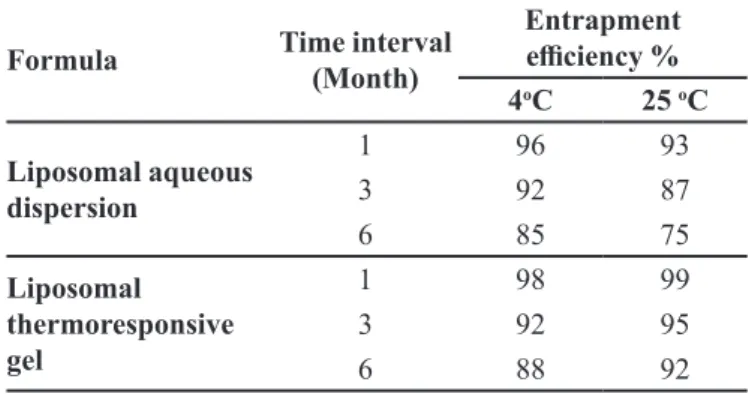
A stability study for six months was performed to assess the ability of the prepared liposomes to maintain its physicochemical characteristics. The optimized liposomal formula (F13), and the in-situ liposomal thermoresponsive gel were stored at 4 and 25 oC. The physical appearance of the liposomes formulations seemed unchanged, neither
sedimentation nor locculation was observed indicating the
high physical stability of the liposomal system in general. The results obtained are tabulated (Table IV) showing that liposomal formula (F13), stored at room temperature (25 oC) showed a faster decrease in EE% than that of refrigerated liposomes (4 oC) that indicate the leakage of the entrapped drug from the vesicular structure. The elevated temperature induced energy-dependent fusion of
liposomes via enhancing its Brownian motion in addition; the phospholipid became more lexible and luid through
increased temperature, thus the entrapped drug leaked easily. Comparable outcomes were obtained by Manosroi
and Podjanasoothon (2002)who studied the effect of temperature on different tranexamic acid liposomal formulations proving that the high temperature led to
the highest leakage of drug. Moreover, liposomal in-situ gelling system was able to maintain drug encapsulation at both temperatures compared to the liposomes dispersion. A more stable formulation was that stored at 25 oC, which might be due to the thermosensitive gel formation at elevated temperature. One can conclude that, liposomes in gel showed better retaining of the drug i.e. better EE% as a two barrier systems not allowing the drug to leak out of the liposomes.
CONCLUSION
In the current study, the potential of liposomes as ketorolac vesicular carrier for ocular delivery was explored. A controlled release ocular dosage form based on ketorolac tromethamine liposomes was successfully formulated, optimized and characterized. It has been shown
that liposomes prepared using ifty percent cholesterol, with low hydrating volume, at pH 4.2, and using a
positive charge-imparting agent at 0.5 mM concentration,
resulted in the highest entrapment eiciency which is the
main key for hydrophilic drug liposomal encapsulation. In addition, It was found that the mechanism of release
from the liposomes to the eye was difusion. Liposomes
dispersed in Pluronic F-127 in situ thermosensitive gel demonstrated a prolonged release over 24 h with more sustainability of the dose due to the dual barrier which the drug has to penetrate. The stability study of liposomes and liposomal gel at 4 and 25 °C for six months, showed that the liposomal gel at room temperature (25 °C) had the best stability. Ketorolac tromethamine-loaded liposomal thermoresponsive-based gel representing a practical stable substituent to the ordinary eye drops via its capability to control and prolong the drug release with reduce frequency
of dosing and in relect augment patient compliance which is a major problem in ocular therapy.
FIGURE 5 - Release profile of ketorolac tromethamine into phosphate bufer saline at 34.5±1oC from marketed eye drops,
liposomes dispersion and thermosensitive liposomal gel.
TABLE IV - Inluence of storage temperature on the entrapment eicacy of ketorolac tromethamine-loaded liposomes
Formula Time interval
(Month)
Entrapment
eiciency %
4oC 25 oC
Liposomal aqueous dispersion
1 96 93
3 92 87
6 85 75
Liposomal thermoresponsive gel
1 98 99
3 92 95
REFERENCES
ABELSON, M.B.; UDELL, I.J.; WESTON, J.H. Normal human tear pH by direct measurement. Arch. Ophthalmol. Chic., v.99, n.2, p.301-304, 1980.
AHUJA, M.; DHAKE, A.S.; SHARMA, S.K.; MAJUMDAR,
D.K. Topical ocular delivery of NSAIDs. AAPS J., v.10, n.2, p.229-241, 2008.
ALSARRA, I.A.; BOSELA, A.A.; AHMED, S.M.; MAHROUS, G.M. Proniosomes as a drug carrier for transdermal delivery
of ketorolac. Eur. J. Pharm. Biopharm., v.59, n.3, p.485-490, 2005.
CARNEY, L.G.; MAUGER, T.F.; HILL, R.M. Buffering in human tears: pH responses to acid and base challenge.
Invest. Ophth. Vis. Sci., v.30, n.4, p.747-754, 1989.
DASH, S.; MURTHY, P.N.; NATH, L.; CHOWDHURY, P.
Kinetic modeling on drug release from controlled drug delivery systems. Acta. Pol. Pharm., v.67, n.3, p.217-23, 2010.
GAUDANA, R.; ANANTHULA, H.K.; PARENKY, A.; MITRA, A.K. Ocular drug delivery. AAPS J., v.12, n.3, p.348-360, 2010.
GHANBARZADEH, S.; VALIZADEH, H.; ZAKERI-MILANI,
P. Application of response surface methodology in
development of sirolimus liposomes prepared by thin ilm
hydration technique. Bioimpacts, v.3, n.2, p.75-81, 2013.
HABIB, F.S.; FOUAD, E.A.; FATHALLA, D. Liposomes as an ocular delivery system of luconazole: In-vitro studies. Bull. Pharm. Sci., v.31, pt.2, p.293-311, 2008.
HOSNY, K.M. Ciprofloxacin as ocular liposomal hydrogel.
AAPS Pharmscitech., v.11, n.1, p.241-246, 2010.
HOU, X.P.; CUI, D.H.; YI, Y.Y.; LI, X.Y.; YANG, L.P. Hydrophobic modiication of water soluble drug and its
reconstitutable liposomes. Acta Pharm. Sinic., v.25, n.11, p.854-858, 1990.
JAIN, R.; SHASTRI, J. Study of ocular drug delivery system
using drug-loaded liposomes. Int. J. Pharm. Invest., v.1, n.1, p.35, 2011.
MA, W. D.; XU, H.; WANG, C.; NIE, S.F.; PAN, W.S. Pluronic
F127-g-poly(acrylic acid) copolymers as in situ gelling vehicle for ophthalmic drug delivery system. Int. J. Pharm., v.350, n.1-2, p.247-256, 2008.
MALHOTRA, M.; MAJUMDAR, D.K. Aqueous, oil, and
ointment formulations of ketorolac: efficacy against
prostaglandin E2-induced ocular inlammation and safety:
a technical note. AAPS Pharmscitech., v.7, n.4, p.96, 2006.
MALHOTRA, M.; MAJUMDAR, D.K. Efect of preservative,
antioxidant and viscolizing agents on in vitro transcorneal permeation of ketorolac tromethamine. Indian J. Exp. Biol., v.40, n.5, p.555-559, 2002.
MANOSROI, A.; PODJANASOONTHON, K. Stability and
release of topical tranexamic acid liposome formulations. J. Cosmet. Sci., v.386, p.375-386, 2002.
MEHANNA, M.M.; ELMARADNY, H.A.; SAMAHA, M.W. Ciproloxacin liposomes as vesicular reservoirs for
ocular delivery: formulation, optimization, and in vitro characterization. Drug Dev. Ind. Pharm., v.35, n.5, p.583-593, 2009.
MEHANNA, M.M.; ELMARADNY, H.A.; SAMAHA, M.W.
Mucoadhesive liposomes as ocular delivery system: physical, microbiological, and in vivo assessment. Drug Dev. Ind. Pharm., v.36, n.1, p.108-118, 2010.
MEHANNA, M.M.; ELMARADNY, H.A.; SAMAHA, M.W.
Thermosensitive liposomal gel as an ocular delivery system: physical, microbiological and in vivo assessments. Pharm. Ind., v.75, n.4, p.681-689, 2013.
MEHANNA, M.M.; ELMARADNY, H.A.; SAMAHA,
M.W. Nanovesicular carrier-mediated transdermal
delivery of tadalail: i-formulation and physicsochemical
characterization. Drug Dev. Ind. Pharm., v.41, n.5, p.714-721, 2014.
NAGARSENKER, M.S.; LONDHE, V.Y.; NADKARNI, G.D.
Preparation and evaluation of liposomal formulations of tropicamide for ocular delivery. Int. J. Pharm., v.190, n.1, p.63-71, 1999.
NANJAWADE, B.K.; MANVI, F.V.; MANJAPPA, A.S. In
RAMANA, L.N.; SETHURAMAN, S., RANGA, U.; KRISHNAN, M.U. Development of a liposomal
nanodelivery system for nevirapine. J. Biomed. Sci., v.17, p.57, 2010.
ROBINSON, J.R.; LEE, V.H. Controlled drug delivery:
fundamentals and applications. 2.ed. New York: Informa Health Care, 2011. 744p.
SAMAD, A.; SULTANA, Y.; AQIL, M. Liposomal drug delivery
systems: an update review. Curr. Drug Deliv., v.4, n.4, p.297-305, 2007.
SANDOVAL, H.P.; CASTRO, L.E.F.; VROMAN, D.T.;
S O L O M O N , K . D . E v a l u a t i o n o f 0 . 4 % k e t o r o l a c tromethamine ophthalmic solution versus 0.5% ketorolac
tromethamine ophthalmic solution after phacoemulsiication
and intraocular lens implantation. J. Ocul. Pharmacol. Ther., v.22, n.4, p.251-257, 2006.
SCHAEFFER, H.E.; KROHN, D.L. Liposomes in topical drug
delivery. Invest. Ophth. Vis. Sci., v.22, n.2, p.220-227, 1982.
SCHUBERT, R. Medical applications of liposomes. Eur. J. Pharm. Biopharm., v.54, n.3, p.358-359, 2002.
SINICO, C.; MANCONI, M.; PEPPI, M.; LAI, F.; VALENTI, D.; FADDA, A.M. Liposomes as carriers for dermal
delivery of tretinoin: in vitro evaluation of drug permeation and vesicle-skin interaction. J. Control. Release, v.103, n.1, p.123-136, 2005.
SNELL, S.R.; LEMP, M.A. Clinical anatomy of the eye. 2.ed. Oxford: Wiley-Blackwell, 1998. 400p.
TAHAA, E.I.; EL-ANAZIA, M.H.; El-BAGORYB, I.M.; BAYOMIA, A.M. Design of liposomal colloidal systems for ocular delivery of ciproloxacin. Saudi. Pharm. J., v.22, n.3, p.231-239, 2014.
THAKOR, S.; VHORA, I.; DESAI, J.; THAKKAR, S.; THAKKAR, H. Physiologically activated phase transition
systems for improved ocular retention of ketorolac tromethamine. J. Pharm. Bioal. Sci., v.4, n.1, p.s6-s7, 2012.
THAKUR, R.R.; KASHIV, M. Modern delivery systems for ocular drug formulations: a comparative overview W.R.T
conventional dosage form.Int. J. Res. Pharm. Biomed. Sci., v.2, n.1, p.8-18, 2011.
VAN DER BIJL, P.; VAN EYK, A.D.; MEYER, D. Efects of
three penetration enhancers on transcorneal permeation of cyclosporine. Cornea, v.20, n.5, p.505-508, 2001.
V O D I T H A L A , S . ; K H AT RY, S . ; S H A S T R I , N . ;
SADANANDAM, M. Formulation and evaluation of ion activated ocular gels of ketorolac tromethamine. Int. J. Curr. Pharm. Res., v.2, n.3, p.33-38, 2010.
WESSMAN, P.; EDWARDS, K.; MAHLIN, D. Structural efects caused by spray- and freeze-drying of liposomes and
bilayer disks. J. Pharm. Sci., v.99, n.4, p.2032-2048, 2010.
WIDJAJA, L.K.; BORA, M.; CHAN, P.N.; LIPIK, V.; WONG, T.T.; VENKATRAMAN, S.S. Hyaluronic
acid-based nanocomposite hydrogels for ocular drug delivery applications. J. Biomed. Mater. Res. A, v.102, n.9, p.3056-3065, 2014.
XU, X.; LONDON, E. The effect of sterol structure on
membrane lipid domains reveals how cholesterol can induce lipid domain formation. Biochem., v.39, n.5, p.843-849, 2000.
ZAKI, R.; HOSNY, M.K.; MOHAMED, A.K.; ABD-ELBARY, A. Ketorolac tromethamine in-situ ocular hydro gel;
preparation, characterization, and in-vivo evaluation. Int. J. Pharm., v.3, n.3, p.535-545, 2011.
Received for publication on 29th June 2016